il-31 is associated with cutaneous lymphocyte antigen–positive skin homing t cells in patients...
TRANSCRIPT
Foodalle
rgy,derm
ato
logic
dise
ase
s,andanaphylaxis
IL-31 is associated with cutaneous lymphocyteantigen–positive skin homing T cells in patientswith atopic dermatitis
Janine Bilsborough, PhD,a Donald Y. M. Leung, MD, PhD,c Mark Maurer, BS,a
Michael Howell, PhD,c Mark Boguniewcz, MD,c Lena Yao, PhD,b Harold Storey, BS,b
Cosette LeCiel, MS,b Brandon Harder, BS,a and Jane A. Gross, PhDa Seattle, Wash,
and Denver, Colo
Background: IL-31 is a newly discovered T-cell cytokine that,
when overexpressed in mice, results in pruritus and skin
dermatitis resembling human atopic dermatitis (AD).
Objective: We sought to investigate the expression of IL-31 and
IL-31 receptor A (IL-31RA) in skin biopsy specimens and
peripheral blood cells from patients with AD and healthy
individuals.
Methods: Expression of IL-31 and IL-31RA was evaluated in
skin biopsy specimens from patients with AD and healthy
individuals by means of immunohistochemistry and RT-PCR.
IL-31 protein production by skin-homing cutaneous
lymphocyte antigen (CLA)–positive T cells was also assessed.
Results: IL-31RA protein was expressed by keratinocytes and
infiltrating macrophages in skin biopsy specimens from
patients with AD. Comparisons between skin from patients
with AD and healthy skin showed IL-31RA expression at higher
levels on epidermal keratinocytes in AD samples. Infiltrating
cells, more numerous in skin from patients with AD compared
with that of healthy individuals, expressed IL31 mRNA.
Histomorphometric analysis of these cells indicated they were
of the lymphocytic lineage, with the majority of cells staining
positive for CLA and CD3. IL31 mRNA and protein expression
is largely restricted to CD45RO1 (memory) CLA1 T cells in
peripheral blood of patients with AD and healthy volunteers.
Moreover, circulating CLA1 T cells from patients with AD, but
not from patients with psoriasis, are capable of producing
higher levels of IL-31 compared with CLA1 T cells from
healthy individuals. However, the average levels of IL-31 were
not significantly different between patients with AD and
healthy individuals.
Conclusion: We provide evidence that IL-31 expression is
associated with CLA1 T cells and might contribute to the
development of AD-induced skin inflammation and pruritus.
(J Allergy Clin Immunol 2006;117:418-25.)
From athe Department of Autoimmunity and Inflammation and bthe
Department of Genetics, ZymoGenetics, Inc, Seattle, and cthe National
Jewish Medical and Research Center, Denver.
This work was funded in total by ZymoGenetics, Inc.
Disclosure of potential conflict of interest: J. Bilsborough, M. Maurer,
L. Yao, H. Storey, C. LeCiel, B. Harder, and J. Gross all have stock in
ZymoGenetics and patent licensing through ZymoGenetics and are
employed by ZymoGenetics. D. Leung helped with the design study and
received money to collect the tissues for the study. All other authors—none
disclosed.
Received for publication September 22, 2005; revised October 27, 2005;
accepted for publication October 28, 2005.
Reprint requests: Janine Bilsborough, PhD, ZymoGenetics, Inc, 1201 Eastlake
Ave E, Seattle, WA, 98102. E-mail: [email protected].
0091-6749/$32.00
� 2006 American Academy of Allergy, Asthma and Immunology
doi:10.1016/j.jaci.2005.10.046
418
Key words: Atopic dermatitis, pruritus, IL-31, IL-31 receptor A,
keratinocytes, macrophages, skin-homing T cells
AD is a chronic inflammatory skin disease associatedwith marked pruritus.1 The cause of pruritus in AD isunknown. IL-31 is a newly described 4-helix bundlecytokine that is expressed by activated T cells and ispreferentially produced by T cells skewed toward aTH2-type cytokine profile.2 Overexpression of IL-31 intransgenic mice induces alopecia and chronic pruritusthat ultimately results in the development of skin lesionsthrough excoriation from excessive scratching, a condi-tion similar to that observed in patients with AD.Histopathology of lesional skin from these animals con-firms the similarities in pathology with skin lesions frompatients with AD.2 Other T cell–derived cytokines, likeIL-4 and IL-13, have been implicated in the pathogenesisof AD, particularly because these cytokines are producedby a subset of T cells that have the distinct capacity tohome to the skin through expression of the skin-homingreceptor cutaneous lymphocyte antigen (CLA).3 CLA isa carbohydrate structure that is expressed on memoryT cells as an epitope of a single cell-surface protein namedP-selectin glycoprotein ligand 1 and facilitates binding ofT cells to E-selectin, an inducible adhesion moleculeexpressed on vascular endothelium.4,5 Approximately90% of infiltrating T cells in inflamed cutaneous sitesexpress CLA, whereas circulating CLA1 T cells representonly approximately 15% of the peripheral T-cell popula-tion.6 In patients with AD, there is a significant increasein circulating CLA1 T cells, and these cells have anaugmented capability to produce IL-4 and IL-13 com-pared with cells from control individuals3 and respondto allergens more vigorously compared with CLA2 cells.Hence CLA1 T cells are probably the most relevant T-cellpopulation in the initiation and maintenance of dysregu-lated immune responses to cutaneous allergen insult inthese patients.7
In the skin cytokines released by T cells interact withcells in the local environment, including keratinocytes andlocal antigen-presenting cells, to induce production ofchemokines, cytokines, or other proinflammatory mole-cules. Keratinocytes in particular can produce a panel ofcytokines and chemokines that not only induce cellularinfiltration1,8,9 but can also influence antigen-presentingcell–mediated skewing of T-cell cytokine profiles.10

J ALLERGY CLIN IMMUNOL
VOLUME 117, NUMBER 2
Bilsborough et al 419
Foodallerg
y,derm
ato
logic
disease
s,andanaphylaxis
Abbreviations usedAD: Atopic dermatitis
CLA: Cutaneous lymphocyte antigen
HPRT: Hypoxanthine-guanine-phosphoribosyl-transferase
IHC: Immunohistochemistry
IL-31RA: IL-31 receptor A
LCM: Laser capture microdissection
MDC: Macrophage-derived chemokine
OSMR: Oncostatin M receptor
TARC: Thymus and activation-regulated chemokine
Dysregulated production of these inflammatory mediatorsis thought to be a contributing factor to the development ofAD. Primary normal human keratinocytes express bothsubunits of the receptor for IL-31, namely IL-31RA andoncostatin M receptor (OSMR), and respond to IL-31stimulation by upregulatingmRNA-encoding chemokinesknown to induce infiltration of immune cells.2 Boththymus and activation-regulated chemokine (TARC;CCL17) and macrophage-derived chemokine (MDC;CCL22) mRNAs were found to be upregulated by kerati-nocytes after stimulation by IL-31. Both chemokines havebeen implicated in the recruitment of T cells into the skinof patients with AD and might contribute to the inflamma-tory process associated with the pathogenesis of AD.11-14
In this study we investigate the potential association ofIL-31 with the pathogenesis of AD in human subjects. Weanalyzed skin biopsy specimens from both patients withAD and healthy individuals for the expression of IL31and IL-31 receptor and measured IL-31 protein producedby peripheral blood T-cell populations, focusing on skin-homing T cells from both patients with AD and controlindividuals.
METHODS
Study subjects
This study was approved by the Institutional Review Board at
National Jewish Medical and Research Center. Twelve patients with
moderate-to-severe AD (median age, 32 years; skin involvement, 5%
to 45%) according to the criteria of Hanifin and Rajka,15 6 patients
with psoriasis (median age, 56 years; skin involvement, 10% to
65%), and 12 healthy individuals (median age, 34 years) were in-
cluded in the study. Informed consent was obtained from all subjects
before the study commenced. None of the patients had previously
received any systemic corticosteroids. All patients were off topical
corticosteroids for 1 week before skin biopsy or blood draws. Two-
millimeter punch biopsy specimens were taken from acute erythem-
atous AD lesions of less than 3 days’ onset and from healthy skin.
The skin samples were immediately frozen at 270�C for immuno-
histochemistry.
T-cell subset isolation from peripheral blood
Human PBMCs from donors were isolated with standard Ficoll-
gradient centrifugation. Total T cells were isolated with the T Cell
Isolation Kit II (Miltenyi Biotec, Auburn, Calif), according to the
manufacturer’s instructions. Separation efficiency was assessed by
using standard flow cytometry, and purity of samples was determined
to be greater than 95% T cells. To separate naive (CD45RA1) T cells
from the memory (CD45RO1) T cells, the total T-cell population was
incubatedwith anti-CD45ROmagnetic microbeads (Miltenyi Biotec)
for 15 minutes at 4�C and isolated with a magnetic column, according
to the manufacturer’s instructions. The naive and memory T-cell
populations were determined to be greater than 90% pure by means
of flow cytometry.
CLA1 T cells were isolated from total T cells by means of mag-
netic bead separationwith a 1:50 dilution of anti-CLA-FITC antibody
(PharMingen, San Diego, Calif) and anti-FITC microbeads (Miltenyi
Biotec), as per the manufacturer’s instructions. The labeled T cells
were determined to be greater than 80% pure by means of flow cyto-
metry, whereas the CLA-depleted T cells were more than 98% pure.
Both CLA1 and CLA2 T cells were collected and cultured
concurrently.
Activation of T cells
CD45RA1 and CD45RO1 T-cell subsets cells were activated
with 2.0 mg/mL plate-bound anti-CD3 and 2.0 mg/mL soluble anti-
CD28 (BD PharMingen) in 24-well tissue-culture plates at 37�C.Cells were plated at a concentration of 2.5 3 106 cells/mL in tissue
culture media (RPMI, 5% FBS, L-glutamine, and sodium pyruvate;
Gibco, Grand Island, NY). After 24 hours, cells were collected, and
conditioned media was stored at 220�C.CLA1 and CLA2 T-cell subsets were activated similarly in
48-well tissue-culture plates. The cells were activated for 24 hours
at 37�C at a concentration of 6.25 3 105 cells/mL. Cells were col-
lected, and conditioned media was stored at 220�C. For suboptimal
activation, CLA1 T cells were cultured in plates precoated with 0.5
mg/mL anti-CD3 antibody.
Analysis of IL-31 mRNA by usingquantitative real-time PCR
Expression of IL-31 mRNA was determined by using multiplex
real-time TaqMan PCR with the ABI PRISM 7900 sequence
detection system (PE Applied Biosystems, Foster City, Calif).
Levels of mRNA were calculated relative to hypoxanthine-guanine-
phosphoribosyl-transferase (HPRT). The following sequences are the
forward, reverse, and probe oligonucleotide primers for IL31 (for-
ward, 5#-TGG ACC TCG CAC TAA AAT CAT TG-3#; reverse,5#-CGA AAG GAA GAG ATG GCC TTA A-3#; probe 5#-TCTGGA GCC CAA CAG GCC ACC A-3#) and for HPRT (forward,
5#-GAC TTT GCT TTC CTT GGT CAG G-3#; reverse, 5#-AGTCTG GCT TAT ATC CAA CAC TTC G-3#; probe, 5#-TTT CAC
CAG CAA GCT TGC GAC CTT GA-3#).
Bioassay for human IL-31 proteinquantification
BaF3, a mouse pre-B-cell line16 expressing IL-31RA, OSMRB,
and KZ134 (a signal transducer and activator of transcription–acti-
vated luciferase reporter), as previously described,2 were grown to
5 3 105 and 1 3 106 cells/mL. Cells were washed and resuspended
at 3 3 105 cell/mL in assay medium (RPMI 1640, 10% FBS, L-glu-
tamine, sodium pyruvate, and Pen/Strep; Gibco). In a 96-well opaque
plate, human IL-31 standards were titered in duplicate from 600
pg/mL to 9.38 pg/mL in assay medium in 100 mL per well, 1:2 serial
dilutions. Quality control standards comprising high (350 pg/mL)
and low (35 pg/mL) concentrations of human IL-31 spiked in human
serum were added in duplicate to the plate in 100 mL. Test samples
were diluted 1:2 or 1:4 and added in duplicate. One hundred microli-
ters of the washed BaF3 cells were then added to each well for a final
concentration of 33 104 cells/well. The plate was then incubated for
16 to 24 hours at 37�C in a 5% CO2 incubator. Luminescence values
were analyzed, and IL-31 levels were calculated and expressed as

J ALLERGY CLIN IMMUNOL
FEBRUARY 2006
420 Bilsborough et al
Foodalle
rgy,derm
ato
logic
dise
ase
s,andanaphylaxis
picograms of IL-31 per 106 cells/mL. Samples of less than the limit
of detection of the assay were designated as 0 pg of IL-31 per 106
cells/mL.
Immunohistochemistry for IL-31RA
Skin biopsy specimens were fixed overnight in 10% normal
buffered formalin and embedded in paraffinwith standard techniques.
Five-micrometer sections were baked at 61�C for 30 minutes for
tissue adhesion. Slides were subsequently dewaxed in 33 5–minute
incubations in xylene and rehydrated 2 3 2 minutes in 100% EtOH
and then 95% EtOH and 13 2 minutes in 70% EtOH. After rinsing,
heat-induced epitope retrieval was performed for 20 minutes under
steam. Slides were cooled to room temperature in 10 mM Tris
and 1 mM EDTA, pH 9.0, and loaded onto a DakoCytomation
Autostainer (Dako, Glostrup, Denmark), rinsed with Tris-buffered
saline Tween buffer, and prepared as recommended by the manu-
facturer. Endogenous biotin was blockedwith a 10-minute incubation
in avidin solution, followed by a 10-minute incubation in biotin
solution. A protein block (phosphate-buffered saline block [PBSB];
0.5% Blocking Powder in PBS; Perkin Elmer, Boston, Mass) was ap-
plied for 30 minutes. Primary antibodies, diluted from 665 ng/mL to
1330 ng/mL for IL-31RA, were applied for 60 minutes in ChemMate
Antibody Dilution Buffer (Ventana Medical systems, Tucson, Ariz).
Slides were washed twice in Tris-buffered saline Tween buffer, incu-
bated for 45 minutes in 750 ng/mL biotinylated goat anti-rabbit anti-
body in PBSB (Vector Labs, Burlingame, Calif), washed twice, and
then incubated with Vectastain Elite ABC Reagent (Vector Labs,
Burlingame, Calif) for 45 minutes and washed twice. Signals were
developed with DAB1 (DakoCytomation, Glostrup, Denmark) for
10 minutes at room temperature. Slides were counterstained in hema-
toxylin (Vector Labs), dehydrated, and placed in coverslips in
VectorMount (Vector Labs). The scoring system for analysis of
IL-31RA expression is based on signal intensity.
For double staining, tissues were incubated with primary and cell
identification marker antibodies simultaneously for 60 minutes in
ChemMate Antibody Dilution Buffer (Ventana Medical Systems).
Tissueswere then incubated for 30minuteswithDakoEnvision1 and
TABLE I. Results of IHC for IL-31RA in skin biopsy
specimens from patients with involved and uninvolved
AD compared with healthy volunteers
Case ID IL-31RA IHC score*y CD3 IHC score*y
AD-1 2-3 0-1
AD-2 2-3 2
AD-3 2-3 1-2
AD-4 3 1
AD-5 2 2
UAD-1 1-2 1
UAD-2 1 0-1
UAD-5 1-2 0-1
UAD-6 2-3 ND
UAD-7 2 1
UAD-8 1 1
UAD-9 1-2 1
UAD-10 2 ND
Normal-1 1 0-1
Normal-2 0-1 0-1
Normal-3 1 0-1
UAD, Uninvolved AD; ND, not done.
*IHC signal was based on signal intensity and scored from 0 (no signal)
to 4 (intense signal).
�Example shown in Figs 1 and 2.
anti-rabbit peroxidase (DakoCytomation), washed twice followed by
an 8-minute incubation of tyramide: alexa555 (Invitrogen, Carlsbad,
Calif), washed, and incubated for 45 minutes in either biotinylated
horse anti-mouse IgG (Vector Labs) or biotinylated goat anti-rat IgG
(Vector Labs). Tissueswere subsequentlywashed in Tris-buffered sa-
line and incubated for 30 minutes in SA:Qdot 705 (Quantum Dot
Corp, Hayward, Calif), rinsed in Tris-buffered saline, counterstained
with DAPI (Invitrogen) for 5 minutes, rinsed again in Tris-buffered
saline and then distilled water, and mounted with Polyvinyl Alcohol
mounting media with DABCO (Sigma, St Louis, Mo).
Isolation of epidermal cells and cellularinfiltrates from skin biopsy specimens
Epidermal cells and cellular infiltrates were isolated from skin
tissue sections on slides by using the PixCell IIe Laser Capture
Microdissection (LCM) System (Arcturus Engineering, Mountain
View, Calif).
Slides were dewaxed in xylene 3 3 5 minutes and incubated for
5 minutes each in 100%, 95%, and 70% EtOH; stained in Gill’s
Hematoxylin formula (Vector H-34010); washed with water; dehy-
drated in graded ethanol solutions (70%, 95%, and 100%) for 5
seconds each; and cleared by 3 incubations in xylene for 5 minutes
each. Approximately 50 infiltrating cells or 500 epidermal cells were
isolated by LCM per HS CapSure cap receptacle. Ambion’s
Optimum FFPE RNA isolation protocol (Ambion, Austin, Tex)
was used with the following modifications: 55 mL of Proteinase
K solution was added to the cap and covered with a 500-mL
microcentrifuge tube. The tubes were inverted and incubated at
37�C overnight in a hybridization oven. After gently vortexing the
cap, the tube was centrifuged at 4000g for 2 minutes. The final vol-
ume of cell lysate in the tube was adjusted to 55 mL with sterile
RNase-free water. One hundred microliters of RNA extract buffer
was added, the tube was vortexed, and then 80 mL of 100% ethanol
was added. Total RNA was isolated after Ambion’s FFPE protocol
and DNase I treatment and eluted in 75�C RNase-free water.
RT-PCR of LCM samples
The LCM RNA was DNAse treated as per the manufacturer’s
instructions (Ambion) and analyzed by means of RT-PCR for IL31
and HPRT. cDNA was synthesized with Invitrogen’s 1st Strand
cDNA kit (Invitrogen). PCR amplification was performed in a 25-
mL reaction, containing 5 mL of the cDNA mix (for IL31) or 1 mL
of the cDNA mix (for HPRT) and BD Biosciences Clontech’s
Advantage2 PCR Mix (BD Biosciences, San Jose, Calif). The
primers for amplification of IL31 producing a 132-bp fragment
were 5# GTC TTG GTA TTT ATG GAA TGC 3# and 5# CCA
GGG AGC ATT GAC AAC TCT TAG 3#, and the primers used
for HPRT producing a 101-bp fragment were 5# GAC TTT GCT
TTC CTT GGT CAG G and 5# AGT CTG GCT TAT ATC CAA
CAC TTC G.
Amplification proceeded for either 27 (HPRT) or 33 (IL31)cycles of 20 seconds each at 94�C, 61�C, and 70�C in the Perkin
Elmer 9600 cycler. PCR products were visualized on a 3% agarose
gel stained with ethidium bromide. Semiquantitative analysis of
IL31 mRNA gene expression was performed with ImageQuant
5.2 software (Amersham Biosciences, Pittsburgh, Pa).
RESULTS
Analysis of IL-31 and IL-31RA in the skinof patients with AD
Our initial studies demonstrated that IL-31 is aclass I cytokine produced by activated T cells and is
J ALLERGY CLIN IMMUNOL
VOLUME 117, NUMBER 2
Bilsborough et al 421
oodallerg
y,derm
ato
logic
isease
s,andanaphylaxis
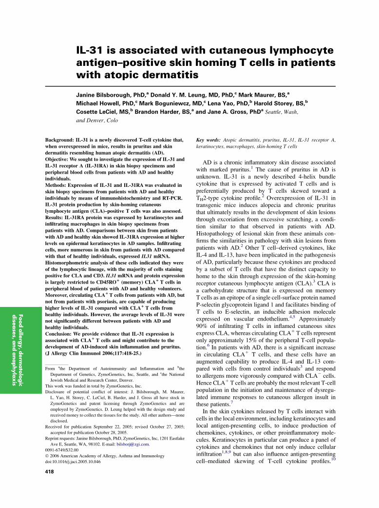
FIG 1. IL-31RA in skin biopsy specimens from patients with AD (A) compared with healthy individuals (B) was
determined bymeans of IHC (403magnification). Immunofluorescent costaining of infiltrating cells present in
skin biopsy specimens from patients with AD with IL-31RA and CD3 (C) or CD68 (D) is shown. These data are
representative of skin biopsy specimens from 5 individual patients with AD and 3 healthy individuals (Table I).
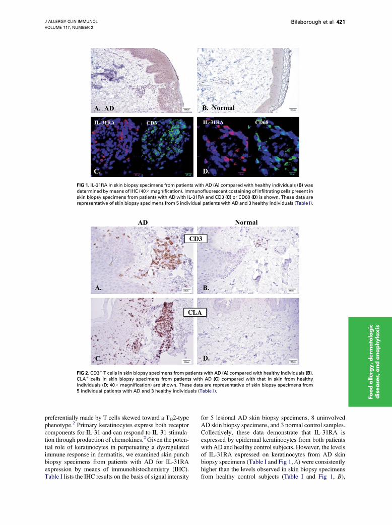
FIG 2. CD31 T cells in skin biopsy specimens from patients with AD (A) compared with healthy individuals (B).
CLA1 cells in skin biopsy specimens from patients with AD (C) compared with that in skin from healthy
individuals (D; 403 magnification) are shown. These data are representative of skin biopsy specimens from
5 individual patients with AD and 3 healthy individuals (Table I).
F d
preferentially made by T cells skewed toward a TH2-typephenotype.2 Primary keratinocytes express both receptorcomponents for IL-31 and can respond to IL-31 stimula-tion through production of chemokines.2 Given the poten-tial role of keratinocytes in perpetuating a dysregulatedimmune response in dermatitis, we examined skin punchbiopsy specimens from patients with AD for IL-31RAexpression by means of immunohistochemistry (IHC).Table I lists the IHC results on the basis of signal intensity
for 5 lesional AD skin biopsy specimens, 8 uninvolvedAD skin biopsy specimens, and 3 normal control samples.Collectively, these data demonstrate that IL-31RA isexpressed by epidermal keratinocytes from both patientswith AD and healthy control subjects. However, the levelsof IL-31RA expressed on keratinocytes from AD skinbiopsy specimens (Table I and Fig 1, A) were consistentlyhigher than the levels observed in skin biopsy specimensfrom healthy control subjects (Table I and Fig 1, B),

J ALLERGY CLIN IMMUNOL
FEBRUARY 2006
422 Bilsborough et al
Foodalle
rgy,derm
ato
logic
dise
ase
s,andanaphylaxis
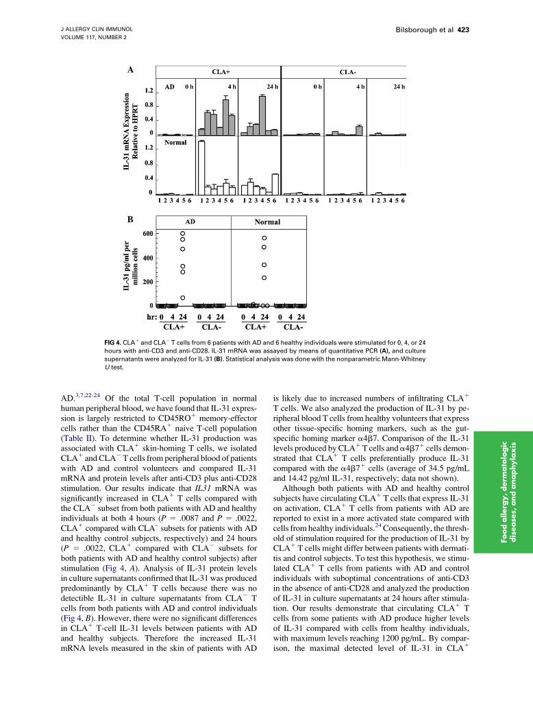
FIG 3. IL-31 mRNA in the epidermal keratinocyte layer (Epi) and infiltrating cells (Inf) of skin biopsy specimens
from patients with AD (A) or the epidermal keratinocyte layer of healthy individuals (B) is shown. Water was
included as a negative control. M, Molecular weight markers. C, Semiquantitative analysis of IL-31 mRNA
gene expression relative to an internal housekeeping gene, HPRT (IL-31/HPRT), is shown.
indicating a potential for increased responsiveness toIL-31 in the context of AD.
IL-31RA was also found to be expressed on a subset ofperivascular infiltrating cells that were more obvious inskin biopsy specimens from patients with AD comparedwith control skin samples (Fig 1, A and B, respectively).Skin biopsy specimens from patients with AD and healthysubjects were double stained with anti-IL-31RA anti-bodies and antibodies specific for cell-subset markers,including CD3 (T lymphocytes), CD68 (macrophages),or a mast cell tryptase (mast cells), to identify the celltype expressing IL-31RA. These costaining experimentsshowed that the perivascular infiltrating IL-31RA1 cellswere CD681 macrophages (Fig 1, D) and not T cells(Fig 1, C) or mast cells (data not shown). This observationsupports previous in vitro studies that demonstrate thatIL-31RA is expressed by macrophages.2,17,18
The presence of skin-infiltrating T cells is a distinguish-ing feature in skin biopsy specimens from patients withAD compared with those from healthy individuals.19
Because IL-31 is a T cell–associated cytokine, we exam-ined whether IL-31 was expressed in skin-infiltrating Tcells in tissue biopsy specimens. First, we confirmed thatskin tissue biopsy specimens from patients with ADshowed increased numbers of CD31 and CLA1 T cells
TABLE II. IL-31 protein production by CD45RO1 and
CD45RA1 peripheral blood T cells in healthy individuals
after 24-hour stimulation with anti-CD3 and anti-CD28
(picograms per milliliter per million cells)
T-cell subset Donor 1 Donor 2 Donor 3
CD45RO1 110.4 145.6 267.1
CD45RA1 13.1 12.7 14.6
by means of IHC compared with those seen in biopsyspecimens from healthy subjects (Fig 2, A and B, andFig 2, C and D, respectively).3,20 We subsequently usedLCM to specifically isolate skin-infiltrating cells for anal-ysis of IL31mRNA bymeans of RT-PCR. Fig 3, A, showsthat IL31 mRNA was expressed by skin-infiltrating cellsisolated from biopsy specimens of patients with AD. Innormal tissues infiltrating cells are not usually found andtherefore could not be analyzed. However, we did analyzethe epidermal keratinocyte layer from bothAD and normalskin for IL31 mRNA (Fig 3, A and B). Semiquantitativeanalysis of IL31 mRNA expression compared with thatof an internal control gene (HPRT) showed that althoughIL31 mRNA levels were not significantly different be-tween samples from patients with AD and samples fromhealthy subjects, there was a trend toward higher IL31expression in skin from patients with AD (Fig 3, C).
Our data demonstrate that IL-31 is produced bothlocally in the skin and by skin-infiltrating cells in patientswith AD compared with control subjects. Local produc-tion of cytokines in tissues by T cells is thought to be a keymechanism for disease pathogenesis in AD, and increasednumbers of T cells both in circulation and in skin isthought to correlate with disease.3,20 Given our previousfinding that IL-31 overexpression in mice results in pruri-tus, these data suggest that IL-31 overexpression in theskin of patients with AD might contribute to the develop-ment, maintenance, or both of disease.
IL-31 is produced by memory T cells witha skin-homing phenotype
CLA1 T cells, which almost always express theCD45RO1 memory-effector phenotype,21 have beenimplicated as a major contributor to the pathology of
J ALLERGY CLIN IMMUNOL
VOLUME 117, NUMBER 2
Bilsborough et al 423
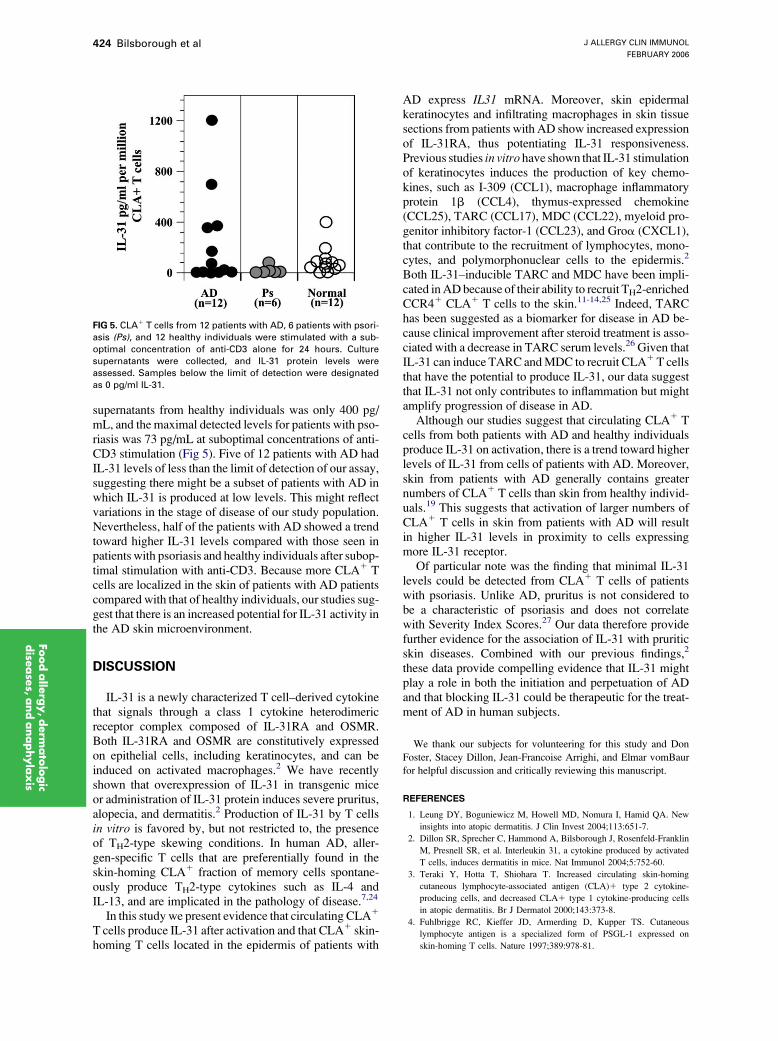
FIG 4. CLA1 and CLA2 T cells from 6 patients with AD and 6 healthy individuals were stimulated for 0, 4, or 24
hours with anti-CD3 and anti-CD28. IL-31 mRNA was assayed by means of quantitative PCR (A), and culture
supernatants were analyzed for IL-31 (B). Statistical analysis was done with the nonparametric Mann-Whitney
U test.
Foodallerg
y,derm
ato
logic
disease
s,andanaphylaxis
AD.3,7,22-24 Of the total T-cell population in normalhuman peripheral blood, we have found that IL-31 expres-sion is largely restricted to CD45RO1 memory-effectorcells rather than the CD45RA1 naive T-cell population(Table II). To determine whether IL-31 production wasassociated with CLA1 skin-homing T cells, we isolatedCLA1 and CLA2T cells from peripheral blood of patientswith AD and control volunteers and compared IL-31mRNA and protein levels after anti-CD3 plus anti-CD28stimulation. Our results indicate that IL31 mRNA wassignificantly increased in CLA1 T cells compared withthe CLA2 subset from both patients with AD and healthyindividuals at both 4 hours (P 5 .0087 and P 5 .0022,CLA1 compared with CLA– subsets for patients with ADand healthy control subjects, respectively) and 24 hours(P 5 .0022, CLA1 compared with CLA2 subsets forboth patients with AD and healthy control subjects) afterstimulation (Fig 4, A). Analysis of IL-31 protein levelsin culture supernatants confirmed that IL-31 was producedpredominantly by CLA1 T cells because there was nodetectible IL-31 in culture supernatants from CLA2 Tcells from both patients with AD and control individuals(Fig 4, B). However, there were no significant differencesin CLA1 T-cell IL-31 levels between patients with ADand healthy subjects. Therefore the increased IL-31mRNA levels measured in the skin of patients with AD
is likely due to increased numbers of infiltrating CLA1
T cells. We also analyzed the production of IL-31 by pe-ripheral blood T cells from healthy volunteers that expressother tissue-specific homing markers, such as the gut-specific homing marker a4b7. Comparison of the IL-31levels produced by CLA1T cells anda4b71 cells demon-strated that CLA1 T cells preferentially produce IL-31compared with the a4b71 cells (average of 34.5 pg/mLand 14.42 pg/ml IL-31, respectively; data not shown).
Although both patients with AD and healthy controlsubjects have circulating CLA1 T cells that express IL-31on activation, CLA1 T cells from patients with AD arereported to exist in a more activated state compared withcells from healthy individuals.24 Consequently, the thresh-old of stimulation required for the production of IL-31 byCLA1 T cells might differ between patients with dermati-tis and control subjects. To test this hypothesis, we stimu-lated CLA1 T cells from patients with AD and controlindividuals with suboptimal concentrations of anti-CD3in the absence of anti-CD28 and analyzed the productionof IL-31 in culture supernatants at 24 hours after stimula-tion. Our results demonstrate that circulating CLA1 Tcells from some patients with AD produce higher levelsof IL-31 compared with cells from healthy individuals,with maximum levels reaching 1200 pg/mL. By compar-ison, the maximal detected level of IL-31 in CLA1
J ALLERGY CLIN IMMUNOL
FEBRUARY 2006
424 Bilsborough et al
Foodalle
rgy,derm
ato
logic
dise
ase
s,andanaphylaxis
supernatants from healthy individuals was only 400 pg/mL, and the maximal detected levels for patients with pso-riasis was 73 pg/mL at suboptimal concentrations of anti-CD3 stimulation (Fig 5). Five of 12 patients with AD hadIL-31 levels of less than the limit of detection of our assay,suggesting there might be a subset of patients with AD inwhich IL-31 is produced at low levels. This might reflectvariations in the stage of disease of our study population.Nevertheless, half of the patients with AD showed a trendtoward higher IL-31 levels compared with those seen inpatients with psoriasis and healthy individuals after subop-timal stimulation with anti-CD3. Because more CLA1 Tcells are localized in the skin of patients with AD patientscompared with that of healthy individuals, our studies sug-gest that there is an increased potential for IL-31 activity inthe AD skin microenvironment.
DISCUSSION
IL-31 is a newly characterized T cell–derived cytokinethat signals through a class 1 cytokine heterodimericreceptor complex composed of IL-31RA and OSMR.Both IL-31RA and OSMR are constitutively expressedon epithelial cells, including keratinocytes, and can beinduced on activated macrophages.2 We have recentlyshown that overexpression of IL-31 in transgenic miceor administration of IL-31 protein induces severe pruritus,alopecia, and dermatitis.2 Production of IL-31 by T cellsin vitro is favored by, but not restricted to, the presenceof TH2-type skewing conditions. In human AD, aller-gen-specific T cells that are preferentially found in theskin-homing CLA1 fraction of memory cells spontane-ously produce TH2-type cytokines such as IL-4 andIL-13, and are implicated in the pathology of disease.7,24
In this study we present evidence that circulating CLA1
T cells produce IL-31 after activation and that CLA1 skin-homing T cells located in the epidermis of patients with
FIG 5. CLA1 T cells from 12 patients with AD, 6 patients with psori-
asis (Ps), and 12 healthy individuals were stimulated with a sub-
optimal concentration of anti-CD3 alone for 24 hours. Culture
supernatants were collected, and IL-31 protein levels were
assessed. Samples below the limit of detection were designated
as 0 pg/ml IL-31.
AD express IL31 mRNA. Moreover, skin epidermalkeratinocytes and infiltrating macrophages in skin tissuesections from patients with AD show increased expressionof IL-31RA, thus potentiating IL-31 responsiveness.Previous studies in vitro have shown that IL-31 stimulationof keratinocytes induces the production of key chemo-kines, such as I-309 (CCL1), macrophage inflammatoryprotein 1b (CCL4), thymus-expressed chemokine(CCL25), TARC (CCL17), MDC (CCL22), myeloid pro-genitor inhibitory factor-1 (CCL23), and Groa (CXCL1),that contribute to the recruitment of lymphocytes, mono-cytes, and polymorphonuclear cells to the epidermis.2
Both IL-31–inducible TARC and MDC have been impli-cated inADbecause of their ability to recruit TH2-enrichedCCR41 CLA1 T cells to the skin.11-14,25 Indeed, TARChas been suggested as a biomarker for disease in AD be-cause clinical improvement after steroid treatment is asso-ciated with a decrease in TARC serum levels.26 Given thatIL-31 can induce TARC andMDC to recruit CLA1T cellsthat have the potential to produce IL-31, our data suggestthat IL-31 not only contributes to inflammation but mightamplify progression of disease in AD.
Although our studies suggest that circulating CLA1 Tcells from both patients with AD and healthy individualsproduce IL-31 on activation, there is a trend toward higherlevels of IL-31 from cells of patients with AD. Moreover,skin from patients with AD generally contains greaternumbers of CLA1 T cells than skin from healthy individ-uals.19 This suggests that activation of larger numbers ofCLA1 T cells in skin from patients with AD will resultin higher IL-31 levels in proximity to cells expressingmore IL-31 receptor.
Of particular note was the finding that minimal IL-31levels could be detected from CLA1 T cells of patientswith psoriasis. Unlike AD, pruritus is not considered tobe a characteristic of psoriasis and does not correlatewith Severity Index Scores.27 Our data therefore providefurther evidence for the association of IL-31 with pruriticskin diseases. Combined with our previous findings,2
these data provide compelling evidence that IL-31 mightplay a role in both the initiation and perpetuation of ADand that blocking IL-31 could be therapeutic for the treat-ment of AD in human subjects.
We thank our subjects for volunteering for this study and Don
Foster, Stacey Dillon, Jean-Francoise Arrighi, and Elmar vomBaur
for helpful discussion and critically reviewing this manuscript.
REFERENCES
1. Leung DY, Boguniewicz M, Howell MD, Nomura I, Hamid QA. New
insights into atopic dermatitis. J Clin Invest 2004;113:651-7.
2. Dillon SR, Sprecher C, Hammond A, Bilsborough J, Rosenfeld-Franklin
M, Presnell SR, et al. Interleukin 31, a cytokine produced by activated
T cells, induces dermatitis in mice. Nat Immunol 2004;5:752-60.
3. Teraki Y, Hotta T, Shiohara T. Increased circulating skin-homing
cutaneous lymphocyte-associated antigen (CLA)1 type 2 cytokine-
producing cells, and decreased CLA1 type 1 cytokine-producing cells
in atopic dermatitis. Br J Dermatol 2000;143:373-8.
4. Fuhlbrigge RC, Kieffer JD, Armerding D, Kupper TS. Cutaneous
lymphocyte antigen is a specialized form of PSGL-1 expressed on
skin-homing T cells. Nature 1997;389:978-81.

J ALLERGY CLIN IMMUNOL
VOLUME 117, NUMBER 2
Bilsborough et al 425
5. Picker LJ, Treer JR, Ferguson-Darnell B, Collins PA, Bergstresser PR,
Terstappen LW. Control of lymphocyte recirculation in man. II. Dif-
ferential regulation of the cutaneous lymphocyte-associated antigen, a
tissue-selective homing receptor for skin-homing T cells. J Immunol
1993;150:1122-36.
6. Santamaria-Babi LF. CLA(1) T cells in cutaneous diseases. Eur J
Dermatol 2004;14:13-8.
7. Santamaria Babi LF, Picker LJ, Perez Soler MT, Drzimalla K, Flohr P,
Blaser K, et al. Circulating allergen-reactive T cells from patients with
atopic dermatitis and allergic contact dermatitis express the skin-selective
homing receptor, the cutaneous lymphocyte-associated antigen. J Exp
Med 1995;181:1935-40.
8. Cookson W. The immunogenetics of asthma and eczema: a new focus on
the epithelium. Nat Rev Immunol 2004;4:978-88.
9. Kupper TS, Fuhlbrigge RC. Immune surveillance in the skin:
mechanisms and clinical consequences. Nat Rev Immunol 2004;4:
211-22.
10. Soumelis V, Reche PA, Kanzler H, Yuan W, Edward G, Homey B, et al.
Human epithelial cells trigger dendritic cell mediated allergic inflamma-
tion by producing TSLP. Nat Immunol 2002;3:673-80.
11. Kakinuma T, Nakamura K, Wakugawa M, Mitsui H, Tada Y, Saeki H,
et al. Serum macrophage-derived chemokine (MDC) levels are closely
related with the disease activity of atopic dermatitis. Clin Exp Immunol
2002;127:270-3.
12. Fujisawa T, Fujisawa R, Kato Y, Nakayama T, Morita A, Katsumata H,
et al. Presence of high contents of thymus and activation-regulated
chemokine in platelets and elevated plasma levels of thymus and
activation-regulated chemokine and macrophage-derived chemokine in
patients with atopic dermatitis. J Allergy Clin Immunol 2002;110:
139-46.
13. Horikawa T, Nakayama T, Hikita I, Yamada H, Fujisawa R, Bito T, et al.
IFN-gamma-inducible expression of thymus and activation-regulated
chemokine/CCL17 and macrophage-derived chemokine/CCL22 in epi-
dermal keratinocytes and their roles in atopic dermatitis. Int Immunol
2002;14:767-73.
14. Vestergaard C, Bang K, Gesser B, Yoneyama H, Matsushima K, Larsen
CG. A Th2 chemokine, TARC, produced by keratinocytes may recruit
CLA1CCR41 lymphocytes into lesional atopic dermatitis skin. J Invest
Dermatol 2000;115:640-6.
15. Hanifin JM, Rajka G. Diagnostic features of atopic dermatitis. Dermatol
Venereol 1980;92:44-7.
16. Palacios R, Steinmetz M. Il-3-dependent mouse clones that express
B-220 surface antigen, contain Ig genes in germ-line configuration, and
generate B lymphocytes in vivo. Cell 1985;41:727-34.
17. Diveu C, Lelievre E, Perret D, Lak-Hal AH, Froger J, Guillet C, et al.
GPL, a novel cytokine receptor related to GP130 and leukemia inhibitory
factor receptor. J Biol Chem 2003;278:49850-9.
18. Ghilardi N, Li J, Hongo JA, Yi S, Gurney A, de Sauvage FJ. A novel
type I cytokine receptor is expressed on monocytes, signals proliferation,
and activates STAT-3 and STAT-5. J Biol Chem 2002;277:16831-6.
19. Leung DY. Atopic dermatitis: the skin as a window into the pathogenesis
of chronic allergic diseases. J Allergy Clin Immunol 1995;96:302-19.
20. Strickland I, Hauk PJ, Trumble AE, Picker LJ, Leung DY. Evidence for
superantigen involvement in skin homing of T cells in atopic dermatitis.
J Invest Dermatol 1999;112:249-53.
21. Picker LJ, Martin RJ, Trumble A, Newman LS, Collins PA, Bergstresser
PR, et al. Differential expression of lymphocyte homing receptors by
human memory/effector T cells in pulmonary versus cutaneous immune
effector sites. Eur J Immunol 1994;24:1269-77.
22. Antunez C, Torres MJ, Mayorga C, Cornejo-Garcia JA, Santamaria-Babi
LF, Blanca M. Different cytokine production and activation marker pro-
files in circulating cutaneous-lymphocyte-associated antigen T cells from
patients with acute or chronic atopic dermatitis. Clin Exp Allergy 2004;
34:559-66.
23. AkdisM, Trautmann A, Klunker S, Daigle I, Kucuksezer UC, DeglmannW,
et al. T helper (Th) 2 predominance in atopic diseases is due to preferential ap-
optosis of circulatingmemory/effector Th1 cells. FASEB J 2003;17:1026-35.
24. Akdis M, Akdis CA, Weigl L, Disch R, Blaser K. Skin-homing, CLA1
memory T cells are activated in atopic dermatitis and regulate IgE by
an IL-13-dominated cytokine pattern: IgG4 counter-regulation by CLA-
memory T cells. J Immunol 1997;159:4611-9.
25. Campbell JJ, Haraldsen G, Pan J, Rottman J, Qin S, Ponath P, et al. The
chemokine receptor CCR4 in vascular recognition by cutaneous but not
intestinal memory T cells. Nature 1999;400:776-80.
26. Hijnen D, De Bruin-Weller M, Oosting B, Lebre C, De Jong E, Bruijn-
zeel-Koomen C, et al. Serum thymus and activation-regulated chemokine
(TARC) and cutaneous T cell- attracting chemokine (CTACK) levels in
allergic diseases: TARC and CTACK are disease-specific markers for
atopic dermatitis. J Allergy Clin Immunol 2004;113:334-40.
27. Nakamura M, Toyoda M, Morohashi M. Pruritogenic mediators in
psoriasis vulgaris: comparative evaluation of itch-associated cutaneous
factors. Br J Dermatol 2003;149:718-30.
Foodallerg
y,derm
ato
logic
disease
s,andanaphylaxis