ihc fundamentals p#1 w pretreatment nsh final revised 10-24
TRANSCRIPT

Immunohistochemistry Fundamentals, Pitfalls and StandardizationNSH October 2007Lisa Paez, HTL, QIHC (ASCP)Sherry Smith, HTL (ASCP)

IHC fundamentals
1. Basic Immunology, Monoclonal, Polyclonal and Rabbit Monoclonal Antibodies
2. Basic IHC techniques, Pretreatments, Fixation, Processing and Detection Methods

1.Basic Immunology, Antibodies and Pretreatments
Attendees will gain a basic knowledge of:
Antigens and Antibodies The concept of the immune reaction Tissue fixation and pretreatments

What is the Purpose of IHC?
To identify and localize proteins or carbohydratesHuman or animal proteins
• In cells or tissuesBacterial cellsViral proteins ?

Who Cares? Why Localize Proteins?
The Central Dogma of Molecular Biology
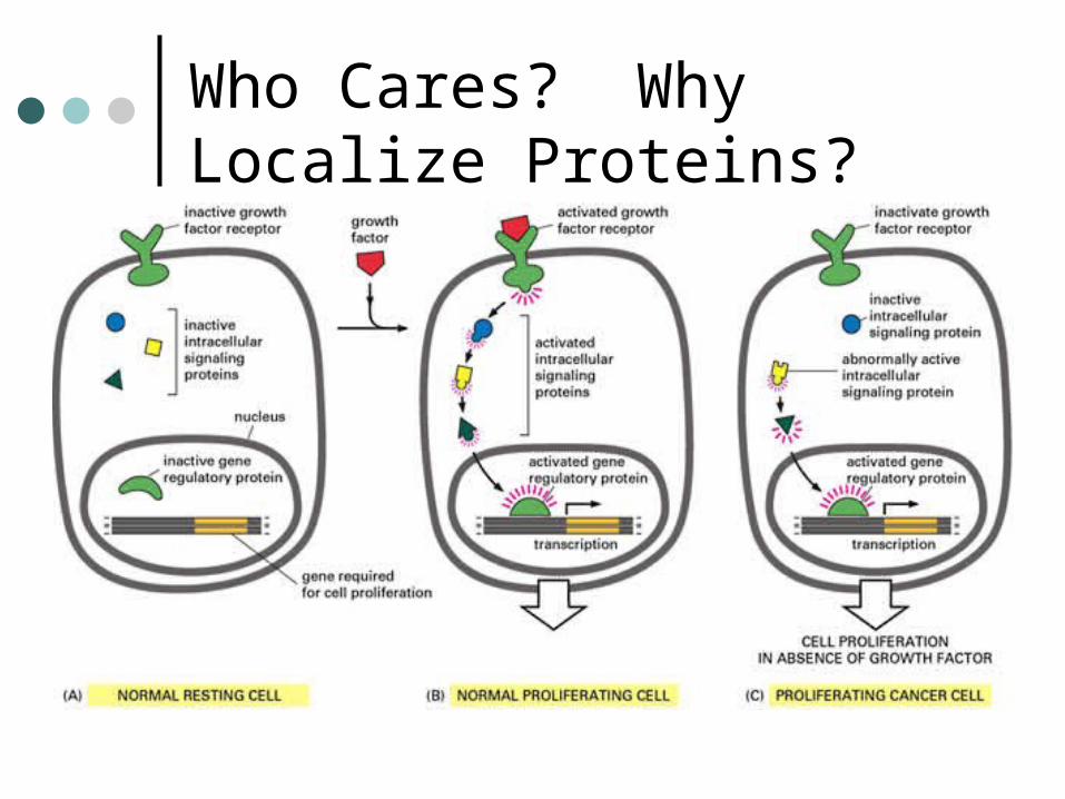
Who Cares? Why Localize Proteins?

How Can We Visualize This Process?
Immunohistochemistry- IHC is currently the most sensitive way to localize proteins within a tissue.
IHC allows us to stain protein expressing cells, cell membranes, cytoplasm, nuclei and even organelles.

1.Basic Immunology, Monoclonal, Polyclonal and Rabbit Monoclonal Antibodies
Immunology is the study of the body’s immune system body’s defense system
• responsible for protecting the body from invading organisms that cause disease
Many cell types involved in this process Belong to the family of blood cells called
white blood cells Circulate through the blood and tissues
looking for “foreign invaders” When they find something that is foreign,
they will surround it and destroy it.

Lymphocytes
Many types of white blood cells White blood cells of interest to
antibody production are the lymphocytesAll lymphocytes originate in the bone
marrow during fetal development. Two main types of lymphocytes
• T-lymphocytes (or T-cells)• B-lymphocytes (or B-cells)
femur

Thymus gland
T-Cells
Lymphocytes that migrate from the bone marrow to the thymus in order to mature there
femur
B-Cells
only B-cells produce antibodies
Lymphocytes that remain in the bone marrow and mature in the bone marrow
+ - + + - - - + + - - + - + + - - - + + - -
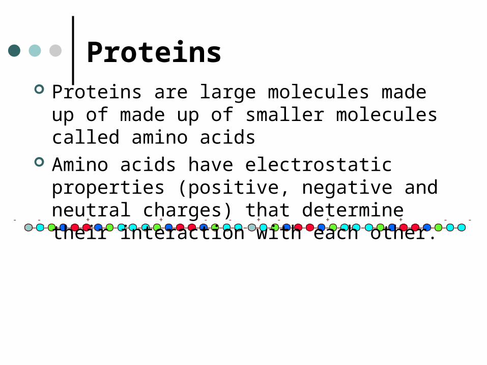
Proteins Proteins are large molecules made up of made
up of smaller molecules called amino acids Amino acids have electrostatic properties
(positive, negative and neutral charges) that determine their interaction with each other.

Protein Structure The amino acids are first linked together in a simple
chain called the primary structure.
The amino acids are then further linked into spirals (helices) or pleats (beta pleated sheets). This called the secondary structure.
The secondary structure chains can then be folded onto themselves and/or linked together to form tertiary structures.
+ - + + - - - + + - - + - + + - - - + + - -

Large protein molecules (called glycoproteins) Belong to the immunoglobulin (immune protein) or Ig
family. Produced by B-cells as part of the body’s defense system
in response to a foreign substance. Each antibody molecule is made up of 2 different types of
protein chains Long chains and short chains Long chains are heavier than the short chains Referred to as heavy chains and light chains,
respectively
Antibodies

Structure of Antibodies
Light chain (L-chain)
Heavy chain (H-chain)
The protein chains are held together by disulfide bonds

Structure of Antibodies
Heavy and Light chains are folded onto themselves in a three-dimensional structuregives a definite physical shape to
the antibody molecule
Light chain Light chain
Heavy chain heavy chain
Light chain
Heavy chain

Heavy and Light Chains
There are five types of heavy chains They are called by their Greek symbols
- gamma - alpha - mu - epsilon - delta
Two types of light chains called by their Greek symbols
- kappa - lambda

H-Chains Within each class of heavy chains the proteins are
strung along the chain in a specific sequence. heavy chains, heavy chains, heavy chains, heavy
chains and heavy chains each have a specific protein sequence.

Antibody Classification Each immunoglobulin is composed of at least
Two heavy (H) chains Two light (L) chains
Antibodies are named according to their heavy chain composition
An immunoglobulin with: heavy chains is called Immunoglobulin G ( IgG ) heavy chains is called Immunoglobulin A ( IgA ) heavy chains is called Immunoglobulin M ( IgM ) heavy chains is called Immunoglobulin E ( IgE ) heavy chains is called Immunoglobulin D ( IgD )
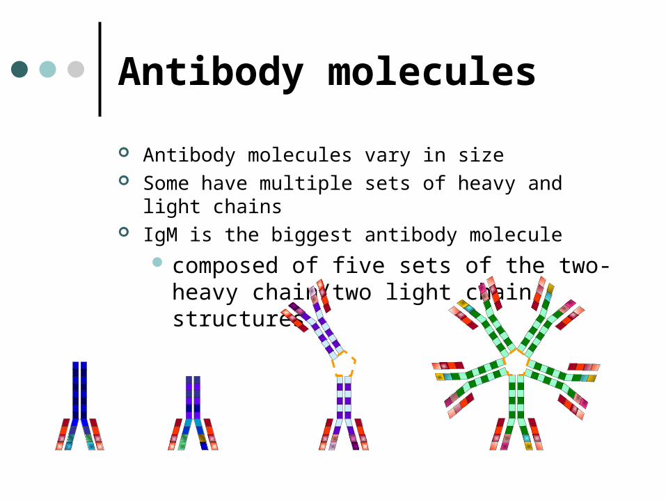
Antibody molecules
Antibody molecules vary in size Some have multiple sets of heavy and light chains IgM is the biggest antibody molecule
composed of five sets of the two-heavy chain/two light chain structures

Antibody function Various classes of antibody molecules have different biological functions Effective at detecting different kinds of invading substances Involved in various types of immune reactions
-IgMfirst antibody type to be produced in the early stages of an immune
reaction-IgA
produced by lymphocytes that “patrol” the gastro-intestinal tissueproduced in response to a parasitic infection
-IgEproduced by lymphocytes in response to an allergic reaction
-IgG is the most common antibody type circulating in the blood and tissues.
Of the five immunoglobulin (Ig) classes, IgG and IgM are the most commonly used for applications in IHC.

Antibody Molecule Together, H-chains and L-chains form two functionally different
parts of the the Ig-molecule The antigen binding fragment (Fab-portion) The crystalline fragment (Fc-portion).
Crystalline Fragment(Fc )
Antigen binding Fragments(Fab )

Antibody/Antigen Antibodies in circulation and on the surface of B-cells react to foreign
substances (“non-self”)-Antigen
To elicit a reaction -must be of a certain size-must usually be made up of protein or carbohydrate
Antibodies do not “recognize” the whole antigen-React to specific physical and chemical structures on the surface
of the antigen -Bind to these structures through a “lock-and-key” type fit-“Closeness” of the fit (or the strength of the binding) depends on
chemical and structural interaction of the antibody and antigen. Antigenic site that binds to the antibody is called the epitope or
determinant The antibody site or structure that reacts specifically with the epitope is
called the idioptype

H-chainL-chain
Antigen
epitope
Antigen Binding Site Three-dimensional shape & chemical properties of the
antigen binding region determines what substances will be able to bind to the antibody.
Antigen binding Fragment (Fab )

Immune Reaction The Antibodies can be either:
Circulating Embedded in the cytoplasmic membrane of B-cells
it is the first exposure to antigen ?If first encountered, there will be no circulating antibodies
Nucleus
Cytoplasm
Cytoplasmic membrane
Antibody molecule
Blood CirculationLymphatic
Circulation

Immune Reaction (First encounter)
If antibody has a “good fit” for the antigen Will bind to the antibody on the surface of the B-cell Trigger the B-cell to produce more of its antibody and release it into the
blood stream Released antibodies will then seek out similar antigens and surround
them (opsonization) Sends a signal that increases the blood flow and causes other white
cells to migrate to the area Other white blood cells (phagocytes) will “chew” up the antigen
(phagocytosis) B-cell will reproduce and make many copies of itself (“clones”) Clones (activated B-cells) will produce more identical antibodies and
release them into circulation. Activated B-cells become super-sensitized to this antigen and have a
very long life-span.("memory” B-cells)

Immune Reaction (First encounter)
1. Antibody on the surface of the B-cell encounters an antigen (bacteria cell)
2. B-cells multiply and produce antibodies
3. Antibodies surround antigens
5. Phagocytes migrate to the area
4. Antibodies bound to the antigen allow the antigen to be detected by phagocytes
6. Phagocytes engulf and dispose of antigen
B-Cell
Phagocyte

Antibodies
Generated (directed) against the antigen of interest, usually a protein i.e.ER, PR, CD45, HER-2/neu
Generated by injecting the protein or a portion of the protein into an animal
Produced by drawing the blood from the animal and processing the blood or cells
Antibodies In order to use antibodies to identify antigens,
the antibody must "recognize" the structure of the particular antigen
Characteristics of a "GOOD" antibody:High Specificity for its target structureHigh affinity (stereochemical fit between
antibody and antigen)High avidity (binding strength between
antibody and antigen)
Antibodies May be
Polyclonal Monoclonal "Cocktail"
May be from different animal species i.e. Polyclonal – rabbit, goat, rat, pig, horse etc. Monoclonal – mouse, rat, etc.
May be whole molecules or fragments Available in different formats

Polyclonal Antibodies Made from serum In vivo method of production Directed against many epitopes Good screening antibodies Produced by
• Generating an immune reaction in an animal• Collecting the blood• Extracting the serum
Most commonly used animals for polyclonal antibody production are rabbit, goat, sheep, horse or donkey
The immunoglobulin type is most typically IgG Many available
S-100 Herceptest

+Human Antigen Inject into animal
Bleed animal Extract serum Containing antibodies
Generate immune reaction with antibody production Several Antibodies directed against several portions of the Antigen
Polyclonal AntibodiesIn Vivo Method of Production:

Polyclonal Antibodies
Definition of Serum:The clear, thin and sticky fluid portion of the blood that remains after coagulation. Serum contains no blood cells, platelets or fibrinogen
Extracting Serum: Obtain blood from the host Allow sample to coagulate Centrifuge blood for separation of:
• Cellular components (red & white blood cells)• Fibrin• Serum
Red Blood cells
White Blood cellsFibrin
Serum

Polyclonal Antibodies
Easy to produce, widely available Production method results in high yields Directed against multiple epitopes, therefore
highly sensitiveGood as "screening" antibodies
Low specificity, tend to have more background Greater variability from lot-to-lot
Consistency relies on availability of the same animal
Field is moving towards monoclonals
Monoclonal Antibodies Directed against a single epitope (determinant)
e.g. Most CD markersResults in greater diagnostic accuracyResults in less background and cleaner slides
In vivo and in vitro methods of productionAscitesCell culture supernatant (90%)Bioreactor
Culture can be maintained indefinitelyGreater consistency from lot-to-lot
Monoclonal Antibodies: Myeloma cell lines
Requires a fusion partner (immortal cell line) Usually a Myeloma cell line grown in culture of that
particular species
Most commonly used species Mouse Rat Rabbit (until recently only mouse and rat myelomas available)
Reference: Rabbit Monoclonal Antibodies: Generating a Fusion Partner to Produce Rabbit-Rabbit HybridomasH Spieker-Polet, P Sethupathi, P Yam, and KL KnightProc. Natl. Acad. Sci. USA. 1995 September; 92(20): 9348 9352

Monoclonal Supernatant
Produced byImmunizing (generating an immune
reaction) in an animalCollecting the B-cellsFusing the B-cells with myeloma cellsGrowing cells in cultureCollecting the secreted antibodies in
the culture fluid

Monoclonal Antibodies
+Human Antigen Inject into animal
serum containing several different antibodies
Induction of immune reaction with production of antibody

Harvest spleen cellsLymphocytic proliferation
Immune reaction generates proliferation of activated cells
Monoclonal Antibodies

Fuse cells with myeloma cell line and grow in culture
+
+
+
+
Monoclonal Antibodies

anti- epitope A
anti- epitope C
anti- epitope B
anti- epitope D
Individual clones grown separately
Each colony is isolated into a separate growing vessel
Monoclonal Antibodies

Monoclonal Antibodies
anti- epitope D
anti- epitope A
anti- epitope C
anti- epitope B
Test clones: The different clones are specific for different epitopes
When a clone has been establisheda manufacturing method must be Selected:
•In Vivo – Ascites•In Vitro - Bioreactor

Monoclonal Antibodies
Purify and package
Each selected clone can be mass produced by the chosen manufacturing method

Monoclonal Ascites
Ascites Definition: an abnormal accumulation of fluid in the abdomen
Produced in Vivo by: Immunizing (generating an immune reaction) in an animal Collecting the B-cells Fusing the B-cells myeloma cells Hybridoma Growing cells in animals (usually rats) Collecting the secreted antibodies in the "tumor" fluid

Monoclonal Antibodies-Ascities
anti- epitope A
anti- epitope D
anti- epitope C
anti- epitope B
Different clones are specific for
different epitopes
Inject into animal
Collect Ascites secreted by tumor
Purify & Package
Generate Tumor

Monoclonal Antibodies- Ascites
Advantages of the Ascites method Produces high concentrations of monoclonal antibody
does not require further concentration Avoids effects of contaminants in in vitro batch-culture fluid
when comparable quantities of monoclonal antibodies are used
Avoids the need to teach cell culture technique Disadvantages of ascites method
Animals must be monitored daily In vivo methods can contain animal proteins and other
contaminants that must be purified Can be expensive Can cause pain or distress to animals used“Monoclonal Antibody Production,” Report of Committee on Methods of Producing Monoclonal Antibodies, Institute for Laboratory Animal Research, National Research Council, 1999

Monoclonal Antibodies: In Vitro Method of Production-Bioreactor
Each cartridge is inoculated with 20 million cells
A pump system provides a continuous flow of fresh media to the cells trapped within the hollow fiber cartridge
Secreted proteins from cells cultured within the hollow-fiber matrix are retained within the cartridge
Five to ten days after the initial inoculation with hybridoma cells, up to 10 mls of antibody rich medium is removed from the cartridge and fresh media is injected
Process can be repeated five times per week

In Vitro Method of ProductionBioreactor
Non-animal alternative to the large scale production of monoclonal antibodies
The antibody concentration from bioreactor fluid is comparable to ascites fluid
Four weeks production can provide an antibody yield comparable to the yield from 32 ascites mice.
Unlike ascites fluid, the bioreactor fluid is free from the contaminating mouse proteins.
Additionally, this system provides an ideal alternative for antibody production from cell lines that do not produce ascites in mice.

Rabbit Monoclonal Antibodies
Rabbits recognize antigens and epitopes that are not immunogenic in mice or rats
Were previously not possible due to lack of fusion partner
Plasmacytoma cell line that could be used as a fusion partner was generated from transgenic rabbits
Stable hybridomas now available

Rabbit Monoclonal Antibodies Higher Affinity
Rabbit anti-sera recognize more epitopes than mouse sera
Higher Specificity Higher Sensitivity Better Development Success
Stable hybridomas Because of the size of the rabbit spleen, more
fusion experiments can be performed, making it a feasible task to screen hybridoma at large scale

Rabbit Monoclonal Antibodies
+Human Antigen Inject into animal
Generate immune reaction
Harvest spleen cells
+
+
+
+
+
+
+
+
Select best clone and grow in culture
Isolate antigen specific B-cell and fuse with plasmacytoma fusion partner

Antibody Cocktails Usually made up of more than one monoclonal antibody
More sensitive than single monoclonal but more specific than polyclonals
Can detect multiple epitopes Can select the epitopes More effective at screening for certain proteins in various
cell types Are often used in combinations that are complementary or
additive More expensive
Requires many clones to achieve the sensitivity of polyclonal antibodies

Antibody Formats
Antibodies may be available in different formats or presentationsConcentratesPredilutesLyophilized
Antibody FormatsConcentrates Usually sold in 1 ml sizes or smaller Need to be diluted using a diluent that can
either be made or purchased. Antibody performance can depend on the
diluent of choice. Buffer component Protein component Preservative
Dilution should be optimized Suggested working concentration is only
a starting point

Antibody FormatsPredilutes
Sold "ready-to-use" Usually in bottles of 5-6 mls. May need “tweaking” to work in your lab Overall performance may depend on
Detection methodDetection sourcePretreatment methodTissue fixation

Antibody Selection Based on:
Clone or antibody properties such as• Specificity, sensitivity, stability
PublicationsApplication (clinical utility)Pathologist preferencePeer recommendationsVendor
Finding information on antibodies can be challenging

Specification Sheets Depending on Regulatory classification of the antibody,
Specification Sheet may provide information on
Antibody specificity Specie Immunogen Clone Isotype
Concentration Ig concentration Suggested Working concentration
Intended Use Clinical utility: Diagnostic vs.
Prognostic
Storage Conditions Temperature and stability
Suggested Protocol Pretreatments Incubation Detection
Regulatory status PMA IVD ASR Research

Specification Sheet: Zymed’s CD20

Specification Sheet: Zymed’s CD20

Specification Sheet: Zymed’s CD20

Diluents
Can make a significant difference to antibody performance
Antibodies from one vendor may not be stable in diluent from another vendorStabilityBackground

DiluentsComposition of a Antibody Diluent: Buffer
TBS (pH is critical - must be pH7.6) PBS (pH 7.2-7.4)
Protein stabilizer FCS Normal serum (10-20*%)
• high concentration of serum in the ab diluent can alter the pH
BSA (0.1- 1% BSA in PBS) Casein .03% in PBS
Preservation 0.002 - 0.1% NaAzide Kathon (Rohm & Haas) Sterilization Filtration

Basic IHC techniques: Fixatives
Common Fixatives IHC techniques are divided into two groups
- Coagulant fixatives such as ethanol, and cross linking fixatives, such as formaldehyde.
- Both can cause changes in the steric configuration of proteins, that can mask antigenic sites (epitopes) and adversly affect binding with antibody

Basic IHC techniques: Fixatives
Formalin has been the standard fixative of use with the most advantages revealed in the course of history:
1. Good preservation of morphology for a variety of tissues2. Formalin is an economic chemical3. Formalin fixation acts to sterilize tissue, especially
containing viruses4. Antigens in Carbohydrates are better preserved 1115. Through cross linking of protein, antigenicity is preserved in
situ, therefore; avoiding leaching out of proteins that may diffuse in alcohol or methanol.
Basic IHC techniques: Pretreatments Heat Unmasking Uses high heat combined with a liquid (usually a buffer) to undo
effects of fixation Microwave to heat the liquid for the purpose of unmasking
antigens Boiling - a beaker over a hot plate etc Steamer Stove top pressure cooker Microwave pressure cooker Electric pressure cooker Autoclave Waterbath
There are also many different buffers, such as Citrate buffer, citrate buffer/urea, citrate buffer/EDTA EDTA, EDTA/urea TRIS Glycine etc. etc. etc.

PretreatmentsHeat Unmasking
Antibody binding site on antigen. Fixation causes bonds
to form across portions of the protein (cross-linking). Antibody cannot bind to site.
Unmasking breaks bonds so that binding site is available for antibody binding.

AR Pitfalls No testing for pH stability in AR buffers Non testing of heating system for AR
Not familiarizing yourself with the following prior to performing AR-IHC staining:
1. The cellular localization of the antigen base2. Specificity of the primary antibody3. Previous IHC staining results from literature, especially from an
experienced laboratory. 4. Any adverse influence on the antigen from tissue fixation,
processing, the necessity of any pretreatment procedures (heat-induced AR)
5. Not reading the package insert! Information regarding reagents, antibody clone, detection systems, manufacturer, recommended concentration, etc.
Pretreatments: Heat Unmasking
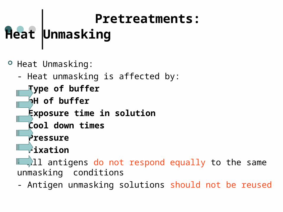
Heat Unmasking: - Heat unmasking is affected by:
Type of bufferpH of bufferExposure time in solutionCool down timesPressureFixation
- All antigens do not respond equally to the same unmasking conditions - Antigen unmasking solutions should not be reused

Pretreatments: Heat Unmasking Heat Unmasking:
- Can increase non-specific staining (background) by exposing previously cross-linked endogenous substances- Needs to be optimized independently for most labs- Solutions used for heat unmasking should never be reused because:
fixative can be dissolved out into the solutioncan become saturated with fixative, so that it fixes
instead of unmaskingpH can drift

Basic IHC techniquesPretreatments: Blockers and Enzymes
Attendees will gain a basic knowledge of: Endogenous Peroxidase Endogenous Biotin Proteases (Enzymes)
And pitfalls to avoid

Basic IHC techniquesPretreatments: Endogenous Peroxidase
Endogenous Peroxidase Facts1. Peroxidase molecules naturally occur, ie, endogenous in bloody
tissue sections fixed in paraffin. These tissue sections will react in the substrate-chromogen step in the detection procedure. Red blood cells will stain when exposed to diaminobenzidine and hydrogen peroxide because of their endogenous peroxidase content.
2. Poor fixation contributes to endogenous peroxidase activity because peroxidase can leach out of the red blood cells into the surrounding tissue increasing the background staining.
3. Note: frozen tissue sections lose their endogenous peroxidase in their red blood cells due to cell lyses in the freezing process.

Basic IHC techniques:Pretreatments: Endogenous Biotin
Highly charged molecules exist within any given tissue as normal components. These molecules may not be the target antigen of a given immunohistochemical protocol. During application of a primary antibody, if the target antigen is present, the primary antibody will bind to it, resulting in a immunospecific reaction. However, in circumstances where the tissue has not been adequately blocked the primary antibody also may combine with non-target sites, resulting in a non-immunospecific reaction. If this happens, the secondary antibody also will bind, leading to background staining.

Blockers Agents that are used to prevent or reduce false-positive or non-
specific staining Staining that is not related to primary antibody binding to the
antigen Also called "background" staining
Non-specific staining has many sources and can vary with Tissue Fixation Pretreatment Antibody Staining protocol Detection system
Selection and application of blocker depends on the source of non-specific staining

Blockers Potential sources of non-specific staining
Protein interactions• General binding of proteins to each other due to compatible
structure and charges Endogenous enzymes and proteins with enzymatic activity
• Peroxidase• Hemoglobin• Alkaline phosphatase
Endogenous Biotin Interstitial Ig Cross-Reactivity of Primary antibody Antigen Diffusion( improper, inadequate or delayed fixation which may
allow the antigen of interest to diffuse from the site of synthesis or storage and disperse throughout the tissue)
Heat unmasking can increase the incidence of all of the above

BlockersProtein Blockers
Protein structures on the surface of the tissue or cells bind the antibody non-specifically
Addition of nonspecific protein, prior to application of primary antibody, blocks non-specific sites.
BB
BB
B B
B
B
BB
B B

BlockersBiotin Blockers
BB
BB
B BB
B B
BB
AA
A A
A
B
B B
B
B B
B
B B
B
B B
B
B B
B
B
B
BEndogenous biotin in the tissue will bind to the avidin/enzyme complex. This will produce color reactions at the site of binding.
Endogenous biotin is blocked by:
1. Addition of free avidin which blocks biotin's one binding site.
2. Addition of free biotin which will block all four avidin binding sites
Inhibition of Endogenous Peroxidase Mechanism
The mixture of Methanol and hydrogen peroxide quenches endogenous peroxidase without altering the subsequent antibody reaction.
Because of the phenomenon of Substrate Inhibition, hydrogen peroxide can act in dual roles, as inhibitor and substrate.
Remember that in the detection system, hydrogen peroxide is the substrate that acts on the peroxidase enzyme to form a colored product.

Inhibition of Endogenous Peroxidase Pitfalls to avoid
Inadequate concentration of hydrogen peroxide to insufficient to irreversibly inhibit the endogenous enzyme. A 100 fold concentration of 0.03% is needed to block endogenous activity.

Blockers
Blocker Type of blocker
H202, sodium azide Peroxidase
Hydrolyzed casein Protein
Levamisole Alkaline Phosphatase
BSA Protein
Free avidin Biotin
Free biotin Avidin
Hydrolyzed casein Protein
Goat Ig Protein

Endogenous Biotin: Pitfalls to avoid Must have both blockers avidin and biotin. Must place avidin blocker on tissue prior to
biotin blocker.
If the end IHC staining results in brown staining covering the entire surface of the tissue, the most probable cause is that biotin blocker was not added after avidin blocker and the detection system linked to the avidin within the tissue.
Basic IHC techniques Pretreatments: Proteases
Proteases1. Enzymatic epitope retrieval is defined as a method used to relax the
rigidity of the protein structure that results from the cross linkages of formalin fixation.
2. Proteolytic enzymes are used in an attempt to restore the immunodominant structure in the epitope of interest. This method makes an epitope available to associate with its antibody.
3. Proteolytic enzymes are thought to cleave proteins at specific locations depending on the specificity of the enzyme. If cleavage points are in proximity to a cross-link, then the resulting effect is a relaxation of the rigid protein structure facilitating contact between the primary antibody and the corresponding antigenic determinant.
Ancillary Methods in Immunohistochemistry, Immunhistochemical Staining Methods, 4th Edition,2006,71.

Proteases
Each enzyme responds to a specific amino acid sequence. Since the specific cleavage sites are usually unpredictable, the procedure is not always successful and sometimes results in the loss of certain epitopes.
Typically enzymatic digestion doesn’t affect epitopes with high carbohydrate content. However, it can be appropriate for glycoprotein-rich targets, such as the epitope for glucagon immunoreactivity in certain tumors.
Proteases
Conditions and enzymes used for unmasking could be different for each antigen.
The optimal temperature for most proteolytic enzymes used for IHC is about 37 C
Lower temperatures are possible and in some cases are preferable because they allow a greater degree of control over the digestive process.
Pretreatments: Protease Enzyme Unmasking
The most commonly used enzymes are Proteases Pronase Proteinase K Pepsin Trypsin Ficin
Each enzyme has a unique enzymatic activity level Activity level varies with:
• Concentration• pH• Temperature

Pretreatments: Proteases Pitfalls to avoid Needs to be controlled very
carefullyCan be harsh
Too little, too much or the wrong one can Prevent staining altogetherResult in inappropriate staining
Pretreatments Pretreatment allows staining of paraffin embedded
tissue with many antibodies over a wide variety of fixation
Improves clinical utility of many primary antibodies Numerous methods are available and they all have
different advantages and disadvantages There is no universal pretreatment and no industry wide
standardization Pretreatments do not just unmask epitopes, but also
expose potential sources of background which may require blocking

Second Half !
Break Time