identification of velamentous cord using … identification of velamentous cord using smi i-series...
TRANSCRIPT
www.medical.toshiba.com.au
IDENTIFICATION OF VELAMENTOUS CORD USING SMI i-Series Case study
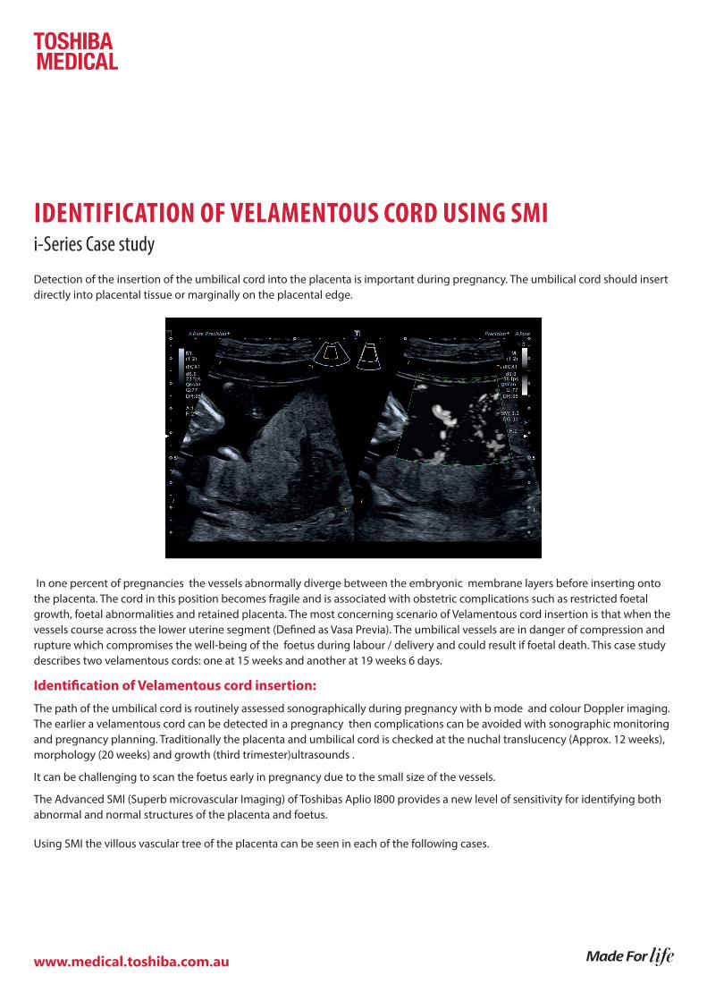
Detection of the insertion of the umbilical cord into the placenta is important during pregnancy. The umbilical cord should insert directly into placental tissue or marginally on the placental edge.
In one percent of pregnancies the vessels abnormally diverge between the embryonic membrane layers before inserting onto the placenta. The cord in this position becomes fragile and is associated with obstetric complications such as restricted foetal growth, foetal abnormalities and retained placenta. The most concerning scenario of Velamentous cord insertion is that when the vessels course across the lower uterine segment (Defined as Vasa Previa). The umbilical vessels are in danger of compression and rupture which compromises the well-being of the foetus during labour / delivery and could result if foetal death. This case study describes two velamentous cords: one at 15 weeks and another at 19 weeks 6 days.
Identification of Velamentous cord insertion:
The path of the umbilical cord is routinely assessed sonographically during pregnancy with b mode and colour Doppler imaging. The earlier a velamentous cord can be detected in a pregnancy then complications can be avoided with sonographic monitoring and pregnancy planning. Traditionally the placenta and umbilical cord is checked at the nuchal translucency (Approx. 12 weeks), morphology (20 weeks) and growth (third trimester)ultrasounds .
It can be challenging to scan the foetus early in pregnancy due to the small size of the vessels.
The Advanced SMI (Superb microvascular Imaging) of Toshibas Aplio I800 provides a new level of sensitivity for identifying both abnormal and normal structures of the placenta and foetus.
Using SMI the villous vascular tree of the placenta can be seen in each of the following cases.
www.medical.toshiba.com.au
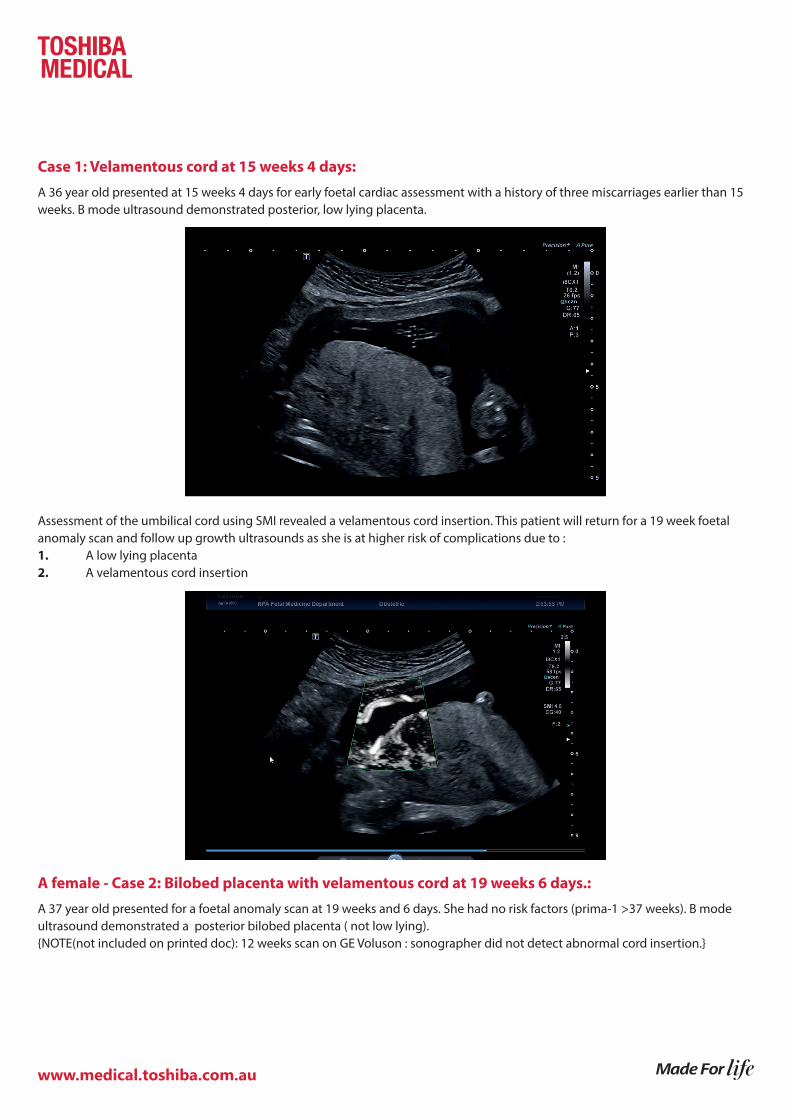
Case 1: Velamentous cord at 15 weeks 4 days:
A 36 year old presented at 15 weeks 4 days for early foetal cardiac assessment with a history of three miscarriages earlier than 15 weeks. B mode ultrasound demonstrated posterior, low lying placenta.
Assessment of the umbilical cord using SMI revealed a velamentous cord insertion. This patient will return for a 19 week foetal anomaly scan and follow up growth ultrasounds as she is at higher risk of complications due to : 1. A low lying placenta2. A velamentous cord insertion
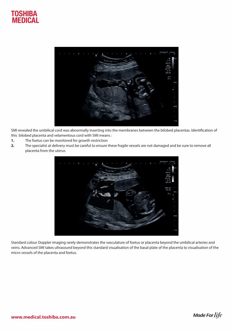
A female - Case 2: Bilobed placenta with velamentous cord at 19 weeks 6 days.:
A 37 year old presented for a foetal anomaly scan at 19 weeks and 6 days. She had no risk factors (prima-1 >37 weeks). B mode ultrasound demonstrated a posterior bilobed placenta ( not low lying). {NOTE(not included on printed doc): 12 weeks scan on GE Voluson : sonographer did not detect abnormal cord insertion.}
www.medical.toshiba.com.au
SMI revealed the umbilical cord was abnormally inserting into the membranes between the bilobed placentas. Identification of this bilobed placenta and velamentous cord with SMI means :1. The foetus can be monitored for growth restriction 2. The specialist at delivery must be careful to ensure these fragile vessels are not damaged and be sure to remove all placenta from the uterus
Standard colour Doppler imaging rarely demonstrates the vasculature of foetus or placenta beyond the umbilical arteries and veins. Advanced SMI takes ultrasound beyond this standard visualisation of the basal plate of the placenta to visualisation of the micro vessels of the placenta and foetus.
www.medical.toshiba.com.au
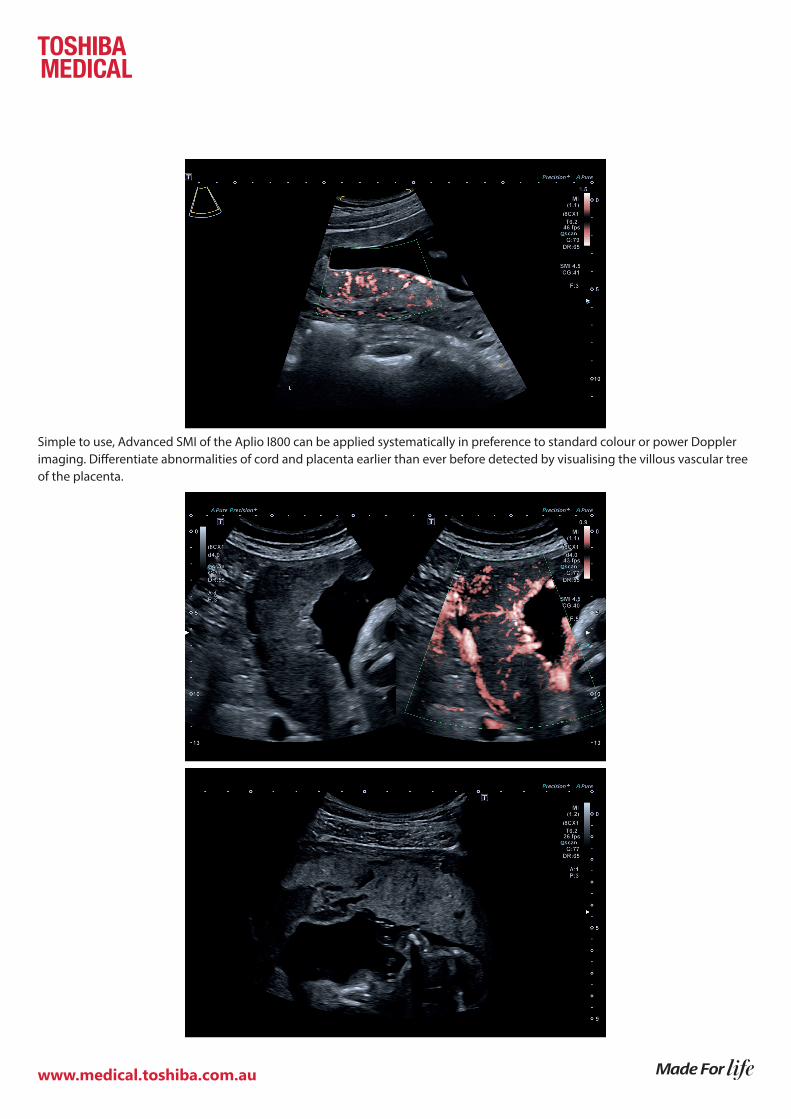
Simple to use, Advanced SMI of the Aplio I800 can be applied systematically in preference to standard colour or power Doppler imaging. Differentiate abnormalities of cord and placenta earlier than ever before detected by visualising the villous vascular tree of the placenta.