identification of the remains of king richard iii of the... · identification of the remains of...
TRANSCRIPT

ARTICLE
Received 5 Aug 2014 | Accepted 21 Oct 2014 | Published 2 Dec 2014
Identification of the remains of King Richard IIITuri E. King1,2, Gloria Gonzalez Fortes3,4,*, Patricia Balaresque5,*, Mark G. Thomas6, David Balding6,
Pierpaolo Maisano Delser1, Rita Neumann1, Walther Parson7,8, Michael Knapp9, Susan Walsh10,11,
Laure Tonasso5, John Holt12, Manfred Kayser11, Jo Appleby2, Peter Forster13,14, David Ekserdjian15,
Michael Hofreiter3,4 & Kevin Schurer16
In 2012, a skeleton was excavated at the presumed site of the Grey Friars friary in Leicester,
the last-known resting place of King Richard III. Archaeological, osteological and radiocarbon
dating data were consistent with these being his remains. Here we report DNA analyses of
both the skeletal remains and living relatives of Richard III. We find a perfect mitochondrial
DNA match between the sequence obtained from the remains and one living relative, and a
single-base substitution when compared with a second relative. Y-chromosome haplotypes
from male-line relatives and the remains do not match, which could be attributed to a false-
paternity event occurring in any of the intervening generations. DNA-predicted hair and eye
colour are consistent with Richard’s appearance in an early portrait. We calculate likelihood
ratios for the non-genetic and genetic data separately, and combined, and conclude that the
evidence for the remains being those of Richard III is overwhelming.
DOI: 10.1038/ncomms6631 OPEN
1 Department of Genetics, University of Leicester, Leicester LE1 7RH, UK. 2 School of Archaeology and Ancient History, University of Leicester, LeicesterLE1 7RH, UK. 3 Department of Biology, University of York, York YO10 5DD, UK. 4 Institute of Biochemistry and Biology, Universitat Potsdam, Karl-Liebknechtstr. 24-25, 14476 Potsdam, Germany. 5 UMR5288-CNRS/Universite Paul Sabatier-Toulouse 3 Laboratoire Anthropologie Moleculaire et Imageriede Synthese Faculte de Medecine Purpam 37, allees Jules Guesde, 31073 Toulouse, France. 6 Research Department of Genetics, Evolution and Environment,University College London, London WC1E 6BT, UK. 7 Institute of Legal Medicine, Innsbruck Medical University, Muellerstra�e 44, A-6020 Innsbruck, Austria.8 Pennsylvania State University, Eberly College of Science, Thomas Bldg, #517, University Park, Pennsylvania 16802, USA. 9 School of Biological Sciences,Bangor University, Bangor LL57 2UW, UK. 10 Molecular Anthropology Laboratory, Department of Anthropology, Yale University, Yale, New Haven,Connecticut 06511, USA. 11 Department of Forensic Molecular Biology, Erasmus MC University Medical Centre Rotterdam, 3000 CA Rotterdam,The Netherlands. 12 Space Research Centre, University of Leicester, Leicester LE1 7RH, UK. 13 McDonald Institute for Archaeological Research, University ofCambridge, Cambridge CB2 3ER, UK. 14 Murray Edwards College, University of Cambridge, Cambridge CB3 0DF, UK. 15 Department of the History of Art andFilm, University of Leicester, Leicester LE1 7RH, UK. 16 Centre for English Local History, University of Leicester, Leicester LE1 7RH, UK. * These authorscontributed equally to this work. Correspondence and requests for materials should be addressed to T.E.K. (email: [email protected]).
NATURE COMMUNICATIONS | 5:5631 | DOI: 10.1038/ncomms6631 | www.nature.com/naturecommunications 1
& 2014 Macmillan Publishers Limited. All rights reserved.

Richard III is one of the most famous and controversialEnglish kings. His ascension to the throne in 1483,following the death of his brother, Edward IV, has been
seen as contentious, involving, as it did, discrediting thelegitimacy of Edward’s marriage and therefore the claim of bothof Edward’s sons to the throne. Later, as yet unproven accusationsarose that Richard had his two nephews murdered to solidify hisown claim. Richard’s death two years later on August 22nd 1485at the Battle of Bosworth marked the end of the Plantagenetdynasty, which had ruled for over 300 years, and the beginning ofthe Tudor period. Richard III was the last English king to bekilled in battle, he became one of Shakespeare’s most notoriousvillains, and is one of the few English monarchs whose preciseresting place was lost: the mystery surrounding the fate of hisremains persisting to the present day.
Historical records report that after Richard III was killed on thebattlefield, age 32, his remains were brought back to Leicester andburied in the medieval church of the Grey Friars1. The friary wasdissolved in 1538 under the orders of King Henry VIII, with mostof the buildings being torn down in the following years.Approximately 125 years later, a rumour arose that RichardIII’s remains had been disinterred during the dissolution of themonasteries and thrown into the river Soar in Leicester2.However, it had long been thought that this rumour wasunsubstantiated and it was therefore expected that the grave ofRichard III should still lie within any remains of the Grey Friarschurch3–5. While historical records and the subsequent analysisthereof have long indicated the approximate location of the GreyFriars friary, and its likely situation in relation to the modernurban landscape of Leicester, the exact site of Richard III’s gravehad been lost in the 527 years since his death3–5.
Although Richard III reigned for only a little over two years,substantial historical information about various features of his lifeand death exists. These include aspects of his physical appearancesuch as having a slim build, one shoulder higher than the otherand that he suffered battle injuries, which resulted in his death6
(see Supplementary Note 1). In September 2012, a skeleton(Skeleton 1) was excavated at the presumed site of the Grey Friarsfriary in Leicester, the last-known resting place of Richard III(ref. 6). The archaeological, osteological and radiocarbon datingevidence were all consistent with the remains being those ofRichard III (ref. 6). The skeleton was that of a male aged 30 to 34years7, with severe scoliosis rendering one shoulder higher thanthe other8, with numerous perimortem battle injuries7. Modelledradiocarbon dating was also consistent (1456–1530AD at 95.4%probability) with these being the remains of an individual whodied in 1485 (refs 6,9). What has been missing to date is thegenetic and genealogical data, and an integrative analysis of boththe genetic and non-genetic lines of evidence. We thereforeconducted ancient and modern DNA analysis, and, for the firsttime, a synthesis of all the evidence together, to come to an overallconclusion about the identity of Skeleton 1.
Analysis of the complete mitochondrial DNA (mtDNA)sequence from Skeleton 1 shows a perfect match with themtDNA sequence of one living female-line relative of Richard IIIand a single substitution when compared with a second livingfemale-line relative. The Y-chromosome haplotype from Skeleton1 does not match that of male-line relatives of Richard III, butthis is not remarkable given that a false-paternity event couldhave occurred in any of the intervening generations. While nocontemporary portraits of Richard III survive, the DNA-predictedhair and eye colour are consistent with Richard’s appearance inone early portrait. Finally, an integrative Bayesian analysis resultsin a conservative overall likelihood ratio of 6.7 million, showingbeyond reasonable doubt that Skeleton 1 is the remains of KingRichard III.
ResultsSex determination of the remains. The sex of the remainswas determined by amplification of segments of the X- andY-chromosomes (see Supplementary Methods and SupplementaryTables 1 and 2)10. The results confirmed the remains being thoseof a male individual, as also suggested by physical analysis of thebones.
DNA identification beyond simply determining the sex of theremains relies on comparison with known relatives. One of thekey problems with deep historical relatedness is that, forrecombining portions of the genome, the sharing of DNAsegments between relatives decays rapidly with the number ofgenerations separating them. Therefore, after several generations,only the uniparentally inherited mitochondrial genome and non-recombining part of the Y-chromosome can be informative aboutrelatedness. As such, only individuals matrilinearly or patrili-nearly related to Richard III would be useful for comparison. AsRichard III left no living descendants, it was necessary to findindividuals related to him through other genealogical links (seeFig. 1, Supplementary Figs 1 and 2 and Supplementary Note 2).
Male-line relatives and Y-chromosome analysis. Male-linerelatives are generally easier to trace than female ones historically,and ennobled and titled lineages are recorded in a number ofpublished sources11. We were able to identify, locate and contactfive such relatives, descended from the 5th Duke of Beaufort(1744–1803), who agreed to take part in the study, providing an,albeit distant (between 24 and 26 generations), set of patrilinearrelatives (see Fig. 1a and Supplementary Fig. 2). It is worth notingthat while easier to trace genealogically, the male line is far moresusceptible to false-paternity than the female line is to false-maternity events12.
Four of the modern relatives were found to belong toY-haplogroup R1b-U152 (x L2, Z36, Z56, M160, M126 andZ192)13,14 with STR haplotypes being consistent with themcomprising a single patrilinear group. One individual (Somerset 3)was found to belong to haplogroup I-M170 (x M253, M223) andtherefore could not be a patrilinear relative of the other fourwithin the time span considered, indicating that a false-paternityevent had occurred within the last four generations.
Sequencing of Y-chromosome single-nucleotide polymorph-isms (SNPs) on Skeleton 1 was carried out by on-array DNAhybridization capture15 of 24 amplified Illumina16 sequencinglibraries, using probes generated to cover SNPs relevant to themajor European Y lineages, followed by sequencing on a single100 SE Illumina Hiseq 2000 sequencing lane. This approachprovided insufficient coverage for some SNPs and further typingwas performed using targeted PCRs with the amplificationproducts sequenced on an Ion Torrent PGM. Finally, we alsogenerated a STR haplotype using the Promega PowerPlex Y23system (see Supplementary Figs 3 and 4 and SupplementaryTables 3–5).
In contrast to the Y-haplotypes of the putative modernrelatives, Skeleton 1 belongs to haplogroup G-P287, with acorresponding Y-STR haplotype. Thus, the putative modernpatrilinear relatives of Richard III are not genetically related toSkeleton 1 through the male line over the time period considered.However, this is not surprising, given an estimated average false-paternity rate of B1–2% (refs 12,17,18). The putative modernrelatives and Richard III are related through a male relative(Edward III) four generations up from Richard III (Fig. 1a andSupplementary Fig. 2), and a false-paternity event could havehappened in any of the 19 generations separating Richard III andthe 5th Duke of Beaufort, on either branch of the genealogydescending from Edward III. Indeed, even with a conservative
ARTICLE NATURE COMMUNICATIONS | DOI: 10.1038/ncomms6631
2 NATURE COMMUNICATIONS | 5:5631 | DOI: 10.1038/ncomms6631 | www.nature.com/naturecommunications
& 2014 Macmillan Publishers Limited. All rights reserved.

false-paternity rate18 (see Supplementary Methods) the chance ofa false-paternity occuring in this number of generations is 16%.
Female-line relatives and mtDNA analysis. In contrast to false-paternity, false-maternity is, for obvious reasons, much less likely.However, historical records of female-line lineages are usuallymore difficult to track over multiple generations due to thechange of surname on marriage. Fortunately, the family trees ofnoble families and other landed elites are often better recordedand a family tree showing an unbroken female lineage tracingfrom Anne of York, Richard’s eldest sister, down to the early 19thcentury was published in a number of sources19 and a moderndescendant family identified20,21. However, as no supportingevidence or documents for these identifications were reported, wecarried out additional genealogical research to fully document thisfirst lineage and, furthermore, traced a second female lineage (seeSupplementary Notes 2 and 3). Thus, we were able to obtainsamples for comparison from Michael Ibsen (ML1), 19generations removed from Richard III on the female line,and from Wendy Duldig (ML2), 21 generations removed fromRichard III (see Fig. 1b and Supplementary Fig. 1). Wendy Duldigand Michael Ibsen are female-line 14th cousins, twice removed(32 meioses). Furthermore, we undertook a reconstruction ofRichard’s kinship network at the time of Bosworth to eliminate,as far as possible, known contemporary relatives sharing acommon inherited mtDNA type (see Supplementary Note 2b).
We carried out mtDNA analyses in two stages. In the firststage, both strands of the mtDNA control region (1,210 bp) weresequenced in duplicate from both ML1 and ML2 using Sangersequencing. No sequence differences were observed betweeneither duplicated samples from the same individual or indivi-duals. Three hypervariable sections (HV1, HV2, HV3) (ref. 22) ofthe mtDNA control region of Skeleton 1 were sequenced fromtwo independent extractions carried out in two different ancientDNA laboratories. Sanger sequencing of cloned PCR productswas also performed and no sequence differences were observedexcept for two that can be attributed to DNA damage patterns
found in ancient DNA23–25. We found a perfect match betweenall three individuals (ML1, ML2 and Skeleton 1), consistent withthese individuals all being matrilinear relatives at the genealogicaltime depth considered.
To determine the full mtDNA similarity, we carried outcomplete mitochondrial genome sequencing on all three samples.For the modern samples (ML1 and ML2), the entire mito-chondrial genome was amplified via two long-range PCRs26
in duplicate, followed by sequencing on an Ion Torrent PGM: allsites differing from the revised Cambridge reference sequence(rCRS)27 were subsequently confirmed by Sanger sequencing onboth strands in both ML1 and ML2 in duplicate.
Whole-genome sequencing of the mtDNA sequence ofSkeleton 1 was carried out using on-array DNA hybridizationcapture15,16 on 24 sequencing libraries generated from 16 extractsusing probes generated from the mtDNA sequence of the twomodern relatives followed by sequencing on a single 100 SEIllumina Hiseq 2000 sequencing lane (see Supplementary Fig. 5).These revealed a perfect whole-genome sequence match withML1 and a single difference (position 8,994) with ML2 consistentwith these individuals being matrilinear relatives over the timeperiod considered28 (see Supplementary Tables 6–8).
Next we investigated the probability that the mtDNA matchbetween Skeleton 1 and ML1 could have occurred by chance. Nomatches with the observed sequence were found in a database of26,127 European complete mtDNA control region sequences(http://empop.org/)29 nor in a database of 1,832 British Islessamples30 covering only positions 16,093–16,320 and 00073–00188, thereby suggesting that the haplotype is rare. MtDNAhaplotype frequencies do not vary greatly across Europe (http://empop.org/) and female mobility among the European nobilitytends to be higher than the general population. Therefore, theabsence of any match among the 26,127 European sequenceswould justify a conservative match probability value of around1 in 10,000. However, to err on the side of caution, we usedonly the smaller, lower-resolution British database to obtaina very conservative match probability value of 2/1,832 (seeSupplementary Methods).
Edward III(1312–1377)
John of Gaunt (1340–1399)
13
Henry Somerset, 5th Duke of Beaufort (1744–1803)
4
2 2
Somerset 2Somerset 1 Somerset 3 Somerset 4 Somerset 5
Male-line relatives
4 3 3
Edmund, Duke of York (1341–1402)
Richard Plantagenet, Earl of Cambridge (1375–1415)
Richard Plantagenet, Duke of York (1411–1460) m. Cecily Neville (1415–1495)
Anne of York (1439–1476)
Anne St Leger (1476–1526)
Catherine Manners (c.1510–c.1547)
Richard III(1452–1485)
Barbara Constable (c.1530–c.1561)
13
Everhilda Constable (c.1535–?)
15
Wendy Duldig
Female-line relatives
Michael Ibsen
a b
1
Figure 1 | Genealogical links between Richard III and modern-day relatives who participated in this study. (a) Genealogical information links the
Somersets to Richard III through an all-male line (left-hand side) through Edward III. Numbers indicate the number of individuals in the tree between
named individuals. Two illegitimacy events where sons born out of wedlock were later legitimized are known to have occurred in the period between John of
Gaunt and Henry Somerset, 5th Duke of Beaufort. (b) Genealogical information links Michael Ibsen and Wendy Duldig to Richard III through a female-only
line (right-hand side) descended from Richard III’s eldest sister, Anne of York. Numbers indicate number of individuals in the tree between named
individuals.
NATURE COMMUNICATIONS | DOI: 10.1038/ncomms6631 ARTICLE
NATURE COMMUNICATIONS | 5:5631 | DOI: 10.1038/ncomms6631 | www.nature.com/naturecommunications 3
& 2014 Macmillan Publishers Limited. All rights reserved.

DNA-predicted hair and eye colour. Genetic data can also beused to infer phenotypic traits such as hair and eye colour31,32.There are no contemporary portraits of Richard III (ref. 33), all ofthem post-dating his death by some 25 years or more(see Supplementary Note 4 and Supplementary Fig. 6).Dendrochronological analysis has confirmed that the earliest ofall known portraits of Richard III to have survived are the Societyof Antiquaries of London (SAL) Arched-Frame portrait and theportrait in the Royal Collection, both thought to date within a fewyears of each other in the 1510s. The SAL portrait is very differentfrom other paintings of the king, which appear to derive from anoriginal type represented by the portrait held by the RoyalCollection. The SAL portrait also has not been subject tosignificant later overpainting33.
Eye and hair colour DNA typing was carried out using probesdesigned for the HIrisPlex31 SNPs and, where necessary, followedby directed PCR using newly designed primers to generateamplicons under 100 bp in length, followed by sequencing on anIon Torrent PGM (see Supplementary Table 9). Phenotypepredictions were produced from the IrisPlex and HIrisplex31
statistical models34. These results show that Skeleton 1 had a 96%probability of having blue eyes together with a 77% probability ofhaving blond hair (see Fig. 2a–b and Supplementary Note 4).Figure 2a–d shows blue eyes of contemporary Europeans whoseDNA predictions fall within the range of high blue probabilityestimated from the Skeleton 1 profile. Similarly, Fig. 2e and fshows blond hair colour within the range of the high blondprobability estimated from the Skeleton 1 profile. However,current hair colour DNA predictions resemble childhood haircolour and it is important to note that in certain blondindividuals, hair colour can darken during adolescence. It istherefore possible that Skeleton 1 had brown hair as representedin Fig. 2g and h as seen in contemporary Europeans with asimilarly high blond probability as obtained for Skeleton 1. Thepainting of Richard III that most closely matches the geneticallypredicted eye and hair colour results is the SAL Arch-Framedportrait (see Fig. 2i and Supplementary Note 5).
Statistical analysis. To obtain a probability that Skeleton 1 is thatof Richard III, we considered the non-genetic data (radiocarbondata6, estimated age at death, sex, presence of scoliosis8 andpresence of perimortem wounds7) together with the genetic data(mtDNA and Y-chromosome). For each data type, we computedlikelihoods for the observed data under hypothesis 1 (H1—thatSkeleton 1 is Richard III) and under hypothesis 2 (H2—thatSkeleton 1 is not Richard III). Although the mtDNA evidencefavours H1, the Y-chromosome evidence provided limitedevidence against H1, and our conservative analysis of thegenetic evidence only gave moderate support for H1 (likelihoodratio, LR¼ 79; see Supplementary Methods). Using a scepticalprior probability of 0.025 that skeleton 1 is that of Richard III, weobtained a posterior probability of 2/3 that H1 is true. On theother hand using a prior probability of 0.5, the genetic evidencelead to almost 99% probability that H1 is true. This analysis ishighly conservative because, first, it used a low rate for false-paternity events, and second, the probability of a mtDNA matchby chance (match probability) used was greater than 0.001, muchhigher than would be suggested by the absence of control regionmatches in the European database (n¼ 26,127, LR¼ 6,847).Furthermore, this analysis does not take into account the whole-genome mtDNA match with one modern relative and a single-base difference with a second. We note that if we ignore theY-chromosome evidence, because of its susceptibility to false-paternity events, the contribution of the genetic data strengthensconsiderably (LR¼ 478). The non-genetic evidence strongly
supports H1 (LR¼ 85,000). All the evidence combined istherefore extremely strong in supporting H1 (LR¼ 6.7 million).This LR leads to a probability that H1 is true between 0.999994(sceptical prior) and 0.9999999 (0.5 prior, see SupplementaryTable 10). All likelihoods were computed under conservativeassumptions (discussed in the Supplementary Methods) andtherefore, these reported values are almost certainly lower thanjustified by the evidence.
DiscussionThe search for the remains of Richard III can be likened to amissing person’s case, with such investigations becoming moredifficult the longer the time between the investigation and thetime of death of the individual35,36. Given the 527 years that hadelapsed since Richard’s death at Bosworth, this case is of specialinterest in that it is the oldest DNA identification case of a knownindividual to date. As with any such case, all quantitative strandsof evidence should be drawn on to reach a conclusion regardingthe identity of any putative candidate. This report is the first thatdraws all such available strands together and estimates thestatistical support for the skeletal remains discovered in 2012being those of the last Plantagenet king, Richard III.
In drawing the evidence together, historical documentsindicated that we would be looking for the remains of anindividual who was described, during his lifetime, as having oneshoulder higher than the other, who, in 1485, aged 32, died in theheat of battle before being brought back to Leicester to be buriedin the choir of the church of the Grey Friars. In September 2012,the remains of an individual fitting all these criteria were found.Subsequently, in addition to the compelling archaeologicalevidence, laboratory analyses provided information on radio-carbon dating6, isotopic analyses9, the degree and nature of thescoliosis8 as well as the injuries sustained7. We present the geneticanalysis of the remains and the only known female-line relativesof Richard III and find a positive mtDNA match. Whilst therewas no Y-chromosome match between the skeletal remains andfive genealogically determined male-line relatives, given theknown possibility of a false-paternity over several generations,this did not prove to be a highly significant factor. One canspeculate that a false-paternity event (or events) at some point(s)in this genealogy could be of key historical significance,particularly if it occurred in the five generations between Johnof Gaunt (1340–1399) and Richard III (see Supplementary Fig. 2).A false-paternity between Edward III (1312–1377) and Johnwould mean that John’s son, Henry IV (1367–1413), and Henry’sdirect descendants (Henry V and Henry VI) would have had nolegitimate claim to the crown. This would also hold true,indirectly, for the entire Tudor dynasty (Henry VII, Henry VIII,Edward VI, Mary I and Elizabeth I) since their claim to the crownalso rested, in part, on their descent from John of Gaunt. Theclaim of the Tudor dynasty would also be brought into question ifthe false paternity occurred between John of Gaunt and his son,John Beaufort, Earl of Somerset. If the false paternity occurred ineither of the three generations between Edward III and Richard,Duke of York, the father of Edward IV and Richard III, thenneither of their claims to the crown would have been legitimate.
Analysing all the available evidence in a Bayesian framework,even using highly conservative measures, we conclude that theevidence is overwhelming that Skeleton 1 from the Grey Friarssite in Leicester is that of Richard III, thereby closing a 500-year-plus missing person case.
MethodsLaboratory locations. All DNA work involving the modern relatives was carriedout at the University of Leicester. Male-line relatives were typed using PromegaPowerPlex Y23 and for SNPs defining the main European Y-haplogroups in
ARTICLE NATURE COMMUNICATIONS | DOI: 10.1038/ncomms6631
4 NATURE COMMUNICATIONS | 5:5631 | DOI: 10.1038/ncomms6631 | www.nature.com/naturecommunications
& 2014 Macmillan Publishers Limited. All rights reserved.
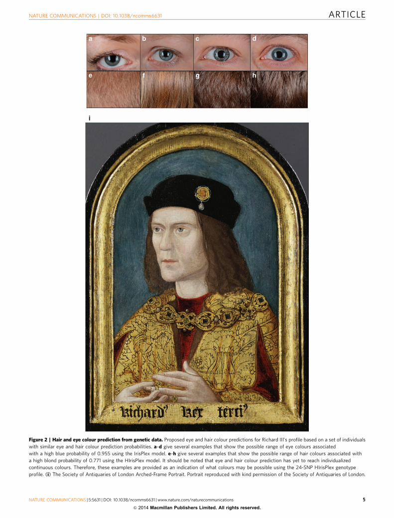
Figure 2 | Hair and eye colour prediction from genetic data. Proposed eye and hair colour predictions for Richard III’s profile based on a set of individuals
with similar eye and hair colour prediction probabilities. a–d give several examples that show the possible range of eye colours associated
with a high blue probability of 0.955 using the IrisPlex model. e–h give several examples that show the possible range of hair colours associated with
a high blond probability of 0.771 using the HIrisPlex model. It should be noted that eye and hair colour prediction has yet to reach individualized
continuous colours. Therefore, these examples are provided as an indication of what colours may be possible using the 24-SNP HIrisPlex genotype
profile. (i) The Society of Antiquaries of London Arched-Frame Portrait. Portrait reproduced with kind permission of the Society of Antiquaries of London.
NATURE COMMUNICATIONS | DOI: 10.1038/ncomms6631 ARTICLE
NATURE COMMUNICATIONS | 5:5631 | DOI: 10.1038/ncomms6631 | www.nature.com/naturecommunications 5
& 2014 Macmillan Publishers Limited. All rights reserved.

Leicester with a subset of the typing being confirmed at the Universite PaulSabatier. Female-line relatives were sequenced for the entire mitochondrial genomeat the University of Leicester. DNA was extracted from ancient teeth and bone atthe University of York and the Universite Paul Sabatier, Toulouse. Library pre-paration and target enrichment were done at the University of York. Single-end100-bp sequencing using a HiSeq 2000 (Illumina, CA, USA) was performed at theCopenhagen Sequencing Facility. Targeted sequencing of both modern and ancientDNA was also carried out at genomic technical platform PlaGe (Genopole, Tou-louse, France) and at the Protein Nucleic Acid Chemistry Laboratory at the Uni-versity of Leicester. Below we provide a condensed version of the methods used.For each step, full information can be found in the Supplementary Methods.Details surrounding the extensive genealogical research carried out for this projectcan be found in the Supplementary Note 2.
Sample collection. DNA was extracted from saliva samples of the modernrelatives of Richard III and all participants were recruited with informed consentfollowing project review by the University of Leicester Research Ethics Committee.
Skeleton 1 was excavated and samples taken under clean conditions37. Everyoneinvolved in the excavation at the Grey Friars site, the clean laboratory in Leicesterand those involved in the laboratories and labwork had their mitochondrial and,for males, Y-chromosomes typed. DNA was extracted from saliva samples and allparticipants were recruited with informed consent.
DNA extraction of ancient samples. DNA was extracted from teeth and bone(femur) samples. All procedures were performed in dedicated ancient DNAlaboratories at the University of York and the Universite Paul Sabatier, Toulousewith appropriate contamination precautions in place. Two extraction blanks wereincluded and treated exactly as if they were extracts throughout the whole process.PCRs and library experiments also included further blank controls.
Sex-typing assay performed on Skeleton 1. A newly designed sex-typing assaycomprising of PCR primers for co-amplification of the SRY fragment with UTXand UTY homologous regions was used. This assay was designed to enablerelatively small sized fragments of SRY, UTX and UTY to be co-amplified fromsamples likely to contain degraded DNA10.
Mitochondrial control region analysis of Skeleton 1. Analysis of the hyper-variable segments (HV1, HV2 and HV3) of the mtDNA control region was carriedout by amplifying and directly sequencing multiple overlapping fragments rangingfrom 153 to 250 bp in size (http://forensic.yonsei.ac.kr/protocol/mtDNA-midi-mini.pdf)22. A selection of amplicons was used for cloning the PCR products in thelab in Toulouse. Sequencing was carried out using the Big-Dye Terminator V3.1cycle sequencing kit (Applied Biosystems) and by capillary electrophoresis on anABI Prism 3730 Genetic Analyser (Applied Biosystems) at the Protein NucleicAcid Chemistry Laboratory at the University of Leicester or at the genomictechnical platform PlaGe (Genopole).
Mitochondrial genome/Y-SNP/HIrisplex typing of Skeleton 1. Libraries werebuilt following Meyer and Kircher16, with the exception that the first filtration stepbetween the blunt end repair and the adapter ligation was substituted by heatinactivation of the enzymes38,39. Two microarrays were designed, one for themtDNA enrichment and another one for nuclear SNP enrichment. DNAenrichment was performed by hybridization capture using the Agilent 244k DNASureSelect microarray (Agilent, Boblingen, Germany). For the nuclear capture,Y-chromosome probes were designed to cover the SNPs relevant to the majorEuropean lineages14. Further probes were designed to cover the SNPs relevant tothe HIrisplex31 markers. These two sets of probes (mitochondrial and SNPs) wereused separately to fill the two different microarray designs of a 1� 244K format.For each microarray, the capture protocol was performed following Hodges et al.15
with the modifications proposed by Zhang et al.40 and Fortes and Paijmans38. Thelibraries were pooled in equimolar quantities and sequenced on two lanes of theIllumina HiSeq 2000 platform in 100 SE mode at the sequencing facility of theUniversity of Copenhagen, Denmark.
The raw reads from each library were sorted based on the six-nucleotide indexused during library preparation. Only reads with a 100% match to the index wereselected for further analyses. Reads shorter than 25 nucleotides were discardedfrom further analysis. The trimmed reads were mapped to autosomes and sexchromosomes from the human reference genome build 37 (GRCh37) and to therCRS (NC_012920.1) using bwa 0.7.5a-r405 (ref. 41). In each alignment, the outputbam files were merged using SAMtools 0.1.19 (ref. 41) and PCR duplicates wereremoved subsequently. The mapped reads were filtered based on a mapping quality429 and their alignment to unique positions along the reference sequence.
Polymorphic positions were identified using SAMtools (SAMtools 0.1.19) andbcftools. Finally, vcfutils.pl was used to filter the list of variants according to aPhred-scaled genotype posterior probability quality 420 and a read depth higherthan 10. To avoid miscalling because of the deamination pattern of ancient DNAmolecules, all the polymorphic positions reported in the vcf output file werechecked by eye. In the case of the mitochondrial genome, the assembly to the
reference was visualized in Tablet42, while the alignment of the reads containingthe SNPs to the reference chromosomes was visualized using IGV43.
SNP typing by PCR. The capture approach yielded insufficient coverage for allHIrisPlex and Y-chromosome SNPs and therefore primers were designed to gen-erate amplicons containing these SNPs as well as two SNPs, which further defineY-chromosome haplogroup G: M285 (G1) and P287 (G2) (ref. 14). These wereamplified as part of multiplex reactions following Rompler et al.44 or singleplexreactions (using 40 cycles and with no secondary amplification) and sequenced onthe Ion Torrent following library preparation using Ion PGM 200 Xpress TemplateKit and PGM 200 Sequencing Kit. To increase coverage, singleplex PCR andsequencing of one marker (rs28777) was carried out according to Binladen et al.45
Typing of the haplogroup G defining SNPs (M201, M285 and P287) wasrepeated in Toulouse using singleplex PCRs. Sequencing of these PCR products wascarried out using Big-Dye Terminator V3.1 cycle sequencing kit (AppliedBiosystems) analysed by capillary electrophoresis on an ABI Prism 3730 GeneticAnalyser (Applied Biosystems) at the genomic technical platform PlaGe(Genopole).
Y-chromosomal haplotype analysis. Ancient and modern samples’ Y-chromo-somal haplotypes were obtained using the PowerPlex Y23 System (Promega) andanalysed by capillary electrophoresis on an ABI Prism 3730 Genetic Analyser(Applied Biosystems) at the genomic technical platform PlaGe (Genopole) and onan ABI Prism 3130xl Genetic Analyser (Applied Biosystems) at the University ofLeicester. For Skeleton 1, this was carried out on three separate extracts (RM2, LM1and LM3) in two different ancient DNA laboratories (York and Toulouse). For themodern relatives, this was carried out on two different extracts in two differentmodern laboratories (Leicester and Toulouse).
Y-chromosomal SNP analysis of modern samples. Following determinationof the Y-haplotype for the modern male-line samples, the predicted haplogroupwas determined using Whit Athey’s haplogroup predictor (http://www.hprg.com/hapest5/hapest5a/hapest5.htm?order=num). Binary markers covering these andrelated lineages were typed in two multiplexes by the SNaPshot minisequencingprocedure (Applied Biosystems) and an ABI3130xl Genetic Analyzer (AppliedBiosystems) followed by confirmation using Sanger sequencing. Somerset 3 wasdetermined to be Hg I (M170þ M223� , M253� )14 derived, further confirmedby the lab in Toulouse. Somersets 1,2,4 and 5 were determined to be derived forR1b-U152. Somersets 1,2,4 and 5 were tested for SNPs subdividing this clade13
(Z56, M126, Z36, Z192, M160 and L2) using Sanger sequencing in both labs.
Modern mtDNA analysis. Both samples were replicated twice.Samples were taken using Oragene DNA Collection kits (DNA Genotek) and
DNA extracted using two different methods: the Qiacube Blood and Body Fluidprotocol (200 ml with 200 ml elution) and the Oragene protocol. To analyse thecontrol region, samples were sequenced twice in both the forward and reversedirection using two overlapping primer sets (15973-296 and 16524-614) using Big-Dye Terminator V 3.1 (Applied Biosystems). No differences were found betweenreplicates or between samples.
Samples were amplified for the complete mitochondrial genome from bothextractions following Meyer et al.26 PCR amplicons were sequenced on an IonTorrent PGM Sequencer on an Ion314 Chip. Libraries were prepared using the IonXpress Plus gDNA Fragment Library Preparation kit, while the templatepreparation and the sequencing were carried out using the Ion PGM 200 XpressTemplate Kit and the Ion PGM 200 Sequencing Kit, respectively. Raw reads weremapped back to the rCRS (NC_012920.1) using TMAP software included in theIon Alignment plugin 3.2.1 (Torrent Suite Software 3.2.1) on the Ion Torrentserver. Duplicate reads removal and variant calling were performed usingSAMtools 0.1.19 (ref. 41) and local realigning was carried out with the GenomeAnalysis Tool Kit46. Variant sites were filtered for Base Quality 20, MappingQuality 50 and Depth of Coverage 30 following which 33 polymorphic sites wereretained. All these sites have been manually checked and confirmed by Sangersequencing in both directions and replicated twice.
Contamination control and quantification. Modern DNA contamination of theancient remains was controlled for by the following methods:
1. Excavation was carried out under clean conditions (see SupplementaryMethods)
2. Samples were stored in clean labs and ancient DNA work carried out only indedicated ancient DNA facilities.
3. Separate ancient samples were processed in separate labs to replicate results.4. All lab members and excavation participants had their mtDNA typed and
Y-chromosome typing was carried out on all men involved. None had amatching mtDNA or Y-chromosome type.
As evidence against significant contamination, DNA analysis of Skeleton 1shows a perfect mtDNA match to ML1 and a single-base difference with ML2.
ARTICLE NATURE COMMUNICATIONS | DOI: 10.1038/ncomms6631
6 NATURE COMMUNICATIONS | 5:5631 | DOI: 10.1038/ncomms6631 | www.nature.com/naturecommunications
& 2014 Macmillan Publishers Limited. All rights reserved.

It also shows a clear Y-STR haplotype, which has been replicated using a number ofextracts generated and tested in two separate labs. Finally, an examination (seeSupplementary Methods) of the substitution pattern in our reads also supports this.
Statistical analysis. Taking a conservative approach at each step, we computed alikelihood for each item of observed evidence under each of two opposinghypotheses: Hypothesis 1 (H1): Skeleton 1 is Richard III, and Hypothesis 2 (H2):Skeleton 1 is not Richard III.
As it was reasonable to assume that all the different lines of evidence wereindependent, the joint likelihood of all the evidence was obtained by multiplicationof the individual likelihoods under each hypothesis. The weight of evidence for H1,called the likelihood ratio (LR), was then given by the ratio of the likelihoodunder H1 to that under H2. We say that an assumption is ‘conservative’ if itreduces the LR.
The LR can be converted into a probability that H1 is true, given a priorprobability. We took as starting point the moment that Skeleton 1 was firstobserved and recognized as a human skeleton, but before any assessments of age,sex, state of health and cause of death were made. At that point, there wassubstantial evidence that a skeleton found in what is believed to have been thelocation of Leicester Grey Friars choir could be that of Richard III. All of theinformation available at the time that Skeleton 1 was unearthed, including itsprecise location and the nature of the grave, was regarded for this analysis asbackground information that can inform the prior probability. On the basis of thatinformation, we believe that a sceptical observer could not reasonably haveassigned a prior probability less than 1 in 40. This value was proposed in a previousanalysis (http://rationalgareth.com/), based on what we judge to be scepticalassessments. The highest probability that could be justified by the prior evidencemight be 1 in 2.
We have used relevant, available data where possible. Inevitably subjectivejudgments are required, for example, the relevant reference populations and aboutthe probabilities of error in reported facts. As far as seemed possible andreasonable, we strived to be conservative in our approach, for example, using apseudocount method to bias the LR towards a neutral value of 1, thus tending toavoid spurious large values from low observed frequencies. Details of the data andmethods used in the statistical analysis of the radiocarbon data, age and sex ofskeleton, presence of scoliosis, presence of perimortem wounds, Y-chromosomeand mtDNA frequency data can be found in the Supplementary Methods. Tosummarize the results: the radiocarbon data yielded a likelihood ratio of 1.84representing limited support for H1. The age and sex data yielded a likelihoodration of 5.25, again representing limited support for H1. The presence of oneshoulder higher than the other, reported during Richard’s lifetime, could beattributed to scoliosis (Skeleton 1 had severe idiopathic adolescent-onset scoliosis)or two other known conditions, Erb’s Palsy and Sprengel’s deformity, both ofwhich are very rare. Under H1, the above rates give an estimated probability of 0.90of observing scoliosis given the description of Richard III’s physical appearance(¼ the scoliosis rate divided by the sum of the three rates), which we multiplied by0.95 to allow for the possibility that the recorded description was incorrect. Thislead to a LR of 212, providing moderately strong support for H1. The presence ofperimortem injuries gave a LR of 42, and so moderate support for H1. TheY-chromosome of Skeleton 1 did not match that of genealogically determinedpatrilineal relatives of Richard III. This could be explained by a false-paternityevent in one or more of the 19 putative father–son links between Richard III andHenry Somerset, fifth Duke of Beaufort. The Y-chromosome results also indicateone further false-paternity event between Henry Somerset and his fivecontemporary, presumed patrilinear descendants. To be conservative, we selected apublished false paternity rate that was (1) lower than any other published rate thatwe considered17,47 and (2) based on genealogical data18. To this we add the false-paternity event in the 19 putative father–son links between Henry Somerset andfive contemporary male Somersets. This gives a probability of at least one falsepaternity event in the 19 putative father–son links between Richard III and HenrySomerset of 0.16. Given that a false-paternity event must have occurred under H1,the population frequency of Skeleton 1’s Y-haplotype is the same under H1 and H2and cancels out in the LR calculation. Thus, the LR is 0.16, representing limitedevidence against H1.
The mtDNA sequences of Skeleton 1 and the presumed 19-meiosis matrilinearrelative of Richard III, Michael Ibsen, matched completely. A 21-meiosis relativealso matched except at one base (8994). The latter observation is equally likelyunder H1 and H2 given the observed sequence of Michael Ibsen, and so cancels outin the LR. Thus, we only need likelihoods for the observation of the sequenceshared by Michael Ibsen and Skeleton 1.
To obtain the likelihood under H1, we require the mtDNA mutation rate, andin this case high estimates are conservative. Parsons et al.28 report 10 control regionmutations in 327 generations using genealogical data. Because this suggests ahigher rate than other published estimates, and is based on genealogical data, weused it to derive a probability of 0.52 for no mutation in 19 meioses.
For the likelihood under H2, we require the population fraction of the Skeleton1 haplotype. Although we obtained the complete mtDNA genome sequence fromSkeleton 1, we identified little published whole-genome comparison data fromEngland. Thus, for the statistical analysis, we used only the mtDNA control regionsbetween positions 16,093 and 16,320 and between 00073 and 00188, for which we
obtained suitable English comparison data from an update of Rohl et al.30,supplemented with mtDNA sequences supplied by Roots for Real (GeneticAncestor Ltd., Clare, Suffolk, UK). Using only these short sections of the controlregion under H2 is conservative, since the population fraction of the observedcontrol region sequences cannot be less than that of the full mtDNA genome.The relevant reference population is over 500 years in the past, but due to thelarge population size over the period considered, we expect populationfrequencies to have changed little over the last five centuries. We found thefrequency of the Skeleton 1 haplotype to be 0 among 1823 in the database, towhich we add the one instance observed in Michael Ibsen. This approach is,again, conservative as Michael was sampled due to his known genealogicalrelationship to Richard III. This leads to an LR of 478 representing moderatelystrong evidence for H1.
We also noted that there were no matches in a database of 26,127 Europeanmitochondrial control region haplotypes (www.empop.org)29. We do not rely onthis database because it is Europe-wide rather than specific to England and becauseof ascertainment issues, but it suggests that the Skeleton 1 haplotype may be muchrarer than can be inferred from our smaller English database. We also note thatfemale mobility among the European nobility is likely to have been much higherthan for the general population, because of marriage practices relating to politicalalliance formation. Such practices would provide some justification for using theEuropean mtDNA database, and so for considering the haplotype found inSkeleton 1 and Michael Ibsen to be extremely rare. In Supplementary Table 10, weshow some illustrative results using the European database to demonstrate theimplications of establishing that the Skeleton 1 haplotype is as rare as suggested bythat database.
The LRs for different combinations of the evidence, and two posteriorprobabilities, are shown in Supplementary Table 10. Using all the evidence, thesupport for H1 is extremely strong with an LR of 6.7 million, so that our scepticwould be driven to the conclusion that the probability that Skeleton 1 is notRichard III is less than 1 in 100,000, while for those taking a 1 in 2 starting positionthat probability is much less than 1 in a million. Taking into account theconservative assumptions underlying our calculation described above, we regardthis as establishing the truth of H1 beyond reasonable doubt.
References1. Rous, J. Joannis Rossi antiquiarii Wawicensis Historia Regum Angliae (Oxford:
Theatro Sheldoniano, 1745 [1486]).2. Speed, J. The History of Great Britaine under the Conquests of ye Romans,
Saxons, Danes and Normans (William Hall & William Beale, 1611).3. Bilson, C. J. Medieval Leicester (Edgar Backus, 1920).4. Baldwin, D. King Richard’s grave in Leicester. Trans. Leicestershire Archaeol.
and Hist. Soc. 60, 21–24 (1986).5. Strange, A. The Grey Friars, Leicester. Ricardian 3, 3–7 (1975).6. Buckley, R. et al. ’The king in the car park’: new light on the death and burial of
Richard III in the Grey Friars church, Leicester, in 1485. Antiquity 87, 519–538(2013).
7. Appleby, J. et al. Perimortem trauma in King Richard III: a skeletal analysis.Lancet doi:10.1016/S0140-6736(14)60804-7 (2014).
8. Appleby, J. et al. The scoliosis of Richard III, last Plantagenet King of England:diagnosis and clinical significance. Lancet 383, 1944 (2014).
9. Lamb, A., Evans, J., Buckley, R. & Appleby, J. Multi-isotope analysisdemonstrates significant lifestyle changes in King Richard III. J. Archaeol. Sci.50, 559–565 (2014).
10. Cadamuro, V. C. et al. Determined about sex: sex-testing in 45 primate speciesusing a 2Y/1X sex-typing assay. Forensic Sci. Int. Gen. 14C, 96–107 (2014).
11. Mosely, C. Burke’s Peerage, Baronetage and Knightage: Clan Chiefs, ScottishFeudal Barons (Boydell & Brewer Ltd., 2003).
12. King, T. E. & Jobling, M. A. Founders, drift and infidelity: the relationshipbetween Y chromosome diversity and patrilineal surnames. Mol. Biol. Evol. 26,1093–1102 (2009).
13. Rocca, R. et al. Discovery of Western European R1b1a2 Y chromosome variantsin 1000 Genomes Project Data: an online community approach. PLoS ONE 7,e41634 (2012).
14. Karafet, T. et al. New binary polymorphisms reshape and increase resolutionof the human Y chromosomal haplogroup tree. Genome Res. 18, 830–838(2008).
15. Hodges, E. et al. Hybrid selection of discrete genomic intervals on custom-designed microarrays for massively parallel sequencing. Nat. Protoc. 4, 960–974(2009).
16. Meyer, M. & Kircher, M. Illumina sequencing library preparation for highlymultiplexed target capture and sequencing. Cold Spring Harb. Protoc.doi:10.1101/pdb.prot5448 (2010).
17. Jobling, M. A., Heyer, E., Dieltjes, P. & de Knijff, P. Y-chromosome-specificmicrosatellite mutation rates re-examined using a minisatellite, MSY1. Hum.Mol. Genet. 8, 2117–2120 (1999).
18. Larmuseau, M. H. D. et al. Low historical rates of cuckoldry in a WesternEuropean human population traced by Y-chromosome and genealogical data.Proc. R. Soc. B 280, 20132400 (2013).
NATURE COMMUNICATIONS | DOI: 10.1038/ncomms6631 ARTICLE
NATURE COMMUNICATIONS | 5:5631 | DOI: 10.1038/ncomms6631 | www.nature.com/naturecommunications 7
& 2014 Macmillan Publishers Limited. All rights reserved.

19. MAHD Heddle de la Caillemotte de Massue de Ruvigny Marquis de Ruvignyand Raineval. in The Anne of Exeter Volume, Containing the Descendants ofAnne-Plantegenet-Duchess of Exeter 2 (T.C.& E.C. Jack, 1907).
20. Ashdown-Hill, J. Alive and well in Canada - The Mitochondrial DNA ofRichard III. Ricardian 16, 113–132 (2006).
21. Ashdown-Hill, J. The Last Days of Richard III (The History Press, 2011).22. Yonsei DNA Profiling Group. Monoplex PCR sets for degraded DNA. Available
at ohttp://forensic.yonsei.ac.kr/protocol/mtDNA-midi-mini.pdf4 (2004–2008).23. Hofreiter, M., Jaenicke, V., Serre, D., Von Haeseler, A. & Paabo, S. DNA
sequences from multiple amplifications reveal artifacts induced by cytosinedeamination in ancient DNA. Nucleic Acids Res. 29, 4793–4799 (2001).
24. Briggs, A. et al. Patterns of damage in genomic DNA sequences from aNeanderthal. Proc. Natl Acad. Sci. USA 104, 14616–14621 (2007).
25. Brotherton, P. et al. Novel high-resolution characterization of ancient DNAreveals C4U-type base modification events as the sole cause of post mortemmiscoding lesions. Nucleic Acids Res. 35, 5717–5728 (2007).
26. Meyer, M., Stenzel, U., Myles, S., Prufer, K. & Hofreiter, M. Targeted high-throughput sequencing of tagged nucleic acid samples. Nucleic Acids Res. 35,e97 (2007).
27. Andrews, R. M. et al. Reanalysis and revision of the Cambridge referencesequence for the human mitochondrial DNA. Nat. Genet. 23, 147 (1999).
28. Parsons, T. J. et al. A high observed substitution rate in the humanmitochondrial DNA control region. Nat. Genet. 15, 363–367 (1997).
29. Parson, W. & Dur, A. EMPOP-a forensic mtDNA database. Forensic Sci. Int.Gen. 1, 88–92 (2007).
30. Rohl, A., Brinkmann, B., Forster, L. & Forster, P. An annotated mtDNAdatabase. Int. J. Legal Med. Aug. 115, 29–39 (2001).
31. Walsh, S. et al. The HIrisPlex system for simulataneous prediction of hair andeye colour from DNA. Forensic Sci. Int.: Gen. 7, 98–115 (2013).
32. Fortes, G. G., Speller, C. F., Hofreiter, M. & King, T. E. Phenotypes from ancientDNA: approaches, insights and prospects. Bioessays 35, 690–695 (2014).
33. Hepburn, F. Portaits of the Later Plantagenets (The Boydell Press, 1986).34. Erasmus MC Forensic Molecular Biology Department HirisPlex Eye and Hair
Colour Phenotyping Webtool. Available at ohttp://www.erasmusmc.nl/fmb/resources/Irisplex_HIrisPlex/4.
35. Jeffreys, A. J., Allen, M. J., Hagelberg, E. & Sonnberg, A. Identification of theskeletal remains of Joseph Mengele by DNA analysis. Forensic Sci. Int. 56,65–76 (1992).
36. Coble, M. D. et al. Mystery solved: the identification of the the two missingRomanov children using DNA analysis. PLoS ONE 4, e4838 (2009).
37. Yang, D. & Watt, K. Contamination controls when preparing archaeologicalremains for ancient DNA analysis. J. Archaeol. Sci. 32, 331–336 (2005).
38. Fortes, G. G. & Paijmans, J. L. A. in Whole Genome Amplicfication(ed. Kroneis, T.) (Humana Press, 2014).
39. Bollongino, R. et al. 2000 years of parallel societies in Stone Age CentralEurope. Science 342, 479–481 (2013).
40. Zhang, H. et al. Morphological and genetic evidence for early Holocene cattlemanagement in northeastern China. Nat. Commun. 4, 2755 (2013).
41. Li, H. & Durbin, R. Fast and accurate short read alignment with Burrows-Wheeler transform. Bioinformatics 25, 1754–1760 (2009).
42. Milne, I. et al. Using Tablet for visual exploration of second-generationsequencing data. Brief. Bioinform. 14, 193–202 (2013).
43. Thorvaldsdottir, H., Robinson, J. & Mesirov, J. Integrative Genomics Viewer(IGV): high-perfomance genomics data visualization and exploration. Brief.Bioinform. 14, 178–192 (2013).
44. Rompler, H. et al. Multiplex amplification of ancient DNA. Nat. Protoc. 1,720–728 (2006).
45. Binladen, J., Gilbert, M. T., Campos, P. F. & Willerslev, E. 50-tailed sequencingprimers improve sequencing quality of PCR products. Biotechniques 42,174–176 (2007).
46. McKenna, A. et al. The Genome Analysis Toolkit: a MapReduce framework foranalyzing next-generation DNA sequencing data. Genome Res. 20, 1297–1303(2010).
47. Foster, E. et al. The Thomas Jefferson paternity case. Nature 397, 32 (1999).
AcknowledgementsThis research was funded by The University of Leicester with support from the Wellcomeand the Leverhulme Trusts. The original excavation that led to the discovery of RichardIII was jointly funded by the University of Leicester, The Richard III Society and LeicesterCity Council. The archaeological excavation was led by Richard Buckley, co-director ofthe University of Leicester Archaeological Service. Mathew Morris acted as site directorand provided information on burials from archaeological sites in Leicester. Thiswork leading to these results has received funding from the Austrian Science Fund
(FWF; P22880-B12), the Netherlands Genomics Initiative (NGI)/Netherlands Organi-zation for Scientific Research (NWO) within the framework of the Forensic GenomicsConsortium Netherlands (FGCN) and was financially supported from the EuropeanUnion Seventh Framework Programme (FP7/2007-2013) under grant agreement no.285487. The Society of Antiquaries of London, the National Trust, the Royal CollectionTrust/Her Majesty Queen Elizabeth and Hatfield House generously provided us withportrait images of Richard III free of charge. The National Portrait Gallery kindlyprovided images at a reduced fee. We thank Anooshka Rawden (SAL), Rosie Jordan,Liz Stacey and Chris Rowlin (NT), Desmond Shawe-Taylor, Agata Rutkowska and KarenLawson (RCT), Vicki Perry and Sarah Whale (HH) and Matthew Bailey (NPG) for theirassistance. Frederick Hepburn also provided information on the portraits. Julie Andrewsand Paul Davies (General Practitioners) and Philip Sell (Consultant Orthopaedic andSpinal Surgeon) provided information on abnormalities leading to having one shoulderhigher than the other. We thank Derek Hamilton of the SUERC Radiocarbon DatingLaboratory for data on the radiocarbon dates previously published in Antiquity. MaartenLarmuseau for data on non-paternity rates previously published in Proc. R Soc B,280:20132400. Deirdre O’Sullivan and Richard Buckley advised on burials in quires(choirs) in friaries. Lynne Moore (Royal Commission on the Ancient and HistoricalMonuments of Wales), Andrew Pye (Principal Project Manager, Exeter City Council )and Mandy Kingdom (PhD Candidate, University of Exeter) provided information onburials in Camarthen Grey Friars and Exeter Dominican excavations respectively.Johanna Paijmans, Catherine Theves, Stephanie Schiavinato, Navdeep Sahota and HeikeSiedel provided technical advice. David Annal, Morris Bierbrier and Bob Matthewshelped with genealogical research. Alec Jeffreys kindly read over a draft of the paper. CarlVivian provided a previously unseen image for the cover. We thank Philippa Langley andJohn Ashdown-Hill (who had previously traced Joy Ibsen and her family) whose faithand perseverance led to the Grey Friars excavation proceeding in the first place. Andfinally, tremendous thanks to the study participants, Michael Ibsen, Wendy Duldig andfive male-line relatives of Richard III (and their families) without whom this researchcould never have taken place.
Author contributionsT.E.K., M.H. and K.S. led the project. Ancient sample: T.E.K. and J.A. carried out theexcavation and sample collection. Samples were stored in the Space Research Centre atthe University of Leicester. J.H. provided space for storage, maintained and monitoredstorage conditions within his clean lab. Ancient DNA extractions were performed byT.E.K., G.G.F. and L.T. PCR amplifications on ancient DNA were carried out by T.E.K.and L.T. Library construction and capture was carried out by T.E.K. and G.G.F. Probeswere designed by T.E.K. and G.G.F. Y-chromosome PCR primers were designed byT.E.K., M.K. and P.B. Analysis of data (mapping, SNP calling, mtDNA assembly,Y-chromosome analysis) was carried out by T.E.K., G.G.F., P.B., P.M.D., M.H., W.P. andR.N. Modern samples: sample collection and DNA extraction was carried out by T.E.K.PCRs were carried out by T.E.K. and L.T. Next-generation sequencing of the mtDNA wascarried out using the Ion Torrent PGM by T.E.K. and R.N. Sequencing of Y-SNPs wascarried out by T.E.K., R.N. and L.T. Analysis of mitochondrial data was carried by T.E.K.,R.N. and P.M.D. Sequencing and analysis of Y-chromosome data was carried out byT.E.K., P.B., R.N. and P.M.D. Data on mtDNA types and mutation rates were providedby W.P., M.H. and P.F. Prediction of hair and eye colour based on HIrisplex allele callswas carried out by S.W. and M.K. Statistical analysis was carried out by M.G.T., T.E.K.and D.B., D.E. provided information and expertise on the portraits. Genealogical researchfor this project was led by K.S., T.K., K.S. and M.H. wrote the majority of the manuscriptwith critical input from G.F., P.B., W.P., P.M.D., M.T., D.B. and all other authors.
Additional informationAccession codes: The mitochondrial DNA sequences generated in this study have beendeposited in GenBank under the accession codes KM676292 to KM676294.
Supplementary Information accompanies this paper at http://www.nature.com/naturecommunications
Competing financial interests: The authors declare no competing financial interests.
Reprints and permission information is available online at http://npg.nature.com/reprintsandpermissions/
How to cite this article: King, T. E. et al. Identification of the remains of KingRichard III. Nat. Commun. 5:5631 doi: 10.1038/ncomms6631 (2014).
This work is licensed under a Creative Commons Attribution 4.0International License. The images or other third party material in this
article are included in the article’s Creative Commons license, unless indicated otherwisein the credit line; if the material is not included under the Creative Commons license,users will need to obtain permission from the license holder to reproduce the material.To view a copy of this license, visit http://creativecommons.org/licenses/by/4.0/
ARTICLE NATURE COMMUNICATIONS | DOI: 10.1038/ncomms6631
8 NATURE COMMUNICATIONS | 5:5631 | DOI: 10.1038/ncomms6631 | www.nature.com/naturecommunications
& 2014 Macmillan Publishers Limited. All rights reserved.