identification of the collagen-binding site of the von
TRANSCRIPT

Identification of the collagen-binding site of the vWF-A3 domain
40
Chapter 2
Identification of the collagen-binding site of the von
Willebrand Factor A3-domain
Roland A.P. Romijn¶†, Barend Bouma¶‡, Winnifred Wuyster†, Piet Gros‡,
Jan Kroon‡, Jan J. Sixma† and Eric G. Huizinga†‡
† Thrombosis and Haemostasis Laboratory, Department of Haematology,
University Medical Center and Institute of Biomembranes, HP G03.647,
PO box 85500, 3508 GA Utrecht, The Netherlands and ‡ Bijvoet Center for Biomolecular Research, Department of Crystal and
Structural Chemistry, Utrecht University, Padualaan 8, 3584 CH Utrecht,
The Netherlands.
§ These authors contributed equally
Published in the Journal of Biological Chemistry, 276 (13), 9985-9991, 2001
Identification of the collagen-binding site of the vWF-A3 domain
41
Summary
Von Willebrand factor (vWF) is a multimeric glycoprotein that mediates platelet adhesion and
thrombus formation at sites of vascular injury. VWF functions as a molecular bridge between
collagen and platelet receptor glycoprotein Ib. The major collagen-binding site of vWF is
contained within the A3 domain, but its precise location is unknown. To localize the collagen-
binding site, we determined the crystal structure of A3 in complex with a Fab fragment of
antibody RU5 that inhibits collagen binding. The structure shows that RU5 recognizes a non-
linear epitope consisting of residues 962-966, 981-997 and 1022-1026. Alanine-mutants were
constructed of residues Arg963, Glu987, His990, Arg1016 and His1023, located in or close to
the epitope. Mutants were expressed as fully processed multimeric vWF. Mutation of His1023
abolished collagen binding, while mutation of Arg963 and Arg1016 reduced collagen binding by
25 – 35%. These residues are part of loops α3β4 and α1β2 and α-helix 3, respectively, and lie
near the bottom face of the domain. His1023 and flanking residues display multiple
conformations in available A3-crystal structures suggesting that binding of A3 to collagen
involves an induced-fit mechanism. The collagen-binding site of A3 is located distant from the
top face of the domain where collagen-binding sites are found in homologous integrin I-domains.
Identification of the collagen-binding site of the vWF-A3 domain
42
Introduction
Platelet adhesion to damaged vessel walls is the first step in the formation of an occluding platelet
plug, which leads to the arrest of bleeding during normal hemostasis. Platelet adhesion can also
cause thrombotic complications such as the occlusion of aetherosclerotic arteries 1. The
multimeric glycoprotein von Willebrand factor (vWF)1 plays an essential role in platelet adhesion
under conditions of high shear stress 2; 3. In this process vWF serves as a molecular bridge that
links collagen exposed by the damaged vessel wall to glycoprotein Ib located on the platelet
surface. Collagens that act as binding sites for vWF include types I and III in perivascular
connective tissue and type VI in the sub-endothelial matrix 3; 4.
Mature vWF consists of a 2,050 residue monomer that contains multiple copies of so
called A, B, C and D-type domains and one CK (cystine knot) domain arranged in the order D'-
D3-A1-A2-A3-D4-B1-B2-B3-C1-C2-CK 1; 3. Disulfide-bond formation between N-terminal D3
domains and between C-terminal CK domains generates vWF multimers that consist of up to 80
monomers. The A1 domain contains the binding site for glycoprotein Ib 5. The A3 domain
(residues 920-1111) contains the major binding site for collagen types I and III 6. The multimeric
structure of vWF is essential for high-affinity collagen binding 7. Multimeric vWF binds collagen
with an apparent Kd of 1-7 nM 8, while a recombinant A3 domain has a much higher Kd of 2 µM 9. Deletion of the A2 and D4 domains, that flank the A3 domain, or deletion of the A1 domain
does not decrease collagen binding of multimeric vWF 6; 8. These data show that a monomeric A3
domain contains a fully active collagen-binding site, the only requirement for tight binding to
collagen being the presence of multiple A3 domains within one vWF multimer.
Integrin I-type domains are homologous to vWF A-type domains 10; 11. I-domains of
integrin α-chains α1, α2, α10 and α11 all possess collagen-binding sites. A crystal structure of
the α2 I-domain reveals binding of a collagen-like peptide to a groove in the surface of the ‘top’
face of the domain 12. This groove contains a so-called metal ion-dependent adhesion site
(MIDAS) 13; 14 which engages a glutamate residue of collagen.
The location of the collagen-binding site in the vWF-A3 domain is not known. Crystal
structures of A3 do not display a collagen-binding groove in the top face, instead, the surface of
A3 is rather smooth 15; 16. Although the MIDAS motif is partly conserved, binding of A3 to
collagen does not require a metal ion 17; 18 and no metal ion is observed in crystal structures of A3.
Moreover, point mutations in the MIDAS motif of A3 do not disrupt collagen binding 8; 16
showing that the motif is not involved in collagen binding, at all. Site-directed mutagenesis

Identification of the collagen-binding site of the vWF-A3 domain
43
studies of other residues in the top face of A3 have yielded conflicting results. Cruz et al. 19
reported in abstract form that amino-acid substitutions D1069R, R1074D, R1090D and E1092R
resulted in a 50% reduction in binding of monomeric A3 to collagen. Van der Plas et al. 8,
however, observed normal collagen binding of fully processed multimeric vWF containing
mutations D1069R, D1069A or R1074A. In the same study, mutations V1040A/V1042A,
D1046A and D1066A also displayed normal collagen binding, suggesting that the collagen-
binding site of vWF-A3 is not located in its top face.
The crystallographic study presented here was conducted to provide new clues on the
location of the collagen-binding site of the vWF-A3 domain. We determined the structure of the
A3 domain in complex with a Fab fragment of monoclonal antibody RU5, which inhibits binding
of vWF to collagen. Site-directed-mutagenesis of residues located in the epitope-region show that
the collagen-binding site is located distant from the top face of A3.
Experimental procedures
Purification of vWF-A3 and RU5
Recombinant seleno-methionine (Se-Met) A3, comprising residues 920 to 1111 of human vWF,
was expressed and purified as described before 15. For production of monoclonal antibody RU5
(IgG2a, κ) hybridoma cells were injected in mice and ascites fluid was collected (Eurogentec,
Seraing, Belgium). IgG was purified on a protein G-Sepharose column and Fab fragments were
generated using an ImmunoPure Fab Kit (Pierce, Rockford IL, USA). RU5-Fab was further
purified with A3-affinity chromatography. For that end, 10 mg of A3 was irreversibly bound via
its N-terminal histidine tag to cobalt(III)-iminodiacetate chelating Sepharose (Pharmacia Biotech,
Uppsala, Sweden), according to a procedure described by Hale 20. RU5 Fab fragment was loaded
onto the A3-affinity column, washed with PBS and subsequently eluted with 50 mM tri-
ethylamine solution (pH 10.0). Some aggregates were removed on a Superdex 75 HR 10/30 gel-
filtration column (Pharmacia, Uppsala, Sweden). Running buffer was 10 mM Tris-HCl pH 8.0, 25
mM NaCl. Next, RU5-Fab was mixed with a twofold molar excess of (Se-Met) A3. The A3-RU5
complex was separated from excess A3 by gel-filtration chromatography. Dynamic-light
scattering measurements on a Dynapro-801 DLS Instrument (Protein Solutions, Charlottesville
VA, USA) indicated the presence of 69-kDa particles in agreement with an expected molecular
weight of about 72 kDa of the complex between A3 and RU5.

Identification of the collagen-binding site of the vWF-A3 domain
44
Determination of RU5 Variable Domain Sequences
The amino-acid sequence of the variable domain of the heavy chain (VH) of RU5 was deduced
from a cDNA nucleotide sequence. Cloning and sequencing was carried out using established
procedures. Total RNA was extracted from 4*106 RU5 hybridoma cells with RNAzol using the
RNA-isolation protocol of the supplier (Campro Scientific, Veenendaal, The Netherlands). First-
strand cDNA was synthesized using Superscript II reverse transcriptase (Gibco, Rockville MD,
USA) in the presence of 3.2 U/µl of RNase-inhibitor RNaseOUT (Gibco, Rockville MD, USA).
cDNA encoding the VH domain was amplified by polymerase chain reaction (PCR) with Pwo
DNA polymerase (Boehringer, Mannheim, Germany). A backward variable-region consensus
primer (B4: CCA GGG GCC AGT GGA TAG ACA AGC TTG GGT GTC GTT TT) was used
together with a forward primer (B3c: CGG ATG GCC AGG T[C/G][A/C] AGC TGC AG[C/G]
AGT C[A/T]G G) that hybridizes with a consensus sequence in the JH region 21. The PCR product
was extended with a 3'-A overhang using Taq DNA polymerase (Promega, Madison WI, USA)
and cloned into the pCR2.1-TOPO vector according to the manufacturer’s protocol (Invitrogen,
Leek, The Netherlands). Nucleotide sequences of three clones were determined2 (figure 1A).
For determination of the sequence of the variable domain (VL) of the RU5 κ-light chain,
the same procedure was used as for the VH domain. For cloning of the VL gene, one backward
variable-region consensus primer (PD1: GAT ATT GTG ATG ACC CAG TCT [C/G]T) and two
forward variable-region consensus primers (PD3: CAG GAA ACA GCT ATG ACC GAG CTC
GTG ATC ACC CAG TCT CC; PD4: TGT AAA ACG ACG GCC AGT TCT AGA TGG TGG
GAA GAT GGA) were used. However, determination of the sequence was hampered by the
abundance of mRNA encoding the light chain of the non-producing myeloma-fusion partner of
the RU5 hybridoma cell, a known drawback of myeloma-cell line P363Ag8.653 22. Figure 1: Amino acid sequences of RU5 VL and VH domains. CDRs (underlined) were defined according to Kabat et al. 23. Strict sequential numbering (#) is used throughout the text. Numbering according to the convention of Kabat et al. 23 (#K) is also shown. A. The amino acid sequence of VH was deduced from cDNA. Residues 1 to 7 could not be
deduced precisely due to the use of a primer complementary to the 5’ coding region. Asn56 in CDRH2 (shaded) is N-glycosylated. The nucleotide sequence of VH has been deposited in the GeneBank database under Accession Number AF286587. B. The amino acid sequence of the VL domain was deduced from the electron density of the A3-RU5 complex and an alignment of 110 Fab VL chains. Side chains of thirteen amino acid residues that could not be determined unambiguously from the electron density and the consensus sequence are shown as lower case characters.
Identification of the collagen-binding site of the vWF-A3 domain
45
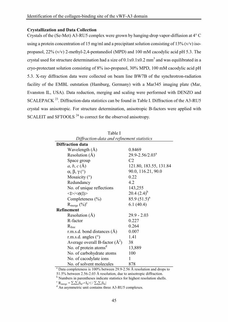
Crystallization and Data Collection Crystals of the (Se-Met) A3-RU5 complex were grown by hanging-drop vapor-diffusion at 4° C
using a protein concentration of 15 mg/ml and a precipitant solution consisting of 13% (v/v) iso-
propanol, 22% (v/v) 2-methyl-2,4-pentanediol (MPD) and 100 mM cacodylic acid pH 5.3. The
crystal used for structure determination had a size of 0.1x0.1x0.2 mm3 and was equilibrated in a
cryo-protectant solution consisting of 8% iso-propanol, 30% MPD, 100 mM cacodylic acid pH
5.3. X-ray diffraction data were collected on beam line BW7B of the synchrotron-radiation
facility of the EMBL outstation (Hamburg, Germany) with a Mar345 imaging plate (Mar,
Evanston IL, USA). Data reduction, merging and scaling were performed with DENZO and
SCALEPACK 23. Diffraction-data statistics can be found in Table I. Diffraction of the A3-RU5
crystal was anisotropic. For structure determination, anisotropic B-factors were applied with
SCALEIT and SFTOOLS 24 to correct for the observed anisotropy.
Table I Diffraction-data and refinement statistics
Diffraction data Wavelength (Å) 0.8469 Resolution (Å) 29.9-2.56/2.03a Space group C2 a, b, c (Å) α, β, γ (°)
121.80, 183.55, 131.84 90.0, 116.21, 90.0
Mosaicity (°) 0.22 Redundancy 4.2 No. of unique reflections 143,255 <I>/<σ(I)> 20.4 (2.4)b Completeness (%) 85.9 (51.5)a Rmerge (%)c 6.1 (40.4)
Refinement Resolution (Å) 29.9 - 2.03 R-factor Rfree
0.227 0.264
r.m.s.d. bond distances (Å) 0.007 r.m.s.d. angles (°) 1.41 Average overall B-factor (Å2) 38 No. of protein atomsd 13,889 No. of carbohydrate atoms 100 No. of cacodylate ions 1 No. of solvent molecules 878
a Data completeness is 100% between 29.9-2.56 Å resolution and drops to 51.5% between 2.56-2.03 Å resolution, due to anisotropic diffraction. b Numbers in parentheses indicate statistics for highest resolution shells. c Rmerge = ∑h∑i|Ihi-<Ih>| / ∑h∑i|Ihi| d An asymmetric unit contains three A3-RU5 complexes.

Identification of the collagen-binding site of the vWF-A3 domain
46
Structure Determination and Refinement
A self-rotation function calculated with POLARRFN 24 and the unit-cell volume suggested the
presence of three A3-RU5 complexes in the asymmetric unit (a.u.) with a VM of 3.3 Å3/Da and a
solvent content of 63%. Initial attempts to solve the structure by molecular replacement failed.
Therefore, a rather weak anomalous signal (<∆Fano/σ(F)> = 1.18) arising from the presence of Se-
Met in A3 was used to locate Se sites from an anomalous Patterson map. Six Se sites could be
assigned to Met947, Met998 and Met1097 of two A3 molecules on the basis of inter-atomic
distances and the observed two-fold non-crystallographic symmetry (n.c.s). The positioning of
two A3 molecules in the a.u. allowed further molecular replacement to proceed in a
straightforward manner. A Fab-fragment of protein data bank entry 2MPA 25; 26 was oriented
using the program AmoRe 27 followed by Patterson correlation refinement using CNS 28; 29. Cross
translation functions calculated with CNS identified the position of three Fab-fragments. A third
A3 molecule was placed on the basis of n.c.s. The a.u. finally contained three A3-RU5 complexes
with an R-factor of 42.6% after rigid body refinement.
For model building, sequences of the constant domain of the light chain (CL) and of the
constant domain of the heavy chain (CH1) of RU5 were taken from IgG2a, κ monoclonal antibody
4-4-20 30; 31. The sequence of residues 1 to 110 of the VL domain of RU5 (Fig. 1B) was derived
from the electron density aided by a consensus sequence based on an alignment of 110 Fab
sequences. For model refinement, cycles of rebuilding using O 32 and positional and B-factor
refinement using CNS were performed until convergence. Cross validation was used throughout
refinement using a 5% test set of reflections. Refinement used the maximum-likelihood algorithm 33. Bulk-solvent correction and anisotropic scaling of diffraction data was applied. During the first
cycles of refinement n.c.s. restraints were used. Based on the behavior of the free R-factor, n.c.s.
restraints were omitted in later stages of refinement. Water molecules were placed in difference
electron-density peaks with a peak height of at least 2.8 σ, a distance of 2.5-3.4 Å to a hydrogen-
bond donor or acceptor and a B-factor smaller than 65 Å2.
Construction of vWF Point Mutants
Point mutations were introduced in the vWF-A3 domain using the Quikchange method
(Stratagene, La Jolla CA, USA) and specific primers (Amersham Pharmacia Biotech, Roosendaal,
The Netherlands). First, a 518 base pair NheI-Csp45I fragment corresponding to amino acid
residues 940 to 1113 of mature vWF was subcloned into pBluescript SKII (Stratagene, La Jolla
CA, USA). To this end a unique Csp45I restriction site was introduced at position 5628 of

Identification of the collagen-binding site of the vWF-A3 domain
47
expression vector pNUT-vWFcas 8 in the following way. The BamHI - EcoRV fragment of
pNUT-vWFcas was ligated into pBluescript SKII. The Csp45I site was introduced using
Quickchange and confirmed by sequencing. The NheI – EcoRV fragment containing the new
Csp45I site was ligated into pNUT-vWFcas generating pNUT-vWFcas2. Next, the NheI - Csp45I
fragment of pNUT-vWFcas2 was made blunt by filling in of 5’ overhangs using Pwo DNA
polymerase. Ligation of this fragment into EcoRI - AccI digested and Pwo treated pBluescript
SKII, produced mutagenesis plasmid pBSvWF-NC. This plasmid retains the NheI and Csp45I
restriction sites. Point mutations R963A, E987A, H990A, R1016A and H1023A were introduced
in pBSvWF-NC. Mutation H1023A causes the disappearance of a NsiI restriction site. The NheI -
Csp45I fragment of this mutant was ligated directly into pNUT-vWFcas2 and confirmed by
restriction analysis. The NheI – Csp45I fragment of the other point mutants were ligated into
pNUT-vWFcas2-H1023A, with reappearance of the NsiI site.
Expression, Purification and Characterization of vWF
vWF was stably expressed in fur-BHK cells, a baby hamster kidney cell line overexpressing furin
necessary for proper removal of vWF propeptide 6; 34. VWF was purified by immuno-affinity
chromatography using monoclonal antibody RU8, which is directed against the D4 domain 6.
VWF-containing fractions were pooled and stored in aliquots at –20 °C until use.
The concentration of vWF was determined by a sandwich ELISA using polyclonal α-vWF
and horseradish peroxidase (HRP) conjugated polyclonal α-vWF (DAKO, Glostrup, Denmark)
for immobilization and detection, respectively 6. Normal pooled plasma from 40 healthy donors
was used as a reference.
The multimeric structure of vWF was analyzed by agarose gel electrophoresis followed by
Western blotting as described by Lawrie et al. 35.
Binding of vWF to monoclonal antibody RU5 was analyzed as follows. Microtiter-plate
wells (Costar, Cambridge MA, USA) were coated with 2.5 µg/ml polyclonal α-D’D3 8 in 50 mM
carbonate buffer pH 9.6 (3 h, 20 oC). Wells were washed with PBS/0.1% Tween-20 (PBS/T) and
blocked with 3 % BSA in PBS/T (1 h, 37 oC). Wells were incubated with 100 ng/ml vWF in
PBS/T including 3% BSA (1 h, 37 oC). After washing, 5 µg/ml RU5 was added (1 h, 37 oC).
Wells were washed and incubated with HRP conjugated swine-anti-rabbit antibody (DAKO,
Glostrup, Denmark) diluted 1:2500 in PBS/T containing 3% BSA (1 h, 37 oC). O-
phenylenediamine was used as substrate for detection.

Identification of the collagen-binding site of the vWF-A3 domain
48
Collagen-Binding Assay
vWF binding to fibrillar human placenta collagen type I (Sigma, St. Louis MO, USA, cat. No. C-
7774) and collagen type III (Sigma, cat. No. C-4407) was studied in a solid-state binding assay
according to Van der Plas et al. 8. Collagen was coated at 50 µg/ml instead of 100 µg/ml as used
previously 8. A vWF concentration of 2.5 µg/ml was used in the binding experiments.
Results
A3-RU5 Structure Determination and Refinement
The structure of the vWF (Se-Met) A3-domain in complex with a Fab-fragment of RU5 was
solved to 2.0 Å resolution. The structure was determined using the anomalous signal from
selenium atoms in (Se-Met) A3 to position two A3 molecules and was completed by molecular
replacement. The a.u. contains three A3-Fab complexes. The structure has been refined to an R-
factor of 22.7% and a free R-factor of 26.4% (Table I).
The sequence of the RU5 VL domain could not be determined from cDNA and was
therefore deduced from electron density aided by an alignment of 110 Fab VL sequences. The
identity of thirteen amino acid residues could not be determined uniquely (see Fig. 1). The
structure displays good model geometry with 88.8% of the residues in the most favored region of
the Ramachandran plot and 10.8% in the additionally allowed region. Residue Thr51 of RU5 VL
domains occurs in the disallowed region of the Ramachandran plot, but its electron density is
convincing.
The a.u. consists of three A3-RU5 complexes that are denoted A, B and C. The overall
structure of the (Se-Met) A3 molecule is the same as the structure of native A3 15; 16. It consists of
a central six-stranded β-sheet on both sides flanked by α-helices. The final model comprises
amino-acid residues 921-1110 of A3 molecules in complexes A and B and residues 920-1110 of
the A3 molecule in complex C. Positions of the N- and C-termini of A3, including disulfide bond
Cys923-Cys1109 that connects these termini, are poorly defined. The model of RU5 consists of
residues 1-211 of the light chains and residues 1-129 and 136-216 of the heavy chains. Residues
130-135 of the heavy chains display very weak electron density and are excluded from the model.
Disorder of this loop is a commonly observed feature in Fab structures 36. Asn56 of the VH
domain is N-glycosylated. Electron density near Asn56 accounted for a GlcNAc(-Fuc)-GlcNAc
Identification of the collagen-binding site of the vWF-A3 domain
49
moiety in complexes A and B and only a GlcNAc-Fuc moiety in complex C. The model contains
one cacodylate ion from the crystallization solution.
Crystal Packing and Differences between Complexes
In the a.u. A3-RU5 complexes B and C form a tightly interacting anti-parallel dimer and are
related by two-fold n.c.s. A3-RU5 complex A forms a similar anti-parallel dimer with a
crystallographically related complex A'. Complexes within dimers B-C and A-A' have large
contact areas of 932 Å2 and 1,454 Å2, respectively (GRASP 37). These large interaction-surface
areas suggest that the dimer of A3-RU5 complexes may also be stable in solution. DLS
measurements, however, clearly indicated that the A3-RU5 complex does not form dimers in
solution. Therefore, dimers observed in the crystal are a result of crystal packing.
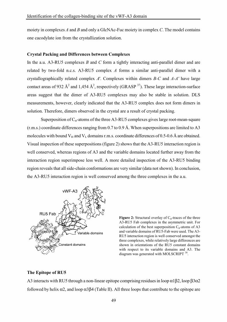
Superposition of Cα-atoms of the three A3-RU5 complexes gives large root-mean-square
(r.m.s.) coordinate differences ranging from 0.7 to 0.9 Å. When superpositions are limited to A3
molecules with bound VH and VL domains r.m.s. coordinate differences of 0.5-0.6 Å are obtained.
Visual inspection of these superpositions (figure 2) shows that the A3-RU5 interaction region is
well conserved, whereas regions of A3 and the variable domains located further away from the
interaction region superimpose less well. A more detailed inspection of the A3-RU5 binding
region reveals that all side-chain conformations are very similar (data not shown). In conclusion,
the A3-RU5 interaction region is well conserved among the three complexes in the a.u.
Figure 2: Structural overlay of Cα-traces of the three A3-RU5 Fab complexes in the asymmetric unit. For calculation of the best superposition Cα-atoms of A3 and variable domains of RU5-Fab were used. The A3-RU5 interaction region is well conserved amongst the three complexes, while relatively large differences are shown in orientations of the RU5 constant domains with respect to its variable domains and A3. The diagram was generated with MOLSCRIPT 38.
The Epitope of RU5
A3 interacts with RU5 through a non-linear epitope comprising residues in loop α1β2, loop β3α2
followed by helix α2, and loop α3β4 (Table II). All three loops that contribute to the epitope are

Identification of the collagen-binding site of the vWF-A3 domain
50
located in the bottom face of A3 (figure 3). The N- and C-termini of A3 that are also located in
the bottom face do not interact with RU5. This observation is in agreement with the fact that RU5
was raised against complete vWF in which the termini connect A3 to flanking A2 and D4
domains. Residues of RU5 that interact with loops α1β2 and α3β4 are located in CDRL1 and
CDRL2 (figure 1). All six CDRs interact with residues in the contiguous segment formed by loop
β3α2 and helix α2. The A3-RU5 interactions involve five hydrogen bonds and one salt bridge.
The buried surface area of A3 molecules in the complex is approximately 1,200 Å2, which is
about 7% of the total surface area of A3. Interestingly, the fucose moiety attached to residue
Asn56 in CDRH2 of complex A interacts with Lys988-Ala989 of A3. The carbohydrate moieties
attached to Asn56 in complexes B and C do not interact with A3.
Conformational Changes in A3
To analyze whether binding of RU5 causes conformational changes in A3 we compared models
of A3 in the A3-RU5 complex with two structures of free A34 15; 16. The two structures of free A3
were determined from different crystal forms with unrelated crystal-packing interactions. In both
crystal forms two molecules are present in the a.u. Since the A3-RU5 complex was obtained with
(Se-Met) A3 we also included the structure of free (Se-Met) A3 in the comparison to detect
possible structural differences caused by Se-Met (E.G. Huizinga, unpublished results). R.m.s.
coordinate differences after pair-wise superimposing Cα-atoms of all eight models range from
0.24 to 0.81 Å. Large differences are restricted to three loops located within the RU5-binding site
and to two loops that are located distant from the epitope (figure 3, Table II).
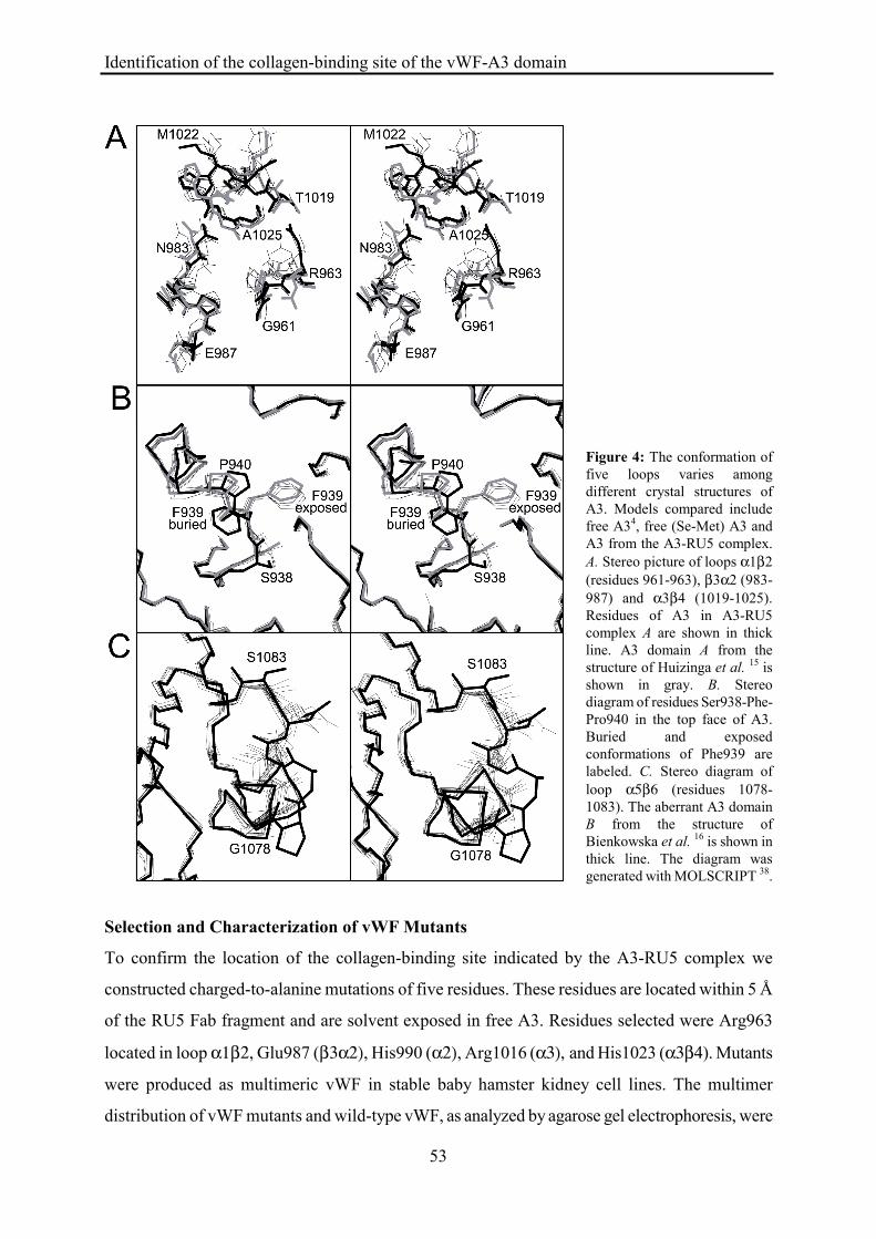
The three conformational diverse loops located in the RU5-epitope are α1β2, β3α2 and
α3β4 (figure 4A). Loop α1β2 has a unique conformation in the A3-RU5 complex, indicating that
this conformation is induced by RU5. Loops β3α2 and α3β4 have considerable conformational
freedom. Conformations observed in the A3-RU5 complex are, however, not systematically
different from the conformations observed in free A3, indicating that the conformations observed
in the complex are not induced by RU5.
Distant from the RU5 epitope conformational variation is observed for loops β1α1 and
α5β6. Loop β1α1 is located in the top face of the domain and contains part of the vestigial
MIDAS motif. Multiple conformations are observed for residues Ser938-Phe-Pro940 (figure 4B).
Phe939 is solvent exposed in A3-RU5 complex C and all models of free A3. In complexes A and
B Phe939 is buried in the hydrophobic core of A3. The buried conformation is likely caused by

Identification of the collagen-binding site of the vWF-A3 domain
51
crystal packing interactions involving A3 residues 940-942. These packing interactions would not
allow for the position of Pro940 observed in models of A3 that have an exposed conformation of
Phe939. Since both the exposed and buried conformations of Phe939 are observed in the three
A3-RU5 complexes the conformation of this loop is certainly not determined by binding of RU5.
Figure 3: Ribbon diagram of A3 and RU5 variable domains. Residues of A3 that are part of the epitope of RU5 are indicated by yellow spheres. Regions of RU5 CDRs that interact with A3 are color-coded in light blue. Regions of A3 that show different conformations among four crystal structures are shown in purple trace. Disulfide bond Cys923-Cys1109 is shown in green ball-and-stick. The α-helices and β-strands of A3 are depicted in blue and red, respectively. For clarity, α-helix 2 is not shown by a ribbon, but in ‘coil’ representation. The VH domain of RU5 is in brown, the VL domain is in green. The diagram was generated with MOLSCRIPT 38 and RASTER3D 39.
click on this link for rotating image
Identification of the collagen-binding site of the vWF-A3 domain
52
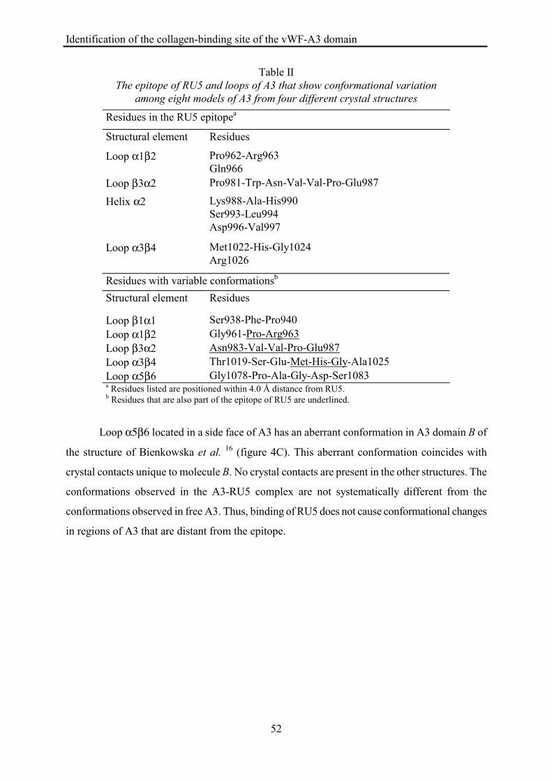
Table II The epitope of RU5 and loops of A3 that show conformational variation
among eight models of A3 from four different crystal structures
Residues in the RU5 epitopea
Structural element Residues
Loop α1β2 Pro962-Arg963 Gln966
Loop β3α2 Pro981-Trp-Asn-Val-Val-Pro-Glu987
Helix α2 Lys988-Ala-His990 Ser993-Leu994 Asp996-Val997
Loop α3β4 Met1022-His-Gly1024 Arg1026
Residues with variable conformationsb Structural element Residues
Loop β1α1 Ser938-Phe-Pro940 Loop α1β2 Gly961-Pro-Arg963 Loop β3α2 Asn983-Val-Val-Pro-Glu987 Loop α3β4 Thr1019-Ser-Glu-Met-His-Gly-Ala1025 Loop α5β6 Gly1078-Pro-Ala-Gly-Asp-Ser1083 a Residues listed are positioned within 4.0 Å distance from RU5. b Residues that are also part of the epitope of RU5 are underlined.
Loop α5β6 located in a side face of A3 has an aberrant conformation in A3 domain B of
the structure of Bienkowska et al. 16 (figure 4C). This aberrant conformation coincides with
crystal contacts unique to molecule B. No crystal contacts are present in the other structures. The
conformations observed in the A3-RU5 complex are not systematically different from the
conformations observed in free A3. Thus, binding of RU5 does not cause conformational changes
in regions of A3 that are distant from the epitope.
Identification of the collagen-binding site of the vWF-A3 domain
53
Figure 4: The conformation of five loops varies among different crystal structures of A3. Models compared include free A34, free (Se-Met) A3 and A3 from the A3-RU5 complex. A. Stereo picture of loops α1β2 (residues 961-963), β3α2 (983-987) and α3β4 (1019-1025). Residues of A3 in A3-RU5 complex A are shown in thick line. A3 domain A from the structure of Huizinga et al. 15 is shown in gray. B. Stereo diagram of residues Ser938-Phe-Pro940 in the top face of A3. Buried and exposed conformations of Phe939 are labeled. C. Stereo diagram of loop α5β6 (residues 1078-1083). The aberrant A3 domain B from the structure of Bienkowska et al. 16 is shown in thick line. The diagram was generated with MOLSCRIPT 38.
Selection and Characterization of vWF Mutants
To confirm the location of the collagen-binding site indicated by the A3-RU5 complex we
constructed charged-to-alanine mutations of five residues. These residues are located within 5 Å
of the RU5 Fab fragment and are solvent exposed in free A3. Residues selected were Arg963
located in loop α1β2, Glu987 (β3α2), His990 (α2), Arg1016 (α3), and His1023 (α3β4). Mutants
were produced as multimeric vWF in stable baby hamster kidney cell lines. The multimer
distribution of vWF mutants and wild-type vWF, as analyzed by agarose gel electrophoresis, were

Identification of the collagen-binding site of the vWF-A3 domain
54
indistinguishable (data not shown). RU5 binding to purified point mutants was not significantly
different from RU5 binding to wild-type vWF (figure 5).
Figure 5: Binding of monoclonal antibody RU5 to vWF mutants. VWF immobilized in microtiter-plate wells via polyclonal αD’D3 was incubated with 5 µg/ml RU5. Bound RU5 was detected by horse radish peroxidase conjugated swine-anti-rabbit antibody as described under “Experimental Procedures”. Wild-type vWF and ∆A3-vWF, which lacks the A3 domain 6, were used as positive and negative controls, respectively. Binding of RU5 to vWF mutants was percentualized against wt-vWF. Each data point represents the mean ± SD of two measurements in duplicate.
Collagen Binding of vWF Mutants
The effect of point mutations on vWF binding to collagen types I and III was investigated by
ELISA (figure 6). Similar results were obtained for both types of collagen. Mutation H1023A
almost completely abolished collagen binding. The level of residual binding observed for this
mutant was similar to binding observed for ∆A3-vWF, a deletion mutant that lacks the entire A3
domain 6. Collagen binding of mutants R963A and R1016A was reduced by 25 - 35%, whereas
collagen binding of mutants E987A and H990A was normal.
Figure 6: Binding of vWF point mutants to collagen types I and III. Collagen type I (white bar) or III (black bar) was coated in microtiter-plate wells. Wells were incubated with vWF at a concentration of 2.5 µg/ml and bound vWF was detected as described under “Experimental Procedures”. ∆A3-vWF was used as a negative control. Bound vWF mutant was percentualized against wt-vWF. Each data point represents the mean ± SD of three independent experiments performed in duplicate.
Identification of the collagen-binding site of the vWF-A3 domain
55
Discussion
The current study was aimed at locating the collagen-binding site of the vWF-A3 domain. For this
purpose we solved the crystal structure of A3 in complex with a Fab fragment of RU5 that
inhibits collagen binding. The structure of the complex shows that RU5 binds to residues within
A3 sequences 962-966, 981-997 and 1022-1026. These residues are located in α-helix 2 and in
loops α1β2, β3α2 and α3β4 at the bottom of one of the side faces of the A3 domain (see figure
3). Comparison of structures of A3 shows that RU5 binding does not induce long range
conformational changes. This excludes a mechanism in which RU5 induced conformational
changes inhibit collagen binding. It seems likely, therefore, that RU5 inhibits collagen binding by
steric hindrance, which implies that the collagen-binding site is located at or close to the RU5
epitope.
To confirm the location of the collagen-binding site we constructed five charged-to-
alanine mutations of residues located in or close to the RU5 epitope. The multimer distribution of
these mutants was similar to that of wild-type vWF. Therefore, observed differences in collagen
binding are not caused by the known dependence of collagen binding on vWF multimer-size 7. All
five mutants bound normally to RU5, which shows that none of the mutated residues plays a
dominant role in the A3-RU5 interaction and, more importantly, that the conformation of A3 in
the neighbourhood of the epitope and the collagen-binding site is not disturbed.
Mutation H1023A abolished collagen binding almost completely, residual-binding being
similar to that observed for ∆A3-vWF, a deletion mutant that lacks the entire A3 domain.
Therefore, His1023 plays a central role in A3-mediated collagen binding. His1023 is located in
loop α3β4 and lies at the edge between the ‘front’ face of the domain, formed by helices α2 and
α3 and strand β3, and the bottom face, which is composed of several loops and contains the N-
and C-termini (see figure 7). A small reduction of collagen binding was observed for mutants
R963A and R1016A located in the bottom and front face of the domain, respectively.

Identification of the collagen-binding site of the vWF-A3 domain
56
Figure 7: Residues of vWF-A3 involved in collagen binding. Stereographic representation of the vWF-A3 domain showing amino-acid residues that have been mutated to alanine in ball-and-stick. Mutation of His1023 (magenta) abolishes collagen binding almost completely. Mutation of Arg963 and Arg1016 (green) reduces collagen binding by 25-35%. Mutation of Glu987 and His990 (gray) does not have an effect on collagen binding. To illustrate the range of conformations of His1023 observed in different crystal forms of A3, two conformations of this residue and the back-bone trace of flanking residues are shown. Residues that have been mutated in previous studies 8; 16; 19 are located in the top face of A3 and are indicated by black spheres. The diagram was generated with MOLSCRIPT 38 and RASTER3D 39.
Interestingly, His1023 and flanking residues display a large variety of conformations
among eight models of A3 (figures 4 and 7). In some A3 structures His1023 protrudes
prominently from the surface of the domain which may be a favorable position for interaction
with collagen. Multiple conformations are also observed for loop β3α2. Like His1023, loop
β3α2 is located at the edge between the front and bottom faces of A3. Since we did not mutate
residues in loop β3α2 its involvement in collagen binding remains to be established. The
observed flexibility of His1023 suggests that collagen binding may involve an induced-fit
mechanism in which significant conformational changes occur in loop α3β4 upon binding of A3
to collagen.
The amino-acid sequence of collagen that is recognized by vWF-A3 has not yet been
identified. We hypothesized previously that negatively charged residues in A3 could interact with
basic residues on collagen 15. Residues now implicated in collagen binding are positively charged.
Therefore, interaction of A3 with negatively charged residues on collagen appears more likely.
In contrast to binding sites of other collagen binding domains, like the α1, α2 I-domains
and the A-domain of S. aureus adhesin 12; 40, the collagen-binding region of A3 does not have a
groove or trench that could accommodate a collagen triple-helix. The front face of the domain,
harboring Arg1016, is rather flat. The bottom face, which contains Arg963 is less smooth, but no
groove is present. Docking of a collagen triple-helix on the A3 domain is not straightforward. In
particular, it is not obvious how His1023, Arg963 and Arg1016 could simultaneously contact a
Identification of the collagen-binding site of the vWF-A3 domain
57
triple-helix in an extended conformation. To define the collagen-binding site more precisely,
characterization of additional mutants will be necessary.
Previously, the collagen-binding site of A3 was proposed to be located at its top face 15; 19
similar to the homologous I-domains of integrins α1β1 and α2β1 12; 41-43. Point mutations
introduced in the top face of an A3 monomer 19 and in multimeric vWF 8 gave conflicting results.
Our results now show conclusively that the collagen-binding site is located close to the bottom
face and not in the top face of A3.
While our results rule out a role for the top face of the A3 domain in collagen binding, this
side of the molecule may still be engaged in other interactions, such as binding of the A1 domain.
Interaction between A1 and A3 has been suggested to play a role in activation of the A1 domain
for binding to platelet receptor glycoprotein Ib 44. Interesting in this respect are the buried and
solvent exposed conformation observed for residue Phe939, which is located close to the vestigial
MIDAS motif in the top face of the domain (figure 4B). Solvent exposure of Phe939 has been
proposed to stabilize the buried Asp934 of the vestigial MIDAS motif in the absence of a bound
metal ion 16. Our observation of a buried conformation shows that exposure of Phe939 is not
critical for structural stability. The two conformations of Phe939 may, however, be relevant for
the putative interaction between A1 and A3, since the shape and hydrophobicity of the upper
surface of A3 differs significantly between the solvent exposed and buried conformation.
In conclusion, the collagen-binding site of vWF-A3 is distinctly different from collagen-
binding sites of I-domains of integrins α1β1 and α2β1. VWF-A3 residues involved in collagen
binding are located close to the bottom face of the domain. His1023 is essential for collagen
binding, while Arg963 and Arg1016 play ancillary roles. Multiple conformations observed for
His1023 and adjacent residues suggest that binding of A3 to collagen involves an induced-fit
mechanism.
Acknowledgements
We thank the staff of the EMBL Outstation DESY (Hamburg, Germany) for assistance in X-ray
data collection and the European Union for support of the work at EMBL (Hamburg, Germany)
through the HCMP Access to Large Installations Project, contract number CHGE-CT-0040.
Identification of the collagen-binding site of the vWF-A3 domain
58
Reference List
1. Vischer UM, De Moerloose P. von Willebrand factor: from cell biology to the clinical management of von
Willebrand's disease. Crit Rev Oncol Hematol. 1999;30:93-109.
2. Ruggeri ZM, Ware J. von Willebrand factor. FASEB J. 1993;7:308-316.
3. Sadler JE. Biochemistry and genetics of von Willebrand factor. Annu Rev Biochem. 1998;67:395-424.
4. Ruggeri ZM. Structure and function of von Willebrand factor. Thromb Haemost. 1999;82:576-584.
5. Mohri H, Yoshioka A, Zimmerman TS, Ruggeri ZM. Isolation of the von Willebrand factor domain
interacting with platelet glycoprotein Ib, heparin, and collagen and characterization of its three distinct
functional sites. J Biol Chem. 1989;264:17361-17367.
6. Lankhof H, van Hoeij M, Schiphorst ME, et al. A3 domain is essential for interaction of von Willebrand
factor with collagen type III. Thromb Haemost. 1996;75:950-958.
7. Fischer BE, Kramer G, Mitterer A, et al. Effect of multimerization of human and recombinant von
Willebrand factor on platelet aggregation, binding to collagen and binding of coagulation factor VIII.
Thromb Res. 1996;84:55-66.
8. Van der Plas RM, Gomes L, Marquart JA, et al. Binding of von Willebrand Factor to Collagen type III:
Role of specific amino acids in the collagen binding domain of vWF and effects of neighbouring domains.
Thromb Haemost. 84, 1005-1011. 2000.
9. Cruz MA, Yuan H, Lee JR, Wise RJ, Handin RI. Interaction of the von Willebrand Factor (vWF) with
collagen. J Biol Chem. 1995;270:10822-10827.
10. Colombatti A, Bonaldo P. The superfamily of proteins with von Willebrand factor type A-like domains:
one theme common to components of extracellular matrix, hemostasis, cellular adhesion, and defense
mechanisms. Blood. 1991;77:2305-2315.
11. Perkins SJ, Smith KF, Williams SC, Haris PI, Chapman D, Sim RB. the secondary structure of the von
Willebrand Factor type A domain in factor B of human complement by Fourier transform infrared
spectroscopy. Its occurence in collagen types VI, VII, XII and XIV, the integrins and other proteins by
avaraged structure predictions. J Biol Chem. 1994;238:104-119.
12. Emsley J, Knight CG, Farndale RW, Barnes MJ, Liddington RC. Structural basis of collagen recognition
by integrin α2β1. Cell. 2000;100:47-56.
13. Lee JO, Rieu P, Arnaout MA, Liddington RC. Crystal structure of the A domain from the α subunit of
integrin CR3 (CD11/CD18). Cell. 1995;80:631-638.

Identification of the collagen-binding site of the vWF-A3 domain
59
14. Michishita M, Videm V, Arnaout MA. A novel divalent cation-binding site in the A domain of the beta 2
integrin CR3 (CD11b/CD18) is essential for ligand binding. Cell. 1993;72:857-867.
15. Huizinga EG, Van der Plas RM, Kroon J, Sixma JJ, Gros P. Crystal structure of the A3 domain of human
von Willebrand factor: implications for collagen binding. Structure. 1997;5:1147-1156.
16. Bienkowska J, Cruz MA, Atiemo A, Handin RI, Liddington R. The von Willebrand Factor A3 domain
does not contain a metal ion-dependent adhesion site motif. J Biol Chem. 1997;272:25162-25167.
17. Pietu G, Fressinaud E, Girma JP, Nieuwenhuis HK, Rothschild C, Meyer D. Binding of human von
Willebrand factor to collagen and to collagen- stimulated platelets. J Lab Clin Med. 1987;109:637-646.
18. Bockenstedt PL, McDonagh j, Handin RI. binding and covalent cross-linking of purified von Willebrand
factor to native monomeric collagen. J Clin Invest. 1986;78:551-556.
19. Cruz MA, Bienkowska J, Mato A., Liddington R, Handin RI. Identification of the collagen binding site in
the vWF-A3 domain by molecular structure and site specific mutagenesis [abstract]. Blood.
1997;Supplement I:23a.
20. Hale JE. Irreversible, oriented immobilisation of antibodies to Cobalt iminodiacetate resin for ue as
immunoaffinity media. Anal Biochem. 1995;231:46-49.
21. Dübel S, Breitling F, Fuchs P, et al. Isolation of IgG antibody Fv-DNA from various mouse and rat
hybridoma cell lines using the polymerase chain reaction with a simple set of primers. J Immunol
Methods. 1994;175:89-95.
22. Carrol WL, Mendel E, Levy S. Hybridoma fusion cel lines containing an aberrant kappa transcript. Mol
Immunol. 1988;25:991-995.
23. Otwinowski Z, Minor O. Processing of X-ray diffraction data collected in oscillation mode. Methods
Enzymol. 1996;276:307-326.
24. Collaborative Computational Project No 4. The CCP4 suite: programs for protein crystallography. Acta
Crystallogr D. 1994;50:760-763.
25. Berman HM, Westbrook J, Feng Z, et al. The protein databank. Nucleic Acids Res. 2000;28:235-242.
26. Van den Elsen JM, Herron JN, Hoogerhout P, et al. Bacterial antibody recognition for a PorA epitope of
Neisseria meningitidid: acrystal structure of a Fab fragment in complex with a fluorescein-confugated
peptide. Proteins. 1997;29:113-125.
27. Navazza J. AMoRe: an automated package for molecular replacement. Acta Crystallogr A. 1994;50:157-
163.

Identification of the collagen-binding site of the vWF-A3 domain
60
28. Brünger AT. Extention of molecular replacement: a new search strategy based on Patterson correlation
refinement. Acta Crystallogr A. 1990;46:46-57.
29. Brünger AT, Adams PD, Clore GM, et al. Crystallography and NMR system: A new software suite for
macromolecular structure determination. Acta Crystallogr D. 1998;54:905-921.
30. Bedzyk WD, Johnson LJ, Riordan GS, Voss Jr. EW. Comparison of variable region primary structures
within an anti-fluorescein idiotype family. J Biol Chem. 1989;265:18615-18620.
31. Herron JN, He X-M, Mason ML, Voss Jr. EW, Edmundson AB. Three dimentional structure of a
fluorescein-Fab complex crystallized in 2-methyl-2,4-pentanediol. Proteins. 1989;5:271-280.
32. Jones TA, Zou JY, Cowan SW, Kjeldgaard M. Improved methods for the building of protein models in
electron density maps and the location of error in these models. Acta Crystallogr A. 1991;47:110-119.
33. Pannu NS, Read RJ. Improved structure refinement through Maximim Likelihood. Acta Crystallogr A.
1996;52:659-668.
34. Graham F, van der Eb A. A new technique for the assay of infectivity of human adenovirus 5 DNA.
Virology. 1973;52:456.
35. Lawrie AS, Horser MJ, Savidge GF. Phast assessment of vWF:Ag multimeric distribution. Thromb
Haemost. 1990;59:369.
36. Stanfield RI, Fieser TM, Lerner RA, Wilson IA. Crystal structure of an antibody to a peptide and its
complex with peptide antigen at 2.8 Angstroms. Science. 1990;248:712-719.
37. Nicholls A, Sharp KA, Honig B. GRASP: Graphical representation and analysis of surface properties.
Biophys J. 1993;64:166-170.
38. Kraulis P. Molscript: a program to produce both detailed and schematic plots of protein structures. J Appl
Crystallogr. 1991;25:649-950.
39. Merritt EA, Bacon DJ. Raster3D: photorealistic molecular graphics. Methods Enzymol. 1997;277:505-
524.
40. Rich RL, Deivanayagam CCS, Owens RT, et al. Trench-shaped binding sites promote multiple classes of
interactions between collagen and the adherence receptors, α1β1 integrin and Staphylococcus aureus Cna
MSCRAMM. J Biol Chem. 1999;274:24906-24913.
41. Kamata T, Puzon W, Takada Y. Identification of putative ligand binding sites within I domain of integrin
alpha 2 beta 1 (VLA-2, CD49b/CD29). J Biol Chem. 1994;269:9659-9663.
42. Kamata T, Takada Y. Direct binding of collagen to the I-domain of integrin α2β1 (Vla-2, CD49b/CD29)

Identification of the collagen-binding site of the vWF-A3 domain
61
in a divalent cation-independent manner. J Biol Chem. 1994;269:26006-26010.
43. Smith C, Estavillo D, Emsley J, Bankston LA, Liddington RC, Cruz MA. Mapping the collagen-binding
site in the I domain of the glycoprotein Ia/IIa (integrin α2β1). J Biol Chem. 2000;275:4205-4209.
44. Obert B, Houllier A, Meyer D, Girma JP. Conformational changes in the A3 domain of von Willebrand
factor modulate the interaction of the A1 domain with platelet glycoprotein Ib. Blood. 1999;93:1959-
1968.