identification of safrole ; precursor...
TRANSCRIPT

Identification of Safrole ; a precursor chemical
Tassaporn Khlaeo-omBureau of Drug and Narcotic
Department of Medical Science, Nonthaburi

What’s Safrole?Safrole is a precursor chemical of
ring-substituted amphetamine-type substances
ex. MDMA or “Ecstasy”MDAMDE

Structure of Safrole
Formula : C10H10O2
Synonym : 3,4-methylenedioxyallylbenzene
5-(2-propenyl)-1,3-benzodioxole
Allyldioxybenzene methylene ether

Physical PropertiesChemical class : aromatic bicyclic phenol etherHabit : colorless to slightly yellow
liquid with an odor of sassafrasSolubility : H2O – insoluble
Propylene glycol- slightly soluble
EtOH, CHCl3, diethyl etherMol Wt. : 162.18 g/molMelting point : 11.2 CBoiling point : 234.5 C at 760 mm Hg
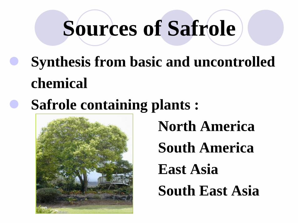
Synthesis from basic and uncontrolledchemical Safrole containing plants :
North AmericaSouth AmericaEast AsiaSouth East Asia
Sources of Safrole

Natural sources - different speciesLauraceae family
( Sassafras albidum, Cinnamomum camphora, Ocotea cymbarum and Ocotea pretiosa)
Piperaceae family( Piper hispidinervium)
Sources of Safrole

Cinnamomum camphora
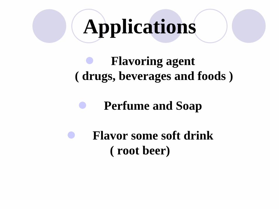
ApplicationsFlavoring agent
( drugs, beverages and foods )
Perfume and Soap
Flavor some soft drink ( root beer)

Law Enforcement
Schedule IV under Narcotic Act, B.E. 2522
(ยาเสพตดิใหโทษประเภท 4 (ลําดบัที่ 15) ประกาศกระทรวงสาธารณสุขฉบับที่ 35 (พ.ศ.2539) พระราชบัญญัติยาเสพตดิ
ใหโทษ พ.ศ. 2522)

Samplesตัวอยางจากสํานักงานคณะกรรมการอาหารและยา
ลกัษณะเปนของเหลวใส ไมมีสี มีตะกอน
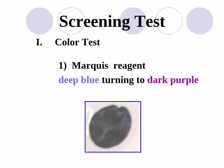
Screening TestI. Color Test
1) Marquis reagentdeep blue turning to dark purple

I. Color Test
2) Gallic acidbrown turning to dark brownish red
Screening Test

Confirmation testII. UV spectrophotometry
wavelength (nm) : 288.20 and 236.00
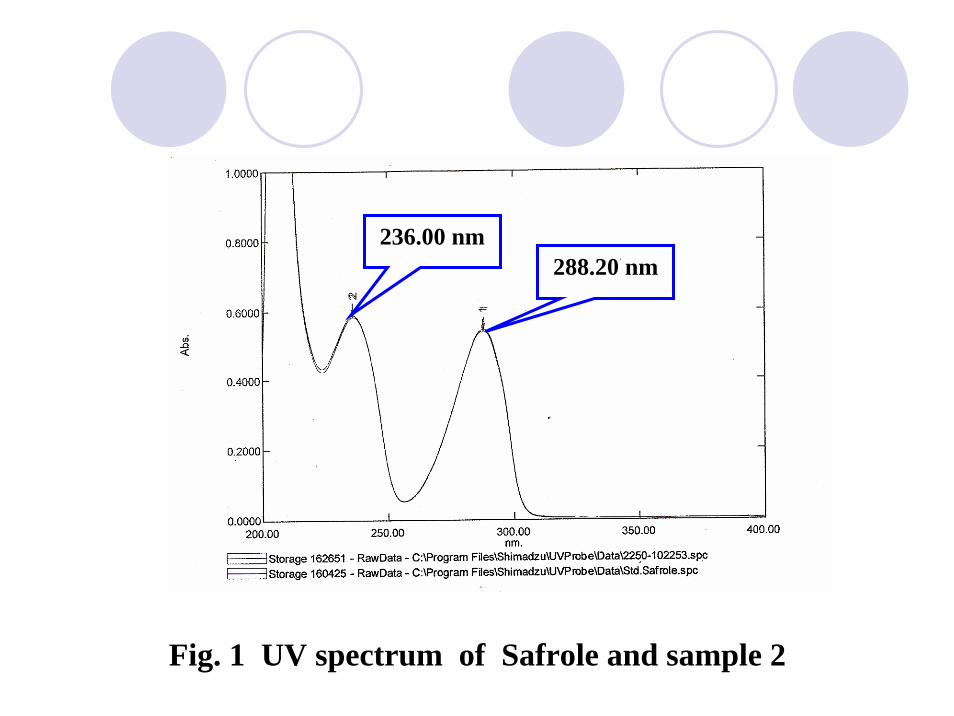
Fig. 1 UV spectrum of Safrole and sample 2
288.20 nm236.00 nm

Fig. 2 UV spectrum of Safrole and sample 2
236.00 nm 288.20 nm
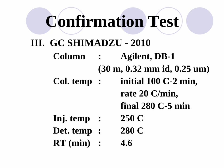
III. GC SHIMADZU - 2010Column : Agilent, DB-1
(30 m, 0.32 mm id, 0.25 um)Col. temp : initial 100 C-2 min,
rate 20 C/min,final 280 C-5 min
Inj. temp : 250 CDet. temp : 280 CRT (min) : 4.6
Confirmation Test

Fig.3 Chromatogram of Safrole standard ( RT 4.682 min )

Fig.4 Chromatogram of sample 1 ( RT 4.665 min )

Fig.4 Chromatogram of sample 2 ( RT 4.670 min )

Confirmation Test
IV. GC-MS
m/z at 162 131 104 77 63 51

Fig.5 Mass spectrum of Safrole standard

Fig.6 Mass spectrum of sample 1

Fig.7 Mass spectrum of sample 2

Fig.8 Mass spectrum of Safrole from library

ConclusionScreening Test :
Color test : Marquis and Gallic acid
Confirmation Test :
UV SpectrophotometryGCGC-MS

THANK YOU FOR
YOUR ATTENTION