identification of rickettsia-like organism (rlo) in the oyster ... · central rii cellece i e ccess...
TRANSCRIPT

CentralBringing Excellence in Open Access
Annals of Clinical Cytology and Pathology
Cite this article: Zhang Y, Fang J, Sun J, Wu X (2017) Identification of Rickettsia-Like Organism (RLO) in the Oyster Crassostrea rivularis Gould. Ann Clin Cytol Pathol 3(2): 1056.
*Corresponding authorXinzhong Wu, South China Sea Institute of Oceanography, Chinese Academy of Sciences, China, Email:
Submitted: 19 January 2016
Accepted: 17 April 2017
Published: 19 April 2017
ISSN: 2475-9430
Copyright© 2017 Wu et al.
OPEN ACCESS
Keywords•Oyster Crassostrea rivularis Gould (also known as
Crassostrea ariakensis)•Rickettsia-like organism (RLO)•16S rDNA•Phylogeny•In situ hybridization
Short Communication
Identification of Rickettsia-Like Organism (RLO) in the Oyster Crassostrea rivularis GouldYang Zhang1#, Jing Fang2#, Jingfeng Sun3, and Xinzhong Wu1,2*1South China Sea Institute of Oceanography, Chinese Academy of Sciences, China2Ocean College, Qinzhou University, China3Aquaculture College, Tianjin Agriculture University, China#Yang Zhang and Jing Fang contributed equally to this work
Abstract
The oyster Crassostrea rivularis Gould (also known as Crassostrea ariakensisis) an important bivalve species cultured in southeastern China. Since 1992, these oysters have suffered high mortality during winter and spring. An intracellular rickettsia-like organism (RLOs) was proposed as the causative agent. In this study, RLOs were purified from the gill and digestive gland of dying oyster (C. rivularis Gould), and their 16S rDNA was amplified from the purified products. To eliminate non-specific bacterial 16S rDNA contamination, the cloned products of bacterial 16S rDNA from gill RLO were screened by the probes of bacterial 16S rDNA amplified from the digestive gland RLO. Finally, the five strongest hybridized dots were picked out and sequenced. The RLO’s 16S rDNA sequence was reconfirmed and the pathogen was found only in epithelia cells by in situ hybridization (ISH) using specific probes. Sequence alignment and phylogenetic analysis indicated the RLO bacterium found in oyster C. rivularis Gould was most similar to Piscirickettsia salmonis, and might be classified into the family of gamma proteobacteria.
ABBREVIATIONSRLP: Rickettsia-Like Prokaryote; RLOs: Rickettsia-Like
Organisms; ISH: in situ Hybridization; PBS: Phosphate-Buffered Saline; min: minutes; h: hours; TEM: Transmission Electron Microscopy; s: seconds; PCR: Polymerase Chain Reaction; SSPE: Saline Sodium Phosphate Ethylenediaminetetraacetic Acid; AP: Alkaline Phosphatase; DIG: Digoxin
INTRODUCTIONThe oyster, Crassostrea rivularis Gould, also known as
Crassostrea ariakensis, is one of the most economically important cultured species in southeastern China, especially in the Guangxi, Guangdong and Fujian provinces. With the large expansion of culturing, mass mortalities have occurred persistently and caused great economic loss since 1992 in the Guangdong province of China. Recent studies suggested oyster culture suffered from severe mortality caused by the pathogen Rickettsia-like organism (RLO) [1,2].
Rickettsias are Gram-negative bacteria, generally described as obligate intracellular pathogens, that have been reported in various fishes, crustaceans [3-15], and mollusks [1,16-20]. Since the first report by Harshbarger et al. in Mercenaria mercenaria, 1977 [21], many mollusk species have been reported to be
infected with RLO, causing mortality and dramatic economic losses, such as the scallop, Pecten maximus [22]; the oyster Crassostrea virginia and the hard clam M. mercenaria [23]; the pearl oyster Pinctada fucata and Pinctada maximum [18,24]; the oyster C. rivularis [1]; the abalone Haliotis rufescens [25]; and the scallop Chlamys farreri (unpublished results). Although RLOs have been recognized as an important pathogen to aquatic organisms, studies have been carried out mostly on a morphological and pathological level, while few studies have identified them on the molecular level.
In this paper, the RLO was purified from the infected oyster C. ariakensis, this bacterium might be classified as a member of gamma-proteobacteria by using 16S rDNA analysis and it is distantly related to Piscirickettsia salmonis by phylogenetic analysis. In situ-hybridization suggested that the pathogen is localized in oyster epithelia cells.
MATERIALS AND METHODS
Sample and processing
The oyster, C. rivularis Gould, 2-3 years old, were collected from Hailing Bay in Yang Xi county, GuangDong province, China, in October 2004 when oyster deaths occurred in the field. The dying oysters were picked out, the bodies cross-sectioned

CentralBringing Excellence in Open Access
Wu et al. (2017)Email:
Ann Clin Cytol Pathol 3(2): 1056 (2017) 2/8
into 5mm thick piece just above the ventricle, and fixed in 4% paraformaldehyde for pathogenic observation. The residual body was stored at -80 until used for RLO purification.
RLO purification
Infected gills and digestive glands were used for RLO purification using renografin density gradient centrifugation [26,27] as described previously with some modifications. Briefly, infected tissues were homogenized in phosphate-buffered saline (PBS, pH7.4: Na2HPO4, 53.9mM; KH2PO4, 12.8mM; NaCl, 72.6mM) and centrifuged at 11000×g for 40 minutes (min) at 4 to remove the fat, then, the pellets were re-suspended in PBS, centrifuged at 700×g for 20 min at 4 and 1100×g for 10 min to remove cell debris. Subsequently, supernatant fluids were collected and re-centrifuged at 11000×g for 40 min. The pellets were then used for density gradient centrifugation after being re-homogenized. About 300mg of the re-homogenized pellets were laid on the top layer of discontinuous renografin gradients (15%, 20%, 25%, 30%, 35% v/v from top to bottom in turn, each layer with about 5.5ml volume and 1.5 cm in depth) and centrifuged at 90,000×g for 2 hours (h) at 4 (Sorvall S80). The particles concentrated at the density interfaces of 20%-25% and 25%-30% were collected, diluted with 5 volumes of PBS, and re-centrifuged at 15000×g for 40 min. Finally, the pellets were diluted to a suitable suspension and stained with Uranyl Acetate and observed using JEOL transmission electron microscopes (TEM).
RLO DNA extraction and 16S rDNA PCR amplification
About 80mg granules purified from gills were used in DNA extraction, as well as granules purified from digestive glands. DNA was extracted according to the methods of Kellner-Cousin [28]. Briefly, purified RLOs were resuspended in TE buffer (Tris-HCl 10 mM, EDTA 1mM, pH 8.0) and incubated for 20 min at 37 with lysozyme (1mg/ml). Then, sodium dodecyl sulfate and proteinase K were added to a final concentration of 0.5% and 100μg/ml respectively and the suspension was incubated at 55 for 3 h. Samples were extracted with phenol-chloroform (twice) and chloroform (once). Nucleic acids were precipitated with 100% ethanol, washed with 70% ethanol (twice), air-dried and dissolved in sterile distilled water. The universal bacterial PCR primers were derived from the highly conserved bacterial 16S region. The forward primer sequence is 5’-gcttaacacatgcaagtcg-3’ (Escherichia coli 16S rDNA positions 39-57), the reverse primer sequence is 5’- actaccgattccgacttca-3’ (E. coli 16S rDNA positions 1322-1344). PCR was performed with 25μl reaction mixtures containing 1μl template DNA, 2.5μl 10×PCR buffer within Mg2+ (TaKaRa, Dalian, China), 1μl of 10mM each dNTPs, 0.8U Taq polymerase (TaKaRa), and 0.5μl each of 25mM universal bacterial 16S rDNA primers. The mixture was denatured at 94 for 2 min before amplification. The amplification profile consisted of 30 seconds (s) at 94, 30 s at 56 and 90 s at 72 cycled 30 times, with an additional 5 min at 72 following the final cycle using Thermal PX2 PCR amplifier (Thermal Ltd.). The PCR products were determined using 1.5% agarose gel electrophoresis and ethidium bromide staining. Expected PCR products (size ~1300bp) were collected from the agarose gel and cloned into a PMD-18T vector (Takara Inc.).
Eliminating unwanted bacterial contamination
To eliminate unwanted bacterial 16S contamination, the 16S fragment amplified from the oyster gill was screened using the probes from the fragments of digestive glands. Sixty-six of 16S fragments from the gill RLO were screened by probes from the digestive glands. The probes were labeled with biotin according to the instructions provided in DIG HIGH prime DNA labeling and Detection starter kit (Roche). The result was recorded by X-ray film (Koda).
Molecular phylogenetic analysis
The nucleotide sequences of the RLO 16S rDNA DQ123914.1 and DQ118733.1 have been blasted within the RDP_SeqDescByOTU_tax_outline.txt. A total of 287 sequences from separate infected oysters with identity >=90% were selected. Then those sequences were further selected by OTU number, only those with OTU number >= 3 and with the best score were selected. The phylogenetic tree was made by MEGA 3.1 [29], with the Neighbor-Joining (NJ) Method.
In situ hybridization
Infected tissues were dissected and fixed with 4% paraformaldehyde in PBS (pH 7.4), dehydrated in an ascending ethanol series (50%, 70%, 80%, 90%, 95%, 100% v/v) for two times, followed by three washes in xylene, embedded in paraffin, sectioned at 5μm, mounted on APES-coated slides, and baked at 55 for 4 h. Then the section was deparaffinized in xylene and re-hydrated in a reverse ethanol series. The rehydrated slides used for in situ hybridization were digested in 30 μg ml-1 Proteinase K solution (pH 8.0) under 37 for about 20 min before hybridization. Slides were then neutralized in 2× saline sodium phosphate ethylenediaminetetraacetic acid (2×SSPE, pH 7.4) buffer for 10 min, and treated with prehybridization solution (0.5mg ml-1 salmon sperm DNA, 5×Denhardt’s reagent, and 2×SSPE) in a moist chamber at 42 for 30 min. After the prehybridization, 100ng of the positive probes and the negative control (NC) probes were separately added onto two different slides and hybridized at 42 for about 16 h in the moist chamber. After hybridization, unbound probes were washed off with 2×SSPE, 1×SSPE, and 0.5×SSPE at 42. Finally, the slides were added into AP-conjugated (Alkaline phosphatase) anti-digoxin (DIG) antibody and detected by NBT/BCIP indication reagent.
The specificity of assumed probe sequences chosen from highly variable regions of the RLO 16S rDNA sequence (GenBank accession number DQ123914) were confirmed by retrieving the sequence within the databases DDBJ-EMBL-GenBank using the BLASTn service. The probe was monolabeled with DIG and the sequence is DIG-5’-aggtagtctgtgaataatgggctactg-3’ at the position from 402-427 in DQ123914. A NC probe was also implemented to monitor the experimental conditions. The NC probe sequence was DIG-5’-gggatgtaggttaataccttgcatctt-3’ with a 12 nucleotides mismatch to the RLO sequence, but less than 12 nucleotides mismatch to other bacterial 16S rDNA sequences by NCBI BLAST database.
Theory/calculation
In this study, it is reported that the RLO bacterium found

CentralBringing Excellence in Open Access
Wu et al. (2017)Email:
Ann Clin Cytol Pathol 3(2): 1056 (2017) 3/8
in oyster Crassostrea rivularis Gould was most similar to Piscirickettsia salmonis and could be classified into a new family of gamma proteobacteria but it necessary to develop more studies. It can provide a theoretical basis for analysis of the death of the oyster C. rivularis Gould and preventing or controlling the RLO.
RESULTS
RLO purification
The purified RLOs mainly existed at band 20%-25% and 25%-30%, which were coincident with the report by Li and Wu (Li and Wu, 2004). Under TEM observation, the purified products displayed particles containing not only the RLOs, but also some cell debris and contaminated bacteria (Figure 1).
Eliminating bacterial contamination and molecular phylogenetic analysis
In density gradient centrifugation, only granules with the same density should be concentrated in the same layer, while particles with differing densities should be eliminated. Among the 66 dots in cross hybrid, the 5 most strongly reactive dots were picked out and sequenced (Figure 2). After being retrieved from the GenBank database, the sequences were found to belong to three known bacteria (Vibrio ordalii, Pseudomonas putida, Serratia marcescens) and one unknown bacterium. By comparing the morphological characters of the three known bacteria described in Bergey’s manual of systematic bacteriology (second edition, 2004) with that of the RLO found in oyster, the morphological character could not fit well. So the unknown sequence was assumed to be the RLO sequence. 48 sequences (Showed in Table 1) were finally selected and used in alignment with two RLO sequences using the clustalX 1.83 software. By sequence alignment analysis, the sequences which are similar to the RLO 16S sequence all belong to the Gamma proteobacteria. Thus it can be inferred that the RLO is a type of gamma proteobactium. By phylogenetic analysis, the sequence was most similar to the 16S sequence of Piscirickettisia salmonis (Figure 3), but the similarity was not high enough to classify these two bacteria into one family. In this study, 1304 sequences of 16S rDNA of RLO were obtained as follows:
1 gcttaacaca tgcaagtcga gcggtaacag gaagagcttg ctctttgctg acgagcggcg
61 gacgggtgag taacgcgtag gaatctgact gtaagagggg gatagcccgg agaaatccgg
121 attaataccg cataacacct aagggtaaaa agaggcactt gtgctactgc ttacagagga
181 gcctgcgttg gattagctag ttggtggggt aaaggcttac caaggcgacg atccatagct
241 gctctgagag gatgatcagc cacactggga ctgagacacg gcccagactc ctacgggagg
301 cagcagtggg gaatattgca caatggggga aaccctgatg cagccatgcc gcgtgtgtga
361 agaaggcttt cgggttgtaa agcactttca gtggtgagga aaggtagtct gtgaataatg
421 ggctactgtg acgttagcca cagaagaagg accggcaaac tccgtgccag
cagccgcggt
481 aatacggagg gtccgagcgt taatcggaat tactgggcgt aaagggtgcg taggcggata
541 tgtaagtggg tagtgaaaga cctgggctca acctgggagg tgctatccaa actgcataac
601 tagagtacag aagaggagtg tggaatttcc tgtgtagcgg tgaaatgcgt agatatagga
661 aggaacaccg gtggcgaagg cggcactctg gtctgatact gacgctgagg tacgaaagcg
721 tggggagcaa acaggattag ataccctggt agtccacgct gtaaacgctg tctactagtc
781 gttgggaact taaaagtttt tagtggcgaa gcaaacgcgc taagtagacc gcctggggag
841 tacggccgca aggttaaaac tcaaatgaat tgacgggggc ccgcacaagc ggtggagcat
901 gtggtttaat tcgacgcaac gcgaagaacc ttacctggtt ttgacatcct cggaatggcg
961 aagagatttg ccagtgcctt cgggagccga gtgacaggtg ctgcatggct gtcgtcagct
1021 cgtgtcgtga gatgttgggt taagtcccgc aacgagcgca acccttatcc
Figure 1 Electron micrograph of RLOs (arrows) from the oyster gill epithelial cell, with 2% uranyl acetate.
Figure 2 Sixty-six of 16S rDNA amplified fragment inserted were detected by probes synthesized from digestive gland purified products. Five of the strongest hybridized dots (indicated by arrows) were picked out and sequenced.

CentralBringing Excellence in Open Access
Wu et al. (2017)Email:
Ann Clin Cytol Pathol 3(2): 1056 (2017) 4/8
Figure 3 Phylogenetic analysis shows that the most similar sequence to RLO 16S rDNA is the 16S sequence of Piscirickettsia Salmonis.
ttatttgcca
1081 gcatgtaaag atgggaactc taaggagact gccggtgaca agccggagga aggtggggac
1141 gacgtcaagt catcatggcc cttacgacca gggctacaca cgtgctacaa tggggcgtac
1201 aaagggaagc gaagcggtga cgtggagcca aacctatcaa agcgcctcgt agtccggatc
1261 gcagtctgca actcgactgc gtgaagtcgg aatcggtagt aatc
In situ hybridization
The specificity of the bacterium probe was identified by comparing the hybridization signal produced using the RLO specific probe and non-specific probe under the same condition. The bacteria were recognized clearly when hybridized with the RLO specific probes, while no signals presented when hybridized with negative control probes (Figure 4A,4B). Meanwhile, ISH

CentralBringing Excellence in Open Access
Wu et al. (2017)Email:
Ann Clin Cytol Pathol 3(2): 1056 (2017) 5/8
Table 1: Sequence name used in alignment and phylogenetic analysis.
AccessionId AlignI taxonomy DESC
AJ704694.1 DED1 Bacteria; ProteoBacteria; DeltaproteoBacteria; Desulfobacterales; Desulfobacteraceae; Desulfobacula
AJ704694.1 marine sediment clone HMMVBeg-47
AY177803.1 DED2 Bacteria; Proteobacteria; Deltaproteobacteria; Desulfobacterales; Desulfobacteraceae; Desulfobacula AY177803.1 Antarctic sediment clone SB4_98
AY465366.1 PAA1 Bacteria; Proteobacteria; Gammaproteobacteria; Pasteurellales; Pasteurellaceae; Actinobacillus AY465366.1 Actinobacillus rossii str. JF2073
AY465368.1 PAA2 Bacteria; Proteobacteria; Gammaproteobacteria; Pasteurellales; Pasteurellaceae; Actinobacillus AY465368.1 Actinobacillus rossii str. P.. 12
AF139582.1 PAP1 Bacteria; Proteobacteria; Gammaproteobacteria; Pasteurellales; Pasteurellaceae; Pasteurella AF139582.1 Pasteurella aerogenes str. JF2039
AY465358.1 PAP2 Bacteria; Proteobacteria; Gammaproteobacteria; Pasteurellales; Pasteurellaceae; Pasteurella
AY465358.1 Pasteurella aerogenes str. 4-97; JF2420
EU341176.1 MOA1 Bacteria; Proteobacteria; Gammaproteobacteria; Pseudomonadales; Moraxellaceae; Acinetobacter
EU341176.1 Evaluation Rapid Technologies Estimate Microbial Burden and Commercial Airline Cabin Air commercial aircraft cabin air clone AV_4R-S-C13
DQ834360.1 MOA2 Bacteria; Proteobacteria; Gammaproteobacteria; Pseudomonadales; Moraxellaceae; Acinetobacter DQ834360.1 Acinetobacter sp. str. BYC2
AY486375.1 PSP1 Bacteria; Proteobacteria; Gammaproteobacteria; Pseudomonadales; Pseudomonadaceae; Pseudomonas AY486375.1 Pseudomonas sp. str. AU2390
AY486377.1 PSP2 Bacteria; Proteobacteria; Gammaproteobacteria; Pseudomonadales; Pseudomonadaceae; Pseudomonas AY486377.1 Pseudomonas sp. str. AU4899
AY498633.1 PIP1 Bacteria;Proteobacteria;Gammaproteobacteria;Thiotrichales;Piscirickettsiaceae;Piscirickettsia AY498633.1 Piscirickettsia salmonis IRE-91A
AY498636.1 PIP2 Bacteria; Proteobacteria; Gammaproteobacteria; Thiotrichales; Piscirickettsiaceae; Piscirickettsia AY498636.1 Piscirickettsia salmonis SCO-95A
EU250940.1 XAP1 Bacteria; Proteobacteria; Gammaproteobacteria; Xanthomonadales; Xanthomonadaceae; Pseudoxanthomonas
EU250940.1 Pseudoxanthomonas sp. str. NFC7-F12
EU177791.1 XAP2 Bacteria; Proteobacteria; Gammaproteobacteria; Xanthomonadales; Xanthomonadaceae; Pseudoxanthomonas
EU177791.1 Pseudoxanthomonas sp. str. Ca7-1J03
AB218877.1 XAS1 Bacteria; Proteobacteria; Gammaproteobacteria; Xanthomonadales; Xanthomonadaceae; Schineria
AB218877.1 Koukoulia aurantiaca str. IAM 15137
EF608545.1 XAS2 Bacteria; Proteobacteria; Gammaproteobacteria; Xanthomonadales; Xanthomonadaceae; Schineria
EF608545.1 predatory Poecilus chalcites their response lab rearing and antibiotic treatment digestive tract ground beetle clone PCD-40
DQ337031.1 IDI1 Bacteria; Proteobacteria; Gammaproteobacteria; Alteromonadales; Idiomarinaceae; Idiomarina
DQ337031.1 subsurface water clone EV818EB5CPSAJJ20
DQ235576.1 IDI2 Bacteria; Proteobacteria; Gammaproteobacteria; Alteromonadales; Idiomarinaceae; Idiomarina
DQ235576.1 biofilm population water pipeline biofilms steel pipelines Gulf Mexico clone 100
DQ899878.1 IDP1 Bacteria; Proteobacteria; Gammaproteobacteria; Alteromonadales; Idiomarinaceae; Pseudidiomarina
DQ899878.1 structure receiving long-term augmentations chromium contaminated wastes landfill sediments Gorwa industrial estate Cr(VI) contamination clone G1DMC-174
DQ234155.2 IDP2 Bacteria; Proteobacteria; Gammaproteobacteria; Alteromonadales; Idiomarinaceae; Pseudidiomarina
DQ234155.2 determined library mangrove clone DS071
DQ899898.1 IDU1 Bacteria; Proteobacteria; Gammaproteobacteria; Alteromonadales; Idiomarinaceae; unclassified_Idiomarinaceae
DQ899898.1 structure receiving long-term augmentations chromium contaminated wastes landfill sediments Gorwa industrial estate Cr(VI) contamination clone G2DMC-116
AY345388.1 IDU2 Bacteria; Proteobacteria; Gammaproteobacteria; Alteromonadales; Idiomarinaceae; unclassified_Idiomarinaceae
AY345388.1 Loihi submarine volcano isolate str. JB11
AY532642.1 INU1 Bacteria; Proteobacteria; Gammaproteobacteria; Alteromonadales; Incertae; unclassified_Incertae AY532642.1 Bugula simplex symbiont
DQ351747.1 INU2 Bacteria; Proteobacteria; Gammaproteobacteria; Alteromonadales; Incertae;unclassified_Incertae
DQ351747.1 Microbial Adherent Sediment Particles Heavy Metal Contaminated North Sea Surface Sediments marine sediments clone Belgica2005/10-120-16
EU399549.1 INM1 Bacteria; Proteobacteria; Gammaproteobacteria; Alteromonadales; Incertae; Marinobacter; Unclassified EU399549.1 Marinobacter sp. str. BR-13

CentralBringing Excellence in Open Access
Wu et al. (2017)Email:
Ann Clin Cytol Pathol 3(2): 1056 (2017) 6/8
DQ015835.1 INM2 Bacteria; Proteobacteria; Gammaproteobacteria; Alteromonadales; Incertae; Marinobacter; Unclassified
DQ015835.1 Antarctic lake water clone ELB19-223
AY394860.1 MOM1 Bacteria; Proteobacteria; Gammaproteobacteria; Alteromonadales; Moritellaceae; Moritella AY394860.1 Moritella sp. str. 762 G
AY380781.1 MOM2 Bacteria; Proteobacteria; Gammaproteobacteria; Alteromonadales; Moritellaceae; Moritella
AY380781.1 Moritella viscosa str. 2002/09/1069-1
AB003190.1 SHS1 Bacteria; Proteobacteria; Gammaproteobacteria; Alteromonadales; Shewanellaceae; Shewanella AB003190.1 Shewanella sp. str. SC2A
AF132875.1 SHS2 Bacteria;Proteobacteria; Gammaproteobacteria; Alteromonadales; Shewanellaceae; Shewanella
AF132875.1 Shewanella frigidimarina str. ACAM 533
AY241547.1 CHR1 Bacteria; Proteobacteria; Gammaproteobacteria; Chromatiales; Chromatiaceae; Rheinheimera
AY241547.1 aggregates water column German Wadden Sea part North Sea isolate str. HP1 HP1
AJ441080.1 CHR2 Bacteria; Proteobacteria; Gammaproteobacteria; Chromatiales; Chromatiaceae; Rheinheimera AJ441080.1 Rheinheimera baltica str. OSBAC1
AY136145.1 ENU1 Bacteria; Proteobacteria; Gammaproteobacteria; Enterobacteriales; Enterobacteriaceae; unclassified_Enterobacteriaceae AY136145.1 Cacopsylla pyri symbiont
AY136162.1 ENU2 Bacteria; Proteobacteria; Gammaproteobacteria; Enterobacteriales; Enterobacteriaceae; unclassified_Enterobacteriaceae
AY136162.1 Uroleucon nigrotuberculatum symbiont
EU134750.1 LEL1 Bacteria; Proteobacteria; Gammaproteobacteria; Legionellales; Legionellaceae; Legionella
EU134750.1 evolutionary between rare and abundant members communty tallgrass soil undisturbed mixed grass prairie preserve clone FFCH14647
EU250248.1 LEL2 Bacteria; Proteobacteria; Gammaproteobacteria; Legionellales; Legionellaceae; Legionella EU250248.1 acid mine drainage clone GXDC-34
AY536230.1 LEU1 Bacteria; Proteobacteria; Gammaproteobacteria; Legionellales; Legionellaceae; unclassified_Legionellaceae AY536230.1 host gut clone LAgut--P18
EU134792.1 LEU2 Bacteria; Proteobacteria; Gammaproteobacteria; Legionellales; Legionellaceae; unclassified_Legionellaceae
EU134792.1 evolutionary between rare and abundant members communty tallgrass soil undisturbed mixed grass prairie preserve clone FFCH4066
EF202341.1 OCU1 Bacteria; Proteobacteria; Gammaproteobacteria; Oceanospirillales; Oceanospirillaceae; unclassified_Oceanospirillaceae
EF202341.1 Matching and function marine one cell time Boothbay Harbor 1m depth clone MS024-3A
EF516584.1 OCU2 Bacteria; Proteobacteria; Gammaproteobacteria; Oceanospirillales; Oceanospirillaceae; unclassified_Oceanospirillaceae EF516584.1 grassland soil clone FCPP727
AY922202.1 OCN1 Bacteria; Proteobacteria; Gammaproteobacteria; Oceanospirillales; Oceanospirillaceae; Nitrincola AY922202.1 whalefall clone 131636
AY567473.1 OCN2 Bacteria; Proteobacteria; Gammaproteobacteria; Oceanospirillales; Oceanospirillaceae; Nitrincola AY567473.1 Nitrumincola lacisaponis str. 4CA
AJ315984.1 SAS1 Bacteria; Proteobacteria; Gammaproteobacteria; Oceanospirillales; Saccharospirillaceae; Saccharospirillum AJ315984.1 Arhodomonas sp. str. EL-201
AJ315983.1 SAS2 Bacteria; Proteobacteria; Gammaproteobacteria; Oceanospirillales; Saccharospirillaceae; Saccharospirillum
AJ315983.1 Saccharospirillum impatiens str. EL-105 = DSM 12546
DQ123914.1 DQ12 Bacteria; Proteobacteria; Gammaproteobacteria; Oceanospirillales; unclassified_Oceanospirillales DQ123914.1 Oceanrickettsia ariakensis
DQ334644.1 OCO1 Bacteria; Proteobacteria; Gammaproteobacteria; Oceanospirillales; unclassified_Oceanospirillales
DQ334644.1 Impact metals on sediments heavy metal polluted marine sediment clone HB2-9-21
AY344367.1 OCO2 Bacteria; Proteobacteria; Gammaproteobacteria; Oceanospirillales; unclassified_Oceanospirillales
AY344367.1 Lake Kauhako 30 m clone K2-30-25
positive signals were often found in the epithelia cells of gill and mantle, and occasionally found in digestive gland cells, but were not observed in hemocytes, muscles or pericardium. The morphology of RLO inclusions were also identified by HE staining method (Figure 5A,5B).
DISCUSSIONThe Oyster, Crassostrea rivularis Gould (also known as
Crassostrea ariakensis), mainly distributed in estuary areas, is a major farmed mollusk species in the Hailing Bay area of Guang Dong province, China. Since 1992, farmed oysters have suffered
from high mortality during winter and spring of every year. An intracellular rickettsia-like prokaryotic parasite was tentatively identified to be the causative agent using histological and ultra structural characteristics. The morphology of individual RLOs consist mostly of a round shape, with occasional short and rod-shaped morphologies, ranging from approximately 0.58 to 1.20 μm in size and with a smooth trilaminar cell wall [1]. Some observations reported that RLOs could form basophilic inclusions [17,30] under H&E staining, while other studies reported that they could form eosinophilic inclusions [1,24], or even two types of inclusions can be observed in the same mollusk [23,31,32]. In
CentralBringing Excellence in Open Access
Wu et al. (2017)Email:
Ann Clin Cytol Pathol 3(2): 1056 (2017) 7/8
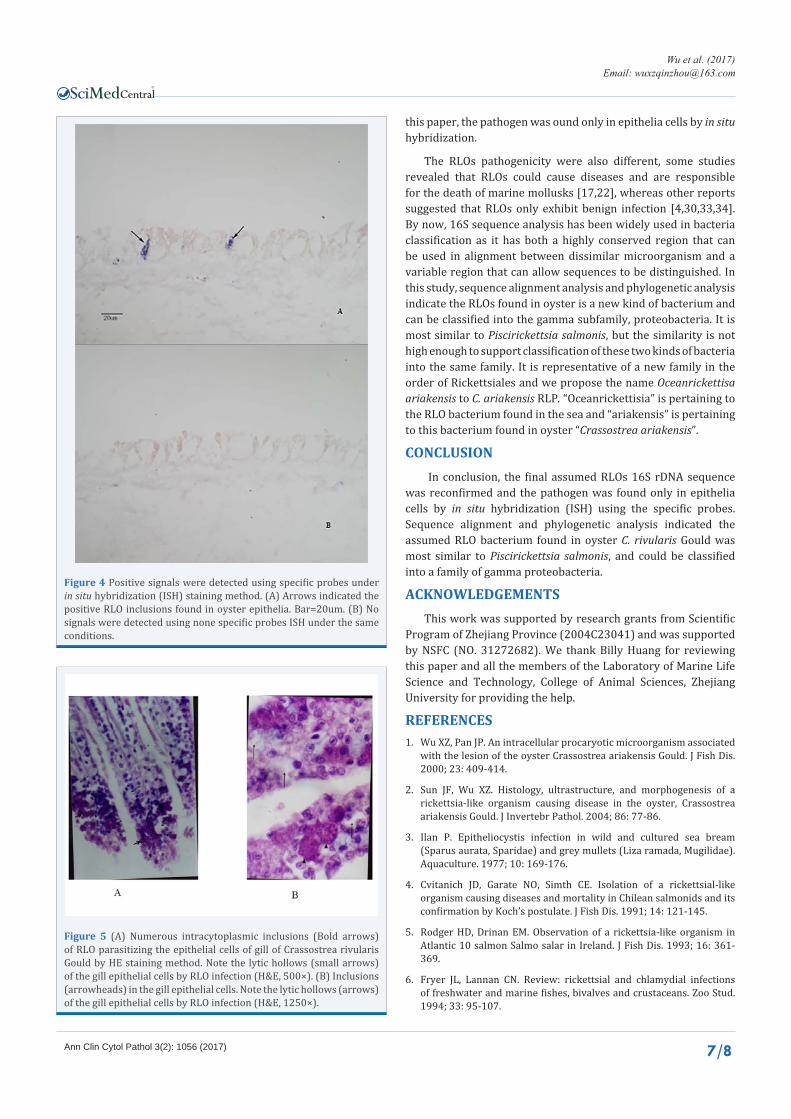
this paper, the pathogen was ound only in epithelia cells by in situ hybridization.
The RLOs pathogenicity were also different, some studies revealed that RLOs could cause diseases and are responsible for the death of marine mollusks [17,22], whereas other reports suggested that RLOs only exhibit benign infection [4,30,33,34]. By now, 16S sequence analysis has been widely used in bacteria classification as it has both a highly conserved region that can be used in alignment between dissimilar microorganism and a variable region that can allow sequences to be distinguished. In this study, sequence alignment analysis and phylogenetic analysis indicate the RLOs found in oyster is a new kind of bacterium and can be classified into the gamma subfamily, proteobacteria. It is most similar to Piscirickettsia salmonis, but the similarity is not high enough to support classification of these two kinds of bacteria into the same family. It is representative of a new family in the order of Rickettsiales and we propose the name Oceanrickettisa ariakensis to C. ariakensis RLP. “Oceanrickettisia” is pertaining to the RLO bacterium found in the sea and “ariakensis” is pertaining to this bacterium found in oyster “Crassostrea ariakensis”.
CONCLUSION In conclusion, the final assumed RLOs 16S rDNA sequence
was reconfirmed and the pathogen was found only in epithelia cells by in situ hybridization (ISH) using the specific probes. Sequence alignment and phylogenetic analysis indicated the assumed RLO bacterium found in oyster C. rivularis Gould was most similar to Piscirickettsia salmonis, and could be classified into a family of gamma proteobacteria.
ACKNOWLEDGEMENTSThis work was supported by research grants from Scientific
Program of Zhejiang Province (2004C23041) and was supported by NSFC (NO. 31272682). We thank Billy Huang for reviewing this paper and all the members of the Laboratory of Marine Life Science and Technology, College of Animal Sciences, Zhejiang University for providing the help.
REFERENCES1. Wu XZ, Pan JP. An intracellular procaryotic microorganism associated
with the lesion of the oyster Crassostrea ariakensis Gould. J Fish Dis. 2000; 23: 409-414.
2. Sun JF, Wu XZ. Histology, ultrastructure, and morphogenesis of a rickettsia-like organism causing disease in the oyster, Crassostrea ariakensis Gould. J Invertebr Pathol. 2004; 86: 77-86.
3. Ilan P. Epitheliocystis infection in wild and cultured sea bream (Sparus aurata, Sparidae) and grey mullets (Liza ramada, Mugilidae). Aquaculture. 1977; 10: 169-176.
4. Cvitanich JD, Garate NO, Simth CE. Isolation of a rickettsial-like organism causing diseases and mortality in Chilean salmonids and its confirmation by Koch’s postulate. J Fish Dis. 1991; 14: 121-145.
5. Rodger HD, Drinan EM. Observation of a rickettsia-like organism in Atlantic 10 salmon Salmo salar in Ireland. J Fish Dis. 1993; 16: 361-369.
6. Fryer JL, Lannan CN. Review: rickettsial and chlamydial infections of freshwater and marine fishes, bivalves and crustaceans. Zoo Stud. 1994; 33: 95-107.
Figure 4 Positive signals were detected using specific probes under in situ hybridization (ISH) staining method. (A) Arrows indicated the positive RLO inclusions found in oyster epithelia. Bar=20um. (B) No signals were detected using none specific probes ISH under the same conditions.
Figure 5 (A) Numerous intracytoplasmic inclusions (Bold arrows) of RLO parasitizing the epithelial cells of gill of Crassostrea rivularis Gould by HE staining method. Note the lytic hollows (small arrows) of the gill epithelial cells by RLO infection (H&E, 500×). (B) Inclusions (arrowheads) in the gill epithelial cells. Note the lytic hollows (arrows) of the gill epithelial cells by RLO infection (H&E, 1250×).

CentralBringing Excellence in Open Access
Wu et al. (2017)Email:
Ann Clin Cytol Pathol 3(2): 1056 (2017) 8/8
Zhang Y, Fang J, Sun J, Wu X (2017) Identification of Rickettsia-Like Organism (RLO) in the Oyster Crassostrea rivularis Gould. Ann Clin Cytol Pathol 3(2): 1056.
Cite this article
7. Fryer JL, Lannan CN. Rickettsila infecions of fish. Annu Rev Fish Dis. 1996; 6: 3-13.
8. Olsen AB, Melby HP, Speilberg L, Evensen O, Haastein T. Piscirickettsia salmonis infection in Atlantic salmon Salmo salar in Norway-epidemiological, pathological and microbiological findings. Dis Aquat Org. 1997; 31: 35-48.
9. Mauel MJ, Miller DL. Piscirickettsiosis and piscirickettsiosis-like infections in fish: a review. Vet Microbiol. 2002; 87: 279-289.
10. Federici BA, Hazard EI, Anthony DW. Rickettsia-like organism causing disease in a crangonid amphipod from Florida. Appl Microbiol. 1974; 28: 885-886.
11. Bonami JR, Pappalardo R. Rickettsial infection in marine crustacea. Experientia. 1980; 36: 180-181.
12. Anderson IG, Shariff M, Nash G, Nash M. Mortalities of juvenile shrimp, Penaeus monodon, associated with Penaeus monodon baculovirus, cytoplasmic reo-like virus, and ricketssial and bacterial infections, from Malaysian brackishwater ponds. Asian Fish Sci. 1987; 1: 47-64.
13. Lightner DV, Redman RM, Bonami JR. Morphological evidence for a single bacterial etiology in Texas necrotizing hepatopancreatitis in Penaeus vannamei (Crustacea: Decapoda). Dis Aquat Org. 1992; 13: 235-239.
14. Kikuchi Y, Sameshima S, Kitade O, Kojima J, Fukatsu T. Novel clade of Rickettsia spp. from leeches. Appl Environ Microbiol. 2002; 68: 999-1004.
15. Wang W, Gu Z. Rickettsia-like organism associated with tremor disease and mortality of the Chinese mitten crab Eriocheir sinensis. Dis Aquat Organ. 2002; 48: 149-153.
16. Comps M, Bonami JR, Vago C. Pathology des invertebre’s e Mise en evidence dune infection rickettsienne chez les Huitres. CR Acad Sc Paris. 1977; 285: 427-429.
17. Gulka G, Chang PW, Marti KA. Prokaryotic infection assocaiated with a mass mortality of the sea scallop, Placopecten magellanicus. J Fish Dis. 1983; 6: 355-364.
18. Wu X, Pan J. Studies on rickettsia-like organism disease of the tropical marine pearl oyster I: the fine structure and morphogenesis of pinctada maxima pathogen rickettsia-like organism. J Invertebr Pathol. 1999; 73: 162-172.
19. Finley CA, Friedman CS. Life history of an exotic sabellid polychaete, Terebrasabella heterouncinata: influence of temperature and fertilization strategy. J Shellfish Res. 2000; 19: 512-513.
20. Wu XZ. Advances in the research of marine-cultured animal diseases in China. In: Lee C-S, Ventura A, editors. Status of Aquaculture in China. 2003; 29-54. Hawaii, USA in Honolulu: The Oceanic Institute.
21. Harshbarger JC, Chang SC. Chlamydiae (with phages), mycoplasmas, and richettsiae in Chesapeake Bay bivalves. Science. 1977; 196: 666-668.
22. Le Gall G, Chagot D, Miahle E. Branchial rickettsiales-like infection associated with mass mortality of sea scallop, Pecten maximus. Dis Aquat Org. 1988; 4: 229-232.
23. Meyers TR. Endemic diseases of cultured shellfish of Long Island. New York: Adult and juvenile American oysters (Crassostrea virginia) and hard clams (Mercenaria mercenaria). Aquaculture. 1981; 22: 305-330.
24. Wu XZ, Pan JP. Studies on Rickettsia-like organism (RLO) diseases of tropical pearl oyster III. Morpholoyg of RLO parasitized in Pinctada fucata. Trop Mar Res. 1997; 5: 110-117.
25. Moore JD, Robbins TT, Friedman CS. The role of a rickettsia-like prokaryote in withering syndrome in California red abalone, Haliotis rufescens. J Shellfish Res. 2000; 19: 525-526.
26. Li DF, Wu XZ. Purification and biological features of rickettsia-like prokaryote from the scallop Argopecten irradians in China. Aquaculture. 2004; 234: 29-40.
27. Wu X, Sun J, Zhang W, Wen B. Purification and antigenic characteristics of a rickettsia-like organism from the oyster Crassostrea ariakensis. Dis Aquat Organ. 2005; 67: 149-154.
28. Kellner-Cousin K, Le Gall G, Despres B, Kahad M, Legoux P, Shire D, et al. Genomic DNA cloning of rikettsia-like organisms (RLO) of Saint-Jacques scallop Pecten maximus: evaluation of pro-karyvote diagnosis by hybridizatin with a non-isotopically labelled probe and polymerase chain reaction. Dis Aquat Org. 1993; 15: 145-152.
29. Wu XZ, Zhang Y. A pair of specific primer for the PCR detection of Rickettsia-like organism (RLO) in the oyster Crassostrea rivularis Gould. The People’s Republic of China, CN 100366754C. Feb 6.
30. Kumar S, Tamura K, Nei M. MEGA3: Integrated software for Molecular Evolutionary Genetics Analysis and sequence alignment. Briefings in Bioinformatics. 2004; 5: 150-163.
31. Elston RA. Occurrence of branchial rickettsiales-like infections in two bivalve molluscs, Tapes japonica and Patinopecten yessoensis, with comments on their significance. J Fish Dis. 1986; 9: 69-71.
32. Norton JH, Shepherd MA, Abdon-Nagutt MR, Lindsay S. Mortalities in the giant clam Hippopus Hippous associated with rickettsia-like organisms. J Invertebr Pathol. 1993; 62: 207-209.
33. Wen CM, Sen T, Kou GH, Chen SN. Rickettsia-like microorganisms in the cultured hard clam (Meretrix lusoria Roding) in Taiwan. J Fish Soc Taiwan. 1994; 20: 347-356.
34. Sparks AK. Synopsis of Invertebrate Pathology Exclusive of Insects. 1985; Elsevier. Amsterdam.