identification of putative ul54 (icp27) transcription ... · identification of putative ul54...
TRANSCRIPT

Identification of putative UL54 (ICP27) transcription regulatory sequences
binding to Oct-1, v-Myb, Pax-6 and Hairy in herpes simplex viruses
Ying-Ying Wang1,2,3,#, Yan-Ning Lyu4,#, Hong-Yi Xin5,#, Jun-Ting Cheng1,2,3, Xiao-
Qin Liu1,2,6, Xian-Wang Wang1,2,7, Xiao-Chun Pen1,2,8, Ying Xiang1,2,3, Victoria W.
Xin9, Cheng-Biao Lu10, Bo-Xu Ren1,8, Yan-Fang Liang11, Jia-Fu Ji12, Zhaowu Ma1,2,3,*,
Shu-Zhong Cui13,*, Hong-Wu Xin1,2,3,*
1. The Second School of Clinical Medicine, Faculty of Medicine, Yangtze University,
Jingzhou, Hubei 434023, China
2. Laboratory of Oncology, Center for Molecular Medicine, School of Basic Medicine,
Faculty of Medicine, Yangtze University, Jingzhou, Hubei 434023, China
3. Department of Biochemistry and Molecular Biology, School of Basic Medicine,
Faculty of Medicine, Yangtze University, Jingzhou, Hubei 434023, China
4. Institute for Infectious Diseases and Endemic Diseases Prevention and Control,
Beijing Center for Diseases Prevention and Control, Beijing, 100013, China 5. Star Array Pte. Ltd., 2 Tukang Innovation Grove, #09-08/09/10, JTC Medtech Hub, Singapore 618305 6. Department of Medical Imaging, School of Basic Medicine, Faculty of Medicine,
Yangtze University, Jingzhou, Hubei 434023, China
7. Department of Laboratory Medicine, School of Basic Medicine, Faculty of Medicine,
Yangtze University, 1 Nanhuan Road, Jingzhou, Hubei 434023, China
8. Department of Pathophysiology, School of Basic Medicine, Faculty of Medicine,
Yangtze University, Jingzhou, Hubei 434023, China
9. Montgomery Blair High School Magnet Program Class of 2020, Silver Spring, MD
20901-2451, USA
10. Laboratory of Neuronal Network and Brain Diseases Modulation, School of
Medicine, Yangtze University, Jingzhou, Hubei Province, China
11. Department of Radiology, Guangzhou Medical University Cancer Hospital,
Guangzhou,China
12. Gastrointestinal Cancer Center, Key Laboratory of Carcinogenesis and
Translational Research (Ministry of Education), Peking University Cancer Hospital and
Institute, Beijing 100142, China
13. Department of Theromotherapy, Guangzhou Medical University Cancer Hospital,
Guangzhou,China
#, Authors contributed equally
*, Corresponding authors
Corresponding authors Zhaowu Ma, MD Laboratory of Oncology
Center for Molecular Medicine
School of Basic Medicine
Faculty of Medicine
Yangtze University,
1 Nanhuan Road, Jingzhou.
Phone: +86 15972188216
Email: [email protected]
Shu-Zhong Cui, MD
Guangzhou Medical University Cancer Hospital
Guangzhou,China
Email: [email protected]
Hong-Wu Xin, PhD, MD
Laboratory of Oncology
Center for Molecular Medicine
School of Basic Medicine
Faculty of Medicine
Yangtze University
1 Nanhuan Road
Jingzhou, Hubei 434023, China Phone:+86 13311055391 Fax: 0716-8062633 Email: [email protected]
ABSTRACT
An oncolytic herpes simplex virus (oHSV) has proven amenable in oncolytic
virotherapy and was approved to treat melanoma. The immediate-early (IE) protein
ICP27 encoded by gene UL54 is essential for HSV infection. Post-transcriptional
modification of UL54 would increase tumor targeting of oHSVs. However, UL54 gene
transcription regulatory sequences and factors were not reported yet. Here we isolated
a new strain LXMW of type 1 HSV (HSV-1-LXMW) in China and found it’s closely
related to HSV-1 strains Patton and H129 in the US by the first and next generation
DNA sequencing viral DNA phylogenetic analysis. Using a weight matrix-based
program Match, we found the UL54 transcription regulatory sequences binding to the
transcription factors Oct-1, v-Myb and Pax-6 in HSV-1-LXMW, while they binding to
Oct-1 and Hairy in a HSV-2 strain. Further validation showed that HSV-1 and HSV-2
shared the common sequence binding to Oct-1, but had unique sequences to bind v-
Myb and Pax-6, or Hairy, respectively, by DNA sequence alignment of total 11 HSV
strains. Publications showed that the expression of transcription factors is consistent
with the tissue tropism of HSV-1 and HSV-2. In the current article a new HSV-1 strain
LXMW was isolated and its putative HSV UL54 transcription regulatory sequences
and factors were identified for the first time. Our findings highlight the new
understanding of the principles of transcriptional regulation in HSV biology and
oncolytic virotherapy.
Keywords:Oncolytic Herpes Simplex Virus (oHSV); Long Unique Region 54 (UL54);
Infected Cell Polypeptide 27 (ICP27); Transcription Regulatory Sequence;
Transcription Factor (TF); RNA polymerase II (RNAP II)
Introduction
Tumors are originated from transformed cells in tissues or organs, which contain
heterogeneous cancer cells, such as tumor stem cells etc [1-5], and tumor stromal cells,
such as immune cells etc., in tumor microenvironment [6]. Chromosome rearrangement
and deletion in cancer cells create cancer-specific fusion genes. Such fusion proteins
and mutated proteins can be targeted therapeutically [7]. Oncolytic viruses have been
used for tumor virotherapy [8-10]. An oncolytic herpes simplex virus (oHSV)
expressing GM-CSF was approved to treat melanoma in 2016. However, novel oHSVs
are necessary to increase their anti-tumor efficiency and safety.
Human herpesviruses have three subfamilies a, b and g [11].
The g herpesviruses include HSV type 1 (HSV-1, causes cold sores) and type 2 (HSV-
2, causes genital herpes) and varicella-zoster virus (causes chickenpox and shingles),
and can establish lifelong latent infections within the peripheral nervous systems of
their hosts [11]. HSVs express immediate–early (IE or α) genes, early (E or β) genes
and late (L or γ) genes, which are important during virus replication [12]. There are five
immediate-early proteins of HSV, such as infected cell polypeptide 0, 4 and 27 (ICP0,
ICP4 and ICP27) [13]. ICP0 is an ubiquitin E3 ligase, which has been reported to be
central for viral evasion of type I IFN responses in human fibroblasts [14]. ICP4 is
responsible for regulatory functions by initiating expression of a set of viral genes,
encoding enzymes necessary for DNA replication, such as the E gene UL23. γ34.5 is a
L gene encoding ICP34.5, important for viral entry, antagonizing antiviral immune
responses and neural virulence [12].
The multifunctional ICP27 is the only protein that has counterparts in all the
herpes virus families [13]. ICP27 is a 512- amino acid IE protein and is post-
translationally modified by phosphorylation and arginine methylation [15, 16]. ICP27
is phosphorylated at serine 63 in its kinase-inducible domain by serine/threonine
kinases, cAMP-dependent protein kinase A, calmodulin-dependent protein kinase I/II,
mitogen-and stress-activated protein kinase and cyclin-dependent kinase 3 (cdk-3). Its
phosphorylation enhances its transactivation and transcriptional activities.
ICP27 can re-localize the cellular protein Upstream Binding Factor (UBF) from the
nucleolus to viral replication compartments (VRCs), which to promote viral DNA
replication [17, 18]. HSV ICP27 functions in all stages of viral mRNA biogenesis from

transcription, RNA processing and export to translation machinery [15, 16]. ICP27
interacts with the C-terminal domain of RNA polymerase II (RNAP II) and recruits
RNAP II to viral replication transcription sites, stimulates viral gene transcription [19].
ICP27 can both enhance and repress expression of HSV early and late genes driven by
ICP0 and ICP4. ICP27 affects post-transcriptional processing by redistributing splicing
components, inhibiting host pre-mRNA splicing and facilitating viral mRNA
processing and trafficking [13]. In the early infection time ICP27 inhibits cellular
splicing resulting in accumulation of un-spliced and unprocessed 3' end pre-mRNAs
transcripts in the nucleus and suppression of host mRNA export [18, 20]. Later in the
infection, ICP27 can bind in vivo to seven HSV transcripts through its RGG box-like
arginine-glycine region and export the RNAs by the interaction of ICP27 leucine-rich
nuclear export signal with the cellular mRNA export receptor TAP/NXF and the mRNA
export adaptor protein Aly/ REF[20, 21]. ICP27 could be crosslinked to poly(A)+ RNA
for RNA export [20]. Nuclear pore glycoprotein p62 (Nup62) provides additional
binding sites of ICP27, promoting viral mRNA export and inhibiting cellular mRNA
export [18]. ICP27 can also regulate the metabolic process of mRNAs. Absence of
ICP27 resulted in decreased accumulation of viral replication proteins and reduction in
viral DNA synthesis [13]. ICP27 may also stimulate translation of viral transcripts,
repress host gene expression, block the cell cycle at the G1 phase and prevents
apoptosis [18].
Specific and dynamic gene expression strongly depends on transcription factors
(TFs) [10, 19]. TFs bind to core promoters, short sequences of ~100 base pair (bp)
surrounding the transcription start-sites at the 5' ends of genes [22]. Synergistic
interaction of several TFs bound to the transcription regulatory sequences or enhancers
and arranged as separate modules, also known as enhanceosomes, is a form of
cooperative regulation of transcription [19]. This dependency leads to an on/off mode
of transcriptional regulation [19].
The UL54 gene encodes the essential protein ICP27 of HSV. Until now only one
study showed that replacing the whole sequence upstream of the UL54 start codon but
downstream of the UL53 stop codon with the telemerase promoter increased the mutant
HSV targeting selectively to tumor cells [23]. There have been no other reports on
UL54 gene transcription regulatory sequences and factors in HSV. Here we isolated a
new HSV-1 strain LXMW, and identified novel putative HSV UL54 transcription
regulatory sequences binding to Oct-1, v-Myb, Pax-6 or Hairy for the first time. These
findings may have significant impact in HSV biology and oncolytic virotherapies.
MATERIALS AND METHODS
Cells
Experiments were performed with Vero cells which were cultured in Dulbecco’s
modified Eagle’s medium (DMEM) with antibiotics and 5% serum at 37 °C. For viral
infection, the vero cells were grown in DMEM with 2% serum. The HSV-1 clinical
strain was grown on Vero cell monolayers.
HSV-1 isolation and identification
Sample collection. A 45 years old male patient with acute oral herpes was admitted for
HSV-1 isolation. The herpes lesion was punctured with sterile syringe, the liquid was
dipped onto sterile cotton swab, the swab was placed in the virus collection tube
(Youkang Keye Biotechnology (Beijing) Co., Ltd., Cat. No. MT0301) and shipped at
4°C to the laboratory.
Virus culture. Vero cells were incubated in a 25 ml cell culture bottle (Corning-Costar
company) to 70%-80% confluence. Then the culture medium was removed and the cells
were washed twice with PBS. A 1.0 ml sample was inoculated in the culture and
incubated at 37°C for 2 hours. The cells were washed with PBS twice and cultured with
6 ml maintenance fluid (2% FBS). The cells were observed daily. After 3 days of culture,
most of the cells became round and floated. The culture medium was collected and
cryo-preserved at -80°C.
HSV-1 identification. A 200 uL of the culture medium supernatant was used to isolate
DNA with DNeasy Blood & Tissue Kit (QIAGEN, Cat No69506) according to the
instruction. PCR primers (forward primer was 5’-GCCAGCGAGACGCTGAT-3’, the
reverse primer was 5’-ACGCAGGTACTCGTGGTGA-3’) were synthesized by
Bioengineering (Shanghai) Company according to the reference[24]. The PCR reaction
was carried out in 25 uL reaction system using Platinum PCR SuperMix premix
(Invitrogen Company, USA, No. 11306-016). Reaction conditions are as following:
pre-denaturation at 95°C for 4 minutes followed by 35 cycles of denaturation at 95°C
for 30 seconds, annealing at 55°C for 30 seconds and extension 72°C for 30 seconds
and followed with a final extention step at 72°C for 7 Min. PCR product 173 bps was
delivered to Bioengineering (Shanghai) Limited for DNA sequencing.
HSV genomic DNA sequencing analysis
High-quality genomic DNA (500 ng) was submitted to Beijing Genomics Institute (BGI,
http://www.genomics.cn) for sequence analysis. The qualified genomic DNA sample
was randomly fragmented by Covaris technology and the fragments of 350 bp were
obtained after fragment selection. The end repair of DNA fragments was performed and
an "A" base was added at the 3'-end of each strand. Adapters were then ligated to both
ends of the end repaired/dA tailed DNA fragments. The ligated fragments were
amplified by ligation-mediated PCR (LM-PCR). The rolling circle amplification (RCA)
was performed to produce DNA Nanoballs (DNBs). The qualified DNBs were loaded
into the patterned nanoarrays and pair-end read were read through on the BGISEQ-500
platform and high-throughput sequencing are performed for each library to ensure that
each sample meet the average sequencing coverage requirement. Sequencing-derived
raw image files were processed by BGISEQ-500 basecalling Software for basecalling
with default parameters and the sequence data of each individual is generated as paired-
end reads, which is defined as "raw data" and stored in FASTQ format. Then
Bioinformatics Analysis Overview was used to analyze the sequencing data (raw data
from the BGISEQ machine). Burrows-Wheeler Aligner (BWA) software was used to
do the alignment. The sequencing depth and coverage for each individual were
calculated based on the alignments. In addition, the strict data analysis quality control
system (QC) in the whole pipeline was built to guarantee qualified sequencing data.
PCR verification of the DNA sequence upstream of the UL54 gene
To verify the DNA sequence upstream of the UL54 gene of HSV-1 by PCR, three pairs
of primers were designed using Snapgene offline software. Forward-1 primer
GCGATTGTGTTATATCGCCGT, and reverse primer-1 AGGCCGAGGTCAATT-
AGCAT, Forward primer-2 TGCGATTGTGTTATATCGCCG, and reverse primer -2
TCCAGGCCGAGGTCAATTAG, Forward primer-3 AGGCGCCTGTTTGATG-
TATGA and reverse primer-3 TGTCCGATTCCAGGTCGTC. The primers were

synthesized at Bioengineering (Wuhan) Company. Reaction conditions are as
following: pre-denaturation at 95°C for 4 minutes followed by 35 cycles of denaturation
at 95°C for 30 seconds, annealing at 55°C for 30 seconds and extension 72°C for 30
seconds and followed with a final extention step at 72°C for 7 Min. The PCR products
667 bps, 671bps and 674 bps for the above primer pairs 1, 2 and 3 were subjected to
gel electrophoresis on 1% agarose gel with and visualized by staining with ethidium
bromide. The agarose gel which contain the target DNA was cut down under ultraviolet
lamp and the PCR product was purified with DNA Gel Extraction Kit from Axygen®
AxyPrep™ according to the instruction. The extracted PCR product was sent to
GENECREATE at Wuhan for DNA sequencing (http://www.genecreate.cn).
Phylogenetic analysis of the sequences upstream of the UL54 gene
For phylogenetic analysis of the sequences upstream of the UL54 gene of 11 HSV
strains, the online program MEGA7 was used. The evolutionary history was inferred
by using the Maximum Likelihood method based on the General Time Reversible
model. The bootstrap consensus tree is taken to represent the evolutionary history of
the taxa analyzed. Branches corresponding to partitions reproduced in less than 50%
bootstrap replicates are collapsed. Initial tree(s) for the heuristic search were obtained
automatically by applying Neighbor-Join and BioNJ algorithms to a matrix of pairwise
distances estimated using the Maximum Composite Likelihood (MCL) approach, and
then selecting the topology with superior log likelihood value.
Prediction of UL54 transcription regulatory sequences and factors
The online program Match was used to predict UL54 transcription regulatory sequences
and factors. Match is a weight matrix-based program for predicting transcription factor
binding sites (TFBS) in DNA sequences. It uses a library of positional weight matrices
from TRANSFAC® Public 6.0. The stringent conditions were set as default by the
online program.
Alignment of the sequences upstream of UL54
The online program ApE (A plasmid Editor) was used to do alignment of the sequences
upstream of UL54 gene. The conserved sequences (>5 base pairs) were marked.

RESULTS
Isolation and DNA sequencing of a New HSV-1 strain LXMW
A new HSV strain from an oral herpes lesion of a patient in Beijing, named as HSV-1
strain LXMW (HSV-1-LXMW), was isolated. LXMW stands for the last name initials
of the authors YNL, HWX, ZWM and YYW representing all the authors. The initial
identification was carried out by PCR amplification and DNA sequencing. The
sequence was determined as follows: GCGTAAAGCCTTTGTGGTGCTGGTTCC-
CAGGGAACCCCCGACGTTCAGTTGCGCCTGACGAGGCCGCAGCTCACCAA
GGTCCTTAACGCGACCGGGGCCGATAGTGCCACGCCCACCACGTTCGAGC
TCGGGGTTAACGGCAAATTTTCCGTGTTCACCACGAGTACCTGCGT. The
sequence has only 1 nucleotide different from HSV-1-17 93603 CTTTGTGGTGC-
TGGTTCCCCAGGGAACCCCCGACGTTCAGTTGCGCCTGACGAGGCCGCAG
CTCACCAAGGTCCTTAACGCGACCGGGGCCGATAGTGCCACGCCCACCAC
GTTCGAGCTCGGGGTTAACGGCAAATTTTCCGTGTTCACCACGAGTACCTG
CGT 93767. However, there is no significant similarity between the sequences of our
HSV-1-LXMW and other HSV-2 strains. Our data support that HSV-1-LXMW is a
HSV-1 strain.
To further identify the new strain, HSV-1-LXMW DNA was then subjected to next
generation genomic DNA sequencing. The DNA sequence (from 113211 to 113752, or
from the stop codon of UL53 to the start codon of UL54) upstream of the UL54 gene
was determined as follows: ACGTCACATCCAGGCCGGCGGAAACCGGAACG-
GCATATGCAAATTGGAAACTGTCCTGTCTTGGGGCCCACCCACCCGACGC
GTCATATGCAAATGAAAATCGGTCCCCCGAGGCCACGTGTAGCCTGGATC
CCAACGACCCCGCCCATGGGTCCCAATTGGCCGTCCCGTTACCAAGACCA
ACCCAGCCAGCGTATCCACCCCCGCCCGGGTCCCCGCCGCGGAAGCGGAA
CGGTGTATGTGATATGCTAATTAAATACATGCCACGTACTTATGGTGTCTG
ATTGGTCCTTGTCTGTGCCGGAGGTGGGGCGGGGGCCCCGCCCGGGGGGC
GGAACGAGGAGGGGTTTGGGAGAGCCGGCCCCGGCACCACGGGTATAAG
GACATCCACCACCCGGCCGGTGGTGGTGTGCAGCCGTGTTCCAACCACGG
TCACGCTTCGGTGCCTCTCCCCGATTCGGGCCCGGTCGCTCGCTACCGGTG
CGCCACCACCAGAGGCCATATCCGACACCCCAGCCCCGACGGCAGCCGAC

CGCCCGGTC. Then the sequence was verified by PCR and first-generation
sequencing again (Figure 1A, B).
A
B
Figure 1: Verification of the DNA sequence upstream of UL54 gene of HSV-1-
LXMW by PCR and sequencing. (A) The three bands in lanes 2-4 were the 542 base pairs
of PCR products. (B) The sequencing result. The three additional nucleotides were in the red box.
Sample 1 Sample 2 Sample 3

Phylogenetic analysis of HSV-1-LXMW together with 10 other HSV strains
To understand the evolutionary relationship of our HSV strain with other HSV-1
and HSV-2 strains, a phylogenetic analysis based on the DNA sequences upstream of
the UL54 gene of HSV-1-LXMW together with 7 HSV-1 strains (17, F, H129, RH2,
SC16, Patton and KOS) and 3 HSV-2 strains (G, HG52 and SD90e) were performed
(Table 1). The DNA sequences upstream of UL54 gene, from the stop codon of UL53
gene to the start codon of UL54 gene, were chosen for the analysis. In Table 1, it
showed that the DNA sequence upstream of UL54 gene of HSV-1-LXMW strain reads
542 base pairs, which is 3 base pairs longer than those (539 bp) of all the other HSV1
strains. The DNA sequences upstream of UL54 gene of HSV-2 strains G, HG 52, SD
90 are all 546 base pairs, 4 base pairs longer than that of HSV-1-LXMW and 7 base
pairs longer than those of all other seven HSV-1 strains. These results support that our
HSV-1-LXMW is a new HSV-1 strain.
Both the phylogenetic tree data (Figure 2A) and neighbor network data (Figure
2B) showed the presence of four groups of clustering structures. The analysis involved
11 nucleotide sequences. The evolutionary history was inferred by using the Maximum
Likelihood method based on the General Time Reversible model. These trees with the
highest log likelihood is shown. The percentage of trees in which the associated taxa
clustered together is shown above the branches. Initial trees for the heuristic search
were obtained automatically by applying Neighbor-Join and BioNJ algorithms to a
matrix of pairwise distances estimated using the Maximum Composite Likelihood
(MCL) approach, and then selecting the topology with superior log likelihood value.
A:The tree is drawn to scale, with branch lengths measured in the number of
substitutions per site. B: The bootstrap consensus tree is taken to represent the
evolutionary history of the taxa analyzed. Evolutionary analyses were conducted in
MEGA7.
HSV-1 strains were clustered into 3 groups and HSV-2 strains were clustered into
1 group. Our new strain HSV-1-LXMW in Beijing, China is close to strains HSV-1-
Patton in New York, US and HSV-1-H129 in Princeton, US, and far from the strain
HSV-1-17 in Glasgow, UK. The data showed a mean distance of approximately 8%
among the strains tested collectively.
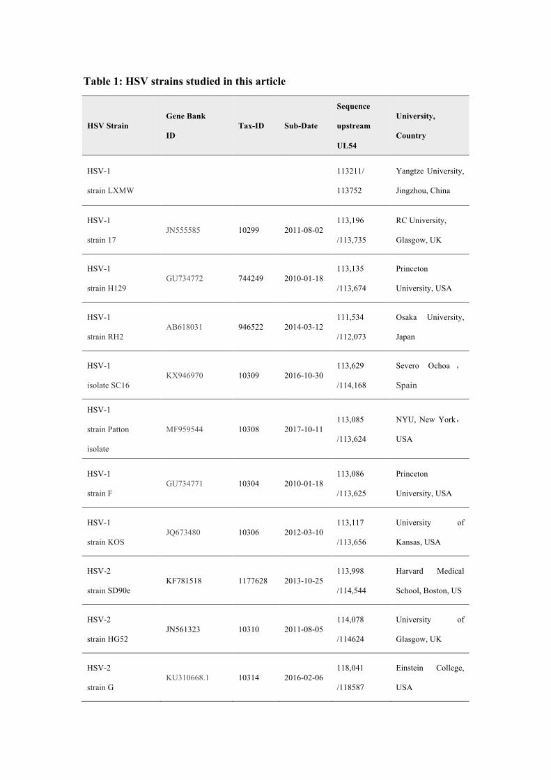
Table 1: HSV strains studied in this article
HSV Strain Gene Bank
ID Tax-ID Sub-Date
Sequence
upstream
UL54
University,
Country
HSV-1
strain LXMW
113211/
113752
Yangtze University,
Jingzhou, China
HSV-1
strain 17 JN555585 10299 2011-08-02
113,196
/113,735
RC University,
Glasgow, UK
HSV-1
strain H129 GU734772 744249 2010-01-18
113,135
/113,674
Princeton
University, USA
HSV-1
strain RH2 AB618031 946522 2014-03-12
111,534
/112,073
Osaka University,
Japan
HSV-1
isolate SC16 KX946970 10309 2016-10-30
113,629
/114,168
Severo Ochoa ,
Spain
HSV-1
strain Patton
isolate
MF959544 10308 2017-10-11 113,085
/113,624
NYU, New York,
USA
HSV-1
strain F GU734771 10304 2010-01-18
113,086
/113,625
Princeton
University, USA
HSV-1
strain KOS JQ673480 10306 2012-03-10
113,117
/113,656
University of
Kansas, USA
HSV-2
strain SD90e KF781518 1177628 2013-10-25
113,998
/114,544
Harvard Medical
School, Boston, US
HSV-2
strain HG52 JN561323 10310 2011-08-05
114,078
/114624
University of
Glasgow, UK
HSV-2
strain G KU310668.1 10314 2016-02-06
118,041
/118587
Einstein College,
USA

Figure 2. Phylogenetic analysis of HSV-1-LXMW together with 10 other HSV
strains. A.The tree is drawn to scale, with branch lengths measured in the number of substitutions
per site. B. The bootstrap consensus tree is taken to represent the evolutionary history of the taxa
analyzed. Evolutionary analyses were conducted in MEGA7.
Identification of the UL54 transcription regulatory sequences and factors
Understanding the UL54 transcription regulatory sequences and factors is important for
HSV biology and transcriptional tumor targeting of oncolytic HSVs. Using the weight
matrix-based program Match, we found seven major UL54 transcription regulatory
sequences of HSV-1-LXMW strain, which bind to transcription factors Oct-1, v-Myb
and Pax-6, and eight major UL54 transcription regulatory sequences of HSV-1-17,
which bind to transcription factors Oct-1, v-Myb and Pax-6 (Table 2, Figures 3). There
were five binding sites for Oct-1/Oct-x, one for v-Myb and one for Pax-6 in HSV-1-

LXMW, and there was an additional binding site for v-Myb in HSV-1-17. The
difference of binding sites for v-Myb would be related to strain-specific features.
The study found only four major UL54 transcription regulatory sequences binding
to transcription factors Oct-1 and Hairy in HSV-2 strain G. There were three sites for
Oct-1 and one for Hairy in HSV-2-G (Table 2, Figures 3). These data suggest that
HSV-1 and HSV-2 would share the common UL54 transcription regulatory sequence
binding to Oct-1, but would have unique sequences binding to v-Myb and Pax-6, or
Hairy, respectively.
Table 2: The UL54 transcription regulatory sequences and factors in HSVs
HSV
strain
Matrix
identifier
Position
strand
Core
match
Matrix
match Sequence
Factor
name
HSV-1
strain
LXMW
V$OCT1_Q6 33(+) 1.000 0.956 gcatatGCAAAttgg Oct-1
V$OCT1_Q6 84(+) 1.000 0.978 tcatatGCAAAtgaa Oct-1
V$OCT_C 86(-) 1.000 0.991 atatGCAAAtgaa OCT-X
V$OCT1_Q6 90(+) 0.893 0.891 gcaaatGAAAAtcgg Oct-1
V$VMYB_01 166(-) 1.000 0.962 tcCCGTTacc v-Myb
V$OCT1_Q6 242(+) 0.883 0.909 tgatatGCTAAttaa Oct-1
V$PAX6_01 428(+) 1.000 0.822 cacggTCACGcttcggtgcct Pax-6
HSV-1
strain 17
V$VMYB_01 26(+) 1.000 0.962 cgtAACGGca v-Myb
V$OCT1_Q6 33(+) 1.000 0.956 gcatatGCAAAttgg Oct-1
V$OCT1_Q6 84(+) 1.000 0.978 tcatatGCAAAtgaa Oct-1
V$OCT_C 86(+) 1.000 0.991 atatGCAAAtgaa OCT-X
V$OCT1_Q6 90(+) 0.893 0.891 gcaaatGAAAAtcgg Oct-1
V$VMYB_01 166(+) 1.000 0.962 tcCCGTTacc v-Myb
V$OCT1_Q6 239(+) 0.883 0.909 tgatatGCTAAttaa Oct-1
V$PAX6_01 425(+) 1.000 0.822 cacggTCACGcttcggtgcct Pax-6
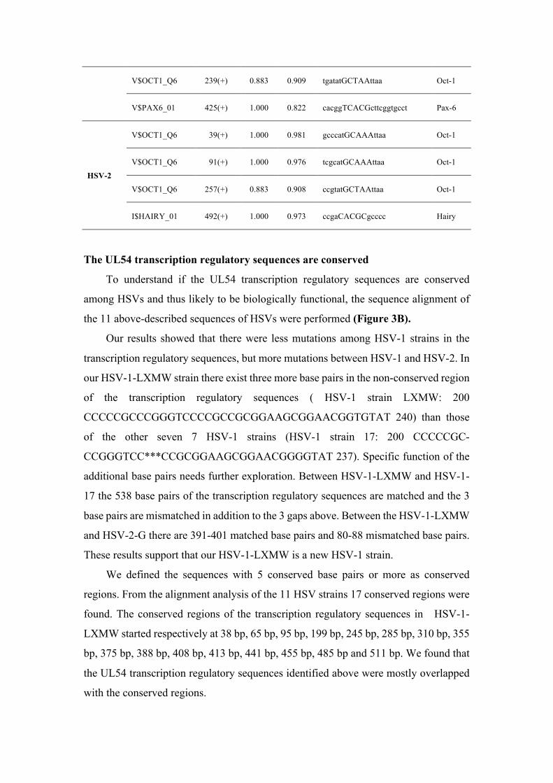
HSV-2
V$OCT1_Q6 39(+) 1.000 0.981 gcccatGCAAAttaa Oct-1
V$OCT1_Q6 91(+) 1.000 0.976 tcgcatGCAAAttaa Oct-1
V$OCT1_Q6 257(+) 0.883 0.908 ccgtatGCTAAttaa Oct-1
I$HAIRY_01 492(+) 1.000 0.973 ccgaCACGCgcccc Hairy
The UL54 transcription regulatory sequences are conserved
To understand if the UL54 transcription regulatory sequences are conserved
among HSVs and thus likely to be biologically functional, the sequence alignment of
the 11 above-described sequences of HSVs were performed (Figure 3B).
Our results showed that there were less mutations among HSV-1 strains in the
transcription regulatory sequences, but more mutations between HSV-1 and HSV-2. In
our HSV-1-LXMW strain there exist three more base pairs in the non-conserved region
of the transcription regulatory sequences ( HSV-1 strain LXMW: 200
CCCCCGCCCGGGTCCCCGCCGCGGAAGCGGAACGGTGTAT 240) than those
of the other seven 7 HSV-1 strains (HSV-1 strain 17: 200 CCCCCGC-
CCGGGTCC***CCGCGGAAGCGGAACGGGGTAT 237). Specific function of the
additional base pairs needs further exploration. Between HSV-1-LXMW and HSV-1-
17 the 538 base pairs of the transcription regulatory sequences are matched and the 3
base pairs are mismatched in addition to the 3 gaps above. Between the HSV-1-LXMW
and HSV-2-G there are 391-401 matched base pairs and 80-88 mismatched base pairs.
These results support that our HSV-1-LXMW is a new HSV-1 strain.
We defined the sequences with 5 conserved base pairs or more as conserved
regions. From the alignment analysis of the 11 HSV strains 17 conserved regions were
found. The conserved regions of the transcription regulatory sequences in HSV-1-
LXMW started respectively at 38 bp, 65 bp, 95 bp, 199 bp, 245 bp, 285 bp, 310 bp, 355
bp, 375 bp, 388 bp, 408 bp, 413 bp, 441 bp, 455 bp, 485 bp and 511 bp. We found that
the UL54 transcription regulatory sequences identified above were mostly overlapped
with the conserved regions.
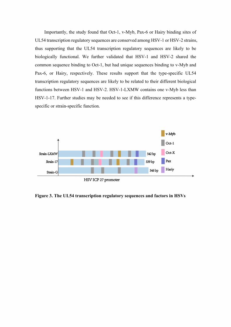
Importantly, the study found that Oct-1, v-Myb, Pax-6 or Hairy binding sites of
UL54 transcription regulatory sequences are conserved among HSV-1 or HSV-2 strains,
thus supporting that the UL54 transcription regulatory sequences are likely to be
biologically functional. We further validated that HSV-1 and HSV-2 shared the
common sequence binding to Oct-1, but had unique sequences binding to v-Myb and
Pax-6, or Hairy, respectively. These results support that the type-specific UL54
transcription regulatory sequences are likely to be related to their different biological
functions between HSV-1 and HSV-2. HSV-1-LXMW contains one v-Myb less than
HSV-1-17. Further studies may be needed to see if this difference represents a type-
specific or strain-specific function.
Figure 3. The UL54 transcription regulatory sequences and factors in HSVs

Figure 4 The UL54 transcription regulatory sequences are conserved. The conserved
regions are marked and numbered as 1-17. The three nucleotides addition CCG in HSV-1-LXMW
was marked In the black box. The conserved transcription regulatory sequences and factors are
shown in different shade colors.
The UL54 transcription regulatory sequences may determine type-specific tissue
tropism
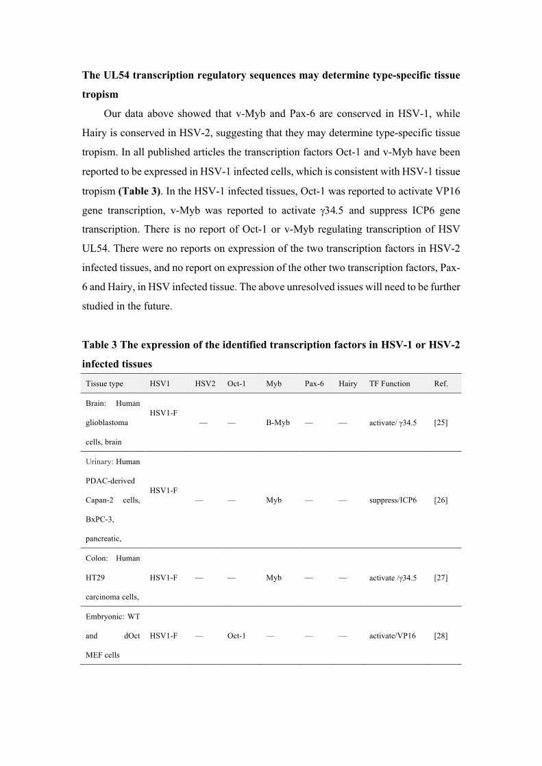
Our data above showed that v-Myb and Pax-6 are conserved in HSV-1, while
Hairy is conserved in HSV-2, suggesting that they may determine type-specific tissue
tropism. In all published articles the transcription factors Oct-1 and v-Myb have been
reported to be expressed in HSV-1 infected cells, which is consistent with HSV-1 tissue
tropism (Table 3). In the HSV-1 infected tissues, Oct-1 was reported to activate VP16
gene transcription, v-Myb was reported to activate g34.5 and suppress ICP6 gene
transcription. There is no report of Oct-1 or v-Myb regulating transcription of HSV
UL54. There were no reports on expression of the two transcription factors in HSV-2
infected tissues, and no report on expression of the other two transcription factors, Pax-
6 and Hairy, in HSV infected tissue. The above unresolved issues will need to be further
studied in the future.
Table 3 The expression of the identified transcription factors in HSV-1 or HSV-2
infected tissues
Tissue type HSV1 HSV2 Oct-1 Myb Pax-6 Hairy TF Function Ref.
Brain: Human
glioblastoma
cells, brain
HSV1-F
— — B-Myb — — activate/ g34.5 [25]
Urinary: Human
PDAC-derived
Capan-2 cells,
BxPC-3,
pancreatic,
HSV1-F
— — Myb — — suppress/ICP6 [26]
Colon: Human
HT29
carcinoma cells,
HSV1-F — — Myb — — activate /g34.5 [27]
Embryonic: WT
and dOct
MEF cells
HSV1-F — Oct-1 — — — activate/VP16 [28]

Genital: HeLa
cells HSV1-F — Oct-1 — — — not described [29]
Digestive: Hep2
cells
HSV-1
KOS — Oct-1 — — — activate /VP16 [30]
Genital: HeLa
cells
HSV-1
KOS — Oct-1 — — — activate /VP16 [31]
Urinary: COS-7
cells
HSV-1
KOS — Oct-1 — — — activate /VP16 [32]
Genital: HFF HSV-1
KOS — Oct-1 — — — activate /VP16 [33]
PDAC: pancreatic ductal adenocarcinoma; MEF: mouse embryonic fibroblas; HFF: human
foreskin fibroblast. COS-7: are fibroblast-like cell lines derived from monkey kidney tissue.
DISCUSSION
Transcriptional modification of UL54 would increase tumor targeting of oHSVs.
However, UL54 gene transcription regulatory sequences and factors were not reported
yet. Here A new strain HSV-1-LXMW was isolated in China and found to be closely
related to HSV-1 strains Patton and H129 in the US. We identified the HSV UL54
transcription regulatory sequences and factors for the first time. Further analysis found
that both HSV-1 and HSV-2 shared the common sequence binding to Oct-1, but had
unique sequences binding to v-Myb and Pax-6, or Hairy, respectively. The study
predicted that the HSV-1 or HSV-2 specific transcription regulatory sequences would
be associated with HSV-1 and HSV-2 tissue tropism. The findings may have significant
impact in HSV biology and oncolytic virotherapy.
Our novel HSV-1 strain and it genetic relationship with other HSV strains may
reflect the real HSV evolution. Clinical studies showed that the mutations in either the
thymidine kinase gene or DNA polymerase gene of HSVs have caused resistance to the
anti-HSV drug aciclovir. Most of the mutations occurred in the thymidine kinase gene
rather than the DNA polymerase gene [34]. The mutation rate in HSV-1 genome is
1.82×10⁻⁸ nucleotide substitution per site per year [35]. With such a mutation rate the
most recent HSV-1 strains could be originated from a common ancestor in ~710,000
years ago.

The functions and their relationship with HSV-1/2 tissue tropism of the identified
transcription factors need to be further studied. We summarized the HSV-1/2 tissue
tropism and the transcription factor expression in different tissues in Table 4. Oct
binding DNA sequence motif ATTTGCAT (octamer) or its reverse complement has
been identified as an evolutionarily conserved element in the promoter region of
immunoglobulin genes, and is an important transcriptional control element [29]. The
motif is located at 36 base pairs upstream of the functional TATA box. Two major
DNA-binding proteins that bind in a sequence-specific manner to the octamer DNA
sequence have been identified in mammalian species-a ubiquitously expressed protein
(Oct-1) and a lymphoid-specific protein (Oct-2) [29]. The ubiquitous expression of Oct-
1 is consistent with our finding that both HSV-1 and HSV-2 contain Oct-1 binding sites.
Table 4 The HSV- 1/2 tissue tropism and the transcription factor expression in different
tissues
System cell HSV1 HSV2 Oct-1 Myb Pax-6 Hairy
Blood system
CD34+ stem cell + — H M L N
721 B lymphoblasts + — H M L N
CD19+ B cell + — H M L H
Leukemia lymphoblastic + — L H M M
Bonemarrow + — H M L M
Pituitariy + — M H M H
Head
Prefrontal Cortex + — H M M H
Pineal + — M H H H
Tongue + — M H L M
Tonsil + — L M L M
Retina + — M H H H
Cerebellum + — L M H M
viscera
Heart + — M M L H
Lung + — M M L H
Liver + — M M L H
Kidney + — L M L H
Smooth Muscles + — M M L H
Adipocyte + — L M L H
Secretory
system
Adrenalgland, + — L M L H
Pancreaticlstet + — M M H H
Genital
system
Placenta + + M H L H
Fetalthyroid + + M M L H
Uterus + + L M L M
Testis + + L M L M
H: high-expression M: middle expression L: little expression N:no-expression
The result from: http://biogps.org. Grading was based on fold increases compared to median
fluorescence intensity on Affymetrix microarray chips at 0-2.5 (L), >2.5-<5 (M), >5 (H).
The viral Myb (v-Myb) regulates proliferation and differentiation of hematopoietic
cells[36]. It contains three domains, a DNA-binding domain (DBD), a transcription
activation domain and a transcriptional repression domain [36]. v-Myb can cooperate
with the CAAT-enhancer binding protein (C/EBP) family, the Ets family, and core
binding factors (CBFs)[36]. v-Myb may block or reverse the differentitation of nearly
mature macrophages into myeloblast-like cells, suggesting v-Myb is an oncogene[36].
Pax proteins are important in organ development and stem cell biology. Several Pax
proteins, including Pax6, possess an additional DNA-binding domain, which
recognizes the sequences containing a TAAT core motif. Hairy is a developmental
repressor pair-rule gene, required for proper body plan and peripheral nervous system
development[37]. Hairy binds to the sequences (ggCACGCGA/CC) with the core Hairy site (CACGCG)[37]. Hairy has transcriptional cofactors Groucho, Drosophila C-
terminal binding protein (dCtBP), and Drosophila silent information regulator 2
(dSir2)[37].
To better understand the significance of our identification of the novel UL54
transcription regulatory sequences and factors, we summarize the UL54 encoded ICP27
function during HSV infection (Figure 5).
oHSV-1 is one of the most promising oncolytic viruses[8, 38]. The human
telomerase reverse transcriptase promoter (hTERT) has been used to replace the
promoters of ICP4 and HSV thymidine kinase (HSV-TK)[10]. The resulting oHSVs
elicited significant antitumor effects[38, 39]. CEA-ICP4 was generated by placing the

ICP4 gene under the CEA promoter[12], and HIF–E6L–HSV and HIF–V6R–HSV were
generated by expressing the ICP4 gene under the hypoxia-inducible factor (HIF)-
responsive promoter E6L or V6R respectively[12]. The whole sequence upstream of
the UL54 start codon but downstream of the UL53 stop codon was replaced with the
telemerase promoter, which increased the oHSV targeting selectively to tumor cells[23].
These recombinant viruses may be a safe and effective therapeutic agents for cancer
treatment, warranting clinical trials in humans[9, 38].
Future studies should further validate the UL54 transcription regulatory sequences
and factors identified here, and their relationship with HSV tissue tropism. These may
lead to our better understanding of HSV biology and better transcriptional targeting of
tumors for oncolytic virotherapy.
Figure 4 : ICP27 function during HSV infection. When a HSV fuses into a cell, viral
cytosolic DNA is sensed mainly through cGAMP synthase cGAS. cGAS catalyzes the production
of a secondary messenger 2′,3′ cGAMP, which binds to and activates the signaling adaptor STING.

Activated STING in turn recruits and activates the transcription factor NF-κB inhibitor IκB kinase
IKK and the kinase TBK1, which leads to activation of NF-κB and the transcription factor IRF3,
respectively. Then type I IFN is expressed and secreted to inhibit host protein translation. ICP27
interacts with the C-terminal domain of RNAP II and recruits RNAP II to viral transcription sites,
which stimulates viral gene transcription. ICP27 affects post-transcriptional processing by
redistributing splicing components, inhibiting host pre-mRNA splicing, and facilitating viral mRNA
processing. ICP27 orchestrates viral mRNA export by interacting with cellular mRNA export
adaptors Aly/REF, SRp20 and 9G8.
Acknowledgement
This work was partly supported by grants from the National Natural Science
Foundation of China (81872412 to XHW, 81772223 and 81670431 to RBX, 81602303
to XY, 31700736 to WXW). We thank Hubei Province Natural Science Foundation of
China (2016CFB180 to WXW), Hubei Province Health and Family Planning Scientific
Research Project (WJ2016Y07 To WXW), Hubei Province Scientific and
Technological Research Project (Q20171306 to XWW), Jingzhou Science and
Technology Development Planning Project (JZKJ15063 to WXW) and Yangtze
University Fellowship to graduate student WYY and Beijing Jing-Meng Stem Cell
Technology, Co. Ltd. (not for intellectual properties or materials). Our new HSV-1
strain HSV-1-LXMW is our protected intellectual property and material.
Reference
1. Xin, H.W., et al., Liver Label Retaining Cancer Cells Are Relatively Resistant
to the Reported Anti-Cancer Stem Cell Drug Metformin. J Cancer, 2016.
7(9): p. 1142-51.
2. Xin, H.W., et al., Wnt and the cancer niche: paracrine interactions with
gastrointestinal cancer cells undergoing asymmetric cell division. J Cancer,
2013. 4(6): p. 447-57.
3. Xin, H.W., et al., Label-retaining liver cancer cells are relatively resistant to
sorafenib. Gut, 2013. 62(12): p. 1777-86.
4. Xin, H.W., et al., Tumor-initiating label-retaining cancer cells in human
gastrointestinal cancers undergo asymmetric cell division. Stem Cells, 2012.
30(4): p. 591-8.
5. Hari, D., et al., Isolation of live label-retaining cells and cells undergoing
asymmetric cell division via nonrandom chromosomal cosegregation from
human cancers. Stem Cells Dev, 2011. 20(10): p. 1649-58.
6. Liu, Y., et al., CD44(+) fibroblasts increases breast cancer cell survival and
drug resistance via IGF2BP3-CD44-IGF2 signalling. J Cell Mol Med, 2017.
21(9): p. 1979-1988.
7. Chen, Z.H., et al., Targeting genomic rearrangements in tumor cells
through Cas9-mediated insertion of a suicide gene. Nat Biotechnol, 2017.
35(6): p. 543-550.
8. Wang, D., et al., CRISPR/Cas9 genome editing technology significantly
accelerated herpes simplex virus research. Cancer Gene Ther, 2018. 25(5-
6): p. 93-105.
9. Wu, Z.J., et al., Oncolytic Viruses for Tumor Precision Imaging and
Radiotherapy. Hum Gene Ther, 2018. 29(2): p. 204-222.
10. Zhang, W., et al., Tumor-selective replication herpes simplex virus-based
technology significantly improves clinical detection and prognostication of
viable circulating tumor cells. Oncotarget, 2016. 7(26): p. 39768-39783.
11. Dai, X. and Z.H. Zhou, Structure of the herpes simplex virus 1 capsid with
associated tegument protein complexes. Science, 2018. 360(6384).
12. Lou, W., et al., Transcriptional retargeting of herpes simplex virus for cell-
specific replication to control cancer. J Cancer Res Clin Oncol, 2018.
13. Qing, G., et al., Research of UL54-specific siRNA on herpes simplex virus
type II replication. Int J Dermatol, 2011. 50(3): p. 362-6.
14. Christensen, M.H., et al., HSV-1 ICP27 targets the TBK1-activated STING
signalsome to inhibit virus-induced type I IFN expression. EMBO J, 2016.
35(13): p. 1385-99.
15. Iversen, M.B., et al., An innate antiviral pathway acting before interferons
at epithelial surfaces. Nat Immunol, 2016. 17(2): p. 150-8.
16. Park, D., et al., Functional comparison of herpes simplex virus 1 (HSV-1)
and HSV-2 ICP27 homologs reveals a role for ICP27 in virion release. J Virol,
2015. 89(5): p. 2892-905.
17. Ouellet Lavallee, G. and A. Pearson, Upstream binding factor inhibits
herpes simplex virus replication. Virology, 2015. 483: p. 108-16.
18. Malik, P., et al., Herpes simplex virus ICP27 protein directly interacts with
the nuclear pore complex through Nup62, inhibiting host
nucleocytoplasmic transport pathways. J Biol Chem, 2012. 287(15): p.
12277-92.
19. Bemer, M., et al., Cross-Family Transcription Factor Interactions: An
Additional Layer of Gene Regulation. Trends Plant Sci, 2017. 22(1): p. 66-
80.
20. Sandri-Goldin, R.M., ICP27 mediates HSV RNA export by shuttling through
a leucine-rich nuclear export signal and binding viral intronless RNAs
through an RGG motif. Genes Dev, 1998. 12(6): p. 868-79.
21. Tian, X., et al., The interaction of the cellular export adaptor protein Aly/REF
with ICP27 contributes to the efficiency of herpes simplex virus 1 mRNA
export. J Virol, 2013. 87(13): p. 7210-7.
22. Zabidi, M.A. and A. Stark, Regulatory Enhancer-Core-Promoter
Communication via Transcription Factors and Cofactors. Trends Genet,
2016. 32(12): p. 801-814.
23. Lee, C.Y., et al., Transcriptional and translational dual-regulated oncolytic
herpes simplex virus type 1 for targeting prostate tumors. Mol Ther, 2010.
18(5): p. 929-35.
24. Liu, J., et al., Development and evaluation of the quantitative real-time PCR
assay in detection and typing of herpes simplex virus in swab specimens
from patients with genital herpes. Int J Clin Exp Med, 2015. 8(10): p. 18758-
64.
25. Chung, R.Y., Y. Saeki, and E.A. Chiocca, B-myb promoter retargeting of
herpes simplex virus gamma34.5 gene-mediated virulence toward tumor
and cycling cells. J Virol, 1999. 73(9): p. 7556-64.
26. Gayral, M., et al., Targeted oncolytic herpes simplex virus type 1 eradicates
experimental pancreatic tumors. Hum Gene Ther, 2015. 26(2): p. 104-13.
27. Nakamura, H., et al., Regulation of herpes simplex virus gamma(1)34.5
expression and oncolysis of diffuse liver metastases by Myb34.5. J Clin
Invest, 2002. 109(7): p. 871-82.
28. Nogueira, M.L., et al., Herpes simplex virus infections are arrested in Oct-

1-deficient cells. Proc Natl Acad Sci U S A, 2004. 101(6): p. 1473-8.
29. Advani, S.J., et al., Oct-1 is posttranslationally modified and exhibits
reduced capacity to bind cognate sites at late times after infection with
herpes simplex virus 1. J Virol, 2003. 77(22): p. 11927-32.
30. Akhova, O., M. Bainbridge, and V. Misra, The neuronal host cell factor-
binding protein Zhangfei inhibits herpes simplex virus replication. J Virol,
2005. 79(23): p. 14708-18.
31. Ottosen, S., et al., Phosphorylation of the VP16 transcriptional activator
protein during herpes simplex virus infection and mutational analysis of
putative phosphorylation sites. Virology, 2006. 345(2): p. 468-81.
32. Misra, V., et al., Conformational alteration of Oct-1 upon DNA binding
dictates selectivity in differential interactions with related transcriptional
coactivators. Mol Cell Biol, 1996. 16(8): p. 4404-13.
33. Suk, H. and D.M. Knipe, Proteomic analysis of the herpes simplex virus 1
virion protein 16 transactivator protein in infected cells. Proteomics, 2015.
15(12): p. 1957-67.
34. Hussin, A., N.S. Md Nor, and N. Ibrahim, Phenotypic and genotypic
characterization of induced acyclovir-resistant clinical isolates of herpes
simplex virus type 1. Antiviral Res, 2013. 100(2): p. 306-13.
35. Norberg, P., et al., A genome-wide comparative evolutionary analysis of
herpes simplex virus type 1 and varicella zoster virus. PLoS One, 2011. 6(7):
p. e22527.
36. Tahirov, T.H., et al., Mechanism of c-Myb-C/EBP beta cooperation from
separated sites on a promoter. Cell, 2002. 108(1): p. 57-70.
37. Bianchi-Frias, D., et al., Hairy transcriptional repression targets and cofactor
recruitment in Drosophila. PLoS Biol, 2004. 2(7): p. E178.
38. Zhang, W., et al., A novel oHSV-1 targeting telomerase reverse
transcriptase-positive cancer cells via tumor-specific promoters regulating
the expression of ICP4. Oncotarget, 2015. 6(24): p. 20345-55.
39. Higashi, K., et al., A novel cancer vaccine strategy with combined IL-18 and
HSV-TK gene therapy driven by the hTERT promoter in a murine colorectal
cancer model. Int J Oncol, 2014. 45(4): p. 1412-20.