identification of protein products encoded by the proto-oncogene int-i
TRANSCRIPT

MOLECULAR AND CELLULAR BIOLOGY, Nov. 1987, p. 3971-3977 Vol. 7, No. 110270-7306/87/113971-07$02.00/0Copyright © 1987, American Society for Microbiology
Identification of Protein Products Encoded by theProto-Oncogene int-i
A. M. C. BROWN,lt J. PAPKOFF,'t Y. K. T. FUNG,1§ G. M. SHACKLEFORD,1 AND H. E. VARMUSl 2*
Departments of Microbiology and Imminologyl* and Biochemistry and Biophysics,2 University of California MedicalCenter, San Francisco, California 94143
Received 20 May 1987/Accepted 7 August 1987
The proto-oncogene int-I is activated by adjacent insertions of proviral DNA in mouse mammary tumorvirus-induced tumors and has transforming activity in certain mammary epithelial cell lines. The gene isnormally expressed in the central nervous system of mid-gestational embryos and in the adult testis. We raisedantibodies against synthetic int-1 peptides and used these to identify protein products of the gene in cellstransfected or infected with retroviral vectors expressing int-1. Four protein species of 36,000, 38,000, 40,000,and 42,000 Mr were immunoprecipitated by antibodies against two different int-1 peptides and were not presentin control cells. Partial degradation with V8 protease showed the four species to be structurally related to eachother and to int-1 polypeptide synthesized in vitro. Treatment of the cells with tunicamycin prevented theappearance of all but the 36,000-Mr species, suggesting that the slower-migrating forms are glycosylatedderivatives. The unglycosylated 36,000-Mr species migrated faster in polyacrylamide gels than the in vitrotranslation product of int-I and has probably undergone cleavage of an amino-terminal signal peptide.
The proto-oncogene int-1 was first identified as a geneactivated by proviral insertions of the mouse mammarytumor virus in the majority of mouse mammary tumorvirus-induced carcinomas in C3H mice (11). While noexpression of the gene is seen in normal mammary glands, alow level of int-1 RNA is detected in the tumors; since theproviral insertions always leave the major open readingframe for int-1 intact (17), expression of the protein productof int-1 is strongly implicated in tumorigenesis. Direct evi-dence suggesting that int-1 contributes to neoplasia has comefrom studies of the phenotypic consequences of expressingexogenous int-1 alleles in cultured cells. We have shown thata retroviral vector expressing int-1 causes morphologicaltransformation and altered growth properties of a mammaryepithelial cell line derived from normal mouse mammarytissue (2). It has also been shown that nontumorigenicrevertants of a tumor-derived mammary cell line can beconverted to a tumorigenic phenotype by the introduction ofan active int-1 gene (13). It is thus firmly established thatint-1 has transforming ability in appropriate target cells,although so far these effects have been demonstrated only incertain epithelial cells of mammary origin.While mammary tumors are the only neoplasms in which
int-1 has so far been implicated, studies of the normalexpression of the gene suggest that it plays important roles inthe development of other tissues. No int-1 RNA is detectedin most adult tissues examined, but the gene is expressed inthe testes of sexually mature mice in postmeiotic cells (5,14). In addition, int-I RNA is expressed during embryo-genesis between days 8.5 and 13.5 of gestation (5), predom-inantly in the developing neural tube (14, 21). This strictspatial and temporal regulation of expression makes int-1 a
* Corresponding author.t Present address: Department of Cell Biology and Anatomy,
Cornell University Medical College, New York, NY 10021.t Present address: Syntex Research, Palo Alto, CA 94304.§ Present address: Department of Hematology and Oncology,
Children's Hospital of Los Angeles, Los Angeles, CA 90027.
strong candidate for a gene that may be functionally involvedin differentiation or development. int-1 also displays a re-markable degree of evolutionary conservation: the predictedamino acid sequences of the mouse and human int-1 geneproducts are 99% identical (16), and the Drosophila homologof int-1, recently identified as the segmental polarity genewingless, encodes a protein with over 50% of its amino acidresidues identical to those in mouse int-1 protein (13a).As a preliminary step toward elucidating the role of int-1 in
mammary tumorigenesis and the function of the gene innormal mammalian tissues, we raised antibodies againstsynthetic int-1 peptides and here report the identification offour int-1 protein products, ranging in apparent molecularweight (Mr) from 36,000 to 42,000. Three of the proteinsrepresent different glycosylated forms of the 36,000-Mr pre-cursor, and all four appear to have undergone cleavage of anN-terminal signal peptide from the 370-amino-acid transla-tion product. The accompanying paper (12) describes furthercharacterization of the int-1 glycoproteins and their sub-cellular location, which indicates that they enter the secre-tory pathway.
MATERIALS AND METHODSPeptides and antibodies. Peptides were synthesized and
kindly provided by R. Lerner and his colleagues (ScrippsClinic, La Jolla, Calif.). A cysteine residue was added to thecarboxy terminus of each peptide. The peptides were cou-pled to keyhole limpet hemocyanin with m-maleimidbenzoylN-hydroxysuccinimide ester as described by Lerner et al. (8)and then injected into rabbits in an emulsion of Freundadjuvant. A mouse hybridoma producing monoclonal anti-bodies against int-1 peptide A (residues 200 to 212) wasgenerously supplied by H. Niman (Scripps Clinic). Ascitesfluid containing the antibody was diluted 1:1 with glyceroland stored at -20°C.
Retroviral vectors and cell culture. Construction of themurine leukemia virus-based retroviral vector MXIN, con-taining int-1 and neo, and of MXfsIN, which carries aframeshift mutant int-1 allele, has been described previously
3971
Dow
nloa
ded
from
http
s://j
ourn
als.
asm
.org
/jour
nal/m
cb o
n 06
Dec
embe
r 20
21 b
y 17
7.19
0.75
.213
.

3972 BROWN ET AL.
(2). pMV7int-1 (P. Pryciak and A. M. C. Brown, unpub-lished data) was constructed by inserting int-1 cDNA fromclone 26 (4) into the retroviral vector pMV-7 (P. T.Kirschmeier, personal communication).7dT cells were derived from an experiment in which
primary Fisher rat embryo fibroblasts prepared as describedby Land et al. (7) were cotransfected with DNA of themurine leukemia virus-based retroviral construct pMXint-l.neo (2) together with the mutant c-Ha-ras gene from thehuman bladder carcinoma line EJ (EJras). Although theexperiment yielded no evidence that int-i could augment alow frequency of transformation by EJras alone in thecotransformation assay, one of the few transformed clones,designated 7dT, was found to express int-i-specific RNAapproximately 20 times more abundantly than mouse mam-mary tumors with proviral insertions at int-1 (G. M.Shackleford, unpublished data).To make MV7int-1/3T3 cells, we inserted full-length int-1
cDNA (clone 26; 4) into the murine sarcoma virus-basedretroviral vector pMV7 (P. T. Kirschmeier, personal com-munication), which carries a bacterial neomycin phospho-transferase gene driven from a herpesvirus tk promoter. Theresulting construct was introduced into the retrovirus pack-aging cell line qj2 (9) by CaPO4 transfection, and helper-freevirus stocks were harvested as described by Brown andScott (1). NIH 3T3 cells were then infected with the MV7int-1 virus in the presence of 8 ,ug of Polybrene per ml, andindividual colonies were selected in 400 ,ug of G418(Geneticin; GIBCO Laboratories, Grand Island, N.Y.) perml.
All cell lines were cultured in Dulbecco modified Eaglemedium supplemented with 10% fetal calf serum. Dexameth-asone (10-7 M) was added to the culture medium of 7dTcells, and 10 jig of insulin per ml was added to that ofC57MG cells (15).
Metabolic labeling, in vitro translation, and immunopre-cipitations. Subconfluent 60-mm dishes of cells were washedwith phosphate-buffered saline and treated for 30 min withserum-free Dulbecco modified Eagle medium lacking cys-teine. The medium was then replaced with 1.5 ml of serum-free Dulbecco modified Eagle medium containing 500 ,uCi of[35S]cysteine (600 Ci/mmol; Amersham Corp., ArlingtonHeights, Ill.), and the dishes were incubated for a further 4 h.After two rinses in phosphate-buffered saline, the cells werelysed at 4°C in 1 ml of RIPA buffer (50 mM NaCl, 25 mM Trishydrochloride [pH 7.5], 0.5% Nonidet P-40, 0.5% sodiumdeoxycholate, 0.1% sodium dodecyl sulfate [SDS], 1%aprotinin [Sigma Chemical Co., St. Louis, Mo.j) containing10 jig of bovine serum albumin per ml. The lysate wascentrifuged at 12,000 x g for 10 min at 4°C, and thesupernatant was either used directly for immunoprecipita-tion or frozen at -70°C. Tunicamycin, when used, wasadded at a final concentration of 5 ,ug/ml 30 min before theaddition of 35S and was present throughout the labelingperiod. In vitro translation of int-1 protein in the presence of[35S]cysteine was performed as described by Fung et al. (4)in a rabbit reticulocyte lysate system, using an int-1 RNAtemplate synthesized in vitro from int-1 cDNA clone 26 (4)with SP6 polymerase (6).
Immunoprecipitations were performed in 500-,ul volumesof RIPA buffer. A 2- to 5-j,l sample of rabbit serum or ascitesfluid was added to the labeled cell extracts or in vitrotranslation reaction and incubated at 0°C for 45 min. (Wheredescribed, the antibodies were blocked by preincubation for30 min with 500 ng of peptide per reaction.) A 25-jil portionof a 50% slurry of protein A-Sepharose (Pharmacia, Inc.,
Piscataway, N.J.) was next added, and the reaction mixtureswere mixed periodically at 0 to 4°C for 45 min. ProteinA-Sepharose-antibody complexes were pelleted by briefcentrifugation and washed three times in RIPA buffer with-out bovine serum albumin. The final pellets were thensuspended in 2 x sample buffer (125 mM Tris hydrochloride[pH 6.8], 4% SDS, 10% ,B-mercaptoethanol, 10% glycerol),boiled for 2 min, and loaded onto SDS-12% polyacrylamidegels. After electrophoresis, the gels were fixed in 10% aceticacid-25% methanol, treated with Amplify (Amersham),dried, and exposed for fluorography.V8 protease mapping. A 60-mm dish of confluent 7dT cells
was labeled for 4.5 h with 800 ,uCi of [35S]cysteine in 1.5 mlof Dulbecco modified Eagle medium lacking cystine andmethionine. A lysate of the cells in RIPA buffer was immu-noprecipitated with 20 ,ul of monoclonal antibody againstpeptide A plus 4 jIl of goat anti-mouse immunoglobulin G asdescribed (12). The final washed immunoprecipitate, as wellas 4 [lI of an int-1 in vitro translation reaction mixture, weresubjected to electrophoresis in a preparative SDS-15% poly-acrylamide gel which was dried directly upon completion.Slices of gel containing each of the int-i products, as well asthe cross-reactive protein X, were excised and divided intothree pieces (1 by 2.5 mm). These were then used forproteolysis with three concentrations of Staphylococcusaureus V8 protease (0, 0.025, and 0.2 jig per gel fragment) asdescribed by Cleveland et al. (3) and were analyzed on anSDS-17.5% polyacrylamide gel.
RESULTS
The previously determined nucleotide sequence of themajor open reading frame in int-1 cDNA clones indicatesthat the primary tran ition product is a polypeptide of 370amino acids (4). ThL principal features of this predictedpolypeptide are shown in Fig. 1A. The first 28 residues at theamino terminus form a strongly hydrophobic domain, sug-gesting that this represents a signal peptide, while thecarboxy-terminal region of the molecule is unusually rich incysteine residues. The amino acid sequence includes fourpotential sites for N-linked glycosylation, three of which areclose to the carboxy terminus (Fig. 1A), and there are alsofour potential sites for cleavage by serine proteases (Fig.1A).
Antipeptide antibodies precipitate int-1 proteins synthesizedin vitro. To identify the translation products of int-1, weraised antisera in rabbits against two synthetic oligopeptides,representing amino acid residues 200 to 212 and 275 to 289and designated A and B in Fig. 1A. In addition, we made useof a mouse monoclonal antibody raised against peptide A (H.Niman and R. Lerner, unpublished data).We have previously shown that when int-i cDNA clones
are transcribed and translated in vitro, they direct synthesisof a single major polypeptide species of 37,000 Mr (4). Theapparent molecular weight of this species is close to thetheoretical value of 41,000 estimated from the predictedamino acid sequence. Enzyme-linked immunosorption as-says showed that the antipeptide sera were reactive againsttheir respective peptides (data not shown), and the availabil-ity of the in vitro translation product allowed us to test aswell whether the antibodies could immunoprecipitate aknown int-i polypeptide. While preimmune rabbit sera didnot precipitate the 37,000-Mr int-i translation product (Fig.1B, lanes 3 and 8), the polypeptide was precipitated by rabbitsera raised against each of the two synthetic peptides (lanes4 and 9). The mouse monoclonal antibody raised against
MOL. CELL. BIOL.
Dow
nloa
ded
from
http
s://j
ourn
als.
asm
.org
/jour
nal/m
cb o
n 06
Dec
embe
r 20
21 b
y 17
7.19
0.75
.213
.

PROTO-ONCOGENE int-1 PROTEIN PRODUCTS 3973
peptide A also precipitated the in vitro translation product(lane 6). In contrast, when the immune rabbit sera andmouse monoclonal antibody were preincubated with therespective immunizing peptides, their abilities to precipitatethe 37,000-Mr int-1 polypeptide were blocked (Fig. 1B, lanes5, 7, and 10). These results imply a specific recognition of theint-1 translation product by the antipeptide antibodies. Sincethe sample of unprecipitated translation product shown inlane 2 represented only one-fifth of the quantity used in eachimmunoprecipitation, the intensity of protein bands in theother lanes of Fig. 1B indicates that the efficiency of precip-itation of the int-1 polypeptide was less than 10%. Never-theless, the specific precipitation of the in vitro translationproduct in these expefiments encouraged us to test whetherthese antibodies would detect authentic int-1 proteins in cellextracts.
Detection of multiple forms of int-I protein in transfectedand infected cells by antipeptide antibodies. To facilitate theinitial identification of int-1 protein in cells, we made use ofa rat fibroblast cell line designated 7dT (see Materials andMethods) which expresses high levels of int-i-specific RNA.7dT cells are stably transfected with DNA of the retroviralexpression construct pMXint-l.neo (2) and contain approx-imately 20-fold more int-1 RNA than mammary tumorsbearing mouse mammary tumor virus proviral insertionsnear int-1 (G. M. Shackleford, unpublished data). The cellswere labeled for 4 h with [35S]cysteine and lysed in RIPAbuffer, and int-i proteins were immunoprecipitated from thecell extract with either polyclonal rabbit serum againstpeptide B or ascites fluid containing the mouse monoclonalantibody against peptide A. Each of these antibodies precip-itated two major protein species of 42,000 and 40,000 Mr, aswell as two minor species of 38,000 and 36,000 Mr (hereafterreferred to as p42, p40, p38, and p36, respectively) (Fig. 2A,lanes 2 and 3). These proteins were not precipitated bypreimmune rabbit serum (lane 1) or when the monoclonalantibody was blocked by preincubation with the appropriatepeptide (lane 4). A 42,000-Mr species in lane 1 is a back-ground band which can be resolved on other gels from the42,000-Mr protein precipitated by the antipeptide sera (datanot shown). The monoclonal antibody against peptide A alsorecognized a protein of around 55,000 Mr (marked X in lane3) whose precipitation is blocked by incubation with peptide.This species is not specifically precipitated by antiseraagainst peptide B, however, and we provide further evidencebelow that the 55,000Mr species is not a product of int-i.
Identification of int-1 proteins in several cell lines. Since theantibodies used in these precipitations of int-i-specific pro-teins from 7dT extracts were raised against two differentint-1 peptides, it is likely that the four protein bands detectedby both antibodies represent translation products of int-i. Toobtain further evidence in support of this, we determinedwhether these proteins are found specifically in other celllines expressing int-1. Since the 7dT cell line was derivedfrom a mixed primary culture of rat embryo cells, it was notpossible to obtain an entirely appropriate control cell line for7dT. We therefore examined int-1 proteins in NIH 3T3 cellsbefore and after infection with a recombinant retrovirus,designated MV7int-1, which expresses full-length int-icDNA (P. Pryciak and A. M. C. Brown, unpublisheddata).The cells were labeled with [35S]cysteine as before,and the lysates were precipitated with antibodies againstpeptides A and B. As with 7dT cell extracts, both antibodiesprecipitated p36, p38, p40, and p42 from extracts of MV7int-1/3T3 cells, and precipitation was blocked by preincubationwith the relevant peptides (Fig. 2B, lanes 2 to 5). In contrast,
A
IR RR RRRI *'I- I1III1
l
B
200 -
A200-212
El
anti-peptide A
a,-
cnEa D Y
ML
B275-289
El370
l-ll Il1 H C
'A Al1'
anti-peptide BI --I-c
E c
_x E a
0
0c0E
97 - -
68- -
C.,
043- GO
43-_ - 37
26- _
18- *1 2 3 4 5 6 7 8 9 10
FIG. 1. Immunoprecipitation of in vitro-synthesized int-1 proteinwith antipeptide antibodies. (A) Predicted structure of int-1 proteinbased on the deduced amino acid sequence (4). The primarytranslation product is 370 amino acids long, with a hydrophobicamino-terminal domain (marked by the stippled box). The protein isrich in cysteine residues (shown by vertical bars), especially in thecarboxy-terminal region. Inverted Ys indicate the four potentialsites for N-linked glycosylation (at residues 29, 316, 346, and 359),and the positions of double or triple basic residues that are potentialsites for cleavage by serine proteases are indicated by R (arginine)and K (lysine). The locations of the synthetic peptides A and B, usedto raise antibodies against int-1 protein, are shown above the map.(B) Test of antipeptide antibodies by immunoprecipitation of int-iprotein synthesized in vitro. A plasmid vector containing int-1cDNA downstream from an SP6 promoter was transcribed in vitrowith SP6 RNA polymerase, and the resulting RNA was translated invitro in a rabbit reticulocyte lysate system in the presence of[35S]cysteine. After immunoprecipitation (under conditions of anti-body excess), the protein was loaded onto an SDS-polyacrylamidegel for electrophoresis and subsequently revealed by fluorography.Lane 1, Molecular weight markers. Lane 2, Unprecipitated in vitrotranslation product. Five times more in vitro product was used foreach immunoprecipitation (lanes 3 to 10) than the quantity loaded inlane 2. Immunoprecipitation was performed with the following: lane3, preimmune serum from rabbit immunized with peptide A; lane 4,serum from rabbit immunized against peptide A; lane 5, rabbitserum against peptide A blocked by preincubation with peptide; lane6, ascites fluid containing mouse monoclonal antibody againstpeptide A; lane 7, monoclonal antibody blocked by preincubationwith peptide A; lane 8, preimmune serum from rabbit immunizedwith peptide B; lane 9, serum from rabbit immunized against peptideB; lane 10, rabbit serum against peptide B blocked by preincubationwith peptide. The position of the 37,000-Mr int-1 translation productis indicated.
-
VOL. 7, 1987
PCt pi
Dow
nloa
ded
from
http
s://j
ourn
als.
asm
.org
/jour
nal/m
cb o
n 06
Dec
embe
r 20
21 b
y 17
7.19
0.75
.213
.

3974 BROWN ET AL.
7dT cells
;_ 0
o:Fw.
A rm
Ec
a C
200- "
97
68 -
x - -.
43 - _42--40,
\36-
26 -
-200
-9-
B3T3 MV7int-1 /13T3
< < v co y
200- Pm,
.-,s3 w~~~~~~~~~~~~~~~~~~~~~~~~~~
-68 68-* ~~~~x- _ _ _
4 3-I..
-26 26 - ..
C ,2 C57MGi
z zz z X z z -uxx x x x x~
P I I P I I
/,4240
2-38\ 36
18-
-1 81 2 3 4
18 -
2 3 4 5 1 2 3 4 5 6
FIG. 2. Identification of int-1 proteins by immunoprecipitation of 35S-labeled cell extracts. Cells were labeled for 4 h with [35S]cysteine andlysed in RIPA buffer. Labeled proteins were immunoprecipitated by incubation with antipeptide antibodies and subsequent addition of proteinA-Sepharose beads. After the immunoprecipitates were washed, the proteins were analyzed by SDS-polyacrylamide gel electrophoresisfollowed by fluorography. (A) 7dT cell extract immunoprecipitated with the following antibodies: lane 1, preimmune rabbit serum; lane 2,rabbit serum against int-1 peptide B; lane 3, monoclonal antibody against int-i peptide A; lane 4, monoclonal antibody blocked bypreincubation with peptide A. The four protein species specifically detected with antibodies against both peptides are indicated between thelanes. (B) Immunoprecipitations of extracts of NIH 3T3 cells infected with MV7int-1 retrovirus. Lane 1, Extract of uninfected cellsprecipitated with mouse monoclonal antibody against peptide A. Lanes 2 to 5, Extract of MV7int-1-infected cells immunoprecipitated withmouse monoclonal antibody against peptide A (lane 2), monoclonal antibody blocked by preincubation with peptide (lane 3), rabbit antiserumagainst peptide B (lane 4), and antiserum against peptide B blocked by preincubation with peptide (lane 5). Positions of the four int-i specificbands are marked. Lane 1 is fourfold overexposed relative to the other lanes to compensate for less efficient labeling of the cells used. (C)Immunoprecipitations of extracts of Q2 cells (lanes 1 to 3) and C57MG cells (lanes 4 to 6) infected with the int-i retrovirus MXIN (lanes 1,2, 4, and 5) or the frameshift control virus MXfsIN (lanes 3 and 6). Precipitations were with rabbit antiserum against peptide B (lanes 2, 3,5, and 6) or with preimmune serum (lanes 1 and 4). The positions of the two major int-l-specific lbands at 40,000 and 42,000 Mr are indicated.
none of these proteins was detected in extracts of uninfectedNIH 3T3 cells (Fig. 2B, lane 1), indicating that they are onlyproduced in cells expressing int-i. A 55,000-Mr proteinspecies (marked X in Fig. 2B) was precipitated from MV7int-1/3T3 cell extracts with the antibody against peptide A, butthis band was also detected in uninfected 3T3 cells and wasagain not recognized by the antibody against peptide B.We also examined expression of these proteins in cell lines
infected with the retroviral vectors MXIN and MXfsIN,which we have previously used to show that expression ofint-1 causes partial transformation of the mammary epithelialcell line C57MG (2). The MXIN virus contains both int-1 andneo, and MXfsIN is identical to MXIN except for a frame-shift mutation near the start of the int-1 open reading frame(2). The levels of int-i-related proteins in cells infected withMXIN appeared to be lower than those in similar cell typesinfected with MV7int-1. Immunoprecipitation of MXIN-infected J2 cells, for example, revealed the p42 and p40proteins detected above, but the p38 and p36 species werenot detectable above background levels (Fig. 2C, lanes 1 and2). To confirm that the putative int-i proteins are expressedin MXIN-infected C57MG cells, we performed immunopre-cipitations of the transformed cells using the rabbit serumagainst int-1 peptide B. Again the abundance of the int-l-related proteins was much lower than in 7dT orMV7int-1/3T3 cells, and a strong background band of 42,000Mr in C57MG cells precluded unequivocal detection of p42.However, the p40 protein species was clearly detected inmorphologically transformed MXIN/C57MG cells (Fig. 2C,
lane 5) and was not seen in C57MG cells infected with theframeshift control virus MXfsIN (Fig. 2C, lane 6).
Confirmation of int-l-specific proteins by digestion with V8protease. While the above data indicate that the proteinbands detected by the antipeptide sera are specific to cellsexpressing an intact int-1 allele, we wished to confirmunequivocally the identity of all four of the species detected.All of these protein species, together with the 55,000-Mrprotein (X) precipitated by the monoclonal antibody againstpeptide A, were therefore gel purified after immunoprecipi-tation of labeled 7dT cell extracts and subjected to partialdegradation with S. aureus V8 protease. The digestionproducts of the various proteins were then compared onSDS-polyacrylamide gels, along with the V8 protease diges-tion products of int-i protein synthesized in vitro. All four ofthe protein species detected in 7dT cells exhibited relatedpatterns of degradation products (Fig. 3), and moreover,many of the proteolytic fragments were common to the int-ipolypeptide translated in vitro. These data demonstrate thatall four proteins are structurally related to one another andare indeed translation products of the int-i gene. In contrast,the 55,000-Mr X protein, which was recognized only by theantibody against peptide A (see above), is unrelated to any ofthe int-i proteins by these criteria.Tunicamycin inhibits synthesis of multiple species of int-i
proteins. The presence of potential sites for N-linked glyco-sylation in the predicted sequence of int-i protein suggestedthat the multiple protein species detected in cells mightrepresent different glycosylated forms of a common precur-
;-. 'N!11, 4 2_"O - 40
MOL. CELL. BIOL.
.-x
Dow
nloa
ded
from
http
s://j
ourn
als.
asm
.org
/jour
nal/m
cb o
n 06
Dec
embe
r 20
21 b
y 17
7.19
0.75
.213
.
PROTO-ONCOGENE int-1 PROTEIN PRODUCTS 3975
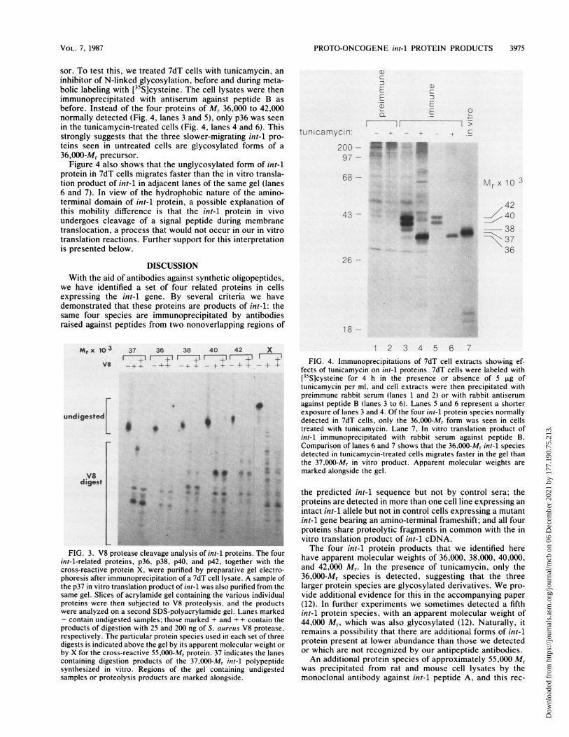
sor. To test this, we treated 7dT cells with tunicamycin, aninhibitor of N-linked glycosylation, before and during meta-bolic labeling with [35S]cysteine. The cell lysates were thenimmunoprecipitated with antiserum against peptide B asbefore. Instead of the four proteins of Mr 36,000 to 42,000normally detected (Fig. 4, lanes 3 and 5), only p36 was seenin the tunicamycin-treated cells (Fig. 4, lanes 4 and 6). Thisstrongly suggests that the three slower-migrating int-I pro-teins seen in untreated cells are glycosylated forms of a36,000-Mr precursor.
Figure 4 also shows that the unglycosylated form of int-1protein ihi 7dT cells migrates faster than the in vitro transla-tion product of int-1 in adjacent lanes of the same gel (lanes6 and 7). In view of the hydrophobic nature of the amino-terminal domain of int-1 protein, a possible explanation ofthis mobility difference is that the int-i protein in vivoundergoes cleavage of a signal peptide during membranetranslocation, a process that would not occur in our in vitrotranslation reactions. Further support for this interpretationis presented below.
DISCUSSIONWith the aid of antibodies against synthetic oligopeptides,
we have identified a set of four related proteins in cellsexpressing the int-1 gene. By several criteria we havedemonstrated that these proteins are products of int-1: thesame four species are immunoprecipitated by antibodiesraised against peptides from two nonoverlapping regions of
Mr x 10 3
Vs
37 36 38 40 42-. - m1 F
_ ~~~~~~~~~~~~~~+ --- _ X., _ +t_
undigested
L I 9
Vsdiaast 9f .4.'
xm
1-
eI ,
'l- t.
0
tunicamycin.
200 -97 -
68 - Mr x 10 3
-- __ - _k
_mil_m
42=g 40
38\937
3626 -
18 -
1 2 3 4 5 6 7FIG. 4. Immunoprecipitations of 7dT cell extracts showing ef-
fects of tunicamycin on int-1 proteins. 7dT cells were labeled with[35Slcysteine for 4 h in the presence or absence of 5 p.g oftunicamycin per ml, and cell extracts were then precipitated withpreimmune rabbit serum (lanes 1 and 2) or with rabbit antiserum
* against peptide B (lanes 3 to 6). Lanes 5 and 6 represent a shorterexposure of lanes 3 and 4. Of the four int-1 protein species normallydetected in 7dT cells, only the 36,000-Mr form was seen in cellstreated with tunicamycin. Lane 7, In vitro translation product ofint-1 immunoprecipitated with rabbit serum against peptide B.Comparison of lanes 6 and 7 shows that the 36,000-Mr int-1 species
* detected in tunicamycin-treated cells migrates faster in the gel than4X the 37.000-Mr in vitro product. Apparent molecular weights are
marked alongside the gel.
rB ~ A.1:*
FIG. 3. V8 protease cleavage analysis of int-1 proteins. The fourint-i-related proteins, p36, p38, p40, and p42, together with thecross-reactive protein X, were purified by preparative gel electro-phoresis after immunoprecipitation of a 7dT cell lysate. A sample ofthe p37 in vitro translation product of int-1 was also purified from thesame gel. Slices of acrylamide gel containing the various individualproteins were then subjected to V8 proteolysis, and the productswere analyzed on a second SDS-polyacrylamide gel. Lanes marked- contain undigested samples; those marked + and + + contain theproducts of digestion with 25 and 200 ng of S. aureus V8 protease,respectively. The particular protein species used in each set of threedigests is indicated above the gel by its apparent molecular weight orby X for the cross-reactive 55,000-Mr protein. 37 indicates the lanescontaining digestion products of the 37,000-Mr int-1 polypeptidesynthesized in vitro. Regions of the gel containing undigestedsamples or proteolysis products are marked alongside.
the predicted int-1 sequence but not by control sera; theproteins are detected in more than one cell line expressing anintact int-I allele but not in control cells expressing a mutantint-1 gene bearing an amino-terminal frameshift; and all fourproteins share proteolytic fragments in common with the invitro translation product of int-1 cDNA.The four int-I protein products that we identified here
have apparent molecular weights of 36,000, 38,000, 40,000,and 42,000 Mr. In the presence of tunicamycin, only the36,000-Mr species is detected, suggesting that the threelarger protein species are glycosylated derivatives. We pro-vide additional evidence for this in the accompanying paper(12). In further experiments we sometimes detected a fifthint-i protein species, with an apparent molecular weight of44,000 Mr, which was also glycosylated (12). Naturally, itremains a possibility that there are additional forms of int-iprotein present at lower abundance than those we detectedor which are not recognized by our antipeptide antibodies.An additional protein species of approximately 55,000 Mr
was precipitated from rat and mouse cell lysates by themonoclonal antibody against int-1 peptide A, and this rec-
VOL. 7, 1987
Dow
nloa
ded
from
http
s://j
ourn
als.
asm
.org
/jour
nal/m
cb o
n 06
Dec
embe
r 20
21 b
y 17
7.19
0.75
.213
.

3976 BROWN ET AL.
ognition was largely inhibited by prior incubation of theantibody with peptide. The 55,000-Mr protein was not rec-ognized by polyclonal antiserum against peptide B, how-ever, and showed no similarity in its V8 protease degrada-tion pattern when compared with the int-i-related proteins.Furthermore, the protein was detected in control NIH 3T3cells which do not express int-1 RNA. We therefore con-clude that the 55,000-Mr protein is not a product of int-i andis not coprecipitated as a complex with int-1 protein; itprobably bears an epitope fortuitously related to that recog-nized by the monoclonal antibody.To identify the protein products of int-1, we made use of
cell lines transfected or infected with retroviral vectorsexpressing high levels of ini-i-specific RNA. Despite this,the less abundant species, p36 and p38, were not alwaysdetectable above background levels in our immunoprecipita-tions (Fig. 2C). In mammary tumors bearing mouse mam-mary tumor virus proviral insertions at the int-i locus, int-imRNA is typically expressed at a level of only 1 to 10 copiesper cell (10), at least 10-fold lower than the levels in the celllines we used in this study. For this reason we did notattempt to characterize int-i proteins in such tumors. Simi-larly, we did not examine tissues in which int-1 is normallyexpressed (postmeiotic cells of the testis and neural tissue ofmid-gestational embryos) because of the difficulties in label-ing such tissues or in obtaining sufficient quantities foranalysis by Western blotting (immunoblotting). This raisesthe possibility that the protein species we detected in fibro-blast cell lines represent nonfunctional forms that differ fromthose normally expressed in vivo or in tumors. However, wedid detect at least the p40 species in MXIN-infected C57MGmammary epithelial cells, a line in which int-i is known to bebiologically active (2).Of the four int-i protein species that we detected in
fibroblast cell lines expressing the gene, none comigrated inpolyacrylamide gels with int-1 polypeptide synthesized invitro. Moreover, the unglycosylated species from cell ex-tracts, p36, migrated faster in the gels than the 37,000-Mr invitro translation product. While there are several possibleexplanations of this discrepancy in apparent molecularweight, based on posttranslational modificationis, the mostlikely explanation seems to be that p36 has undergonecleavage of an amino-terminal signal peptide. The first 48amino acids in the predicted int-i protein sequence arepredominantly hydrophobic (4, 17), but there are polaramino acids at positions 29, 30, and 31 and a charged residueat position 33. Together these define the end of a shorteruninterrupted hydrophobic domain at the amino terminus.The amino acid sequence and characteristics of this domainare eminently compatible with the consensus rules for signalsequences governing membrane translocation (18-20), andthere is a potential signal peptidase cleavage site after aminoacid 27 (Ala-Leu-Ala l Ala) at the end of the hydrophobicregion. Von Heijne (20) has devised an algorithm to identifysignal peptides and predict probable signal cleavage siteswhich has a predictive accuracy of at least 75%. In the caseof int-1, the algorithm predicts a signal cleavage site betweenresidues 27 and 28 with a score of 13.7 (R. Colgrove,personal communication). Since the scores calculated forknown signal peptide cleavage sites have a 95% range from4 to 14, with a modal score around 9 (20), this result indicatesa very high probability that the amino-terminal domain ofint-1 protein is indeed a signal peptide that undergoescleavage at this site. Final proof of this, however, willrequire amino-terminal sequence analysis of the intracellularint-i proteins.
ACKNOWLEDGMENTS
We are particularly grateful to Richard Lerner and Henry Nimanfor the synthesis of peptides and the provision of the monoclonalantibody. We also thank Robin Colgrove for invaluable help withcomputer analysis, Peter Pryciak for constructing pMV7int-1,Randy Schatzman for useful advice, and Mario Chamorro for helpwith antibody production.A.M.C.B. was a special fellow of the Leukemia Society of
Anierica, G.M.S. was a feilow of the Damon Runyon-WalterWinchell Cancer Fund, and H.E.V. is an American Cancer Societyresearch professor. This work was supported by a grant from theNational Institutes of Health.
LITERATURE CITED
1. Brown, A. M. C., and M. R. D. Sc6tt. 1987. Retroviral vectors,p. 189-212. In D. Glover (ed.), DNA cloning-a functionalapproach, vol. 2. IRL Press, Oxford.
2. Brown, A. M. C., R. S. Wildin, T. J. Prendergast, and H. E.Varmus. 1986. A retrovirus vector expressing the putativemammary oncogene int-1 causes partial transformation of amammary epithelial cell line. Cell 46:1001-1009.
3. Cleveland, D. W., S. G. Fischer, M. W. Kirschner, and U. K.Laemmli. 1977. Peptide mapping by limited proteolysis in so-dium dodecyl sulfate and analysis by gel electrophoresis. J.Biol. Chem. 252:1102-1106.
4. Fung, Y.-K. T., G. M. Shackleford, A. M. C. Brown, G. S.Sanders, and H. E. Varmus. 1985. Nucleotide sequence andexpression in vitro of cDNA derived from mRNA of int-1, aprovirally activated mouse mammary oncogene. Mol. Cell.Biol. 5:3337-3344.
5. Jakobovits, A., G. M. Shackleford, H. E. Varmus, and G. R.Martin. 1986. Two proto-oncogenes implicated in mammaryoncogenesis, int-1 and int-2, are independently regulated duringmouse development. Proc. Natl. Acad. Sci. USA 83:7806-7810.
6. Krieg, P. A., and D. A. Melton. 1984. Functional messengerRNAs are produced by SP6 in vitro transcription of clonedcDNAs. Nucleic Acids Res. 12:7057-7070.
7. Land, H., L. F. Parada, and R. A. Weinberg. 1984. Cellularoncogenes and multistep carcinogehsis. 1984. Science 222:771-778.
8. Lerner, R. A., J. G. Sutcliffe, alnd T. M. Shinnick. 1981.Antibodies to chemically synthesized peptides predict-ed fromDNA sequences as probes of gene expression. Cell 23:309-310.
9. Mann, R., R. C. Mulligan, and D. Baltimore. 1983. Constructionof a retrovirus packaging mutant and its use to produce helper-free defective retrovirus. Cell 33:153-159.
10. Nusse, R., A. van Ooyen, D. Cos, Y. K. Fung, and H. E. Varmus.1984. Mode of proviral activation of a putative mammaryontogene (int-1) on mouse chromosome 15. Nature (London)307:131-136.
11. Nusse, R., and H. E. Varmus. 1982. Many tumors induced by themouse mammary tumor virus contain a provirus integrated inthe same region of the host genome. Cell 31:99-109.
12. Papkoff, J., A. M. C. Brown, and H. E. Varmus. The int-1proto-oncogene products are glycoproteins that appear to enterthe secretory pathway. Mol. Cell. Biol. 7:3978-3984.
13. Rijsewijk, F., L. van Deemter, E. Wagenaar, A. Sonnenberg, andR. Nusse. 1987. Transfection of the int-1 mammary oncogene incuboidal RAC mammary cell line results in morphologicaltransformation and tumorigenicity. EMBO J. 6:127-131.
13a.RijsewiJk, F., M. Schuermann, E. Wagenaar, P. Parren, D.Weigel, and R. Nusse. 1987. The Drosophila homolog of themouse mammary oncogene int-1 is identical to the segmentpolarity gene wingless. Cell 50;649-657.
14. Shackletord, G. M., and H. E. Varmus. 1987. Expression of theproto-oncogene, int-1, is restricted to postmeiotic male germcells and the neural tube of midgestational embryos. Cell 50:89-95.
15. Vaidya, A. B., E. Y. Lasfargues, J. B. Sheffield, and W. G.Coutinho. 1978. Murine mammary tumor virus (MuMTV) infec-tion of an epithelial cell line established from C57BL/6 mouse
MOL. CELL. BIO0L.
Dow
nloa
ded
from
http
s://j
ourn
als.
asm
.org
/jour
nal/m
cb o
n 06
Dec
embe
r 20
21 b
y 17
7.19
0.75
.213
.

PROTO-ONCOGENE int-1 PROTEIN PRODUCTS 3977
mammary glands. Virology 90:12-22.16. van Ooyen, A., V. Kwee, and R. Nusse. 1985. The nucleotide
sequence of the human int-1 mammary oncogene; evolutionaryconservation of coding and noncoding sequences. EMBO J. 4:2905-2909.
17. van Ooyen, A., and R. Nusse. 1984. Structure and nucleotidesequence of the putative mammary oncogene int-1; proviralinsertions leave the protein-encoding domain intact. Cell39:233-240.
18. von Heijne, A. 1984. How signal sequences maintain cleavage
specificity. J. Mol. Biol. 173:243-251.19. von Heijne, A. 1984. Analysis of the distribution of charged
residues in the N-terminal region of signal sequences: implica-tions for protein export in prokaryotic and eukaryotic cells.EMBO J. 3:2315-2318.
20. von Heijne, A. 1986. A new method for predicting signalcleavage sites. Nucleic Acids Res. 14:4683-4690.
21. Wilkinson, D. G., J. A. Bailes, and A. P. McMahon. 1987.Expression of the proto-oncogene int-1 is restricted to specificneural cells in the developing mouse embryo. Cell 50:79-88.
VOL. 7, 1987
Dow
nloa
ded
from
http
s://j
ourn
als.
asm
.org
/jour
nal/m
cb o
n 06
Dec
embe
r 20
21 b
y 17
7.19
0.75
.213
.