identification of novel inducers for ll-37 ... of novel inducers for ll-37 expression - assay...
TRANSCRIPT

From THE DEPARTMENT OF LABORATORY MEDICINE
Karolinska Institutet, Stockholm, Sweden
IDENTIFICATION OF NOVEL INDUCERS FOR LL-37 EXPRESSION - ASSAY
DEVELOPMENT AND MECHANISTIC STUDIES
Frank Nylén
Stockholm 2016

All previously published papers were reproduced with permission from the publisher.
Published by Karolinska Institutet.
Printed by E-Print AB 2016
© Frank Nylén, 2016
ISBN 978-91-7676-303-2

Identification of Novel Inducers for LL-37 Expression - Assay Development and Mechanistic Studies THESIS FOR DOCTORAL DEGREE (Ph.D.)
By
Frank Nylén
Principal Supervisor:
Associate Professor Peter Bergman
Karolinska Institutet
Department of Laboratory medicine
Division of Clinical Microbiology
Co-supervisor(s):
Professor Birgitta Agerberth
Karolinska Institutet
Department of Laboratory medicine
Division of Clinical Microbiology
Professor Guðmundur Hrafn Guðmundsson
University of Iceland
Biomedical Center
Reykjavik, Iceland
Opponent:
Professor Henk Haagsman
Utrecht University
Department of Infectious Diseases and
Immunology
Division of Molecular Host Defense
Utrecht, The Netherlands
Examination Board:
Professor Petter Höglund
Karolinska Institutet
Department of Medicine
Center for Hematology and Regenerative Medicine
Professor Jan Johansson
Karolinska Institutet
Department of Neurobiology, Care Sciences
and Society
Division of Neurogeriatrics
Professor Ulrich Theopold
Stockholm University
Department of Molecular bioscience,
The Wenner-Gren Institute


Till mamma och pappa


ABSTRACT
The remaining reservoir of effective antibiotics is running out and more and more pathogens
are gaining resistance, even to the last line of antibiotics. Therefore, novel treatment regimens
against bacterial infections are urgently needed. Antimicrobial peptides (AMPs) are
expressed at mucosal surfaces and protect the host against invasive microbes. Not only are
they efficient microbial killers but they also orchestrate additional immune responses,
including chemotaxis and wound healing. The focus in the current work has been on the
human cathelicidin LL-37 (encoded by the CAMP gene), which can be considered as a
marker for the AMP-system in general. Since several AMPs with different mechanism of
action are released simultaneously, the risk of developing bacterial resistance is very low.
Here we propose a concept where drug-like molecules that induce the expression of AMPs
can be used to prevent or treat infections. We suggest that this novel approach designated
‘Host Directed Therapy’ can be used alone or together with traditional antibiotics for
treatment of infections.
The overall aim with this thesis was to identify novel AMP inducing compounds and to
dissect the regulatory pathways involved in their mechanism of action.
We set out to establish a cell-based reporter-gene-assay under the control of regulatory
elements of the CAMP gene. Following a successful evaluation for high throughput screening
purposes, six out of 1200 tested compounds were validated as novel inducers. One of these
was the histone deacetylase (HDAC) inhibitor entinostat, which was found to be a potent
inducer of LL-37 expression. Several derivatives of entinostat were synthesized and
evaluated for LL-37 inducing activity. This strategy demonstrated which part of the molecule
that was important for the induction.
Entinostat was shown to regulate LL-37 via activation of the transcription factors STAT3 and
HIF-1α. Notably, HIF-1α was found to directly bind and initiate transcription of the CAMP
gene. In contrast, no direct binding of STAT3 to the CAMP gene promoter could be
demonstrated. However, STAT3 was clearly involved in the regulation of LL-37 expression,
since macrophages from a patient carrying a mutated STAT3 gene were unable to upregulate
LL-37 upon entinostat treatment.
Several additional regulatory pathways were discovered utilizing label free quantitative mass
spectrometry, where protein levels were compared between unstimulated cells and cells
treated with the AMP-inducing compounds lactose and phenylbutyrate. Next, the proteomic
profiles were mapped to known pathways utilizing bioinformatics tools. The identified
pathways were validated using the cell based reporter assay. The thyroid hormone receptor
pathway, the eicosanoid signaling pathway and the steroid biosynthesis pathway could all be
attributed to LL-37 expression.
Together these results indicate that AMPs are strictly regulated both in homeostasis and
during infection. The novel AMP-inducing compounds identified here should be further
evaluated as Host Directed Therapy against infections in cellular and animal models as well
as in relevant disease cohorts.

LIST OF SCIENTIFIC PAPERS
The following articles form the basis of this thesis and are referred to with their
corresponding Roman numeral.
I. Nylén F*, Miraglia E*, Cederlund A, Ottosson H, Strömberg R,
Gudmundsson G H, Agerberth B. Boosting innate immunity: Development
and validation of a cell-based screening assay to identify LL-37 inducers
Innate Immun. 2014, 20(4):364-76
*These authors contributed equally to this study
II. Ottosson H, Nylén F, Sarker P, Miraglia E, Bergman P, Gudmundsson G H,
Raqib R, Agerberth B, Strömberg R. Highly Potent Inducers of Endogenous
Antimicrobial Peptides for Host Directed Therapy of Infections. Manuscript
III. Miraglia E*, Nylén F*, Johansson K, Arnér E, Cebula M, Farmand S,
Ottosson H, Strömberg H, Gudmundsson G H, Agerberth B#, Bergman P
#.
Entinostat up-regulates the CAMP gene encoding LL-37 via activation of
STAT3 and HIF-1α transcription factors. Manuscript
*#These authors contributed equally to this study
IV. Cederlund A, Nylén F, Miraglia E, Bergman P, Gudmundsson G H,
Agerberth B. Label-free quantitative mass spectrometry reveals novel
pathways involved in LL-37 expression. J Innate Immun. 2014;6(3):365-76


CONTENTS
1 Background ...................................................................................................................... 1
1.1 Introduction ............................................................................................................ 1
1.2 The immune system............................................................................................... 1
1.3 Innate immunity ..................................................................................................... 1
1.3.1 Innate sensing ............................................................................................ 2
1.3.2 Innate effectors .......................................................................................... 3
1.3.3 Innate immune cells ................................................................................ 10
1.4 Gene regulation.................................................................................................... 12
1.4.1 Regulation of antimicrobial peptides ...................................................... 13
1.4.2 Dysregulation of antimicrobial peptides in disease................................ 17
1.5 Transcription factors involved in the regulation of LL-37 ................................. 18
1.5.1 Vitamin D Receptor ................................................................................ 18
1.5.2 STAT3 ..................................................................................................... 18
1.5.3 HIF-1 ....................................................................................................... 19
1.5.4 PU.1 ......................................................................................................... 19
1.5.5 Thyroid Receptor .................................................................................... 19
1.5.6 Estrogen Receptor ................................................................................... 20
1.6 HDACs and HDAC inhibitors ............................................................................ 20
1.6.1 Entinostat ................................................................................................. 21
1.7 Identification of novel drugs ............................................................................... 23
1.7.1 Assay development ................................................................................. 23
1.7.2 Chemical libraries ................................................................................... 25
2 Aims of this thesis ......................................................................................................... 27
3 Methodology.................................................................................................................. 29
3.1 Molecular cloning ................................................................................................ 29
3.2 Chromatin Immuno Precipitation ....................................................................... 32
3.3 Proteomics coupled to pathway analyses ........................................................... 33
4 Results and Discussion .................................................................................................. 35
4.1 Paper I: Boosting innate immunity: Development and validation of a cell-
based screening assay to identify LL-37 inducers .............................................. 35
4.2 Paper II: Highly potent inducers of endogenous antimicrobial peptides for
host directed therapy of infections ...................................................................... 39
4.3 Paper III: Entinostat up-regulates the CAMP gene encoding LL-37 via
activation of STAT3 and HIF-1 transcription factors ........................................ 42
4.4 Paper IV: Label-free quantitative mass spectrometry reveals novel
pathways involved in LL-37 expression ............................................................. 45
5 Conclusions ................................................................................................................... 47
6 Future experiments or things I would have liked to do ................................................ 48
7 Acknowledgements ....................................................................................................... 49
8 References ..................................................................................................................... 52

LIST OF ABBREVIATIONS
ADME Absorption, distribution, metabolism, and excretion
AMP Antimicrobial peptide
APD Aroylated phenylene diamine
ASA Acetylsalicylic acid
Bcl2 B-cell lymphoma 2
bp Base pairs
CAMP gene Cathelicidin antimicrobial peptide gene
ChIP Chromatin immuno precipitation
ChIP-seq ChIP with subsequent deep sequencing
COX Cyclooxygenase
DAMP Damage associated molecular patterns
DMSO Dimethyl sulphoxide
ER Estrogen hormone receptor
Foxp3 Forkhead box P3
GR Glucocorticoid receptor
HAT Histone acetyl transferase
hBD Human β defensin
hCAP-18 Human cationic antimicrobial protein 18 kDa
HD5-6 Human alpha defensin 5-6
HDAC Histone deacetylase
HDACi Histone deacetylase inhibitor
HIES Hyper IgE syndrome
HIF-1 Hypoxia induced factor 1
HIV Human immunodeficiency virus
HMGB1 High mobility group box 1
HNP1-4 Human neutrophil peptide-1-4, alpha defensin 1-4
HPD HIF prolyl-hydroxylase
HTS High throughput screening
IKB Ingenuity knowledgebase
IL Interleukin

IPA Ingenuity pathway analyzer
JAK Janus kinase
kb kilo base pairs
KDAC Lysine deacetylase
LCA Litocholic acid
LPS Lipopolysaccharides
LTB4 Leukotriene B4
MAPK Mitogen-activated protein kinases
MIC Minimal inhibitory concentrations
MRSA Methicillin-resistant Staphylococcus aureus
MS Mass spectrometry
Mtb Mycobacterium tuberculosis
NET Neutrophil extracellular trap
NF-κB Nuclear factor κ light chain enhancer of activated B-cells
NK-cells Natural Killer cells
NOD-like receptor Nucleotide-binding oligomerization domain receptor
Nrf2 Nuclear related factor 2
P2X7 P2X purino-receptor 7
PAMP Pathogen associated molecular patterns
PBA Phenylbutyrate
pDC Plasmacytoid dendritic cell
PGE2 Prostaglandin E2
PLS Papillon-Lefèvre syndrome
PMN Polymorphonuclear Granulocytes
PRR Pattern recognition receptors
pVHL von Hippel-Lindau tumor suppressor protein
qPCR Quantitative PCR
Reg Regenerating islet-derived protein
RNS Reactive Nitrogen Species
ROS Reactive Oxygen Species
RXR Retinoid X receptor

shRNA Short hairpin RNA
STAT Signal transducer and activator of transcription
T3 Triiodothyronine
T4 Thyroxine
TLR Toll like receptor
TR Thyroid hormone receptor
VDR Vitamin D receptor

1
1 BACKGROUND
1.1 INTRODUCTION
With the arrival of antibiotics during World War II, came a weapon against microbes and
today, we are not used to people dying from ‘simple’ bacterial infections. However, the
microbes are winning ground and more bacterial pathogens are gaining resistance even to our
most advanced antibiotics. Therefore, to prevent the return of the pre-antibiotic era, it is of the
utmost importance to develop novel antibiotics and treatment regimes. This thesis describes
the field of ‘Host Directed Therapy’, where we aim to treat infections by aiding the immune
system in order to eliminate pathogens. By treating the host with compounds that induce
expression of a wide variety of antimicrobial peptides (AMPs), enhancing ROS and RNS
production and by activating autophagy, the host can efficiently destroy invading pathogens,
inhibit their growth and prevent attachment. Moreover, induction of multiple AMPs with
different mechanisms of action are likely more effective and also less prone to cause the
development of microbial resistance, which is a relevant point given the emerging spread of
multidrug resistant bacteria (Marr et al., 2006; Singh et al., 2000).
1.2 THE IMMUNE SYSTEM
The human immune system is typically described as two separate parts, the innate immune
system, which we share with all multicellular organisms and the adaptive immune system,
which is exclusive to jawed vertebrates. The adaptive immune system developed from innate
immunity and the two systems are intertwined with each other meaning that the separation
between the two parts is not exact, and separately, neither would be enough to defend us nor
function in an optimal manner. The immune system is a complex entity and has always been
at the forefront of evolution to keep up with all microbes in the surrounding. The innate
system is the first line of defense and stops most invaders at the barrier of our body. The
microbes that breach the barrier will be recognized by receptors on the cells of the adaptive
system and a clonal expansion will start. The adaptive immune system is slower in action
than innate immunity but on the other hand is much more specific and efficient when it is
fully matured. The expansion of T- and B- cells may take up to a week for full functionality
and during that time the innate immune system needs to hold all invaders at bay.
1.3 INNATE IMMUNITY
Innate immunity is the inherited defense against microbial threats towards the host and it is
immediately activated when needed. It is evolutionary conserved and is the only immune-
defense in plants, fungi and invertebrates. In vertebrates, including humans, innate immunity
functions as the first line of defense but also activates and recruit cells of the adaptive
immune system. All organisms need ways to discriminate between self and non-self, hence
there is a need for sensing of foreign molecules, such as microbial patterns and the ability to
distinguish them from self. The sensing is followed by a response with direct antimicrobial

2
action, tissue-repair or the recruitment of clotting cells. The innate system is therefore divided
in a sensing part and an effector part.
1.3.1 Innate sensing
An array of recognition receptors is included in innate immunity with two main classes; the
Toll-like receptors (TLRs), which are situated in the cell-membrane and in the membrane of
the lysosomes and the nucleotide-binding oligomerization domain receptors (NOD-like
receptors), which are located inside the cells. There are also additional innate receptors, such
as the RAGE receptor and the RIG-1-like receptors (Kierdorf and Fritz, 2013; Loo and Gale,
2011; Xie et al., 2013). All receptors recognize certain structures or molecules, which signal
infection or injury (Matzinger, 2002; Medzhitov, 2007, 2009). These receptors are jointly
called pattern recognition receptors (PRRs) and they have specificity towards different
molecular patterns. These patterns can be divided in pathogen associated molecular patterns
(PAMPs) and damage associated molecular patterns (DAMPs) (Matzinger, 1994; Matzinger,
2002). PAMPs are generally unique and constitute vital parts of microbes, such as
lipopolysaccharides (LPS), lipoteichoic acid, peptidoglycans, in addition to double stranded
RNA, single stranded DNA and unmethylated CpGs originated from microbes (Mogensen,
2009).
DAMPs - also known as alarmins - are released from cells under stress and consist of several
molecules or patterns. The classical DAMP is the protein High mobility group box 1
(HMGB1), which binds to the RAGE-receptor. Additional alarmins are self-DNA, heat-shock
proteins, uric acid and S100 proteins (Foell et al., 2007; Lotze et al., 2007). In addition,
AMPs can also function as alarmins. For example, LL-37 can bind to self DNA and form a
complex, which is recognized by Toll like receptor 9 (TLR9) in plasmacytoid dendritic cells
(pDCs), while the human neutrophil peptide-1 (HNP1) also known as α-defensin 1 regulates
IL-1β post translationally via the P2X purino-receptor 7 (P2X7) (Chen et al., 2014; Lande et
al., 2007).
In response to the different stimuli described above, pro-inflammatory pathways are
triggered. The best described pathway involves activation of ‘nuclear factor κ light chain
enhancer of activated B cells’ (NF-κB). In addition, there are several other important
pathways in innate immunity, such as the mitogen-activated protein kinases (MAPK) P38 and
ERK1/2 pathways. The signal cascade of the pro-inflammatory pathways leads to an
increased expression and release of cytokines and chemokines but also triggers expression of
additional defense molecules, such as AMPs and mucins (Enss et al., 2000; Perez-Vilar and
Hill, 1999) (O'Neil et al., 1999). However, the specific response will be determined by two
major factors; the ligand and in which cell-type the response occurs (Bianchi, 2007).

3
1.3.2 Innate effectors
1.3.2.1 Reactive Oxygen Species (ROS) and Reactive Nitrogen Species (RNS)
Both ROS and RNS are produced as a part of the innate defenses. The molecules exhibit a
high reactivity by virtue of single electrons, which react with proteins, lipids and
carbohydrates. NADPH oxidase is an enzyme, which is stored in the granules of neutrophils
and can generate both ROS and RNS. In addition to direct microbial killing, both ROS and
RNS are prominent signal molecules and activate several pathways of immunity, by
activating redox sensitive proteins, such as tyrosine phosphatases which regulate the MAPK
pathway (Neish, 2013). NF-κB is a redox sensitive transcription factor which is closely
associated to many immune genes and functions (Gostner et al., 2013).The determination of
Th1 or Th2 regulatory T cells is linked to the redox potential in the milieu around the naive
T-cell, where an oxidative milieu promotes Th1 cells and versa reducing environment
appears to promote Th2-cells (Gostner et al., 2013). Another major transcription factors
regulated by ROS is the hypoxia induced factor 1 (HIF-1). To protect the host from damage,
several antioxidant systems are in use, such as superoxide dismutases and the thioredoxins
(Nathan and Cunningham-Bussel, 2013).
1.3.2.2 Autophagy
The mechanism of autophagy is how the cells dispose of old proteins and broken organelles
in order to prevent them from intervening with the normal processes in the cell. Autophagy is
a key component to keep the homeostasis of protein production and degradation.
Interestingly, the process of autophagy recycles all material and it is therefore vital during
starvation when less important proteins and organelles can be degraded and used for building
parts and nutrients. However, it is also a very important pathway for clearance of intracellular
pathogens, such as Mycobacterium tuberculosis (Mtb). In addition, autophagy have been
demonstrated to modulate both MHC class I and class II antigen presentation (Crotzer and
Blum, 2009; Glick et al., 2010).
1.3.2.3 Antimicrobial peptides
AMPs are often referred to as gene encoded antibiotics and are typically produced and
released upon infection. They are cationic and amphipathic and as such have a preference for
microbial cell-membranes and the main mode of action is disruption of cell-membranes
(Zasloff, 2002). AMPs are ancient molecules in evolution and important effector molecules
of the innate immune system. They are found throughout all branches of life, including
archaea. The halocin S8 from Haloarchaea (Price and Shand, 2000) is an example of an
AMP from a very primitive organism. Moreover, also bacteria express molecules with
antimicrobial action against other bacterial species. These molecules are called bacteriocins
and are important for the bacterial competition at mucosal surfaces (Dobson et al., 2012).
The first antimicrobial molecule found in humans was isolated by Alexander Fleming from
nasal secretions in 1922 and this antimicrobial protein was named ‘lysozyme’ (Fleming,

4
1922). It is highly expressed in neutrophils (PMN’s) and also present in tears and breast milk.
This finding set off an era of novel discoveries in the field. Between 1920 and 1950 several
articles described the isolation of molecules with antimicrobial activity from many different
organisms, Bombinin from the frog Bombina variegate, lactoferrin from cow milk and
purothionins from wheat, (Fernandez de Caleya et al., 1972; Groves et al., 1965; Kiss and
Michl, 1962) . Technical limitations of the time hindered full characterization of the
respective molecules but in 1980, the first AMPs were characterized and reported by the
group of Hans G. Boman. These small peptides were isolated from the silk moth, Hyalophora
cecropia and were subsequently named as ‘cecropins’ (Hultmark et al., 1980; Steiner et al.,
1981). In the beginning of the eighties the defensins were isolated from rabbit macrophages
by the group of Robert Lehrer (Selsted et al., 1983). At the end of the 80’s Michael Zasloff
isolated and characterized the Magainins from the African clawed frog Xenopus laevis
(Zasloff, 1987). These discoveries indicated that AMPs were widespread in nature. To date
2684 AMPs and proteins from different species and kingdoms have been described and in
humans there are 112 reported antimicrobial peptides and proteins (Wang, 2014; Wang et al.,
2016b).
AMPs share some common denominators across species. Typically, they are small (12–50
amino acids), have a positive net-charge with an amphipathic character and are usually
defined by their secondary structure; α-helical, β-sheets or random coils, rich in arginine and
prolines (Lai and Gallo, 2009).
1.3.2.3.1 Defensins
The defensins represent an ancient family of AMPs and in contrast to the cathelicidins they
are more than 30 different defensin genes identified in the human genome. However, only ten
of them have been characterized on the peptide level. Defensins have been found in animals,
plants, fungi and in some species of myxobacteria (Lehrer and Ganz, 2002; Mygind et al.,
2005; Vriens et al., 2014; Wong et al., 2007; Zhu, 2007, 2008). The defensins are synthesized
as preproproteins and are activated by cleavage into peptides with sizes between18-45 amino
acids. These peptides contain conserved cysteines, six in mammals and birds, eight in plants
and vertebrates, forming disulphide bonds, which stabilize a β-sheet conformation (Carvalho
Ade and Gomes, 2009; Ganz, 2003). The S-S bonds are however paired differently between
the cysteine residues, giving rise to the division of the defensins into α, β and θ defensins
(Ganz, 2003). The defensins, as a group, have been shown to have a broad and potent
antimicrobial activity against both gram positive and gram negative bacteria, fungi and some
enveloped viruses (Ganz, 2003). The concentration of enteric defensins can reach levels of
above 10 mg/ml in the intestinal crypts and between 10 and 100 µg/ml in other epithelial
cells, as determined by analysis of human skin and porcine tongue (Ganz, 2003). In a
publication by Salzman et al. in 2003, defensins were for the first time shown to be important
in vivo, where human α defensin-5 (HD5) enabled protection against enteric salmonellosis
(Salzman et al., 2003). Like most AMPs, defensins act through the disruption of microbial
cell membranes, but it has also been demonstrated that they interfere with both RNA and

5
DNA synthesis (Ganz, 2003). Interestingly, it has been reported that human β defensin-1
(hBD-1) increases its antimicrobial activity upon reduction of its S-S bridges, most likely via
thioredoxin, which co-localizes with hBD-1 in epithelial cells (Schroeder et al., 2011). For
HD5, on the other hand, reduction of its S-S bonds reduces its ability to kill microbes in vitro
and instead induces the LPS neutralizing effect and turns the peptide into an effective zinc
chelator (Wang et al., 2016a; Zhang et al., 2013). Defensins are more than just active
antimicrobials; some also function as alarmins, regulators of cytokines and chemo-attractants
of monocytes and T-cells (Chen et al., 2014; Chertov et al., 1996; Sakamoto et al., 2005;
Semple and Dorin, 2012; Territo et al., 1989).
α-defensins
There are six α defensins expressed in humans. Four of them are known under the name of
human neutrophil peptide (HNP1-4). The HNPs are highly concentrated in the primary
granules of neutrophils and expressed in B-, T- and NK-cells as well as in monocytes
(Agerberth et al., 2000; Ganz, 2003). The human defensins 5 and 6 (HD5 and HD6) are
expressed by Paneth cells in the small intestine in a constitutive manner. They are stored in
granules that upon stimulation are released into the intestinal crypts, where they can reach
concentrations of more than10 mg/ml (Bevins and Salzman, 2011; Ganz, 2003; Ouellette,
2011). HD5 and HD6 complement each other by virtue of their separate mechanisms in the
innate immune system. HD5 has a direct antimicrobial action by lysing bacteria, while HD6,
interestingly, can self-assemble or aggregate into what can be defined as “nanonets”, with the
ability to ensnare bacteria (Bevins, 2013; Chu et al., 2012; Ouellette and Selsted, 2012).
HD5 is constitutively expressed in the female genital tract and induced in urethra by
Neisseria gonorrhoeae infection. HD6 has also been implicated in the female genital tract
upon the same infection (Klotman et al., 2008; Porter et al., 2005; Svinarich et al., 1997).
Interestingly, HD5 and 6 as well as Neisseria gonorrhoeae infection have been suggested to
facilitate HIV transmission (Klotman et al., 2008; Rapista et al., 2011).
β-defensins
The human β-defensins are mainly expressed in the skin and at mucosal barriers (Ganz, 2003;
Lai and Gallo, 2009). Despite many genes encoding β-defensins in the human genome, only
four peptide products have been characterized. hBD-1 is mainly constitutively expressed,
while the other three β-defensins, hBD2-4, are regulated by PAMPs or DAMPs, e.g. by
bacterial infection or injury (Nuding et al., 2013). However, we and others have demonstrated
that also hBD-1 can be regulated, both by bacterial infection and by small molecular
compounds (paper III) (Sorensen et al., 2005). In addition, β-defensins are expressed in the
genital tract and interestingly hBD2-4 are down-regulated as a response to unprotected
vaginal intercourse (Nakra et al., 2016).

6
θ-defensins
θ-defensins are interesting molecules that only exists in old world primates. θ-defensins are
cyclic peptides and as such the only cyclic peptides found in mammals, where they seem to
have evolved from α-defensins. The active θ-defensin is formed by ligation of two copies of
the θ-defensin peptide; i.e. two copies of the precursor protein are needed for one θ-defensin.
The precursor protein is interestingly closely related to the α-defensins (Tang et al., 1999).
Humans, chimpanzees and gorillas do not express θ-defensins, however the genes for θ-
defensins can be found in the genomes of these species. In humans the mRNA is transcribed,
but a mutation has introduced a premature stop codon and thus the active peptide cannot be
translated. The θ-defensins have been studied for their potential as anti-viral compounds,
especially in the treatment of HIV (Gallo et al., 2006; Munk et al., 2003).
1.3.2.3.2 Cathelicidins
Cathelicidins are generally considered mammalian AMPs and at least one cathelicidin gene
have been identified within all mammals examined. However, they have also been found in
snakes and catfish. They are synthesized as pre-pro-peptides and the signal sequence (pre) is
directing the pre-pro-peptide towards the designated compartment, usually destined for
exocytosis (Gudmundsson et al., 1996). The conserved cathelin domain (pro) has a high
similarity to the cathepsin L inhibitor cathelin and the variable C-terminal domain is the part
that confer the antimicrobial activity (Zanetti et al., 1995). This is conferred by a four exon
gene which in human is entitled Cathelicidin AntiMicrobial Peptide or CAMP (Gudmundsson
et al., 1996). It is located on the third chromosome and is only ~2 kilo base pairs (kb) long
including introns (Figure 1). Interestingly, the variable active peptide is always located in the
fourth exon, potentially explaining why the C-terminal and active peptides of the
cathelicidins are not evolutionary conserved (Tomasinsig and Zanetti, 2005). Humans only
carry one cathelicidin gene and this holds true also for mouse, rat and rabbit. The peptides in
these species are all α-helical in their secondary structure, implying that these species can
work as model systems for studies on the human cathelicidin. The α-helical secondary
structure of the human C-terminal domain adopts this conformation upon contact with
microbes, experimentally demonstrated using micelles (Li et al., 2006). Possibly avoiding
host damage by undertaking a random coil structure until activity is needed. Furthermore it
has been shown that the helical structure is also stabilized under mimicking of in vivo
conditions (Johansson et al., 1998).
Cathelicidin is vital for resistance to several pathogens. For example, in mice it has been
demonstrated that the mouse cathelicidin CRAMP is pivotal for resistance against C. albicans
in the gut and against a sensitive strain of Group A Streptococcus in the skin (Fan et al., 2015;
Nizet et al., 2001).
The human cathelicidin peptide was discovered in 1995 on cDNA level and the putative
antimicrobial peptide was named FALL-39 (Agerberth et al., 1995). However, when the
active peptide was isolated and characterized from granulocytes it was found to be two
residues shorter. Thus, the peptide was named LL-37, after the two leucines in the N-terminal

7
end and from the length of 37 amino acid residues. The LL-37 peptide sequence is as follows.
NH2-LLGDFFRKSKEKIGKEFKRIVQRIKDFLRNLVPRTES-COOH (Gudmundsson et al.,
1996). The proform was designated as human cationic antimicrobial protein weighing 18 kDa
or hCAP-18.
Figure 1. Schematic representation of the CAMP gene and its translational product. Transcription factors activate
RNA polymerase II and mediate transcription. The CAMP gene is transcribed from four exons, where Exon 1-3
encode the cathelin domain including a sequence that signals for exocytosis. Exon four encodes the peptide with the
antimicrobial activity, LL-37, shown by dual colors, representing the amphipathicity of the peptide. The signal
sequence is removed after direction for exocytosis. The remaining pro-peptide is designated hCAP-18 and the active
peptide is cleaved off by protease 3 in neutrophils and adopts an α-helical conformation.
LL-37 has been shown to be potent against many different microbial species (Turner et al.,
1998), and is considered especially potent against gram negative bacteria because of the high
affinity to LPS. LL-37 actually binds LPS with a 2:1 stoichiometry that is cooperative to its
nature (Turner et al., 1998). Minimal inhibitory concentrations (MIC) values of LL-37 varies
with bacteria and method used, it is however not unusual with sub-µM concentrations
(Larrick et al., 1995). Furthermore, the activity based on the ability to achieve α-helical
structure of LL-37 is enhanced in the presence of anions and by increasing the pH value
(Johansson et al., 1998). It has been demonstrated that apart from the microbial killing, LL-37
is responsible for additional effects, such as angiogenesis, apoptosis and as already
mentioned, endotoxin neutralizing (Table 1).

8
Table 1. Additional functions of LL-37 apart from the antimicrobial effects.
Function Mechanism Reference
Chemotaxis By activation of formyl peptide
receptor-like 1 (FPRL1) and
CXCR2 (IL-8 receptor)
(De et al., 2000; Zhang et al., 2009)
Apoptosis Both inducing apoptosis and
increase life span of cells. Action
suggested via FPRL1, P2X7, P53,
Bax ,Bak and Bcl-2
(Barlow et al., 2006; Lau et al.,
2006; Nagaoka et al., 2006; Ren et
al., 2012)
Angiogenic Via FPRL1 (Koczulla et al., 2003)
Neutralization of endotoxin Direct LPS binding and
competitive binding to P2X7
(Hu et al., 2014; Turner et al.,
1998)
Wound healing LL-37 induce keratinocyte
migration via EGFR activation
(Heilborn et al., 2003; Shaykhiev et
al., 2005; Tokumaru et al., 2005)
Histamine release Via G protein-phospholipase C-
dependent manner
(Niyonsaba et al., 2001)
Leukotriene B4 release Via activation of FPR2/ALX (Wan et al., 2011; Wan et al., 2007;
Wan et al., 2014a)
Increase in ROS production and
release of α defensins
By enhanced production of IL-8 (Zheng et al., 2007)
Modulation of dendritic cell
function
Via regulation of TLR ligands (Kandler et al., 2006)
Macrophage differentiation (van der Does et al., 2010)
Induction of autophagy (Rekha et al., 2015)
Increase phagocytosis Via activation of integrin Mac-1 (Wan et al., 2014b; Zhang et al.,
2016)
1.3.2.3.3 Antimicrobial proteins
The S100 calcium proteins are a large group of structurally related proteins of which several
have demonstrated to exhibit antimicrobial activity. Calprotectin consists as a dimer of

9
S100A8 and A9 and is highly abundant in the cytoplasm of neutrophils (Edgeworth et al.,
1991). The mechanism of action is attributed to its chelating properties, where it deprives the
microbes of essential minerals (Clohessy and Golden, 1995; Damo et al., 2013; Sohnle et al.,
2000). Another S100 protein with antimicrobial activity is psoriasin, which was first isolated
from psoriatic lesions, where it is drastically induced. Interestingly, psoriatic lesions are
seldom infected despite the prolonged exposure to possible invasive pathogens, likely due to
chelating of divalent zinc ions (Glaser et al., 2005). Psoriasin is also expressed during
differentiation of mammary epithelial cells, and it is associated with poor prognosis in
estrogen negative breast cancer tumors (Emberley et al., 2003; Vegfors et al., 2012).
Lactotransferrin is another chelating protein that was first identified to act as a transporter for
iron in milk and blood. However, it is now known to be active against viruses, bacteria and
fungi, and the main mechanism is sequestering of irons from the milieu and hence to starve
the microbes (Bullen et al., 1972; Jenssen and Hancock, 2009).
Contrary to AMPs, many antimicrobial proteins are negatively charged, hence the chelating
properties, which seem to be a general route of antimicrobial activity. In fact all metal
sequestering proteins can be considered to be antimicrobial by nature, disregarding their
actual function.
Regenerating islet-derived proteins (Regs) from the superfamily of C-type lectins have been
detected in many different cell-types and are often associated with differentiation and
proliferation (Parikh et al., 2012). Generally, the Regs are thought of as intestinal proteins and
have been demonstrated to keep the small intestinal epithelial cells sterile (Vaishnava et al.,
2011). In mice it has been demonstrated that signal transducer and activator of transcription 3
(STAT3) is a key regulator of Reg3γ in both intestinal and lung epithelia, and that Reg3γ is
vital in MRSA pneumonia clearance (Choi et al., 2013; Lee et al., 2012; Murano et al., 2014).
Also in humans it has been demonstrated that several Reg genes are upregulated upon
STAT3 activation (Murano et al., 2014). The killing mechanism of Reg3γ action have not yet
been determined (Gallo and Hooper, 2012). Reg3α is bactericidal for Gram-positive but not
Gram-negative bacteria, which is explained by the fact that LPS blocks the ability of Reg3α
to form pores in bacterial membranes (Mukherjee et al., 2014). Reg4 is less studied but is
strongly associated with colorectal cancer (Kawasaki et al., 2015).
Histones are proteins that are known to aid in the packaging of DNA in the cell nucleus. In
addition, they also have antibacterial properties, where peptides derived from the larger
histone proteins have been demonstrated to inhibit bacteria in vitro. They are also a major
part of the neutrophil extracellular traps (NETs) and as such also inhibit infection (Kawasaki
and Iwamuro, 2008).
Ribonucleases are responsible for degradation of RNA in the cell and six out of eight have
been reported to be involved in host defenses, RNases 2, 3, 5, 7, and 8 (Wang, 2014).

10
1.3.2.4 Linking innate and adaptive immunity
In addition to their capacity to directly kill microbes, the innate effectors (AMPs, ROS and
RNS) can all be described as linkers to the adaptive immune system, which via the activation
of cytokines, chemokines and specific AMPs continue to strengthen defenses by recruiting
cells of both the innate and adaptive immune system.
1.3.3 Innate immune cells
1.3.3.1 Epithelial cells
The first innate defenses that any microbe will face are the physical barriers of the host, i.e.
the skin, the eyes, the mouth, lungs and the lining of the gut. A part of this defense is
epithelial cells, which are able to mount an effective defense against any microbial attack.
Epithelial cells are covered by a layer of mucins, building the mucous membrane, as in the
gut lining (Perez-Vilar and Hill, 1999). The mucin layer harbors several effector molecules,
such as AMPs that are able to eliminate potential pathogens. The mucin layer consists of the
highly glycosylated mucin proteins, which forms a virtually impassible gel-like structure. The
mucus layer is replenished by special epithelial cells designated goblet cells. The result of this
replenishment from below is a near sterile environment closest to the epithelial cells. The fact
is that very few of the microbes we encounter every day will ever be in proximity to the
epithelial cells. Even in the small intestine, where the mucus layer is thin, it has been
demonstrated that bacteria is kept at distance from the epithelial cells by secretion of the
antimicrobial protein Reg3γ (Vaishnava et al., 2011). If the epithelial layer or the mucosal
layer is damaged, a pathogen may reach a site, where it can infect or invade. Epithelial cells
are decorated with PRRs, which recognize PAMPs or DAMPs and expression and release of
cytokines and chemokines is initiated. This leads to recruitment and activation of other
immune cells, such as antigen presenting cells. Interestingly, TLR4 in intestinal epithelial
cells are located on the basal side of the cells to avoid unnecessary activation by LPS (Hornef
et al., 2003). Most of our barriers are covered by commensal bacteria specific to the place of
colonization, helping the host by competing with pathogens for both nutrients and habitation.
Moreover, it has been demonstrated that many of the commensal bacteria are inducing the
expression of AMPs to which the commensal bacteria are resistant to and in that way keep
pathogenic competitors at bay (Bevins, 2005; Kamada et al., 2013). The number of species in
the natural flora of the gut increases with the distance from the stomach and in the colon the
number of commensal bacteria reaches 10 to the power of 13 (Sartor, 2008). In contrast, the
human skin barrier is more arid of commensals. Instead of the mucus layer seen in eyes,
intestine and lung, the skin has the apoptotic layer of keratinocytes, which stops invading
pathogens with its hard exterior. In skin, the AMPs are mainly produced when the barrier is
broken, damaged or infected. For example, cathelicidin and hBD1 is constitutively expressed
at low levels but may be induced together with hBD2-4 upon infection or damage (Kenshi
and Richard, 2008).

11
1.3.3.2 Polymorphonuclear Granulocytes (PMNs)
PMNs makes up 50-70 % of all leukocytes in humans and are named for their small vesicles
or granules, which can be detected upon Hematoxylin staining of blood smear (Borregaard et
al., 1987). Of the granulocytes around 99% are neutrophils and 1% consists of eosinophils,
basophils and mast cells. The neutrophils are the first immune cells recruited to a site of
infection or injury, where they immediately destroy microbes by phagocytosis or by releasing
their granular content. The granules contain high concentrations of AMPs (α-defensins and
hCAP-18) and additional proteins with antimicrobial activity, such as
bactericidal/permeability-increasing protein and Lysozyme. The AMP concentration in the
phagocytic vacuoles reaches concentration above 10 mg/ml (Ganz, 2003). The granules also
contain proteases such as protease 3, which has been shown to cleave the human cathelicidin
pro-form hCAP-18 into the cathelin domain and the active peptide LL-37 (Sorensen et al.,
2001). The neutrophils, besides their granules, carry disassembled NADPH oxidase 2
separated in the cytosol and in the membrane which upon stimuli readily can be assembled
and generate an oxidative burst as a general defense. Neutrophils can undergo what is called
NETosis, where they in a suicidal fashion spew out NETs, a sticky entity made up of
chromatin and decorated with AMPs, trapping microbes like flies in a spider web (Brinkmann
and Zychlinsky, 2007; Sorensen et al., 2001).
1.3.3.3 Macrophages
Monocytes are circulating in the blood stream and lymphatic system, and depending on
stimuli they differentiate into either macrophages or myeloid dendritic cells.
Macrophages are found in many tissues as specialized cells with phagocytic properties. They
have different names depending on the tissue; e.g.Kupffer cells in the liver, Langerhans cells
in the skin and alveolar macrophages in the lungs. The macrophages are professional
phagocytes of bacteria and present antigens from engulfed and degraded bacteria. Thus,
macrophages can sense danger signals and convey immunological signals as well as engulf
and kill bacteria. The circulating macrophages traverse the body to sites of infection upon
stress signals. There are also stationary macrophages present in the tissues, which are often
the first cells that a pathogen encounters after invasion. Upon recognition they start to express
and release cytokines, leading to recruitment of PMNs and cells from the adaptive immune
system (Medzhitov, 2007). Macrophages can be divided in two subtypes, the M1 and M2
types, where the M1 is more pro-inflammatory and M2 ameliorate inflammation and drive
tissue repair (tolerogenic phenotype) (Mills, 2012). Macrophages can also be used as
intracellular reservoirs for certain bacteria, such as Mtb, which after phagocytosis is able to
block the intracellular killing machinery. Notably, it has been shown by our group that Mtb
downregulates the expression of the human cathelicidin LL-37 in macrophages (Rekha et al.,
2015).
12
1.3.3.4 Th17 cells
Th17 cells are a subset of T-helper cells with the ability to produce IL-17, hence the name of
the cell. In addition to IL-17, Th17 cells also produce IL-22 and together these cytokines
orchestrate the mucosal immune-response to many pathogens. Th17 cells have been
implicated in both induced clearance of pathogens and autoimmune diseases. Interestingly,
various microbial species seem to induce the differentiation of naive T-cells into the Th17
cell type where, Propionibacterium acnes and Klebsiella pneumoniae in addition to the fungi
Candida albicans are three examples (Korn et al., 2009). In psoriatic plaques it has been
demonstrated that Th17 cells are increased and considered to correlate with the severity of the
disease (Pène et al., 2008). For Th17 differentiation to take place, STAT3 is central.
Interestingly, patients with Hyper IgE syndrome (HIES) have a loss of function mutation in
the gene encoding STAT3 and consequently lack IL-17 producing T helper cells (Bevins,
2005). Since Th17-cytokines have been shown to induce AMP-expression in epithelial cells,
a lack of these cytokines may explain why infections with specific bacteria, including
Staphylococcus aureus are so frequent in HIES patients (Milner et al., 2008).
1.3.3.5 Natural Killer cells, Dendritic cells, Eosinophils, Basophils and Mast cells
NK-cells are responsible for elimination of autologous (endogenous) cells that have been
infected with a virus or turned into dangerous tumor cells (Vivier et al., 2008). Dendritic cells
are antigen presenting cells that constantly patrol the tissues and sample antigens, which are
presented. DCs express most PRR’s and are excellent antigen presenting cells, which rapidly
may reach a lymph node, where they present the antigens to B- and T-cells (Medzhitov,
2007). Eosinophils, basophils and mast cells are part of PMNs but carry other defensive
molecules in their granules. They are all closely associated to inflammation and allergy and
they are regulators of both innate and adaptive immunity. They are also important in the
defense against parasites such as helminths and ticks. Interestingly, eosinophils as well as
mast cells are able to produce NETs (Rosenberg et al., 2013; Stone et al., 2010; Voehringer,
2013; Wernersson and Pejler, 2014).
1.4 GENE REGULATION
In each cell, DNA is stored that have the blueprint to make up an entirely new, albeit
“cloned” individual. In humans this blueprint contains some 20 000 genes and not all of them
can be expressed at the same time. The active genes in a cell are quite different if you
compare for example a macrophage to an adipocyte. Subsequently the question arrives, what
determines, which genes should be transcribed in time and in space?
DNA is packed in the nucleus wrapped around histones. This histone-DNA complex is
designated nucleosomes, which can be further condensed until it reaches its most compact
form, which can be seen during mitosis, the metaphase chromosome. During this phase no
transcription can take place.

13
The active genes are sitting in the relaxed chromatin structure designated as euchromatin,
while the more rigid structure with inactive genes is named heterochromatin or the 30 nm
fiber.
How tightly wrapped the DNA is around the histones depends on epigenetic factors, usually
acetyl- and methyl-groups on the different histones. For DNA to be transcribed it needs to be
accessible to transcription factors (TFs), which are proteins that bind DNA and initiate or
repress transcription of genes. Therefore, almost all active genes in a cell are located in
stretches of euchromatin and the promoters of the genes are located between two
nucleosomes (Consortium, 2012; Thurman et al., 2012).
DNA is transcribed into RNA by RNA polymerases I-III. RNA polymerase I and III produce
the different RNA subunits of the ribosomes, while RNA polymerase II is the one responsible
for transcription of all genes destined for translation.
RNA polymerase II is not able to initiate transcription by itself instead it needs additional
proteins to coordinate and activate transcription, including TFs. TFs contain a DNA binding
domain, recognizing specific nucleotide-sequences on the DNA string, so called TF binding
motifs. In the simplest scenario, a TF binds the designated site and recruits RNA polymerase
II and additional co-activators, which will initiate transcription. Less simple scenarios include
TFs functioning as repressors or recruitment of large complexes that can both unwind and
condense the chromatin.
Most TFs are activated through posttranslational modifications or simply by their induced
expression. Some TFs, however, need an endogenous or exogenous ligand to induce
binding/transcription. These are called nuclear receptors and several of them seem to be
essential for CAMP gene expression (paper IV) (Gombart et al., 2005; Kulkarni et al., 2016;
Park et al., 2014).
What ultimately decides which genes should be transcribed to mRNA are a complex system,
consisting of chromatin condensation, transcription factors and various stimuli.
1.4.1 Regulation of antimicrobial peptides
The identified regulatory pathways of AMP expression is depending on whether the peptide
is needed constantly or only during challenge. Thus, some AMPs are constitutively
expressed, some are inducible and some are both. Several mechanisms that govern AMP-
expression will be discussed below.
The regulatory pathways of AMPs depend on several factors, for instance, which PRR is
activated, which microbe is infecting and the responsive tissue. For example, hBD1 is kept at
a constitutive expression by HIF-1 in the intestine, where oxygen pressure often reaches 25 %
of normoxia (Kelly et al., 2013). Another tissue specific pathway is the activation of the taste
receptor for bitterness, T2R38, which results in induced expression of hBD2 in the upper
airway epithelium and in the oral cavity (Gil et al., 2015).

14
Several post-translational modifications are responsible for AMP regulation. Processing is for
instance a vital way of regulating AMPs. LL-37 needs to be cleaved from the pro-peptide
hCAP-18 to reach its active state. Recently Koro et al. demonstrated that LL-37 can be
carbamylated at 7 different residues and that the carbamylation has profound effects on the
peptide and its signaling pathway (Koro et al., 2016).
Citrullination is another post translational modification that can alter the effects of LL-37. It
has been demonstrated to reduce the binding capacity to LPS, hence reduce the beneficial
effects of LL-37 during sepsis (Koziel et al., 2014). Further, the antimicrobial effect against
Staphylococcus aureus, Streptococcus pneumoniae, and Haemophilus influenzae is reduced
by citrullination. Citrullinated LL-37 has been found to be more efficient in recruiting
monocytes and is also more likely to be degraded by proteases (Kilsgard et al., 2012).
There is a difference in storage and expression of AMPs between neutrophils and epithelial
cells. In neutrophils there are minor transcription and translation going on after maturation,
instead all active polypeptides are stored in granules and are released upon specific stimuli. In
epithelial cells there are no granules that can keep the peptides from harming the cell, so
instead they are expressed and continuously released. AMP activation usually occurs upon
infection, inflammation and wounding (Hancock and Diamond, 2000; Sorensen et al., 2003).
This is facilitated by recognition of PAMP and/or DAMP molecules (Matzinger, 2002). This
recognition will trigger a pro-inflammatory response, recruiting immune cells and initiate the
expression of several immune genes. It has also been demonstrated that AMPs can be
induced by wound-healing factors, such as insulin-like growth factor I and transforming
growth factor α, probably in an effort to keep the damaged barrier sterile until the repair is
finalized (Sorensen et al., 2003).
LL-37 is also regulated by parathyroid hormone (Muehleisen et al., 2012) and by several
steroid hormones, such as vitamin D (Wang et al., 2004b), thyroid hormone (paper IV),
estrogen (Park et al., 2014) and glucocorticoids (paper I) (Jensen et al., 2011; Kulkarni et al.,
2016). The short chain fatty acid butyrate, a bacterial fermentation product, is readily
produced in the gut and able to induce cathelicidin expression both in vitro and in vivo in a
rabbit model (Raqib et al., 2006; Schauber et al., 2003).
1.4.1.1 Host Directed Therapy
A highly effective immune-system needs to be tightly regulated, in order to prevent attack on
“self” or unnecessary wasting of energy after the intruder is eliminated. Therefore we have
several regulatory pathways that keep the AMPs at bay or release them upon invading
pathogens. Usually this regulation is exactly what the body requires, however, the peptides
can be dysregulated in various diseases. We work on a concept that we designate “host
directed therapy” based on the application of small molecular compounds that can induce
AMPs in various tissues.
Conceptually it has been shown that supplementation of vitamin D to primary immune
deficient patients significantly reduced the use of antibiotics in the treated group (Bergman et

15
al., 2012). Furthermore, adjunctive therapy with phenylbutyrate (PBA) and vitamin D to
tuberculosis patients significantly reduced the time to sputum conversion and the clinical
score (Mily et al., 2015). Recently, it was shown that the levels of AMPs was increased in
saliva and plasma after 14 weeks of vitamin D supplementation in athletes (He et al., 2016).
Host directed therapy is meant to be an alternative or complement to classical antibiotic
treatment and hence the question arise, what happens if the pathogens gain resistance to the
AMPs? This is a valid question which should be contemplated. The fact is that many
pathogens today are resistant to specific AMPs, at least during in vitro conditions, when a
single peptide is tested alone (Cole and Nizet, 2016). However, the system is evolutionary
ancient and multi-resistance towards the combined action of many AMPs has not been found.
Our concept of inducing multiple AMPs with different mechanisms of action, rather than
administering a single peptide would be more efficient from a resistance point of view. This
is supported in several reports showing that the peptides are co-regulated (paper III) (Aberg et
al., 2008; Hau et al., 2013; Ong et al., 2002; Sarker et al., 2011).
Small molecular compounds that are known to regulate AMP expression are not only vitamin
D and PBA but also, for example, mimosine extracted from the Leucaena leucocephala seed,
which stabilizes HIF-1α and thereby increase LL-37 expression (paper III) (Fan et al., 2015).
Endoplasmic reticulum stress is also a potent inducer of LL-37 expression and molecules like
tunicamycin induce endoplasmic reticulum stress and LL-37 (Park et al., 2011). Small
molecule AMP regulators are listed in Table 2.
.

16
Table 2. List of small molecular compounds regulating AMPs.
Compound ↕AMP Mechanism Reference Type of compound
Mimosine ↑LL-37 Stabilizes HIF-1α (Fan et al., 2015) Alkaloid
Vitamin D ↑LL-37, ↑hBD1 Activates VDR (Wang et al., 2004b) Steroid hormone
Butyrate, PBA and
trichostatin
↑LL-37 HDAC inhibition,
Activates PU.1 and
VDR. Discussed in
paper IV
(Kulkarni et al., 2015;
Schauber et al., 2003)
HDAC inhibitors,
butyrate is a
fermentation product
of gut microbiota
Tunicamycin and
thapsigargin
↑LL-37 Activation of
endoplasmic reticulum
stress response
(Park et al., 2011)
T3 and T4 ↑LL-37 Activation of the
thyroid hormone
pathway.
Paper IV Active form of thyroid
hormone and its
precursor form,
respectively
Tiratricol, clobetasol,
auranofin, isovaleric-
and isobutyric-acid
↑LL-37 Paper I T3 analogue,
corticosteroid,
antirheumatic agent,
PBA derivatives
Litocholic acid ↑LL-37 VDR and FXR
activation
(Termen et al., 2008) Secondary bile acid
Lactose ↑LL-37 Mechanism discussed
in paper IV
(Cederlund et al., 2013) Sugar
Entinostat ↑LL-37 and
↑hBD1
Activation of STAT3
and HIF-1α
Paper II and III Aroylated phenylene
diamine, HDAC
inhibitor
Betamethasone
valerate, triamcinolone
acetonide
↓hBD2-3,
↓psoriasin,
↓RNase 7 and
↓LL-37
(Jensen et al., 2011) Corticosteroid
Pimecrolimus ↓hBD3 and
↓LL-37
(Jensen et al., 2011) Immunomodulatory
Dexamethasone ↓LL-37 (Kulkarni et al., 2016) Corticosteroid

17
1.4.2 Dysregulation of antimicrobial peptides in disease
Antimicrobial peptides have been reported to be dysregulated in diseases ranging from
autoimmune diseases to bacterial infections as well as in various cancers. During shigellosis
the Shigella bacteria downregulate the expression of LL-37 and hBD1and 3 and thereby
evade killing and growth inhibition, facilitating further invasion (Islam et al., 2001; Sperandio
et al., 2008). This downregulation can, however, be overcome by the use of an inducer, such
as PBA (Sarker et al., 2011). Mtb is an intracellular pathogen able to inhibit autophagy, a
mechanism important for elimination of intracellular bacteria. Rekha et al. reported that the
active form of vitamin D and PBA re-activate autophagy in an LL-37 dependent fashion
(Rekha et al., 2015). Neisseria gonorrhoeae is also able to downregulate the expression of
LL-37 and the effect seems to be correlate with the virulence of the strain (Bergman et al.,
2005). Patients with Papillon-Lefèvre syndrome (PLS) syndrome have a mutation in the
cathepsin C gene. Cathepsin C is activating neutrophil elastase, cathepsin G, neutrophil serine
protease 4, and protease 3. Protease 3 is the main processing enzyme of LL-37 in neutrophils
(Sorensen et al., 2001). These patients are characterized by palmar hyperkeratosis and by
severe periodontitis. Interestingly, PLS patients have the same number of neutrophils
recruited to infections as healthy controls. However, there is a complete lack of LL-37 in the
neutrophils of PLS patients but the inactive precursor protein, hCAP-18 is abundant (Eick et
al., 2014). Morbus Kostmann disease is also characterized with severe periodontitis and it has
been demonstrated that there is reduced levels of both LL-37 and HNP1-3 in the neutrophils
of these patients (Putsep et al., 2002).
Patients with autoimmune diseases often have increased expression of AMPs, which is
considered to be a consequence of the increase of DAMPs from the injured tissue. For
example, in psoriatic lesions the cathelicidin LL-37 is upregulated, possibly as a response to
tissue stress (Frohm et al., 1997). As discussed earlier, AMPs are also acting as DAMPs or
alarmins and LL-37 can bind to self DNA in pDCs and as a complex act as an activator of
TLR9, inducing the expression and release of type 1 interferon and hence drive the
inflammation (Lande et al., 2007). In rosacea and systemic lupus, LL-37 is also upregulated
and is proposed to drive the inflammation by recruitment of mast cells (Muto et al., 2014;
Yamasaki et al., 2007). Another disorder with dysregulation of AMPs is Crohn's disease. This
is especially associated with the expression of enteric α-defensins in the gut mucosa
(Wehkamp et al., 2004) and to an altered microbiota in addition to a genetic disorder of the
nucleotide-binding oligomerization domain-containing protein 2 (NOD2) receptor (Hugot et
al., 2001; Kobayashi et al., 2005). The sensing mechanism exerted by NOD2 is
malfunctioning and the normal antibacterial response is downregulated. The barrier function
is disturbed and activation of compensatory pro-inflammatory pathways occur, however
leading to an overcompensation, with a chronic inflammation in the gut as a result (Philpott et
al., 2014).

18
1.5 TRANSCRIPTION FACTORS INVOLVED IN THE REGULATION OF LL-37
1.5.1 Vitamin D Receptor
The vitamin D receptor (VDR) is a nuclear receptor which dimerizes with retinoid X receptor
(RXR) to initiate transcription. The main activating molecule is 1,25-dihydroxyvitamin D3 or
calcitriol, a metabolite of the pro-hormone, vitamin D (Miller, 2016). It has also been
demonstrated that VDR can use the secondary bile acid, litocholic acid (LCA) as well as PBA
as a ligand (Adachi et al., 2005; Kulkarni et al., 2015). VDR is the most studied TF when it
comes to regulation of the antimicrobial peptide LL-37 and binds to the promotor of the
CAMP gene that has been shown in several chromatin immune precipitation (ChIP)
experiments (Gombart et al., 2005; Seuter et al., 2013; Termen et al., 2008; Wang et al.,
2004b) and CAMP expression is induced in vitro upon 1,25-dihydroxyvitamin D3 treatment
(Gombart et al., 2005; Wang et al., 2004b). Vitamin D has also been demonstrated to induce
the gene encoding human β-defensin 2 (hBD2) (Wang et al., 2004b).
Interestingly, only the human cathelicidin gene CAMP is equipped with a VDR binding site,
a late evolutionary trait that possibly could reflect the connection between immunity, ambient
UV-exposure and the subsequent increase in vitamin D levels that the loss of fur facilitated.
1.5.2 STAT3
Signal transducer and activator of transcription (STAT) 3 has been associated with various
cancers, including solid tumors and blood-cancer (lymphoma, leukemia). Since STAT3 is
activated in a number of tumor types, it has been associated with cancer in general. A list of
STAT3 associated cancer types can be found in Buettner et.al. (Buettner et al., 2002).
Activation of STAT3 usually occurs through activation of Janus Kinases (JAKs) by
cytokines, and JAK in turn activates STAT3 through phosphorylation. Phosphorylated
STAT3 will dimerize and translocate to the nucleus, where it regulates genes important for
proliferation, apoptosis and differentiation (Hirano et al., 2000; Li et al., 2010; Sherry et al.,
2009). STAT3 is also regulated via many different cytokines and is thereby involved in the
regulation and control of both adaptive and innate immunity. The general consensus is that
STAT3 exhibits anti-inflammatory properties and thereby promotes tumor evasion by
directing the anti-tumor immune response towards tolerance. Inhibition of STAT3 has been
proposed as a novel anti-cancer-treatment by activating the immune-response against the
tumor (Wang et al., 2004a). More recent data have indicated that gain of function mutations
in STAT3 leads to an early onset of autoimmune diseases and that loss of function results in
immunodeficiency (Casanova et al., 2012; Forbes et al., 2016; Milner et al., 2015).One
example of loss of function mutations in the STAT3 gene is the causative mutation of HIES
(Holland et al., 2007). This disease is characterized with skeletal malformations, recurrent
staphylococcal skin abscesses, eczema and pulmonary infections as well as intestinal
problems (Freeman and Holland, 2008, 2010). In a report from our group, Cederlund et.al
found that HIES patient failed to produce and release AMP during colonization and bacterial
infection in the nasopharynx (Cederlund et al., 2011).

19
STAT3 is known to regulate several AMPs, including the Regs from the superfamily of C-
type lectins, as well as the S100A7 protein (Choi et al., 2013; Hulse et al.; Lee et al., 2012;
Murano et al., 2014). We report the novel finding that entinostat-mediated LL-37 expression
is regulated via STAT3 (paper III).
1.5.3 HIF-1
Hypoxia inducible factor 1 is a transcription factor, which is activated upon oxygen
deprivation and is responsible for the cells emergency response. It has also been linked to
immunity, wound repair and regeneration (Andrikopoulou et al., 2011; Cramer et al., 2003;
Peyssonnaux et al., 2005; Zhang et al., 2015). HIF-1 functions as a heterodimer with α and β
subunits. The β -subunit is constitutively expressed and not activated upon oxygen
deprivation. The α-subunit is on the other hand inducible on transcriptional level and also
stabilized during low oxygen levels. The α-subunit has a proline residue that is hydroxylated
by HIF prolyl-hydroxylases (HPDs). The hydroxylated proline residue will readily be
ubiquitinated and, hence, HIF-1α will be degraded. HPD uses oxygen as a cofactor and,
therefore, during hypoxia, HIF-1α will be stabilized, e.g. not degraded, and hypoxia induced
transcription will be initiated. HIF-1 can however be activated also when the oxygen level is
sufficient. Usually this occurs through inhibition of the HPDs, which is the case for the HPD-
inhibitor mimosine (Warnecke et al., 2003). Binding of STAT3 to HIF-1α will competitively
stop ubiquitination by the von Hippel-Lindau tumor suppressor protein (pVHL) and hence
stabilizes HIF-1α, as discussed in paper III and in Jung et al. (Jung et al., 2008). We also
reveal that STAT3 activation increases the transcription of HIF-1α. Notably HIF-1α is further
stabilized by acetylation of the lysine residue, K709 (Geng et al., 2012), a posttranslational
modification that possibly may facilitate through the use of histone deacetylase (HDAC)
inhibitors (HDACi). The constitutive expression of intestinal hBD1 has been demonstrated to
be entirely dependent on HIF-1(Kelly et al., 2013).
1.5.4 PU.1
PU.1 is a transcription factor encoded by the SPI1 gene and is tissue specific to myeloid cells
and generally only active during immune cell development, such as during macrophage
differentiation from human peripheral blood monocytes (Celada et al., 1996). This
differentiation usually takes place after PRR signaling, when there is a need for macrophages
with elevated AMP expression (Mosser and Edwards, 2008).
PU.1 binds to an element designated as a PU-box, which can be found in the promoter of the
CAMP gene and ChIP analysis revealed binding of PU.1 to this site (Termen et al., 2008).
1.5.5 Thyroid Receptor
Thyroid hormones and their receptors have been described as a part in the regulation of
immunity (De Vito et al., 2011), including regulation of the CAMP gene (paper IV). It also
regulates chemotaxis, phagocytosis, reactive oxygen species (ROS) production, and cytokine
synthesis and release (De Vito et al., 2011). The thyroid hormone receptor (TR) is most

20
active, when it dimerizes with RXR (Zhang and Lazar, 2000). Interestingly, the TRα subunit
can be acetylated at three lysine residues in the DNA binding domain, which increases
binding affinity. The acetylation occurs upon treatment with the ligand T3 (Sanchez-Pacheco
et al., 2009) a posttranslational modifications that possibly can be facilitated through the use
of HDAC inhibitors.
1.5.6 Estrogen Receptor
There are two genes encoding the estrogen receptor (ER) subunits, ESR1 and ESR2 encoding
ERα and ERβ. These subunits are tissue specifically expressed and the functioning receptor
can be both a homo- and hetero- dimer. The active form of estrogen and the ligand of ER is
17β-estradiol.
Estrogen hormone and ERβ have been demonstrated to be involved in the regulation of the
cathelicidin LL-37 and also regulate immune cells (Kovats, 2015; Park et al., 2014). In paper
IV, the ERα pathway was identified, suggesting an involvement in the regulation of the
CAMP gene.
1.6 HDACS AND HDAC INHIBITORS
Post translational modifications of proteins are another way of regulating protein turnover
and function. The most common modifications are phosphorylation, acetylation, N-linked
glycosylation, amidation, hydroxylation and methylation (Khoury et al., 2011). However,
there are many other post translational modifications, just to mention a few, SUMOylation,
ubiquitination and adenylation (Falkenberg and Johnstone, 2014). Acetylation either occurs at
the N-terminal end of a protein or at a lysine residue. Removing the lysine acetylation is
carried out by a class of enzymes designated as HDACs or KDACs (histone deacetylases or
lysine deacetylases) (Falkenberg and Johnstone, 2014). HDACs have the main function of
removing an acetyl group on the lysine residue of proteins as opposed to histone acetyl
transferases (HATs). A majority of the known acetylated lysine containing proteins is de
facto histones and the deacetylation of these proteins condenses the chromatin structure,
resulting in decreased transcription. HATs work on the same proteins but in the opposite way,
leading to equilibrium of acetylated and non-acetylated histones (Falkenberg and Johnstone,
2014). Typically, inhibition of the HDACs is thought to increase gene expression by altering
the equilibrium towards a more open chromatin structure. This is however not the case since
almost the same amount of genes are both up- and down-regulated upon treatment with an
HDAC inhibitor (Falkenberg and Johnstone, 2014). The interplay of histone modifications is
intricate and HDACs would probably be viewed in the context as a part of chromatin
remodeling multiprotein complexes, where the action is dependent on the complex rather
than on the specific HDAC. These remodeling complexes are often recruited by TFs and
hence initiate histone remodeling (Lai and Wade, 2011; Muchardt and Yaniv, 1999) (Figure
2). There are 18 mammalian HDACs, which are subdivided into class I, IIa and b, III and IV
depending on sequence homology to the original yeast enzymes. All but the class III enzymes
are Zinc dependent for their activity, instead class III is dependent on NAD+. The protein

21
names are HDAC followed by a number except for the class III again, which are designated
as sirtuins. Interestingly, the gene regulation is specific for the different HDACs, for example,
HDACs of class I are generally associated with regulation of innate immune genes, while
class IIa regulates adaptive immune genes. The enzymes HDAC1-3 of class I are inhibiting
pro-inflammatory cytokine production and TLR signaling. HDAC3 for instance inhibits NF-
κB signaling through deacetylation of the p65 NF-κB subunit (Chen et al., 2001). In contrast
class I HDACs are associated with interferon signaling. Interestingly, this is tightly
interwoven with the STAT enzymes (see paper III) (Falkenberg and Johnstone, 2014). A
specific HDAC6 inhibitor of class IIb induces ROS production and increase bacterial
clearance in macrophages (Ariffin et al., 2015). The inhibition of HDACs can hence have
opposite effects with both pro- and anti-inflammatory activities. Moreover, HDAC inhibitors
do not only act on histones of the chromatin but also modifies other proteins. Transcription
factors are often acetylated, and one example is STAT3, which is discussed in paper III and
in (Yuan et al., 2005). Acetylated Lys685 in STAT3 will aid in dimerization and thereby
increase transcription of STAT3 regulated genes. ERα as well as HIF-1α also contain lysine
residues, which have been shown to increase transcription if acetylated (Wang et al., 2001).
HDAC inhibitors are being evaluated as adjunctive cancer treatment by virtue of their
potentiation of cytostatic drugs. In addition, they are relevant for other indications, such as
neurodegeneration and inflammation (Falkenberg and Johnstone, 2014). HDAC inhibitors are
also implicated in attempts to cure HIV, due to their potential to activate viral replication in
cellular reservoirs (Shan et al., 2014).
1.6.1 Entinostat
Entinostat is an HDAC inhibitor of the benzamide-type. However, chemically it can also be
defined as an aroylated phenylene diamine (APD), which is further discussed in paper II.
Entinostat is selective against class I HDACs, i.e. HDAC1-3, with an IC50 of sub µM
concentration (Lauffer et al., 2013). Entinostat is currently in clinical trials as adjunctive
treatment in a variety of cancer indications, such as blood cancers and solid tumors.

22
Figure 2. Gene regulation and transcription factors regulating the CAMP gene. Heterochromatin inhibits
transcription by hiding the genes in tightly packed nucleosomes (a). A transcription factor can bind the DNA and
facilitates de-condensation by recruiting special chromatin remodeling complexes. Inhibition of HDACs can also
facilitate the less dense heterochromatin (b). Transcription requires recruitment of several factors to form an
initiation complex that directs RNA polymerase II (c). Transcription factors known to regulate CAMP gene expression
are STAT3, thyroid receptor (TR), estrogen receptor (ER), vitamin D receptor (VDR), retinoid X receptor (RXR),
hypoxia inducible factor 1 (HIF-1), PU.1, C/EBP and glucocorticoid receptor (GR) (d).

23
1.7 IDENTIFICATION OF NOVEL DRUGS
Novel small molecular compounds to be utilized as drugs are difficult to find. The number of
molecules that can be synthesized is vast and the numbers of targets of these compounds are
large. Therefore, combining the large amount of compounds with the large number of targets
in a rapid, simple and efficient way is desirable. Drugs are being developed in an ‘intelligent’
way designed to fit the active site in a given enzyme. This is, however, a difficult task, where
a crystal structure is needed and thus does not represent the dynamic structure of a protein.
With computer-aided drug design the limitation of the crystal structure can to some extent be
circumvented by modeling the structure of a specific enzyme in a computer program and
even apply dynamic properties to the structure (Sliwoski et al., 2014; Van Drie, 2007).
Although these computer programs are getting better, protein modeling takes huge processing
power and the result is difficult to validate. However, when the structure is available, it is
possible with additional programs to analyze a massive number of compounds for binding to
the enzyme of interest. This is designated as virtual high throughput screening (HTS)
(Sliwoski et al., 2014; Van Drie, 2007). For information about methods of computer-aided
drug design and virtual HTS and the drugs that have been discovered by these methods,
please see the excellent overviews by Van Drie and Sliwoski et.al. (Sliwoski et al., 2014; Van
Drie, 2007). The use of various bioinformatic tools is a necessity in research today, but
nothing beats ‘reality’ and the majority of drugs discovered in the last thirty or forty years and
also today is through the use of ‘reality’ HTS or by serendipity. However, a good and reliable
assay is necessary in order to search for novel compounds with biological activity.
1.7.1 Assay development
Assays come in many shapes, but there are some general requirements outlined below.
Overall, the main aim of the selected assay is to reach a result in an easy, fast and cheap
manner.
The choice of assay also depends on the amount of information available about the final
target. For example, in the current work we aimed for induction of a specific peptide without
knowing which intracellular signaling pathways that are involved. We therefore designed an
assay as close to in vivo conditions as possible; in this case a so called ‘cell-based assay’.
Alternatively, if we had been searching for inhibitors of a specific enzyme, it would have
been better to first produce the enzyme in large amounts. Next, the enzyme would have been
mixed with compounds from a chemical library to search for inhibition of the chemical
reaction, as measured by a chromogenic substrate changing the color or absorbance, i.e. a
‘biochemical assay’ and in this case a ‘colorimetric assay’.
All techniques can be streamlined and the method chosen must be weighed against a number
of considerations. What is the biology of the target? Is it an enzyme, a receptor or a gene?
What equipment is available in the laboratory? What is the background and experience of the
scientist who is developing the assay? Should the target be inhibited or induced? How many
compounds shall be screened? The following considerations in assay development are

24
adopted from Huges et.al (Hughes et al., 2011) and they were used when the Luciferase
construct was designed in paper I:
1. Consider whether the assay is relevant from a pharmacological point of view. Are
there known compounds with activity on the target, that can be used for evaluation? If
so, use these compounds to evaluate whether the assay is responding as in previous
methods and can be entrusted to identify novel compounds within the desired potency
range.
2. Consider that the assay must be stable enough to work within the program of a HTS
environment. HTS is often done over the time of several days, and a drug screening
program often lasts for years. It is therefore vital that the assay is reproducible enough
to give the same response to the controls across both time and plates.
3. Consider the cost per compound, reagents are expensive and so are plates. The assay
should be able to be run in microtiter plates. Plates with 96-wells up to 1536-wells are
available and with more wells less reagents per compound are needed. However, with
the higher number of wells per plate comes a higher cost in form of automation and
plate readers.
4. Consider to evaluate the quality of the assay, which is usually done, by assessing the
Z’-factor (Zhang et al., 1999). The Z’-factor method measures the span, where a hit
compound can be detected and contemplates the variance in both positive and
negative control signals or simply put, it gives a statistical value on the chance of
detecting a hit. The Z’ factor is a number between zero and one, where 0.5-1 denotes
an excellent assay. Before the screening endeavor the Z’-factor would be determined
but also during the run of a HTS the quality is assessed using the Z’-factor by adding
controls to each plate. This adds the opportunity to follow the quality of the assay in
time and between plates. The use of the Z’-factor to determine assay quality is also
discussed in paper I and in the methodology section.
5. Consider the concentration of solvents and of the library compounds, and how these
factors influence the assay. Compounds of the library are usually stored and shipped
in a solvent. This means that in the assay there will be two external parameters
affecting the system. Therefore the solvent needs to be tested for effect prior to the
HTS. In the case of dimethyl sulphoxide (DMSO) potential effects are usually not
observed up to 1 % in cell based assays, while up to 10 % can be used in biochemical
assays. To test compounds in a library at different concentrations are inefficient and
the concentration used must be carefully considered. Typically, 1 or 10 µM is used as
the final concentration in the assay, however, both higher and lower concentrations
may be used depending on the library, the target and the expected outcome. Lower
concentrations often give fewer hits, albeit stronger activity upon the target (more
potent) and reciprocally, higher concentrations give more hits with less activity (less
potent).

25
1.7.2 Chemical libraries
The number of possible conformations of molecules almost reaches infinity and hence there
are very large compound libraries available within the pharma-industry but also commercial
libraries are available. What library to use is a matter of where the screening takes place, the
cost per screened compound and whether the intellectual property is well covered.
The use of a ‘proper’ HTS library challenges the target with compounds far from each other
in chemical space and often includes a huge number of molecules. More often libraries of
focused screens are used. A ‘Lipinski's rule of five’ library is an example of a focused library
and is often utilized to remove the cost of testing molecules that most likely never will be
used as drugs (Lipinski et al., 1997). The Lipinski's rule of five hints whether a molecule is
drug-like or falls outside the boundaries set for oral drugs regarding absorption, distribution,
metabolism, and excretion (ADME). Other focused libraries can be made up of molecules
that are known to act on targets of similar background or derivatives of a compound known to
act on the target of interest. The latter case is often the step after the HTS, the hit-to-lead
phase, where a hit compound is slightly altered with combinatorial chemistry to find an even
better hit or a lead compound (Hughes et al., 2011). In paper I we used the Prestwick
chemical library of which all compounds are known to activate various targets. All
compounds have been through regulatory controls and the safety profile is known for each
compound. The compounds have also been selected to be of high diversity in molecular
space. This combination will give the library a high probability for hits but also for hits that
can be further developed into drugs. Importantly, the Prestwick compounds can also be
directly turned into repurposed drugs. Repurposing of a drug is efficient from a medical point
of view, since all the safety studies already have been performed. The perspective of
patenting the re-purposed drug is however more difficult.


27
2 AIMS OF THIS THESIS
The main aim of this thesis has been to identify novel inducers and to elucidate the
mechanism of action of AMP expression in general and more specifically the expression of
the human cathelicidin LL-37.
The specific aims are:
To establish a methodology for rapid identification of AMP inducers and to identify
novel inducers and validate them in vitro. (Paper I)
To evaluate a novel group of AMP inducing compounds and to elucidate how their
effectiveness are related to the chemical structures. (Paper II)
To study the LL-37 inducing mechanism of entinostat, a compound identified in
paper II. (Paper III)
To investigate the mechanism and signaling pathways involved in PBA and lactose
mediated induction of the CAMP gene through the use of label free mass
spectrometry coupled to proteomic analysis. (Paper IV)

29
3 METHODOLOGY
General aspects: Most methods utilized in this thesis are well established and described
elsewhere. Western blot and quantitative real-time PCR, immunohistochemistry and cell
culture work is described in (Kai-Larsen et al., 2007; Sarker et al., 2011) as well as in papers
I, II, III and IV. Isolation and differentiation of mononuclear cells to macrophages is
described in paper III as well as how cell lysate fractionation was performed.
3.1 MOLECULAR CLONING
Molecular cloning is a collective terminology of methods used in the manipulation of genetic
material, often combined from different species and by utilizing enzymes with the ability to
specifically alter DNA. Replication of the genetic material is crucial and almost always
performed in a host organism, such as Escherichia coli. In this thesis molecular cloning has
been used to produce the MN8CampLuc cell line and is described in detail in paper I.
Before early 1970’s, molecular biology and genetics were laden with the fact that genes and
other genetic information could not be isolated. This notion changed dramatically with the
finding of bacterial restriction endonucleases and with the identification of the DNA ligases.
These findings enabled the first joining of recombinant DNA in 1972 and 1973 (Cohen,
2013; Cohen et al., 1973; Jackson et al., 1972). The amount of discoveries and advances that
this methodology has brought to science in general cannot be overestimated.
The starting point of any molecular cloning experiment is the vector or the plasmid, a
standard DNA backbone with all essential elements needed for replication in bacteria. To this
backbone other traits are inserted, such as a selection cassette, i.e. an antibiotic resistance
gene, and a ‘multiple cloning’ site for easy insertion of novel DNA. The variants of the
backbone are almost endless; there are special backbones for experiments on microRNA and
special backbones for overexpression experiments and others to be used for bacterial protein
expression. We utilized a plasmid, pGL-4.26, with a luciferase cassette used for reporter gene
assays. The plasmid includes an ampicillinR cassette for selection of clones in the bacterial
host and a hygromycinR
cassette for selection in mammalian cells.
Normally, for the construction of a reporter gene assay, only a small part of the gene
promoter is inserted in front of the luciferase reporter gene. We, however, had clear evidence
that part of the regulation of the CAMP gene was located to intron 1. There was also minor
information on the regulatory elements upstream of the gene and to cover as much of the
regulatory elements as possible it was decided to keep the reporter gene under as much
endogenous CAMP gene control as possible. Molecular cloning, especially the steps
regulating transformation (bacterial uptake) and transfection (mammalian cell uptake) is
dependent on the size of the plasmid. Transfection efficiency declines exponentially with
plasmid size above 10 kb. The backbone of the pGL-4.26 plasmid is ~ 6 kb, which left us
with 4 kb to work with. Fortunately, the CAMP-gene is a rather small gene of only 2 kb,
including introns. Therefore we decided to incorporate the entire gene into the plasmid and

30
we included a 3 kb stretch of promoter DNA upstream of the gene (Figure 3) and although
this reduced the transfection efficiency (final size ~10.7 kb), we did not want to miss
potentially important regulatory elements. For the cloning we utilized a seamless cloning kit
allowing for insertion of fragments into a plasmid without the use of restriction enzymes.
Instead we introduced long overhangs by PCR on both the insert and the plasmid (A1-A4 in
Figure 3). These fragments were mixed with an enzyme that facilitated recombination, i.e.
seamless cloning. This technique allowed for insertion of the CAMP gene fragment in frame
with the luciferase. It also permitted us to easily remove the stop codon of the CAMP gene
and the start codon from the luciferase gene. The final product would produce a fusion
enzyme consisting of proLL37-Luciferase under control of all regulatory elements of the
CAMP gene (Figure 3). By removing the start codon of the luciferase gene we asserted a low
background luminescence signal, since all other possible start codons were located in the
CAMP gene. Before the genomic integration procedure we transiently transfected the, human
colonic epithelial, cell-line HT-29 with the construct and verified that the construct indeed
was able to produce functioning luciferase. Stable integration demands a linear DNA
fragment and a circular plasmid will randomly be linearized prior to integration. By actively
linearizing the plasmid prior to transfection the chances of a successful experiment were
highly enhanced. The vector utilized could be linearized within a 3.1 kb stretch of DNA. If
the linearization would have been allowed to occur randomly, only 29 % of all integration
events would have been functioning and 68 % of all hygromycin resistant clones would be
identified as false positives. We used the restriction enzyme Spe1 for linearization (Figure 3).

31
Figure 3. Schematic representation of the vector used in paper I, including genomic integration, transcriptional and
translational end product. The pGL-CampLuc vector was produced by fusion of the CAMP gene into a pGL-4.26
vector (a). After stable transfection and selection using hygromycin, the fusion gene produces fusion CampLuc mRNA
under endogenous-like CAMP gene control (b). The ensuing fusion protein was designated proLL37-Luciferase and
expression can be quantified utilizing Luciferin and a luminometer (b).
At the end of our experiment we obtained a high success rate and could choose between
several clones with different phenotypes. The MN8CampLuc cell line was propagated from a
clone of moderate background and with a potent response to known LL-37 inducers.
The MN8CampLuc cell line has been invaluable to all of the following work in this thesis.
However, any reporter cell line has certain drawbacks, which needs to be taken into
consideration. The stable integration is not under endogenous control and the place of
genomic insertion is random. Thus, stable integration can alter the phenotype of the cells and
affect specific responses. It has therefore been important to always verify our results in the

32
parental cell line HT-29. At the time of the construction, a novel technique of zinc finger
nucleases, which allowed for exact insertion into the genome, arrived on the market. This
method enabled the possibility to insert luciferase in frame with the endogenous CAMP gene
and actually have full endogenous control over the reporter gene insertion site. The technique
was however very expensive and time consuming. Therefore, we did not continue with site
specific integration of the luciferase-reporter construct. Today, on the other hand, with the
rapid development of the CRISPR/Cas9 technology, site specific integration would be
possible in a fast and easy way (Cong et al., 2013; Jinek et al., 2012; Jinek et al., 2013; Mali
et al., 2013).
3.2 CHROMATIN IMMUNO PRECIPITATION
Chromatin Immuno Precipitation (ChIP) coupled to qPCR is a technique developed to
analyze the recruitment of proteins to DNA or to additional proteins bound to DNA. In short,
the method allows for discriminating between increased or decreased binding of a protein to a
general specified part of DNA. The prerequisites demand that a protein of interest and a
stretch of DNA is selected for analysis of differential binding.
In this work we used ChIP in paper III for determining whether STAT3 and HIF-1α were
recruited to the CAMP gene promoter upon entinostat stimulation. Cells were treated with
entinostat or the vehicle control. In this case cells were fixed using formaldehyde, which
caused covalent binding of DNA and proteins in proximity to each other. Next, cells were
lysed and the lysates, which contained a chromatin mixture of DNA and bound proteins, was
sonicated. The sonication broke DNA into random fragments of approximately 400-500 bp.
The size of the fragments can be regulated by altering sonication cycles, pulse lengths and
sound intensity. These 400-500 bp DNA fragments carried bound proteins of various sorts
and by utilizing a specific antibody, in this case against HIF-1α and STAT3, only fragments
that carried these proteins were fished out. This procedure resulted in an enrichment of
fragments with bound HIF-1α or STAT3. Next, the fixation was reversed and the DNA
fragments were purified and separated from the proteins. By utilizing primers close to the
putative binding site of the particular protein, we determined if there was an increase in
binding in treated cells compared to unstimulated cells.
The technique is invaluable when it comes to determine if a recruited protein occurs at the
promoter of your gene of interest. However, there are some important limitations to this
technique. First, the protein of interest must be known and there has to be specific antibodies
against it. The general binding site would also have been identified. There is the possibility to
construct several primer pairs that cover a large part of the DNA if the binding site is
unknown. The method does require some material though and therefore the number of primer
pairs cannot be too large. Another drawback with ChIP-analysis is that it cannot distinguish
between stationary and recruited transcription factors. For example, the VDR is considered to
be stationary on the CAMP gene, i.e. it acts as an inhibitor of transcription until it is activated
by the ligand calcitriol (Seuter et al., 2013). This means that although the gene is regulated by
VDR, the recruitment seen as the difference between stimulated and unstimulated cells will

33
not be visible, since there is no increase of VDR binding. Finally, ChIP-analysis is highly
sensitive to methodological errors, due to many washing steps, small volumes and the risk of
losing material during the many experimental parts. Thus, there is a need to be careful and to
include proper controls for all steps in order to obtain reliable and reproducible results.
Utilizing ChIP with subsequent deep sequencing (ChIP-seq) will increase costs, but the
problem with stationary TFs will be circumvented, since the difference is compared to both
the average signal and to the unstimulated cells. Interestingly, read densities, utilizing ChIP-
seq is indicative of the protein/DNA binding affinity. This allows for comparison of
preferential protein binding to different promotors and could possibly be used to compare the
binding of, for example, VDR to different genes encoding AMPs. The binding sites that are
needed to be predicted before ChIP-qPCR can instead be observed in the results from ChIP-
seq, and the exact binding site can be determined within 10 bp (Jothi et al., 2008). ChIP-seq is
limited to sequencing read lengths and to repetitive regions in the genome and of course to
the cost. The limitations are likely to be circumvented with the new generation of sequencing
techniques in addition to more powerful computers and software.
Finally, most datasets from deep sequencing projects are uploaded to public repositories and
are free of charge for other scientists to use. Much information can be gained from these
repositories and should be utilized.
3.3 PROTEOMICS COUPLED TO PATHWAY ANALYSES
In paper IV we utilized label-free quantitative mass spectrometry (MS) to illuminate the
proteome of cells and how it differs with treatment. The benefit of analyzing proteins and
protein levels are to circumvent all method errors that follow transcription dependent
methods. With quantitative MS there is no translational regulation or transcript stability to
account for and moreover, proteins are the functional end product. Utilizing quantitative MS
over gel based methods, such as 2D-DIGE is rapidly winning ground. The hands on time and
the time to obtain results are significantly shorter and 2D-DIGE is unable to map
hydrophobic, high molecular weight proteins with extreme isoelectric points. However, the
ability of quantitative MS to identify full length proteins and especially to separate between
post translational modifications is still difficult (Arentz et al., 2015). Another drawback is the
inability to identify ‘absence’ in quantitative MS; i.e. it is very difficult to prove the presence
of ‘on-off’ proteins. Comparing label free MS method to labeling MS methods, such as stable
isotope labeling by amino acids in cell culture (SILAC) or isotope tags for relative and
absolute quantification (iTRAQ) mostly favors label free MS. As with any labeling chemistry
there is always the preference for certain reactions, hence an introduced error in labeled
entities and in results. The number of experimental steps is also much lower in the label free
setting, which reduces costs. The normality in growing cells with ‘unnatural’ isotopes can
also be discussed. The only drawback of label free quantitative MS is the need of an
instrument with high enough resolution (Bantscheff et al., 2007; Distler et al., 2014).

34
Finally, the results we obtained were analyzed by the use of QIAGEN’s Ingenuity® Pathway
Analysis. This way of allowing a computer program to map and analyze the vast data set
streamlined the whole process and rapidly allowed for identification of potential regulatory
pathways. We utilized the MN8CampLuc cell line to rapidly sort the pathways into LL-37
unrelated but affected pathways and of signaling pathways regulating the expression of the
CAMP gene.

35
4 RESULTS AND DISCUSSION
Antimicrobial peptides were early appreciated for their involvement against infection and
inflammation. Therefore, much effort has been focused on possible therapeutic applications
using the peptides as treatment per se. However, this approach has so far not been very
successful. There have been several attempts to administer the peptides topically in various
clinical settings, however, despite several attempts none has reached the market so far (Fox,
2013; Moore, 2003).
If the body instead could be ‘forced’ to produce more of the peptides at the site of infection
many drawbacks of exogenous administration could be avoided. Since many AMPs are
regulated together (Sarker et al., 2014) it is tempting to search for inducers as a novel
approach against infections. It is known that PBA can induce LL-37 and was initially
developed to treat urea cycle disorders and thus has been evaluated for safety in humans. This
means that PBA can be ‘repurposed’ on a new indication without the need of safety
evaluation and in that manner be introduced on the market.
The exact mechanisms behind the regulation of AMPs are still largely unknown. With a
better understanding of the mechanisms we can find more effective inducers and choose the
inducer that activates a preferred pathway and hence optimize effects and reduce side effects.
4.1 PAPER I: BOOSTING INNATE IMMUNITY: DEVELOPMENT AND VALIDATION OF A CELL-BASED SCREENING ASSAY TO IDENTIFY LL-37 INDUCERS
Rationale: To search for novel inducers of LL-37 expression in an efficient manner, therefore
it was important to develop a simple, fast and cheap screening-system.
Aim: To develop a cell-based luciferase reporter-system, which could be used for HTS of
chemical libraries.
Results: The colonic epithelial cell line HT-29 was stably transfected with a Luciferase
construct, where the CAMP gene was seamlessly integrated with the luciferase gene; i.e.
luciferase will only be transcribed together with the CAMP gene. The regulation will
resemble the endogenous regulatory machinery, since a large (3 kb) part of the promotor as
well as all introns were preserved. The construct was verified to be fully integrated and with
only one copy into the genome. The expected protein product was a fusion protein of proLL-
37 (hCAP-18) and Luciferase. This new cell line was designated MN8CampLuc and was
tested for responsiveness to known inducers of the CAMP gene. The cell line responded in a
similar manner as the parental cell line HT-29 to stimulation by vitamin D, LCA, lactose,
butyrate and PBA. During the work, a new paper by Park et.al showed that endoplasmic
reticulum stress induced LL-37 (Park et al., 2011). Thus, addition of tunicamycin, an
endoplasmic reticulum stress inducing compound, to the MN8CampLuc cell line resulted in
induced expression of LL-37, which was in line with the reported result.

36
After having confirmed that the cell line indeed was responding as expected to known
inducers, a small panel of HDAC inhibitors and derivatives thereof was screened. All tested
compounds were able to induce the expression of proLL37-Luciferase to the same extent or
more prominent than the positive control PBA. To further confirm the validity of these novel
inducers, the parental cell-line HT-29 was screened with these compounds and similar results
were obtained.
Prior to HTS, the assay needed to be quality assessed, utilizing the industrial standard Z’-
factor (described in 1.7.1 Assay development). The MN8CampLuc cells scored 0.7 as Z’-
factor, which indicated that the assay was well adjusted for HTS purposes. We decided to use
the assay in in 96 well plate format in order to save time, reagents and hands-on-time.
The Prestwick library consists of 1200 compounds and was delivered in 15 plates with
column 1 and 12 empty for controls. Negative and positive controls were added to the empty
columns. Cells in 15 different 96-well plates were treated with the compounds in a final
concentration of 10 µM and incubated for 24 hours. The next day each well in the 15 plates
were inspected under the microscope and marked in case for signs of cytotoxicity. The cells
were then washed, lysed and measured for luciferase activity. Fold induction for each
compound was calculated for each plate by dividing the signal of the compound with the
average of the four vehicle controls present on each plate. Hits were determined to be outside
of three standard deviations of the average fold induction for all compounds. The temperature
in the plate reader slowly increased with time, which correlated to an increase in luciferase
signal intensity. To circumvent this, the calculation of the hit threshold was performed on the
fold inductions, as described in 1.7.1 Assay development, rather than on the raw signal. We
got 18 positive hits which is a 1.5 % hit rate, a typical number from this type of focused
libraries.
The continued quality assessment of the assay during the screening-procedure gave 15
Z’-factor values ranging between 0.7 and 0.91 which was more than adequate (Figure 4).
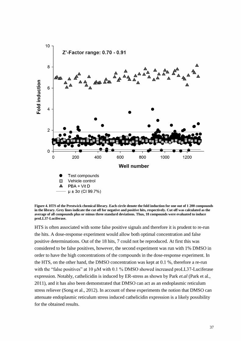
37
Figure 4. HTS of the Prestwick chemical library. Each circle denote the fold induction for one out of 1 200 compounds
in the library. Grey lines indicate the cut off for negative and positive hits, respectively. Cut off was calculated as the
average of all compounds plus or minus three standard deviations. Thus, 18 compounds were evaluated to induce
proLL37-Luciferase.
HTS is often associated with some false positive signals and therefore it is prudent to re-run
the hits. A dose-response experiment would allow both optimal concentration and false
positive determinations. Out of the 18 hits, 7 could not be reproduced. At first this was
considered to be false positives, however, the second experiment was run with 1% DMSO in
order to have the high concentrations of the compounds in the dose-response experiment. In
the HTS, on the other hand, the DMSO concentration was kept at 0.1 %, therefore a re-run
with the “false positives” at 10 µM with 0.1 % DMSO showed increased proLL37-Luciferase
expression. Notably, cathelicidin is induced by ER-stress as shown by Park et.al (Park et al.,
2011), and it has also been demonstrated that DMSO can act as an endoplasmic reticulum
stress reliever (Song et al., 2012). In account of these experiments the notion that DMSO can
attenuate endoplasmic reticulum stress induced cathelicidin expression is a likely possibility
for the obtained results.

38
Since the assay is a cell based system with a reporter construct there is a chance that the
responses are due to a new phenotype introduced by the transfection. Therefore it is important
that the hits can be verified in the parental cell line. The validation was performed on seven of
the 11 confirmed hits. Four hits, 5-Azacytidine, Azathioprine, Thioguanosine and
Mercaptopurine, were rejected for further analysis since their safety profile would make them
unsuitable to treat infections. Azathioprine and Mercaptopurine are used to induce immune
suppression and the other two for treatment of late stage cancers, in which the safety profile is
not that restricted. The seven remaining compounds were used to stimulate the parental HT-
29 cell line in a dose-range around the optimal concentrations obtained in the reporter cell
line dose response experiment. Three out of these seven compounds were confirmed as
inducers of the CAMP gene also in the parental cells.
In total, the two small screening-experiments utilizing the Prestwick library and selected
HDAC-inhibitors produced five novel compounds with potent CAMP-inducing capacity:
tiratricol, clobetasol and auranofin from the Prestwick library along with isovaleric- and
isobutyric-acid from the screening of HDAC inhibitors.
Tiratricol is an analog of T3, the active form of thyroid hormone, and the thyroid hormone
pathway was found to be upregulated as described in paper IV, where we also could show
that T3 in fact induced CAMP gene expression. We know that many immune genes are
regulated by thyroid hormones, however despite several attempts we have not been able to
reproduce the T3 results on additional cells responding to T3, such as primary PBMC’s and
macrophages.
Clobetasol is a corticosteroid also known to activate the glucocorticoid receptor (GR), which
has been shown to downregulate AMP expression (Kulkarni et al., 2016). Notably, out of 17
compounds in the Prestwick library with glucocorticoid activity, only clobetasol induced LL-
37. Thus, the induction exerted by clobetasol is likely very specific. The mechanism is still
unknown and whether the action is mediated through the GR is not determined at this point
and will be the subject of follow-up experiments in our laboratory.
Lastly, we also identified auranofin, an organo-gold compound used to relive symptoms in
rheumatoid arthritis. Auranofin is known to act on both nuclear related factor 2 (Nrf2) and
NF-κB (Kim et al., 2010; Nakaya et al., 2011), however, neither of these pathways are likely
to induce LL-37 to any excess. NF-κB has been demonstrated to regulate cathelicidin (Li et
al., 2009; Park et al., 2011), however, in paper III, the pTRAF vector was used to identify
entinostat as an inducer of HIF-1α. The same experimental setup also enabled determination
of NF-κB and Nrf2-activation. Utilizing additional compounds that activated these pathways
more than auranofin, we could not observe any induction of CAMP gene expression (data not
shown), hence auranofin most likely increases LL-37 expression via a pathway separate from
NF-κB and Nrf2. Interestingly, auranofin has been found to be effective against both
methicillin-resistant Staphylococcus aureus (MRSA) and Mtb. A likely mode of action of
auranofin against MRSA is through the activation of the redox pathway (Harbut et al., 2015),

39
but increased AMP-expression may be a possible explanation (Dean et al., 2011; Pence et al.,
2015; Sass et al., 2008).
4.2 PAPER II: HIGHLY POTENT INDUCERS OF ENDOGENOUS ANTIMICROBIAL PEPTIDES FOR HOST DIRECTED THERAPY OF INFECTIONS
Rationale: Most compounds initially tested in the MN8CampLuc cell line showed moderate
inducing ability, and somewhat higher than for PBA. However, one compound, Entinostat,
exhibited potent AMP-inducing properties. This prompted further investigation if compounds
with similar structural features, (aroylated phenylene diamines, APDs) also were potent
inducers. Therefore it was of importance to investigate structural requirements and to produce
inducers with alternative structures in order to be able to select candidates with a suitable
activity vs toxicity profile as well as to obtain proof of concept in an animal infection model.
Aims: 1) To develop a number of different APDs and related compounds. 2) To obtain
information on how structural features affect the ability to induce LL-37 by utilizing the
MN8CampLuc cell line as the first assay. 3) To obtain information if the activity is
reproducible in the parental cell line HT-29 and to investigate if addition of vitamin D affects
the induction. 4) To obtain in vivo data in a rabbit model of Shigellosis, showing the potential
efficacy of entinostat.
(Numbers refer to compounds depicted in paper II and in Figure 5.
During the evaluation process of the MN8CampLuc cells in paper I, several experiments with
different possible inducers were performed. Butyrate, PBA and the two newly identified
HDAC inhibitors from paper I proved that HDAC-inhibition can be associated to LL-37
induction. Therefore, the structurally unrelated second generation HDAC inhibitors,
vorinostat and entinostat (5), were tested in the MN8CampLuc cell line. Despite being an
HDACi that is several orders of magnitude more potent than PBA, vorinostat (not depicted)
induced proLL37-Luciferase expression to approximately the same level as PBA. Entinostat
(5), however, outperformed all previously identified inducers of LL-37 expression.
Furthermore, the effect occurred at a concentration about 1000 times lower than any other
inducer hitherto tested.
A number of different compounds with similar structures were synthesized with variation of
substituents and other structural features. Thereafter these were analyzed for proLL37-
Lucifease induction and the consensus was that the general structure needed for induction
was the APD moiety (boxed molecule in Figure 5). Thus, a structurally new class of inducers,
APDs, was discovered. It was found that many of the novel APD-compounds were 15-20
times more potent than PBA (1) to induce proLL37-Lucifease and at 100-1000 times lower
concentration.
From the structure-activity studies it became clear that the substituents of the aroyl moiety (2-
4, 6-9) influenced the inducing ability. Also the diamine moiety was crucial for induction

40
since neither nitrogen could be exchanged for oxygen without complete loss of activity (10,
11, 13). Neither could the ring structure be exchanged for an aliphatic diamine (14, 15).
Substitutions in the aroyl moiety were more forgiving with respect to loss of activity. On the
other hand, removal of the carbonyl group resulted in a pronounced reduction in activity (16).
One of the derivatives was able to induce expression of proLL37-Luciferase to somewhat
higher levels than entinostat, albeit at a higher concentration. This could be due to a less toxic
substituent for this compound, allowing the dose response to rise above that of entinostat.
Compound 8 lacked the aroylated substituent and had reduced potency, and was less toxic
than 5. However, potency is often rejected for less toxic compounds and both compound 8
and 9 were considered to be possible leads for future drug development.
To obtain in vivo data, entinostat was used to treat rabbits in a model of shigellosis. The
results clearly demonstrated that entinostat improved clinical symptoms in the rabbit, reduced
the infectious burden and induced cathelicidin-expression in the gut epithelial layer, as shown
by immunohistochemistry. It should be mentioned that entinostat also is known to activate
other parts of the immune system i.e. to reduce the levels of forkhead box P3 (Foxp3) in
regulatory T-cells and hence activate a more pro-inflammatory and possibly a more
antibacterial response (Shen et al., 2012). Entinostat has also been demonstrated to regulate
immunity by inducing the unfolded protein response pathway, also called endoreticulum
stress (Gameiro et al., 2016). Whether the mechanism behind the beneficial effects is due to
induction of AMPs or involves a combination of additional immune functions needs to be
explored. However, our comparison with even more potent HDAC inhibitors suggests that
HDAC inhibition is not the major mechanism behind the induced expression of LL-37.

41
Figure 5. Chemical structures of the APD compounds tested for induction of LL-37. Number 1 is PBA and number 5
is entinostat. The boxed structure in the left top corner indicates the general structure of an APD.

42
4.3 PAPER III: ENTINOSTAT UP-REGULATES THE CAMP GENE ENCODING LL-37 VIA ACTIVATION OF STAT3 AND HIF-1 TRANSCRIPTION FACTORS
Rationale: Entinostat was found to be our most potent inducer of LL-37 expression.
However, the mechanism behind this induction has not been investigated. If the involved
transcription-factors could be identified, we could learn more details of LL-37 induction but
also obtain insight how additional AMPs are regulated by the same pathway.
Aim: To identify transcription factors involved in entinostat-mediated induction of LL-37 and
potential co-regulation with other AMPs.
Results: The entinostat inducing effect was discovered during the evaluation of the
MN8CampLuc cells in paper I. These results were confirmed and further evaluated for dose-
response and kinetics in the parental cell line HT-29. The optimal time for induction was
found to be 24 h, while the optimal dose was defined as 2.5 µM, based on the balance
between optimal effect and minimal toxicity. We also hypothesized that entinostat would
upregulate several AMPs and therefore we investigated expression-levels for human β
defensins. A combined action of several AMPs with different mechanism of action, leading
to a multiple attack to the pathogen would minimize the development of bacterial resistance.
Notably, we found that entinostat induced hBD1, an important peptide in epithelial defense
(Prado-Montes de Oca, 2010), whereas HBD2 was not affected.
Entinostat has been shown to have potent immunomodulatory effects. Shen et.al reported that
entinostat inhibits the effect of T regulatory cells by acetylation and activation of STAT3,
which in turn de-activated Foxp3, the master regulator of T regulatory cell development (Hori
et al., 2003; Shen et al., 2012). To elucidate whether STAT3 could be involved in AMP-
expression, the CAMP gene was scanned for STAT3 binding elements. Interestingly, several
STAT3 binding sites were identified in the promoter of the CAMP gene.
Further, the induction of CAMP gene expression could be repressed using both short hairpin
RNA (shRNA) and a STAT3-inhibitor. Additional evidence for STAT3-activation by
entinostat was obtained by the observation of increased expression of ‘B-cell lymphoma 2’
(Bcl2), a STAT3 downstream target gene (Carpenter and Lo, 2014) and by Western blot
analysis on nuclear extracts, where STAT3 appeared to be accumulated in the nucleus after
entinostat-treatment. However, no binding of STAT3 to the CAMP gene could be detected,
utilizing the ChIP technique. Hence, it was concluded that STAT3 was a part of the
regulatory pathway of LL-37-expression, albeit it was acting as a second mediator rather than
binding directly to the CAMP gene promoter.
Given that HIF-1α, a transcription factor involved in cellular response to hypoxia, has been
shown to regulate LL-37 expression, we hypothesized that HIF-1α might be activated by
entinostat treatment and thereby increase expression of LL-37. Indeed, we found binding
elements for HIF-1α in the CAMP gene promoter and stimulation by entinostat caused
induced expression of HIF-1α mRNA in HT-29 cells. To test whether this increase in mRNA
was associated with an increase in active HIF-1, the vector pTRAF was utilized (Johansson et

43
al., 2015). This vector contains the gene for a fluorescent protein under the control of tandem
repeats of HIF-1 transcription factor binding sites. Therefore, entinostat was used to stimulate
HEK293 cells transfected with the pTRAF vector, which revealed that HIF-1 activation was
indeed increased upon entinostat treatment. In addition, utilizing ChIP, HIF-1α was shown to
be enriched on the CAMP gene upon stimulation with entinostat.
Furthermore, shRNA experiments indicated that knockdown of HIF-1α completely abrogated
entinostat elicited induction of the CAMP gene. Interestingly, shRNA directed towards
STAT3 inhibited the entinostat induced HIF1A gene expression, suggesting an important
cross-talk between STAT3 and HIF-1α.
The following mechanism of LL-37 induction elicited by entinostat is suggested. Entinostat
inhibits deacetylation of STAT3 and hence increase the amount of active STAT3, which
translocates to the nucleus where it acts as a dual activator by binding and activating
transcription of the HIF1A gene but also by stabilization of the HIF-1α subunit, which then in
turn will bind to the promoter of the CAMP gene and initiate transcription (see Figure 7).
Finally, we had access to whole-blood derived macrophages from a HIES-patient, with a
causative STAT3 mutation. Interestingly, entinostat did not induce CAMP gene-expression in
these cells as compared to two healthy control individuals, which lend further support to the
observation that STAT3 is important for LL-37 expression (Figure 6).
Figure 6. Entinostat elicited induction of CAMP gene expression is reduced in macrophages derived from a HIES
patient compared to healthy individuals.

44
Figure 7. Mechanism of entinostat induced LL-37 expression. Entinostat inhibit HDAC activity, which shifts STAT3
towards a more active state (a). Acetylated STAT3 is translocated to the nucleus and activates transcription of the
HIF1A gene (b). Increased transcription of HIF1A will lead to more HIF-1α translated protein (c). Acetylated STAT3
inhibits ubiquitination and hence degradation of HIF-1α (d). Stabilized HIF-1α then binds to the CAMP gene and
initiates transcription (e), leading to downstream translation of the pro-peptide of LL-37, hCAP-18 (f).

45
4.4 PAPER IV: LABEL-FREE QUANTITATIVE MASS SPECTROMETRY REVEALS NOVEL PATHWAYS INVOLVED IN LL-37 EXPRESSION
Rationale: It was found that co-stimulation with lactose and PBA, exhibited a synergistic
effect on AMP-inducing properties. However, there was no information on the mechanism
governing this potent induction. Identifying the mechanism could potentially lead to novel
inducing compounds.
Aim: To perform a proteomic screen on cell lysates from HT-29 cells stimulated with
vehicle, lactose, PBA or a combination of PBA-lactose and to use bioinformatic methods to
map the proteomic data on regulatory pathways.
In previous work from our group it was demonstrated that a synergistic induction of CAMP
gene expression occurred upon combined stimulation with PBA and lactose (Cederlund et al.,
2013).
To elucidate the mechanism behind this induction, cells were treated with a combination of
PBA and lactose or with each compound, separately. Next, the cell lysates were prepared and
analyzed, utilizing Label-Free Quantitative MS to determine the complete proteome of these
cells. More than 1300 proteins were identified, quantified and protein quantities were
normalized to the protein content of untreated cells. Proteins recognized to be up- or
downregulated were mapped to the ingenuity knowledgebase (IKB) and pathways were
predicted, utilizing the ingenuity pathway analyzer (IPA). IPA also assigned significance
values to the suggested pathways and ranked them accordingly. Utilizing the MN8CampLuc
cell line developed in paper I 11 pathways were suggested as activated by IPA and were
evaluated by treating the cells with either agonists or antagonists, to crucial components of
the identified pathways. Three pathways were induced or blocked with specific agonists and
antagonists, utilizing MN8CampLuc cells: i.e. the thyroid hormone receptor/retinoid X
receptor (TR/RXR) pathway, the eicosanoid signaling pathway and the steroid biosynthesis
pathway. Stimulation of MN8CampLuc cells with the active thyroid hormone (T3) and the
proform (T4) resulted in increased luminescent signals. Moreover, the combination of lactose
with T3 and T4 indicated an additive effect, whereas PBA and T3 exhibited a more
synergistic effect. Notably, the parental cell line HT-29, did not respond toT4-stimulation,
while T3 induced CAMP gene expression within the short span of 6 h of stimulation. Thus,
these results indicated an important difference between the reporter cell-line and the parental
cells. mRNA degradation is a regulatory pathway often applied to immune genes and it is
possible that the CAMP gene is regulated this way and that the extended mRNA of the
CampLuc fusion gene is more stable and hence prolong the optimal time of induction (Schott
and Stoecklin, 2010). Also consider the time lag between transcription and translation as well
as the stability of mRNA versus protein. At this point, we cannot explain why T4 failed to
produce a response in the parental cells, but we plan to elucidate the details in future projects.
T3 is a ligand of the thyroid hormone receptor (TR), and it is known that HDAC inhibitors
can acetylate various transcription factors. Sanchez-Pacheco et al. reported that the TR can be

46
activated via acetylation, which could explain the synergistic effect of T3 combined with the
HDAC inhibitor PBA (Sanchez-Pacheco et al., 2009).
The Eicosanoid signaling pathway was examined using several antagonists of eicosanoid
synthesis. MN8CampLuc cells were treated with PBA and lactose separately or in
combination and mixed with the antagonists. We found that inhibition of cyclooxygenase-2
(COX-2) was enough to significantly reduce PBA/lactose induction of proLL37-Luciferase
expression and this could be validated in the parental cell line HT-29, utilizing the COX-1/2
antagonist acetylsalicylic acid (ASA). Given that COX-2 inhibition would deplete the levels
of PGE2, addition of downstream prostaglandin E2 (PGE2) would rescue CAMP gene
expression. However, we could not prove this hypothesis in our experiments. One
explanation for this unexpected result is that the eicosanoid signaling pathway is a
downstream target with an AMP inducing feed-back mechanism since cathelicidin has been
demonstrated to induce the production of both COX-2 and PGE2 (Chamorro et al., 2009).
Another explanation is that there are other COX-2 generated lipid products that are
responsible for the regulation. Bernard and Gallo have demonstrated that hBD2 and hBD3
also are COX-2 regulated and that the lipid products PGD2 and 15-deoxy-∆12,14-PGJ2 is
inducing these defensins (Bernard and Gallo, 2010). Interestingly, Park et al. introduced
sphingosine-1-phosphate as a key inducer of human cathelicidin and there are indications that
sphingosine-1-phosphate increases expression of COX-2 (Hsu et al., 2015; Park et al., 2016;
Park et al., 2014; Rumzhum et al., 2016; Volzke et al., 2014). There are additional links
between lipid mediators and LL-37, including the interconnected circuits of activation and
production of LL-37 and leukotriene B4 (Wan et al., 2011; Wan et al., 2007; Wan et al.,
2014a).
The steroid biosynthesis pathway was also probed by inhibition studies on the MN8CampLuc
cells. PBA and lactose, separately and in combination, were co-incubated with Simvastatin,
an inhibitor of HMG-CoA reductase, which would block the production of cholesterol and
isoprenoids. Simvastatin inhibited CAMP gene induction by PBA/lactose and the effect of
PBA per se, but not the effect of lactose. The results were validated in the parental cell line
HT-29, with the difference that the effect of lactose mediated induction also could be
inhibited by simvastatin. The pathway was independent of the downstream products
mevalonate and cholesterol since addition of these compounds did not rescue induction of the
CAMP gene. These experiments suggest a HMG-CoA dependent but cholesterol-independent
regulation of LL-37 expression. More experiments must be performed to prove this fact but
there are indications that statins may exhibit anti-inflammatory effects. Statins have been
shown to affect several signaling pathways, including the NF-κB, AP-1, and HIF-1 pathways,
which all are important for the regulation of different AMPs. Interestingly, all these
pathways are down-regulated by statins, which is somewhat in line with our results (Ganz,
2003; Kida et al., 2006; Peyssonnaux et al., 2005; Rius et al., 2008; Tousoulis et al., 2014).

47
5 CONCLUSIONS
In conclusion, our results indicate that LL-37 is part of a complex regulatory network and is
not merely a response to infection and wounding. It seems that LL-37 and other AMPs are
carefully regulated also during non-stress situations in order to reach homeostasis between
the host, the commensal and the pathogenic microbiota.
Paper I: A well functioning assay was produced and 5 novel inducers of LL-37
expression were reported.
Paper II: Entinostat was found to be a potent inducer of LL-37 expression, however,
the inducing mechanism was only in part due to HDAC inhibition.Altering the
chemical structure of entinostat displayed which parts of the molecule that was
important for achiving induction.
Paper III: By combining several classical molecular techniques we could conclude
that LL-37 was regulated by entinostat through the transcription factors HIF-1 and
STAT3. We also showed that entinostat induced LL-37 expression was impaired in a
HIES patient with a known STAT3-mutation.
Paper IV: Utilizing label free mass spectrometry we have gained full insight into how
different stimuli alter the proteome of the cell populations. Strategic use of proteomic
pathway analysis tools and quick examination of pathways utilizing the
MN8CampLuc cells showed that this methodology is a resonable alternative to large
HTS. We could demonstrate that three of several proposed pathways indeed regulated
LL-37 expression.

48
6 FUTURE EXPERIMENTS OR THINGS I WOULD HAVE LIKED TO DO
There are several experiments that could be performed to gain more insight into the
regulating pathways of AMPs in general and for LL-37 expression in particular.
1. Since HDAC inhibitors are potent inducers of LL-37 but the detailed mechanism of action
remains elusive, I suggest that a screen of a larger HDAC inhibitor library is investigated.
Thereby, a better and more specific understanding of the role of HDAC-inhibition in the
regulation of AMP-expression can be gained.
2. Another way to further dissect the mechanism could be to alter the chemical structure of
entinostat to obtain a compound with retained LL-37 inducing activity but with no HDAC-
inhibitory activity.
3. The development of CRISPR/CAS9 is a true revolution in molecular biology and enables
rapid gene-editing. This technique could be used to both knockdown and knock in genes
important for AMP expression, which will greatly advance the knowledge of AMP
regulation.
4. To obtain additional information on transcription-factor of relevance for AMP-expression,
there are several new and powerful techniques available. For example, The use of enChIP
coupled to MS, a method for capturing specific regions of DNA, utilizing CRISPR/CAS9 and
ChIP, followed by analysis of the protein composition in this region could also be of great
value to deciphering which factors drive or inhibit AMP transcription (Fujita and Fujii, 2013).
In addition, ChIP-seq is another powerful method to find transcription-factors. Many
experiments using this method have been performed where different stimuli and different
antibodies have been used, all relevant for AMP expression. Notably, these data are often
made available and can be collected from various web-based depositories. These data-sets
can be re-evaluated with regard to AMP-expression. Most likely, there is a wealth of relevant
information available in silico.

49
7 ACKNOWLEDGEMENTS
The work in this thesis has been carried out at the Karolinska Institutet, Department of
Medical Biochemistry and Biophysics, Chemistry I and II and at the Department of
Laboratory Medicine, Clinical Microbiology. I would like to thank the institute and
both departments for providing a stimulating environment for research and aiding me
in my work and thesis writing.
I would like to thank all co-authors, donors and patients, their contributions have been
invaluable to my work.
I was told, I should start with my acknowledgements, “you don’t want to forget anyone
because you are feeling stressed in the end”. Well, sorry if I have left anyone out!
I’ve been fortunate to have two main supervisors during my PhD, and even though Birgitta
Agerberth isn't my main supervisor on paper any more, she has not lost interest in me. She
took me in and has smoothly guided me through these last 5? years and sculpted me in her
own image. Now everything is ‘novel’ to me and ‘furthermore’, I have done more ChIPs than
I want to remember, ‘however’ with a great mind and always being friendly, she is the image
of the scientist I want to be! I will always be grateful to you for all your help, guidance and
support, many thanks!
My main super-dupervisor Peter Bergman’s picture is in Wikipedia filed under ‘Effective’.
Apparently, he published a paper on amyloids and wrote another about fruit flies while I was
writing chapter 1.6.1. No need for Google when he’s around, he knows everything.
Additionally, he’s professional, friendly and importantly, always takes to new ideas with
vigor! Your help and guidance has been invaluable. Thank you!
I would also like to thank my Icelandic Viking-like co-supervisor Guðmundur Hrafn
Guðmundsson. A man teeming with ideas and like all respectable scientists, he knows his
way around molecular cloning. My only regret is that I never got to work in his lab in
Iceland, apparently they don't use incubators there; they just have flasks hanging at
predetermined lengths over volcanos.
Erica Miraglia, I could only have dreamt of a better co-worker. I wouldn’t have wanted to
do this without you and you made coming to work so much more fun (you other guys were
also fun)! We were a great team, miss you! Oh, I once made her blow into a USB connector
on the back of her computer to get her internet going…
Rekah Sultana Rokeya or Rokeya Rekah Sultana or Rokeya Sultana Rekah, we just call
her Rekha. Or Rokeya. We don’t really know. She’s the best of us. Runs ELISAs because
they’re fun!
From Torpet, Monica Lindh, the one to lean on and ask how to calculate molarity. Now she
mostly grows stuff or when I think about, she mostly throws stuff. Matte had to help her for
50
an entire day. Well she too needs some help because she helps me the rest of the time. Thank
you Monica!
We got a new roommate and group member, Sim Salah ‘Zangenah’ Bim var han där. A man
with many interests, a hairy chest and he only listens to KENT. Thanks for all the lunches
and interesting discussions about science, but mostly about society. And flowers. And
hedges. And razorblades. And excavators, cameras, cars, trailers, and people.
Andreas, haxball and wordfeud master. Most of all, thank you for everything. Kobi lomba.
Terese, Terese, Terese.
All the other people at the AMP lab. Ylva Kajjan Larsson, whisperer of not so secret stuff.
Protim, teacher of bad words and he only eats yellow snow food. Cina, Linda, Mislav,
Savitha, Christophf, Sadik, and a few more I’m sure I’ve missed.
From Preussen, Michael LandrEh, will be remembered for the thickest, thesis on fanily guy
science, ever. But he thought It was going to say something different. Its nice that you are
coming home. I’ve missed you.
Limpan, computer genius who has probably been infected with a computer virus at some
point.
Åke and Essam, best friends and forever my friends.
Åke, Susi, Susanne and fellow teachers at MBB.
Elias, Marcus and Katarina from MBB
Min, Jia, Dolores and the rest who were present at Chemistry 2 when I was there.
Some administrative people who are special to me are Svante, Chad, Torbjörn och
Johanna så klart. Tack för festerna och all dans.
Thanks Ella, Carina, Marie and Gunvor, and to the rest of you at former Chemistry 1 and
MBB, thanks.
Our friends from the physiology department, Lennart, Anne and Ellinor.
Roger and Håkan from BioNut.
Everyone at the new place, LabMed, and thanks to the welcoming committee of Las Frelin,
Matti Sällberg, Anders Sönnerborg, Andrej Weintraub, Anna and Annica Karlsson.
I also want to mention some people I’ve talked to a little extra, Shuba, Marjan, Xioshan,
Kerstin, Sepöjde, Mohammed, Markus, Sophie, Marita, and many more I realize now.
Thanks to you all.
Jovanka, for helping out in the very last minute.

51
My comrade Babbi and friend BomBomLas from Whatsapp. Thanks for all the selfies and
laughs and for being my friends, I hope to see you both IRL someday soon.
A big hug and a big thank you, to my mentor and friend Ylva Terelius. She is the reason why
I got in to science in the first place. I was hoping that maybe you wanted to continue
mentoring me?
To all my friends from and around Fyfa, Anna P, or R, let us call her Kråkan or Kajjan.
She is dear to me and therefore has many names. Magda with the greatest laugh in the world!
Gukken, världens bästa Gukken! Gukkens friend Larsa, fun at parties. Björn, who left for
Göteborg. Torun, also left for Göteborg. Peder Peppar! Calle, Maria, Aki, hon i fickan,
Annica, Sara, Klas, Marcus, Lullan, Pixi, Etienne, Isa, and many more. I forgot Alvin.
Thanks!
Tack till alla mina andra kompisar, och mina kära kusiner. Ni är bäst!
Alla andra i hela släkten också!
Christine och Per som försett mig med en sån fin och väluppfostrad kvinna.
Min tuffa syster Katja som fortfarande är min stora idol och mina fans Ragnar och Birger.
Vi ska nog se till att det blir vetenskapsmän av er också.
Mina älskade föräldrar, Lisbeth och Jarl eller mamma och pappa som jag kallar dom, är
mina största beundrare och utan er så hade det så klart inte blivit nånting, Jag älskar er!
Min lilla Elsie, det bästa som någonsin har hänt mig, eller någon annan heller för den delen.
Du lyser upp världen omkring dig och jag älskar dig så mycket att du omöjligen kan förstå
det. Ska bara skriva klart det här, sen kommer jag hem!
Jag avslutar min lista med att stolt nämna, Sara, min vackra kvinna! Utan dig vill jag inte
vara. Tack för all din uppmuntran och hjälp under den här tiden, Jag älskar dig och ser fram
emot vår bästa sommar någonsin. Puss!

52
8 REFERENCES
Aberg, K.M., Man, M.Q., Gallo, R.L., Ganz, T., Crumrine, D., Brown, B.E., Choi, E.H.,
Kim, D.K., Schroder, J.M., Feingold, K.R., and Elias, P.M. (2008). Co-regulation and
interdependence of the mammalian epidermal permeability and antimicrobial barriers. J
Invest Dermatol 128, 917-925.
Adachi, R., Honma, Y., Masuno, H., Kawana, K., Shimomura, I., Yamada, S., and
Makishima, M. (2005). Selective activation of vitamin D receptor by lithocholic acid acetate,
a bile acid derivative. Journal of lipid research 46, 46-57.
Agerberth, B., Charo, J., Werr, J., Olsson, B., Idali, F., Lindbom, L., Kiessling, R., Jornvall,
H., Wigzell, H., and Gudmundsson, G.H. (2000). The human antimicrobial and chemotactic
peptides LL-37 and alpha-defensins are expressed by specific lymphocyte and monocyte
populations. Blood 96, 3086-3093.
Agerberth, B., Gunne, H., Odeberg, J., Kogner, P., Boman, H.G., and Gudmundsson, G.H.
(1995). FALL-39, a putative human peptide antibiotic, is cysteine-free and expressed in bone
marrow and testis. Proc Natl Acad Sci U S A 92, 195-199.
Andrikopoulou, E., Zhang, X., Sebastian, R., Marti, G., Liu, L., Milner, S.M., and Harmon,
J.W. (2011). Current Insights into the role of HIF-1 in cutaneous wound healing. Curr Mol
Med 11, 218-235.
Arentz, G., Weiland, F., Oehler, M.K., and Hoffmann, P. (2015). State of the art of 2D DIGE.
Proteomics. Clinical applications 9, 277-288.
Ariffin, J.K., das Gupta, K., Kapetanovic, R., Iyer, A., Reid, R.C., Fairlie, D.P., and Sweet,
M.J. (2015). Histone Deacetylase Inhibitors Promote Mitochondrial Reactive Oxygen
Species Production and Bacterial Clearance by Human Macrophages. Antimicrobial agents
and chemotherapy 60, 1521-1529.
Bantscheff, M., Schirle, M., Sweetman, G., Rick, J., and Kuster, B. (2007). Quantitative mass
spectrometry in proteomics: a critical review. Analytical and bioanalytical chemistry 389,
1017-1031.
Barlow, P.G., Li, Y., Wilkinson, T.S., Bowdish, D.M., Lau, Y.E., Cosseau, C., Haslett, C.,
Simpson, A.J., Hancock, R.E., and Davidson, D.J. (2006). The human cationic host defense
peptide LL-37 mediates contrasting effects on apoptotic pathways in different primary cells
of the innate immune system. J Leukoc Biol 80, 509-520.
Bergman, P., Johansson, L., Asp, V., Plant, L., Gudmundsson, G.H., Jonsson, A.B., and
Agerberth, B. (2005). Neisseria gonorrhoeae downregulates expression of the human
antimicrobial peptide LL-37. Cell Microbiol 7, 1009-1017.
Bergman, P., Norlin, A.C., Hansen, S., Rekha, R.S., Agerberth, B., Bjorkhem-Bergman, L.,
Ekstrom, L., Lindh, J.D., and Andersson, J. (2012). Vitamin D3 supplementation in patients
with frequent respiratory tract infections: a randomised and double-blind intervention study.
BMJ Open 2.
Bernard, J.J., and Gallo, R.L. (2010). Cyclooxygenase-2 enhances antimicrobial peptide
expression and killing of Staphylococcus aureus. J Immunol 185, 6535-6544.
Bevins, C.L. (2005). Events at the host-microbial interface of the gastrointestinal tract. V.
Paneth cell alpha-defensins in intestinal host defense. American journal of physiology.
Gastrointestinal and liver physiology 289, G173-176.

53
Bevins, C.L. (2013). Innate immune functions of alpha-defensins in the small intestine.
Digestive diseases (Basel, Switzerland) 31, 299-304.
Bevins, C.L., and Salzman, N.H. (2011). Paneth cells, antimicrobial peptides and
maintenance of intestinal homeostasis. Nat Rev Microbiol 9, 356-368.
Bianchi, M.E. (2007). DAMPs, PAMPs and alarmins: all we need to know about danger. J
Leukoc Biol 81, 1-5.
Borregaard, N., Miller, L.J., and Springer, T.A. (1987). Chemoattractant-regulated
mobilization of a novel intracellular compartment in human neutrophils. Science 237, 1204-
1206.
Brinkmann, V., and Zychlinsky, A. (2007). Beneficial suicide: why neutrophils die to make
NETs. Nat Rev Micro 5, 577-582.
Buettner, R., Mora, L.B., and Jove, R. (2002). Activated STAT signaling in human tumors
provides novel molecular targets for therapeutic intervention. Clin Cancer Res 8, 945-954.
Bullen, J.J., Rogers, H.J., and Leigh, L. (1972). Iron-binding proteins in milk and resistance
to Escherichia coli infection in infants. Br Med J 1, 69-75.
Carpenter, R.L., and Lo, H.W. (2014). STAT3 Target Genes Relevant to Human Cancers.
Cancers (Basel) 6, 897-925.
Carvalho Ade, O., and Gomes, V.M. (2009). Plant defensins--prospects for the biological
functions and biotechnological properties. Peptides 30, 1007-1020.
Casanova, J.L., Holland, S.M., and Notarangelo, L.D. (2012). Inborn errors of human JAKs
and STATs. Immunity 36, 515-528.
Cederlund, A., Kai-Larsen, Y., Printz, G., Yoshio, H., Alvelius, G., Lagercrantz, H.,
Stromberg, R., Jornvall, H., Gudmundsson, G.H., and Agerberth, B. (2013). Lactose in
human breast milk an inducer of innate immunity with implications for a role in intestinal
homeostasis. PLoS One 8, e53876.
Cederlund, A., Olliver, M., Rekha, R.S., Lindh, M., Lindbom, L., Normark, S., Henriques-
Normark, B., Andersson, J., Agerberth, B., and Bergman, P. (2011). Impaired release of
antimicrobial peptides into nasal fluid of hyper-IgE and CVID patients. PLoS One 6, e29316.
Celada, A., Borras, F.E., Soler, C., Lloberas, J., Klemsz, M., van Beveren, C., McKercher, S.,
and Maki, R.A. (1996). The transcription factor PU.1 is involved in macrophage
proliferation. J Exp Med 184, 61-69.
Chamorro, C.I., Weber, G., Gronberg, A., Pivarcsi, A., and Stahle, M. (2009). The human
antimicrobial peptide LL-37 suppresses apoptosis in keratinocytes. J Invest Dermatol 129,
937-944.
Chen, L.-f., Fischle, W., Verdin, E., and Greene, W.C. (2001). Duration of Nuclear NF-κB
Action Regulated by Reversible Acetylation. Science 293, 1653-1657.
Chen, Q., Jin, Y., Zhang, K., Li, H., Chen, W., Meng, G., and Fang, X. (2014). Alarmin
HNP-1 promotes pyroptosis and IL-1beta release through different roles of NLRP3
inflammasome via P2X7 in LPS-primed macrophages. Innate Immun 20, 290-300.
Chertov, O., Michiel, D.F., Xu, L., Wang, J.M., Tani, K., Murphy, W.J., Longo, D.L., Taub,
D.D., and Oppenheim, J.J. (1996). Identification of defensin-1, defensin-2, and
CAP37/azurocidin as T-cell chemoattractant proteins released from interleukin-8-stimulated
neutrophils. J Biol Chem 271, 2935-2940.

54
Choi, S.-M., McAleer, J.P., Zheng, M., Pociask, D.A., Kaplan, M.H., Qin, S., Reinhart, T.A.,
and Kolls, J.K. (2013). Innate Stat3-mediated induction of the antimicrobial protein Reg3γ is
required for host defense against MRSA pneumonia. The Journal of Experimental Medicine
210, 551-561.
Chu, H., Pazgier, M., Jung, G., Nuccio, S.-P., Castillo, P.A., de Jong, M.F., Winter, M.G.,
Winter, S.E., Wehkamp, J., Shen, B., et al. (2012). Human α-Defensin 6 Promotes Mucosal
Innate Immunity Through Self-Assembled Peptide Nanonets. Science 337, 477-481.
Clohessy, P., and Golden, B. (1995). Calprotectin‐Mediated Zinc Chelation as a Biostatic
Mechanism in Host Defence. Scandinavian journal of immunology 42, 551-556.
Cohen, S.N. (2013). DNA cloning: A personal view after 40 years. Proceedings of the
National Academy of Sciences 110, 15521-15529.
Cohen, S.N., Chang, A.C., Boyer, H.W., and Helling, R.B. (1973). Construction of
biologically functional bacterial plasmids in vitro. Proc Natl Acad Sci U S A 70, 3240-3244.
Cole, J.N., and Nizet, V. (2016). Bacterial Evasion of Host Antimicrobial Peptide Defenses.
Microbiol Spectr 4.
Cong, L., Ran, F.A., Cox, D., Lin, S., Barretto, R., Habib, N., Hsu, P.D., Wu, X., Jiang, W.,
Marraffini, L.A., and Zhang, F. (2013). Multiplex genome engineering using CRISPR/Cas
systems. Science 339, 819-823.
Consortium, T.E.P. (2012). An integrated encyclopedia of DNA elements in the human
genome. Nature 489, 57-74.
Cramer, T., Yamanishi, Y., Clausen, B.E., Forster, I., Pawlinski, R., Mackman, N., Haase,
V.H., Jaenisch, R., Corr, M., Nizet, V., et al. (2003). HIF-1alpha is essential for myeloid cell-
mediated inflammation. Cell 112, 645-657.
Crotzer, V.L., and Blum, J.S. (2009). Autophagy and its role in MHC-mediated antigen
presentation. Journal of immunology (Baltimore, Md. : 1950) 182, 3335-3341.
Damo, S.M., Kehl-Fie, T.E., Sugitani, N., Holt, M.E., Rathi, S., Murphy, W.J., Zhang, Y.,
Betz, C., Hench, L., Fritz, G., et al. (2013). Molecular basis for manganese sequestration by
calprotectin and roles in the innate immune response to invading bacterial pathogens.
Proceedings of the National Academy of Sciences 110, 3841-3846.
De Vito, P., Incerpi, S., Pedersen, J.Z., Luly, P., Davis, F.B., and Davis, P.J. (2011). Thyroid
hormones as modulators of immune activities at the cellular level. Thyroid : official journal
of the American Thyroid Association 21, 879-890.
De, Y., Chen, Q., Schmidt, A.P., Anderson, G.M., Wang, J.M., Wooters, J., Oppenheim, J.J.,
and Chertov, O. (2000). LL-37, the neutrophil granule- and epithelial cell-derived
cathelicidin, utilizes formyl peptide receptor-like 1 (FPRL1) as a receptor to chemoattract
human peripheral blood neutrophils, monocytes, and T cells. J Exp Med 192, 1069-1074.
Dean, S.N., Bishop, B.M., and van Hoek, M.L. (2011). Natural and synthetic cathelicidin
peptides with anti-microbial and anti-biofilm activity against Staphylococcus aureus. BMC
Microbiology 11, 1-13.
Distler, U., Kuharev, J., and Tenzer, S. (2014). Biomedical applications of ion mobility-
enhanced data-independent acquisition-based label-free quantitative proteomics. Expert Rev
Proteomics 11, 675-684.
Dobson, A., Cotter, P.D., Ross, R.P., and Hill, C. (2012). Bacteriocin Production: a Probiotic
Trait? Applied and Environmental Microbiology 78, 1-6.

55
Edgeworth, J., Gorman, M., Bennett, R., Freemont, P., and Hogg, N. (1991). Identification of
p8,14 as a highly abundant heterodimeric calcium binding protein complex of myeloid cells.
Journal of Biological Chemistry 266, 7706-7713.
Eick, S., Puklo, M., Adamowicz, K., Kantyka, T., Hiemstra, P., Stennicke, H., Guentsch, A.,
Schacher, B., Eickholz, P., and Potempa, J. (2014). Lack of cathelicidin processing in
Papillon-Lefevre syndrome patients reveals essential role of LL-37 in periodontal
homeostasis. Orphanet journal of rare diseases 9, 148.
Emberley, E.D., Niu, Y., Njue, C., Kliewer, E.V., Murphy, L.C., and Watson, P.H. (2003).
Psoriasin (S100A7) expression is associated with poor outcome in estrogen receptor-negative
invasive breast cancer. Clin Cancer Res 9, 2627-2631.
Enss, M.L., Cornberg, M., Wagner, S., Gebert, A., Henrichs, M., Eisenblatter, R., Beil, W.,
Kownatzki, R., and Hedrich, H.J. (2000). Proinflammatory cytokines trigger MUC gene
expression and mucin release in the intestinal cancer cell line LS180. Inflammation research :
official journal of the European Histamine Research Society ... [et al.] 49, 162-169.
Falkenberg, K.J., and Johnstone, R.W. (2014). Histone deacetylases and their inhibitors in
cancer, neurological diseases and immune disorders. Nat Rev Drug Discov 13, 673-691.
Fan, D., Coughlin, L.A., Neubauer, M.M., Kim, J., Kim, M.S., Zhan, X., Simms-Waldrip,
T.R., Xie, Y., Hooper, L.V., and Koh, A.Y. (2015). Activation of HIF-1alpha and LL-37 by
commensal bacteria inhibits Candida albicans colonization. Nature medicine 21, 808-814.
Fernandez de Caleya, R., Gonzalez-Pascual, B., Garcia-Olmedo, F., and Carbonero, P.
(1972). Susceptibility of phytopathogenic bacteria to wheat purothionins in vitro. Applied
microbiology 23, 998-1000.
Fleming, A. (1922). On a Remarkable Bacteriolytic Element Found in Tissues and
Secretions. Proceedings of the Royal Society of London. Series B, Containing Papers of a
Biological Character 93, 306-317.
Foell, D., Wittkowski, H., Vogl, T., and Roth, J. (2007). S100 proteins expressed in
phagocytes: a novel group of damage-associated molecular pattern molecules. J Leukoc Biol
81, 28-37.
Forbes, L.R., Milner, J., and Haddad, E. (2016). Signal transducer and activator of
transcription 3: a year in review. Curr Opin Hematol 23, 23-27.
Fox, J.L. (2013). Antimicrobial peptides stage a comeback. Nat Biotech 31, 379-382.
Freeman, A.F., and Holland, S.M. (2008). The Hyper IgE Syndromes. Immunology and
allergy clinics of North America 28, 277-viii.
Freeman, A.F., and Holland, S.M. (2010). Clinical Manifestations of Hyper IgE Syndromes.
Disease markers 29, 123-130.
Frohm, M., Agerberth, B., Ahangari, G., Ståhle-Bäckdahl, M., Lidén, S., Wigzell, H., and
Gudmundsson, G.H. (1997). The Expression of the Gene Coding for the Antibacterial
Peptide LL-37 Is Induced in Human Keratinocytes during Inflammatory Disorders. Journal of
Biological Chemistry 272, 15258-15263.
Fujita, T., and Fujii, H. (2013). Efficient isolation of specific genomic regions and
identification of associated proteins by engineered DNA-binding molecule-mediated
chromatin immunoprecipitation (enChIP) using CRISPR. Biochem Biophys Res Commun
439, 132-136.

56
Gallo, R.L., and Hooper, L.V. (2012). Epithelial antimicrobial defence of the skin and
intestine. Nature reviews. Immunology 12, 503-516.
Gallo, S.A., Wang, W., Rawat, S.S., Jung, G., Waring, A.J., Cole, A.M., Lu, H., Yan, X.,
Daly, N.L., Craik, D.J., et al. (2006). Theta-defensins prevent HIV-1 Env-mediated fusion by
binding gp41 and blocking 6-helix bundle formation. J Biol Chem 281, 18787-18792.
Gameiro, S.R., Malamas, A.S., Tsang, K.Y., Ferrone, S., and Hodge, J.W. (2016). Inhibitors
of histone deacetylase 1 reverse the immune evasion phenotype to enhance T-cell mediated
lysis of prostate and breast carcinoma cells. Oncotarget.
Ganz, T. (2003). Defensins: antimicrobial peptides of innate immunity. Nat Rev Immunol 3,
710-720.
Geng, H., Liu, Q., Xue, C., David, L.L., Beer, T.M., Thomas, G.V., Dai, M.-S., and Qian,
D.Z. (2012). HIF1α Protein Stability Is Increased by Acetylation at Lysine 709. Journal of
Biological Chemistry 287, 35496-35505.
Gil, S., Coldwell, S., Drury, J.L., Arroyo, F., Phi, T., Saadat, S., Kwong, D., and Chung,
W.O. (2015). Genotype-specific regulation of oral innate immunity by T2R38 taste receptor.
Mol Immunol 68, 663-670.
Glaser, R., Harder, J., Lange, H., Bartels, J., Christophers, E., and Schroder, J.M. (2005).
Antimicrobial psoriasin (S100A7) protects human skin from Escherichia coli infection. Nat
Immunol 6, 57-64.
Glick, D., Barth, S., and Macleod, K.F. (2010). Autophagy: cellular and molecular
mechanisms. The Journal of pathology 221, 3-12.
Gombart, A.F., Borregaard, N., and Koeffler, H.P. (2005). Human cathelicidin antimicrobial
peptide (CAMP) gene is a direct target of the vitamin D receptor and is strongly up-regulated
in myeloid cells by 1,25-dihydroxyvitamin D3. The FASEB Journal 19, 1067-1077.
Gostner, J.M., Becker, K., Fuchs, D., and Sucher, R. (2013). Redox regulation of the immune
response. Redox report : communications in free radical research 18, 88-94.
Groves, M.L., Peterson, R.F., and Kiddy, C.A. (1965). Polymorphism in the Red Protein
isolated from Milk of Individual Cows. Nature 207, 1007-1008.
Gudmundsson, G.H., Agerberth, B., Odeberg, J., Bergman, T., Olsson, B., and Salcedo, R.
(1996). The human gene FALL39 and processing of the cathelin precursor to the antibacterial
peptide LL-37 in granulocytes. Eur J Biochem 238, 325-332.
Hancock, R.E., and Diamond, G. (2000). The role of cationic antimicrobial peptides in innate
host defences. Trends Microbiol 8, 402-410.
Harbut, M.B., Vilchèze, C., Luo, X., Hensler, M.E., Guo, H., Yang, B., Chatterjee, A.K.,
Nizet, V., Jacobs, W.R., Schultz, P.G., and Wang, F. (2015). Auranofin exerts broad-
spectrum bactericidal activities by targeting thiol-redox homeostasis. Proceedings of the
National Academy of Sciences 112, 4453-4458.
Hau, C.S., Kanda, N., Noda, S., Tatsuta, A., Kamata, M., Shibata, S., Asano, Y., Sato, S.,
Watanabe, S., and Tada, Y. (2013). Visfatin enhances the production of cathelicidin
antimicrobial peptide, human beta-defensin-2, human beta-defensin-3, and S100A7 in human
keratinocytes and their orthologs in murine imiquimod-induced psoriatic skin. Am J Pathol
182, 1705-1717.
He, C.-S., Fraser, W.D., Tang, J., Brown, K., Renwick, S., Rudland-Thomas, J., Teah, J.,
Tanqueray, E., and Gleeson, M. (2016). The effect of 14 weeks of vitamin D3

57
supplementation on antimicrobial peptides and proteins in athletes. Journal of Sports Sciences
34, 67-74.
Heilborn, J.D., Nilsson, M.F., Kratz, G., Weber, G., Sorensen, O., Borregaard, N., and Stahle-
Backdahl, M. (2003). The cathelicidin anti-microbial peptide LL-37 is involved in re-
epithelialization of human skin wounds and is lacking in chronic ulcer epithelium. J Invest
Dermatol 120, 379-389.
Hirano, T., Ishihara, K., and Hibi, M. (2000). Roles of STAT3 in mediating the cell growth,
differentiation and survival signals relayed through the IL-6 family of cytokine receptors.
Oncogene 19, 2548-2556.
Holland, S.M., DeLeo, F.R., Elloumi, H.Z., Hsu, A.P., Uzel, G., Brodsky, N., Freeman, A.F.,
Demidowich, A., Davis, J., Turner, M.L., et al. (2007). STAT3 mutations in the hyper-IgE
syndrome. The New England journal of medicine 357, 1608-1619.
Hori, S., Nomura, T., and Sakaguchi, S. (2003). Control of regulatory T cell development by
the transcription factor Foxp3. Science 299, 1057-1061.
Hornef, M.W., Normark, B.H., Vandewalle, A., and Normark, S. (2003). Intracellular
recognition of lipopolysaccharide by toll-like receptor 4 in intestinal epithelial cells. J Exp
Med 198, 1225-1235.
Hsu, C.K., Lee, I.T., Lin, C.C., Hsiao, L.D., and Yang, C.M. (2015). Sphingosine-1-
phosphate mediates COX-2 expression and PGE2 /IL-6 secretion via c-Src-dependent AP-1
activation. J Cell Physiol 230, 702-715.
Hu, Z., Murakami, T., Suzuki, K., Tamura, H., Kuwahara-Arai, K., Iba, T., and Nagaoka, I.
(2014). Antimicrobial Cathelicidin Peptide LL-37 Inhibits the LPS/ATP-Induced Pyroptosis
of Macrophages by Dual Mechanism. PLoS ONE 9, e85765.
Hughes, J.P., Rees, S., Kalindjian, S.B., and Philpott, K.L. (2011). Principles of early drug
discovery. British Journal of Pharmacology 162, 1239-1249.
Hugot, J.P., Chamaillard, M., Zouali, H., Lesage, S., Cezard, J.P., Belaiche, J., Almer, S.,
Tysk, C., O'Morain, C.A., Gassull, M., et al. (2001). Association of NOD2 leucine-rich
repeat variants with susceptibility to Crohn's disease. Nature 411, 599-603.
Hulse, K.E., Singh, R., Chaung, K., Norton, J., Harris, K., Conley, D., Chandra, R., Kern, R.,
Peters, A., Grammer, L., et al. STAT3 and NF-kB Regulate S100A7 Expression in Human
Bronchial Epithelial Cells. Journal of Allergy and Clinical Immunology 129, AB68.
Hultmark, D., Steiner, H., Rasmuson, T., and Boman, H.G. (1980). Insect immunity.
Purification and properties of three inducible bactericidal proteins from hemolymph of
immunized pupae of Hyalophora cecropia. Eur J Biochem 106, 7-16.
Islam, D., Bandholtz, L., Nilsson, J., Wigzell, H., Christensson, B., Agerberth, B., and
Gudmundsson, G. (2001). Downregulation of bactericidal peptides in enteric infections: a
novel immune escape mechanism with bacterial DNA as a potential regulator. Nat Med 7,
180-185.
Jackson, D.A., Symons, R.H., and Berg, P. (1972). Biochemical method for inserting new
genetic information into DNA of Simian Virus 40: circular SV40 DNA molecules containing
lambda phage genes and the galactose operon of Escherichia coli. Proc Natl Acad Sci U S A
69, 2904-2909.
Jensen, J.M., Ahrens, K., Meingassner, J., Scherer, A., Brautigam, M., Stutz, A., Schwarz, T.,
Folster-Holst, R., Harder, J., Glaser, R., and Proksch, E. (2011). Differential suppression of

58
epidermal antimicrobial protein expression in atopic dermatitis and in EFAD mice by
pimecrolimus compared to corticosteroids. Experimental dermatology 20, 783-788.
Jenssen, H., and Hancock, R.E.W. (2009). Antimicrobial properties of lactoferrin. Biochimie
91, 19-29.
Jinek, M., Chylinski, K., Fonfara, I., Hauer, M., Doudna, J.A., and Charpentier, E. (2012). A
programmable dual-RNA-guided DNA endonuclease in adaptive bacterial immunity. Science
337, 816-821.
Jinek, M., East, A., Cheng, A., Lin, S., Ma, E., and Doudna, J. (2013). RNA-programmed
genome editing in human cells. eLife 2, e00471.
Johansson, J., Gudmundsson, G.H., Rottenberg, M.E., Berndt, K.D., and Agerberth, B.
(1998). Conformation-dependent antibacterial activity of the naturally occurring human
peptide LL-37. J Biol Chem 273, 3718-3724.
Johansson, K., Cebula, M., Rengby, O., Dreij, K., Carlstrom, K.E., Sigmundsson, K., Piehl,
F., and Arner, E.S. (2015). Cross-talk in HEK293 cells between Nrf2, HIF and NF-kappaB
activities upon challenges with redox therapeutics characterized with single-cell resolution.
Antioxid Redox Signal.
Jothi, R., Cuddapah, S., Barski, A., Cui, K., and Zhao, K. (2008). Genome-wide identification
of in vivo protein-DNA binding sites from ChIP-Seq data. Nucleic Acids Res 36, 5221-5231.
Jung, J.E., Kim, H.S., Lee, C.S., Shin, Y.-J., Kim, Y.-N., Kang, G.-H., Kim, T.-Y., Juhnn, Y.-
S., Kim, S.-J., Park, J.-W., et al. (2008). STAT3 inhibits the degradation of HIF-1[alpha] by
pVHL-mediated ubiquitination. Exp Mol Med 40, 479-485.
Kai-Larsen, Y., Bergsson, G., Gudmundsson, G.H., Printz, G., Jornvall, H., Marchini, G., and
Agerberth, B. (2007). Antimicrobial components of the neonatal gut affected upon
colonization. Pediatr Res 61, 530-536.
Kamada, N., Chen, G.Y., Inohara, N., and Nunez, G. (2013). Control of pathogens and
pathobionts by the gut microbiota. Nat Immunol 14, 685-690.
Kandler, K., Shaykhiev, R., Kleemann, P., Klescz, F., Lohoff, M., Vogelmeier, C., and Bals,
R. (2006). The anti-microbial peptide LL-37 inhibits the activation of dendritic cells by TLR
ligands. Int Immunol 18, 1729-1736.
Kawasaki, H., and Iwamuro, S. (2008). Potential roles of histones in host defense as
antimicrobial agents. Infectious disorders drug targets 8, 195-205.
Kawasaki, Y., Matsumura, K., Miyamoto, M., Tsuji, S., Okuno, M., Suda, S., Hiyoshi, M.,
Kitayama, J., and Akiyama, T. (2015). REG4 is a transcriptional target of GATA6 and is
essential for colorectal tumorigenesis. Scientific Reports 5, 14291.
Kelly, C.J., Glover, L.E., Campbell, E.L., Kominsky, D.J., Ehrentraut, S.F., Bowers, B.E.,
Bayless, A.J., Saeedi, B.J., and Colgan, S.P. (2013). Fundamental role for HIF-1alpha in
constitutive expression of human beta defensin-1. Mucosal Immunol 6, 1110-1118.
Kenshi, Y., and Richard, L.G. (2008). Antimicrobial peptides in human skin disease.
European journal of dermatology : EJD 18, 11-21.
Khoury, G.A., Baliban, R.C., and Floudas, C.A. (2011). Proteome-wide post-translational
modification statistics: frequency analysis and curation of the swiss-prot database. Scientific
reports 1, 90.
59
Kida, Y., Shimizu, T., and Kuwano, K. (2006). Sodium butyrate up-regulates cathelicidin
gene expression via activator protein-1 and histone acetylation at the promoter region in a
human lung epithelial cell line, EBC-1. Mol Immunol 43, 1972-1981.
Kierdorf, K., and Fritz, G. (2013). RAGE regulation and signaling in inflammation and
beyond. J Leukocyte Biol 94, 55-68.
Kilsgard, O., Andersson, P., Malmsten, M., Nordin, S.L., Linge, H.M., Eliasson, M.,
Sorenson, E., Erjefalt, J.S., Bylund, J., Olin, A.I., et al. (2012). Peptidylarginine deiminases
present in the airways during tobacco smoking and inflammation can citrullinate the host
defense peptide LL-37, resulting in altered activities. American journal of respiratory cell and
molecular biology 46, 240-248.
Kim, N.H., Oh, M.K., Park, H.J., and Kim, I.S. (2010). Auranofin, a gold(I)-containing
antirheumatic compound, activates Keap1/Nrf2 signaling via Rac1/iNOS signal and mitogen-
activated protein kinase activation. Journal of pharmacological sciences 113, 246-254.
Kiss, G., and Michl, H. (1962). Uber das Giftsekret der Gelbbauchunke, Bombina variegata
L. Toxicon 1, 33-34.
Klotman, M.E., Rapista, A., Teleshova, N., Micsenyi, A., Jarvis, G.A., Lu, W., Porter, E., and
Chang, T.L. (2008). Neisseria gonorrhoeae-induced human defensins 5 and 6 increase HIV
infectivity: role in enhanced transmission. J Immunol 180, 6176-6185.
Kobayashi, K.S., Chamaillard, M., Ogura, Y., Henegariu, O., Inohara, N., Nunez, G., and
Flavell, R.A. (2005). Nod2-dependent regulation of innate and adaptive immunity in the
intestinal tract. Science 307, 731-734.
Koczulla, R., von Degenfeld, G., Kupatt, C., Krotz, F., Zahler, S., Gloe, T., Issbrucker, K.,
Unterberger, P., Zaiou, M., Lebherz, C., et al. (2003). An angiogenic role for the human
peptide antibiotic LL-37/hCAP-18. J Clin Invest 111, 1665-1672.
Korn, T., Bettelli, E., Oukka, M., and Kuchroo, V.K. (2009). IL-17 and Th17 Cells. Annual
review of immunology 27, 485-517.
Koro, C., Hellvard, A., Delaleu, N., Binder, V., Scavenius, C., Bergum, B., Główczyk, I.,
Roberts, H.M., Chapple, I.L., Grant, M.M., et al. (2016). Carbamylated LL-37 as a modulator
of the immune response. Innate Immunity 22, 218-229.
Kovats, S. (2015). Estrogen receptors regulate innate immune cells and signaling pathways.
Cell Immunol 294, 63-69.
Koziel, J., Bryzek, D., Sroka, A., Maresz, K., Glowczyk, I., Bielecka, E., Kantyka, T., Pyrc,
K., Svoboda, P., Pohl, J., and Potempa, J. (2014). Citrullination alters immunomodulatory
function of LL-37 essential for prevention of endotoxin-induced sepsis. J Immunol 192,
5363-5372.
Kulkarni, N.N., Gunnarsson, H.I., Yi, Z., Gudmundsdottir, S., Sigurjonsson, O.E., Agerberth,
B., and Gudmundsson, G.H. (2016). Glucocorticoid dexamethasone down-regulates basal and
vitamin D3 induced cathelicidin expression in human monocytes and bronchial epithelial cell
line. Immunobiology 221, 245-252.
Kulkarni, N.N., Yi, Z., Huehnken, C., Agerberth, B., and Gudmundsson, G.H. (2015).
Phenylbutyrate induces cathelicidin expression via the vitamin D receptor: Linkage to
inflammatory and growth factor cytokines pathways. Mol Immunol 63, 530-539.
Lai, A.Y., and Wade, P.A. (2011). Cancer biology and NuRD: a multifaceted chromatin
remodelling complex. Nat Rev Cancer 11, 588-596.

60
Lai, Y., and Gallo, R.L. (2009). AMPed up immunity: how antimicrobial peptides have
multiple roles in immune defense. Trends Immunol 30, 131-141.
Lande, R., Gregorio, J., Facchinetti, V., Chatterjee, B., Wang, Y.-H., Homey, B., Cao, W.,
Wang, Y.-H., Su, B., Nestle, F.O., et al. (2007). Plasmacytoid dendritic cells sense self-DNA
coupled with antimicrobial peptide. Nature 449, 564-569.
Larrick, J.W., Hirata, M., Zhong, J., and Wright, S.C. (1995). Anti-microbial activity of
human CAP18 peptides. Immunotechnology 1, 65-72.
Lau, Y.E., Bowdish, D.M., Cosseau, C., Hancock, R.E., and Davidson, D.J. (2006).
Apoptosis of airway epithelial cells: human serum sensitive induction by the cathelicidin LL-
37. American journal of respiratory cell and molecular biology 34, 399-409.
Lauffer, B.E., Mintzer, R., Fong, R., Mukund, S., Tam, C., Zilberleyb, I., Flicke, B., Ritscher,
A., Fedorowicz, G., Vallero, R., et al. (2013). Histone deacetylase (HDAC) inhibitor kinetic
rate constants correlate with cellular histone acetylation but not transcription and cell
viability. J Biol Chem 288, 26926-26943.
Lee, K.S., Kalantzis, A., Jackson, C.B., O'Connor, L., Murata-Kamiya, N., Hatakeyama, M.,
Judd, L.M., Giraud, A.S., and Menheniott, T.R. (2012). Helicobacter pylori CagA Triggers
Expression of the Bactericidal Lectin REG3γ via Gastric STAT3 Activation. PLoS ONE 7,
e30786.
Lehrer, R.I., and Ganz, T. (2002). Defensins of vertebrate animals. Curr Opin Immunol 14,
96-102.
Li, G., Domenico, J., Jia, Y., Lucas, J.J., and Gelfand, E.W. (2009). NF-κB-Dependent
Induction of Cathelicidin-Related Antimicrobial Peptide in Murine Mast Cells by
Lipopolysaccharide. International Archives of Allergy and Immunology 150, 122-132.
Li, G.H., Wei, H., Lv, S.Q., Ji, H., and Wang, D.L. (2010). Knockdown of STAT3 expression
by RNAi suppresses growth and induces apoptosis and differentiation in glioblastoma stem
cells. International journal of oncology 37, 103-110.
Li, X., Li, Y., Han, H., Miller, D.W., and Wang, G. (2006). Solution structures of human LL-
37 fragments and NMR-based identification of a minimal membrane-targeting antimicrobial
and anticancer region. Journal of the American Chemical Society 128, 5776-5785.
Lipinski, C.A., Lombardo, F., Dominy, B.W., and Feeney, P.J. (1997). Experimental and
computational approaches to estimate solubility and permeability in drug discovery and
development settings. Advanced Drug Delivery Reviews 23, 3-25.
Loo, Y.-M., and Gale, M. (2011). Immune signaling by RIG-I-like receptors. Immunity 34,
680-692.
Lotze, M.T., Zeh, H.J., Rubartelli, A., Sparvero, L.J., Amoscato, A.A., Washburn, N.R.,
Devera, M.E., Liang, X., Tor, M., and Billiar, T. (2007). The grateful dead: damage-
associated molecular pattern molecules and reduction/oxidation regulate immunity. Immunol
Rev 220, 60-81.
Mali, P., Yang, L., Esvelt, K.M., Aach, J., Guell, M., DiCarlo, J.E., Norville, J.E., and
Church, G.M. (2013). RNA-guided human genome engineering via Cas9. Science 339, 823-
826.
Marr, A.K., Gooderham, W.J., and Hancock, R.E. (2006). Antibacterial peptides for
therapeutic use: obstacles and realistic outlook. Curr Opin Pharmacol 6, 468-472.
61
Matzinger, P. (1994). Tolerance, danger, and the extended family. Annu Rev Immunol 12,
991-1045.
Matzinger, P. (2002). The Danger Model: A Renewed Sense of Self. Science 296, 301-305.
Medzhitov, R. (2007). Recognition of microorganisms and activation of the immune
response. Nature 449, 819-826.
Medzhitov, R. (2009). Approaching the asymptote: 20 years later. Immunity 30, 766-775.
Miller, W.L. (2016). Genetic Disorders Of Vitamin D Biosynthesis and Degradation. The
Journal of steroid biochemistry and molecular biology.
Mills, C.D. (2012). M1 and M2 Macrophages: Oracles of Health and Disease. Critical
reviews in immunology 32, 463-488.
Milner, J.D., Brenchley, J.M., Laurence, A., Freeman, A.F., Hill, B.J., Elias, K.M., Kanno,
Y., Spalding, C., Elloumi, H.Z., Paulson, M.L., et al. (2008). Impaired TH17 cell
differentiation in subjects with autosomal dominant hyper-IgE syndrome. Nature 452, 773-
776.
Milner, J.D., Vogel, T.P., Forbes, L., Ma, C.A., Stray-Pedersen, A., Niemela, J.E., Lyons,
J.J., Engelhardt, K.R., Zhang, Y., Topcagic, N., et al. (2015). Early-onset lymphoproliferation
and autoimmunity caused by germline STAT3 gain-of-function mutations. Blood 125, 591-
599.
Mily, A., Rekha, R.S., Kamal, S.M., Arifuzzaman, A.S., Rahim, Z., Khan, L., Haq, M.A.,
Zaman, K., Bergman, P., Brighenti, S., et al. (2015). Significant Effects of Oral
Phenylbutyrate and Vitamin D3 Adjunctive Therapy in Pulmonary Tuberculosis: A
Randomized Controlled Trial. PLoS One 10, e0138340.
Mogensen, T.H. (2009). Pathogen Recognition and Inflammatory Signaling in Innate
Immune Defenses. Clinical Microbiology Reviews 22, 240-273.
Moore, A. (2003). The big and small of drug discovery. EMBO Reports 4, 114-117.
Mosser, D.M., and Edwards, J.P. (2008). Exploring the full spectrum of macrophage
activation. Nature reviews. Immunology 8, 958-969.
Muchardt, C., and Yaniv, M. (1999). ATP-dependent chromatin remodelling: SWI/SNF and
Co. are on the job. Journal of molecular biology 293, 187-198.
Muehleisen, B., Bikle, D.D., Aguilera, C., Burton, D.W., Sen, G.L., Deftos, L.J., and Gallo,
R.L. (2012). PTH/PTHrP and vitamin D control antimicrobial peptide expression and
susceptibility to bacterial skin infection. Sci Transl Med 4, 135ra166.
Mukherjee, S., Zheng, H., Derebe, M.G., Callenberg, K.M., Partch, C.L., Rollins, D.,
Propheter, D.C., Rizo, J., Grabe, M., Jiang, Q.X., and Hooper, L.V. (2014). Antibacterial
membrane attack by a pore-forming intestinal C-type lectin. Nature 505, 103-107.
Munk, C., Wei, G., Yang, O.O., Waring, A.J., Wang, W., Hong, T., Lehrer, R.I., Landau,
N.R., and Cole, A.M. (2003). The theta-defensin, retrocyclin, inhibits HIV-1 entry. AIDS
research and human retroviruses 19, 875-881.
Murano, T., Okamoto, R., Ito, G., Nakata, T., Hibiya, S., Shimizu, H., Fujii, S., Kano, Y.,
Mizutani, T., Yui, S., et al. (2014). Hes1 promotes the IL-22-mediated antimicrobial response
by enhancing STAT3-dependent transcription in human intestinal epithelial cells. Biochem
Biophys Res Commun 443, 840-846.

62
Muto, Y., Wang, Z., Vanderberghe, M., Two, A., Gallo, R.L., and Di Nardo, A. (2014). Mast
cells are key mediators of cathelicidin initiated skin inflammation in rosacea. The Journal of
investigative dermatology 134, 2728-2736.
Mygind, P.H., Fischer, R.L., Schnorr, K.M., Hansen, M.T., Sonksen, C.P., Ludvigsen, S.,
Raventos, D., Buskov, S., Christensen, B., De Maria, L., et al. (2005). Plectasin is a peptide
antibiotic with therapeutic potential from a saprophytic fungus. Nature 437, 975-980.
Nagaoka, I., Tamura, H., and Hirata, M. (2006). An antimicrobial cathelicidin peptide, human
CAP18/LL-37, suppresses neutrophil apoptosis via the activation of formyl-peptide receptor-
like 1 and P2X7. J Immunol 176, 3044-3052.
Nakaya, A., Sagawa, M., Muto, A., Uchida, H., Ikeda, Y., and Kizaki, M. (2011). The gold
compound auranofin induces apoptosis of human multiple myeloma cells through both down-
regulation of STAT3 and inhibition of NF-kappaB activity. Leukemia research 35, 243-249.
Nakra, N.A., Madan, R.P., Buckley, N., Huber, A.M., Freiermuth, J.L., Espinoza, L., Walsh,
J., Parikh, U.M., Penrose, K.J., Keller, M.J., and Herold, B.C. (2016). Loss of Innate Host
Defense Following Unprotected Vaginal Sex. J Infect Dis 213, 840-847.
Nathan, C., and Cunningham-Bussel, A. (2013). Beyond oxidative stress: an immunologist's
guide to reactive oxygen species. Nat Rev Immunol 13, 349-361.
Neish, A.S. (2013). Redox signaling mediated by the gut microbiota. Free radical research 47,
950-957.
Niyonsaba, F., Someya, A., Hirata, M., Ogawa, H., and Nagaoka, I. (2001). Evaluation of the
effects of peptide antibiotics human beta-defensins-1/-2 and LL-37 on histamine release and
prostaglandin D(2) production from mast cells. Eur J Immunol 31, 1066-1075.
Nizet, V., Ohtake, T., Lauth, X., Trowbridge, J., Rudisill, J., Dorschner, R.A., Pestonjamasp,
V., Piraino, J., Huttner, K., and Gallo, R.L. (2001). Innate antimicrobial peptide protects the
skin from invasive bacterial infection. Nature 414, 454-457.
Nuding, S., Antoni, L., and Stange, E.F. (2013). The Host and the Flora. Digestive Diseases
31, 286-292.
O'Neil, D.A., Porter, E.M., Elewaut, D., Anderson, G.M., Eckmann, L., Ganz, T., and
Kagnoff, M.F. (1999). Expression and regulation of the human beta-defensins hBD-1 and
hBD-2 in intestinal epithelium. J Immunol 163, 6718-6724.
Ong, P.Y., Ohtake, T., Brandt, C., Strickland, I., Boguniewicz, M., Ganz, T., Gallo, R.L., and
Leung, D.Y. (2002). Endogenous antimicrobial peptides and skin infections in atopic
dermatitis. N Engl J Med 347, 1151-1160.
Ouellette, A.J. (2011). Paneth cell alpha-defensins in enteric innate immunity. Cell Mol Life
Sci 68, 2215-2229.
Ouellette, A.J., and Selsted, M.E. (2012). HD6 Defensin Nanonets. Science 337, 420-421.
Parikh, A., Stephan, A.-F., and Tzanakakis, E.S. (2012). Regenerating proteins and their
expression, regulation and signaling. Biomolecular concepts 3, 57-70.
Park, K., Elias, P.M., Oda, Y., Mackenzie, D., Mauro, T., Holleran, W.M., and Uchida, Y.
(2011). Regulation of cathelicidin antimicrobial peptide expression by an endoplasmic
reticulum (ER) stress signaling, vitamin D receptor-independent pathway. J Biol Chem 286,
34121-34130.

63
Park, K., Ikushiro, H., Seo, H.S., Shin, K.O., Kim, Y.I., Kim, J.Y., Lee, Y.M., Yano, T.,
Holleran, W.M., Elias, P., and Uchida, Y. (2016). ER stress stimulates production of the key
antimicrobial peptide, cathelicidin, by forming a previously unidentified intracellular S1P
signaling complex. Proc Natl Acad Sci U S A.
Park, K., Kim, Y.-I., Shin, K.-O., Seo, H.S., Kim, J.Y., Mann, T., Oda, Y., Lee, Y.-M.,
Holleran, W.M., Elias, P.M., and Uchida, Y. (2014). The dietary ingredient, genistein,
stimulates cathelicidin antimicrobial peptide expression through a novel S1P-dependent
mechanism. The Journal of Nutritional Biochemistry 25, 734-740.
Pence, M.A., Haste, N.M., Meharena, H.S., Olson, J., Gallo, R.L., Nizet, V., and Kristian,
S.A. (2015). Beta-Lactamase Repressor BlaI Modulates Staphylococcus aureus Cathelicidin
Antimicrobial Peptide Resistance and Virulence. PLoS ONE 10, e0136605.
Pène, J., Chevalier, S., Preisser, L., Vénéreau, E., Guilleux, M.-H., Ghannam, S., Molès, J.-
P., Danger, Y., Ravon, E., Lesaux, S., et al. (2008). Chronically Inflamed Human Tissues Are
Infiltrated by Highly Differentiated Th17 Lymphocytes. The Journal of Immunology 180,
7423-7430.
Perez-Vilar, J., and Hill, R.L. (1999). The Structure and Assembly of Secreted Mucins.
Journal of Biological Chemistry 274, 31751-31754.
Peyssonnaux, C., Datta, V., Cramer, T., Doedens, A., Theodorakis, E.A., Gallo, R.L.,
Hurtado-Ziola, N., Nizet, V., and Johnson, R.S. (2005). HIF-1alpha expression regulates the
bactericidal capacity of phagocytes. J Clin Invest 115, 1806-1815.
Philpott, D.J., Sorbara, M.T., Robertson, S.J., Croitoru, K., and Girardin, S.E. (2014). NOD
proteins: regulators of inflammation in health and disease. Nat Rev Immunol 14, 9-23.
Porter, E., Yang, H., Yavagal, S., Preza, G.C., Murillo, O., Lima, H., Greene, S., Mahoozi,
L., Klein-Patel, M., Diamond, G., et al. (2005). Distinct Defensin Profiles in Neisseria
gonorrhoeae and Chlamydia trachomatis Urethritis Reveal Novel Epithelial Cell-Neutrophil
Interactions. Infection and Immunity 73, 4823-4833.
Prado-Montes de Oca, E. (2010). Human beta-defensin 1: a restless warrior against allergies,
infections and cancer. Int J Biochem Cell Biol 42, 800-804.
Price, L.B., and Shand, R.F. (2000). Halocin S8: a 36-Amino-Acid Microhalocin from the
Haloarchaeal Strain S8a. Journal of Bacteriology 182, 4951-4958.
Putsep, K., Carlsson, G., Boman, H.G., and Andersson, M. (2002). Deficiency of
antibacterial peptides in patients with morbus Kostmann: an observation study. Lancet
(London, England) 360, 1144-1149.
Rapista, A., Ding, J., Benito, B., Lo, Y.T., Neiditch, M.B., Lu, W., and Chang, T.L. (2011).
Human defensins 5 and 6 enhance HIV-1 infectivity through promoting HIV attachment.
Retrovirology 8, 45.
Raqib, R., Sarker, P., Bergman, P., Ara, G., Lindh, M., Sack, D.A., Nasirul Islam, K.M.,
Gudmundsson, G.H., Andersson, J., and Agerberth, B. (2006). Improved outcome in
shigellosis associated with butyrate induction of an endogenous peptide antibiotic.
Proceedings of the National Academy of Sciences 103, 9178-9183.
Rekha, R.S., Rao Muvva, S.S., Wan, M., Raqib, R., Bergman, P., Brighenti, S.,
Gudmundsson, G.H., and Agerberth, B. (2015). Phenylbutyrate induces LL-37-dependent
autophagy and intracellular killing of Mycobacterium tuberculosis in human macrophages.
Autophagy 11, 1688-1699.

64
Ren, S.X., Cheng, A.S., To, K.F., Tong, J.H., Li, M.S., Shen, J., Wong, C.C., Zhang, L.,
Chan, R.L., Wang, X.J., et al. (2012). Host immune defense peptide LL-37 activates caspase-
independent apoptosis and suppresses colon cancer. Cancer Res 72, 6512-6523.
Rius, J., Guma, M., Schachtrup, C., Akassoglou, K., Zinkernagel, A.S., Nizet, V., Johnson,
R.S., Haddad, G.G., and Karin, M. (2008). NF-kappaB links innate immunity to the hypoxic
response through transcriptional regulation of HIF-1alpha. Nature 453, 807-811.
Rosenberg, H.F., Dyer, K.D., and Foster, P.S. (2013). Eosinophils: changing perspectives in
health and disease. Nat Rev Immunol 13, 9-22.
Rumzhum, N.N., Rahman, M.M., Oliver, B.G., and Ammit, A.J. (2016). Effect of
Sphingosine 1-Phosphate on Cyclo-Oxygenase-2 Expression, Prostaglandin E2 Secretion,
and beta2-Adrenergic Receptor Desensitization. American journal of respiratory cell and
molecular biology 54, 128-135.
Sakamoto, N., Mukae, H., Fujii, T., Ishii, H., Yoshioka, S., Kakugawa, T., Sugiyama, K.,
Mizuta, Y., Kadota, J., Nakazato, M., and Kohno, S. (2005). Differential effects of alpha- and
beta-defensin on cytokine production by cultured human bronchial epithelial cells. Am J
Physiol Lung Cell Mol Physiol 288, L508-513.
Salzman, N.H., Ghosh, D., Huttner, K.M., Paterson, Y., and Bevins, C.L. (2003). Protection
against enteric salmonellosis in transgenic mice expressing a human intestinal defensin.
Nature 422, 522-526.
Sanchez-Pacheco, A., Martinez-Iglesias, O., Mendez-Pertuz, M., and Aranda, A. (2009).
Residues K128, 132, and 134 in the thyroid hormone receptor-alpha are essential for receptor
acetylation and activity. Endocrinology 150, 5143-5152.
Sarker, P., Ahmed, S., Tiash, S., Rekha, R.S., Stromberg, R., Andersson, J., Bergman, P.,
Gudmundsson, G.H., Agerberth, B., and Raqib, R. (2011). Phenylbutyrate Counteracts
Shigella Mediated Downregulation of Cathelicidin in Rabbit Lung and Intestinal Epithelia: A
Potential Therapeutic Strategy. PLoS ONE 6, e20637.
Sarker, P., Mily, A., Mamun, A.A., Jalal, S., Bergman, P., Raqib, R., Gudmundsson, G.H.,
and Agerberth, B. (2014). Ciprofloxacin Affects Host Cells by Suppressing Expression of the
Endogenous Antimicrobial Peptides Cathelicidins and Beta-Defensin-3 in Colon Epithelia.
Antibiotics 3, 353-374.
Sartor, R.B. (2008). Microbial Influences in Inflammatory Bowel Diseases. Gastroenterology
134, 577-594.
Sass, V., Pag, U., Tossi, A., Bierbaum, G., and Sahl, H.-G. (2008). Mode of action of human
β-defensin 3 against Staphylococcus aureus and transcriptional analysis of responses to
defensin challenge. International Journal of Medical Microbiology 298, 619-633.
Schauber, J., Svanholm, C., Termen, S., Iffland, K., Menzel, T., Scheppach, W., Melcher, R.,
Agerberth, B., Luhrs, H., and Gudmundsson, G.H. (2003). Expression of the cathelicidin LL-
37 is modulated by short chain fatty acids in colonocytes: relevance of signalling pathways.
Gut 52, 735-741.
Schott, J., and Stoecklin, G. (2010). Networks controlling mRNA decay in the immune
system. Wiley interdisciplinary reviews. RNA 1, 432-456.
Schroeder, B.O., Wu, Z., Nuding, S., Groscurth, S., Marcinowski, M., Beisner, J., Buchner,
J., Schaller, M., Stange, E.F., and Wehkamp, J. (2011). Reduction of disulphide bonds
unmasks potent antimicrobial activity of human beta-defensin 1. Nature 469, 419-423.

65
Selsted, M.E., Brown, D.M., DeLange, R.J., and Lehrer, R.I. (1983). Primary structures of
MCP-1 and MCP-2, natural peptide antibiotics of rabbit lung macrophages. J Biol Chem 258,
14485-14489.
Semple, F., and Dorin, J.R. (2012). β-Defensins: Multifunctional Modulators of Infection,
Inflammation and More? Journal of Innate Immunity 4, 337-348.
Seuter, S., Heikkinen, S., and Carlberg, C. (2013). Chromatin acetylation at transcription start
sites and vitamin D receptor binding regions relates to effects of 1alpha,25-dihydroxyvitamin
D3 and histone deacetylase inhibitors on gene expression. Nucleic Acids Res 41, 110-124.
Shan, L., Xing, S., Yang, H.C., Zhang, H., Margolick, J.B., and Siliciano, R.F. (2014).
Unique characteristics of histone deacetylase inhibitors in reactivation of latent HIV-1 in Bcl-
2-transduced primary resting CD4+ T cells. The Journal of antimicrobial chemotherapy 69,
28-33.
Shaykhiev, R., Beisswenger, C., Kandler, K., Senske, J., Puchner, A., Damm, T., Behr, J.,
and Bals, R. (2005). Human endogenous antibiotic LL-37 stimulates airway epithelial cell
proliferation and wound closure. Am J Physiol Lung Cell Mol Physiol 289, L842-848.
Shen, L., Ciesielski, M., Ramakrishnan, S., Miles, K.M., Ellis, L., Sotomayor, P., Shrikant,
P., Fenstermaker, R., and Pili, R. (2012). Class I histone deacetylase inhibitor entinostat
suppresses regulatory T cells and enhances immunotherapies in renal and prostate cancer
models. PLoS One 7, e30815.
Sherry, M.M., Reeves, A., Wu, J.K., and Cochran, B.H. (2009). STAT3 is required for
proliferation and maintenance of multipotency in glioblastoma stem cells. Stem Cells 27,
2383-2392.
Singh, P.K., Tack, B.F., McCray Jr, P.B., and Welsh, M.J. (2000). Synergistic and additive
killing by antimicrobial factors found in human airway surface liquid. American Journal of
Physiology - Lung Cellular and Molecular Physiology 279, L799-L805.
Sliwoski, G., Kothiwale, S., Meiler, J., and Lowe, E.W. (2014). Computational Methods in
Drug Discovery. Pharmacological Reviews 66, 334-395.
Sohnle, P.G., Hunter, M.J., Hahn, B., and Chazin, W.J. (2000). Zinc-Reversible
Antimicrobial Activity of Recombinant Calprotectin (Migration Inhibitory Factor—Related
Proteins 8 and 14). Journal of Infectious Diseases 182, 1272-1275.
Song, Y.M., Song, S.O., Jung, Y.K., Kang, E.S., Cha, B.S., Lee, H.C., and Lee, B.W. (2012).
Dimethyl sulfoxide reduces hepatocellular lipid accumulation through autophagy induction.
Autophagy 8, 1085-1097.
Sorensen, O.E., Cowland, J.B., Theilgaard-Monch, K., Liu, L., Ganz, T., and Borregaard, N.
(2003). Wound healing and expression of antimicrobial peptides/polypeptides in human
keratinocytes, a consequence of common growth factors. J Immunol 170, 5583-5589.
Sorensen, O.E., Follin, P., Johnsen, A.H., Calafat, J., Tjabringa, G.S., Hiemstra, P.S., and
Borregaard, N. (2001). Human cathelicidin, hCAP-18, is processed to the antimicrobial
peptide LL-37 by extracellular cleavage with proteinase 3. Blood 97, 3951-3959.
Sorensen, O.E., Thapa, D.R., Rosenthal, A., Liu, L., Roberts, A.A., and Ganz, T. (2005).
Differential regulation of beta-defensin expression in human skin by microbial stimuli. J
Immunol 174, 4870-4879.

66
Sperandio, B., Regnault, B., Guo, J., Zhang, Z., Stanley, S.L., Jr., Sansonetti, P.J., and
Pedron, T. (2008). Virulent Shigella flexneri subverts the host innate immune response
through manipulation of antimicrobial peptide gene expression. J Exp Med 205, 1121-1132.
Steiner, H., Hultmark, D., Engstrom, A., Bennich, H., and Boman, H.G. (1981). Sequence
and specificity of two antibacterial proteins involved in insect immunity. Nature 292, 246-
248.
Stone, K.D., Prussin, C., and Metcalfe, D.D. (2010). IgE, Mast Cells, Basophils, and
Eosinophils. The Journal of allergy and clinical immunology 125, S73-S80.
Svinarich, D.M., Wolf, N.A., Gomez, R., Gonik, B., and Romero, R. (1997). Detection of
human defensin 5 in reproductive tissues. American Journal of Obstetrics and Gynecology
176, 470-475.
Tang, Y.Q., Yuan, J., Osapay, G., Osapay, K., Tran, D., Miller, C.J., Ouellette, A.J., and
Selsted, M.E. (1999). A cyclic antimicrobial peptide produced in primate leukocytes by the
ligation of two truncated alpha-defensins. Science 286, 498-502.
Termen, S., Tollin, M., Rodriguez, E., Sveinsdottir, S.H., Johannesson, B., Cederlund, A.,
Sjovall, J., Agerberth, B., and Gudmundsson, G.H. (2008). PU.1 and bacterial metabolites
regulate the human gene CAMP encoding antimicrobial peptide LL-37 in colon epithelial
cells. Mol Immunol 45, 3947-3955.
Territo, M.C., Ganz, T., Selsted, M.E., and Lehrer, R. (1989). Monocyte-chemotactic activity
of defensins from human neutrophils. J Clin Invest 84, 2017-2020.
Thurman, R.E., Rynes, E., Humbert, R., Vierstra, J., Maurano, M.T., Haugen, E., Sheffield,
N.C., Stergachis, A.B., Wang, H., Vernot, B., et al. (2012). The accessible chromatin
landscape of the human genome. Nature 489, 75-82.
Tokumaru, S., Sayama, K., Shirakata, Y., Komatsuzawa, H., Ouhara, K., Hanakawa, Y.,
Yahata, Y., Dai, X., Tohyama, M., Nagai, H., et al. (2005). Induction of keratinocyte
migration via transactivation of the epidermal growth factor receptor by the antimicrobial
peptide LL-37. J Immunol 175, 4662-4668.
Tomasinsig, L., and Zanetti, M. (2005). The cathelicidins--structure, function and evolution.
Curr Protein Pept Sci 6, 23-34.
Tousoulis, D., Psarros, C., Demosthenous, M., Patel, R., Antoniades, C., and Stefanadis, C.
(2014). Innate and Adaptive Inflammation as a Therapeutic Target in Vascular Disease: The
Emerging Role of Statins. J Am Coll Cardiol 63, 2491-2502.
Turner, J., Cho, Y., Dinh, N.-N., Waring, A.J., and Lehrer, R.I. (1998). Activities of LL-37, a
Cathelin-Associated Antimicrobial Peptide of Human Neutrophils. Antimicrobial Agents and
Chemotherapy 42, 2206-2214.
Vaishnava, S., Yamamoto, M., Severson, K.M., Ruhn, K.A., Yu, X., Koren, O., Ley, R.,
Wakeland, E.K., and Hooper, L.V. (2011). The antibacterial lectin RegIIIgamma promotes
the spatial segregation of microbiota and host in the intestine. Science 334, 255-258.
van der Does, A.M., Beekhuizen, H., Ravensbergen, B., Vos, T., Ottenhoff, T.H., van Dissel,
J.T., Drijfhout, J.W., Hiemstra, P.S., and Nibbering, P.H. (2010). LL-37 directs macrophage
differentiation toward macrophages with a proinflammatory signature. J Immunol 185, 1442-
1449.
Van Drie, J.H. (2007). Computer-aided drug design: the next 20 years. J Comput Aided Mol
Des 21, 591-601.

67
Vegfors, J., Petersson, S., Kovács, A., Polyak, K., and Enerbäck, C. (2012). The Expression
of Psoriasin (S100A7) and CD24 Is Linked and Related to the Differentiation of Mammary
Epithelial Cells. PLoS ONE 7, e53119.
Vivier, E., Tomasello, E., Baratin, M., Walzer, T., and Ugolini, S. (2008). Functions of
natural killer cells. Nature immunology 9, 503-510.
Voehringer, D. (2013). Protective and pathological roles of mast cells and basophils. Nat Rev
Immunol 13, 362-375.
Volzke, A., Koch, A., Meyer Zu Heringdorf, D., Huwiler, A., and Pfeilschifter, J. (2014).
Sphingosine 1-phosphate (S1P) induces COX-2 expression and PGE2 formation via S1P
receptor 2 in renal mesangial cells. Biochim Biophys Acta 1841, 11-21.
Vriens, K., Cammue, B.P., and Thevissen, K. (2014). Antifungal plant defensins:
mechanisms of action and production. Molecules (Basel, Switzerland) 19, 12280-12303.
Wan, M., Godson, C., Guiry, P.J., Agerberth, B., and Haeggstrom, J.Z. (2011). Leukotriene
B4/antimicrobial peptide LL-37 proinflammatory circuits are mediated by BLT1 and
FPR2/ALX and are counterregulated by lipoxin A4 and resolvin E1. FASEB J 25, 1697-
1705.
Wan, M., Sabirsh, A., Wetterholm, A., Agerberth, B., and Haeggstrom, J.Z. (2007).
Leukotriene B4 triggers release of the cathelicidin LL-37 from human neutrophils: novel
lipid-peptide interactions in innate immune responses. FASEB J 21, 2897-2905.
Wan, M., Soehnlein, O., Tang, X., van der Does, A.M., Smedler, E., Uhlen, P., Lindbom, L.,
Agerberth, B., and Haeggstrom, J.Z. (2014a). Cathelicidin LL-37 induces time-resolved
release of LTB4 and TXA2 by human macrophages and triggers eicosanoid generation in
vivo. FASEB J 28, 3456-3467.
Wan, M., van der Does, A.M., Tang, X., Lindbom, L., Agerberth, B., and Haeggstrom, J.Z.
(2014b). Antimicrobial peptide LL-37 promotes bacterial phagocytosis by human
macrophages. J Leukoc Biol 95, 971-981.
Wang, C., Fu, M., Angeletti, R.H., Siconolfi-Baez, L., Reutens, A.T., Albanese, C., Lisanti,
M.P., Katzenellenbogen, B.S., Kato, S., Hopp, T., et al. (2001). Direct Acetylation of the
Estrogen Receptor α Hinge Region by p300 Regulates Transactivation and Hormone
Sensitivity. Journal of Biological Chemistry 276, 18375-18383.
Wang, C., Shen, M., Zhang, N., Wang, S., Xu, Y., Chen, S., Chen, F., Yang, K., He, T.,
Wang, A., et al. (2016a). Reduction Impairs the Antibacterial Activity but Benefits the LPS
Neutralization Ability of Human Enteric Defensin 5. Sci Rep 6, 22875.
Wang, G. (2014). Human Antimicrobial Peptides and Proteins. Pharmaceuticals 7, 545-594.
Wang, G., Li, X., and Wang, Z. (2016b). APD3: the antimicrobial peptide database as a tool
for research and education. Nucleic Acids Research 44, D1087-D1093.
Wang, T., Niu, G., Kortylewski, M., Burdelya, L., Shain, K., Zhang, S., Bhattacharya, R.,
Gabrilovich, D., Heller, R., Coppola, D., et al. (2004a). Regulation of the innate and adaptive
immune responses by Stat-3 signaling in tumor cells. Nat Med 10, 48-54.
Wang, T.T., Nestel, F.P., Bourdeau, V., Nagai, Y., Wang, Q., Liao, J., Tavera-Mendoza, L.,
Lin, R., Hanrahan, J.W., Mader, S., and White, J.H. (2004b). Cutting edge: 1,25-
dihydroxyvitamin D3 is a direct inducer of antimicrobial peptide gene expression. Journal of
immunology (Baltimore, Md. : 1950) 173, 2909-2912.

68
Warnecke, C., Griethe, W., Weidemann, A., Jurgensen, J.S., Willam, C., Bachmann, S.,
Ivashchenko, Y., Wagner, I., Frei, U., Wiesener, M., and Eckardt, K.U. (2003). Activation of
the hypoxia-inducible factor-pathway and stimulation of angiogenesis by application of
prolyl hydroxylase inhibitors. FASEB J 17, 1186-1188.
Wehkamp, J., Harder, J., Weichenthal, M., Schwab, M., Schaffeler, E., Schlee, M.,
Herrlinger, K.R., Stallmach, A., Noack, F., Fritz, P., et al. (2004). NOD2 (CARD15)
mutations in Crohn's disease are associated with diminished mucosal alpha-defensin
expression. Gut 53, 1658-1664.
Wernersson, S., and Pejler, G. (2014). Mast cell secretory granules: armed for battle. Nat Rev
Immunol 14, 478-494.
Wong, J.H., Xia, L., and Ng, T.B. (2007). A review of defensins of diverse origins. Curr
Protein Pept Sci 8, 446-459.
Xie, J., Mendez, J.D., Mendez-Valenzuela, V., and Aguilar-Hernandez, M.M. (2013).
Cellular signalling of the receptor for advanced glycation end products (RAGE). Cellular
signalling 25, 2185-2197.
Yamasaki, K., Di Nardo, A., Bardan, A., Murakami, M., Ohtake, T., Coda, A., Dorschner,
R.A., Bonnart, C., Descargues, P., Hovnanian, A., et al. (2007). Increased serine protease
activity and cathelicidin promotes skin inflammation in rosacea. Nat Med 13, 975-980.
Yuan, Z.-l., Guan, Y.-j., Chatterjee, D., and Chin, Y.E. (2005). Stat3 Dimerization Regulated
by Reversible Acetylation of a Single Lysine Residue. Science 307, 269-273.
Zanetti, M., Gennaro, R., and Romeo, D. (1995). Cathelicidins: a novel protein family with a
common proregion and a variable C-terminal antimicrobial domain. FEBS Lett 374, 1-5.
Zasloff, M. (1987). Magainins, a class of antimicrobial peptides from Xenopus skin:
isolation, characterization of two active forms, and partial cDNA sequence of a precursor.
Proc Natl Acad Sci U S A 84, 5449-5453.
Zasloff, M. (2002). Antimicrobial peptides of multicellular organisms. Nature 415, 389-395.
Zhang, J.H., Chung, T.D., and Oldenburg, K.R. (1999). A Simple Statistical Parameter for
Use in Evaluation and Validation of High Throughput Screening Assays. J Biomol Screen 4,
67-73.
Zhang, J.S., and Lazar, M.A. (2000). The mechanism of action of thyroid hormones. Annu
Rev Physiol 62, 439-466.
Zhang, X., Bajic, G., Andersen, G.R., Christiansen, S.H., and Vorup-Jensen, T. (2016). The
cationic peptide LL-37 binds Mac-1 (CD11b/CD18) with a low dissociation rate and
promotes phagocytosis. Biochim Biophys Acta 1864, 471-478.
Zhang, Y., Cougnon, F.B., Wanniarachchi, Y.A., Hayden, J.A., and Nolan, E.M. (2013).
Reduction of human defensin 5 affords a high-affinity zinc-chelating peptide. ACS chemical
biology 8, 1907-1911.
Zhang, Y., Strehin, I., Bedelbaeva, K., Gourevitch, D., Clark, L., Leferovich, J., Messersmith,
P.B., and Heber-Katz, E. (2015). Drug-induced regeneration in adult mice. Sci Transl Med 7,
290ra292-290ra292.
Zhang, Z., Cherryholmes, G., Chang, F., Rose, D.M., Schraufstatter, I., and Shively, J.E.
(2009). Evidence that cathelicidin peptide LL-37 may act as a functional ligand for CXCR2
on human neutrophils. Eur J Immunol 39, 3181-3194.

69
Zheng, Y., Niyonsaba, F., Ushio, H., Nagaoka, I., Ikeda, S., Okumura, K., and Ogawa, H.
(2007). Cathelicidin LL-37 induces the generation of reactive oxygen species and release of
human alpha-defensins from neutrophils. Br J Dermatol 157, 1124-1131.
Zhu, S. (2007). Evidence for myxobacterial origin of eukaryotic defensins. Immunogenetics
59, 949-954.
Zhu, S. (2008). Discovery of six families of fungal defensin-like peptides provides insights
into origin and evolution of the CSalphabeta defensins. Mol Immunol 45, 828-838.