identification of myocardial infarction tissue...
TRANSCRIPT
IDENTIFICATION OF MYOCARDIAL INFARCTION TISSUE
BASED ON TEXTURE ANALYSIS FROM ULTRASOUND IMAGES
NAZORI
A thesis submitted in fulfilment of the
requirements for the award of the degree of
Doctor of Philosophy (Electrical Engineering)
Faculty of Electrical Engineering
Universiti Teknologi Malaysia
MARCH 2007
iii
To my parents, my beloved wife and sons, Rimico, Septiadi and Madani for their
supports and understandings
iv
ACKNOWLEDGEMENT
All praise to Allah, the most Gracious and most Merciful, Who has created
the mankind with knowledge, wisdom and power.
I would like to thank my supervisors, Assoc. Prof. Dr. Syed Abdul Rahman
Al-Attas and Prof. Dr. Ir. Sheikh Hussain b. Sheikh Salleh not only for providing the
opportunity for this research but also for their continuous support, advice and
encouragement. I received significant help during my pleasant stay at Computer
Vision, Video and Image Processing (CVVIP), Fakulti Kejuruteraan Elektrik (FKE),
Universiti Teknologi Malaysia (UTM).
I am grateful to Kasih Hanggoro,MBA and Drs. Djaetun HS from Yayasan
Pendidikan Budi Luhur who helped me in innumerable ways for their continuing
interest and financial support during my study. Thanks also to all my colleagues at
Faculty of Electrical Engineering, Universitas Budi Luhur.
I would like to express my gratitude to all the technicians in
Echocardiography Laboratory at Universiti Kebangsaan Malaysia Hospital, who
have generously given their assistance, guidance and help in supplying the collection
of data. The friendship and the time we shared will be remembered for a long time.
Special gratitude to other members of CVVIP Research Group at Microelectronics &
Computer Engineering (MICE), FKE, UTM especially Rudi, Musa and Usman for
their inputs, advice, motivation and provided me with new ideas and very useful
comments during of my research.
I cannot thank my parents enough for their endless love and support. Their
live have taught me to understand the value of hard work and sharing. Finally, I

v
thank my wife, Lihaimi for her understanding and support, and my three sons,
Rimico, Septiadi and Madani for giving me smiles and fond loves.
vi
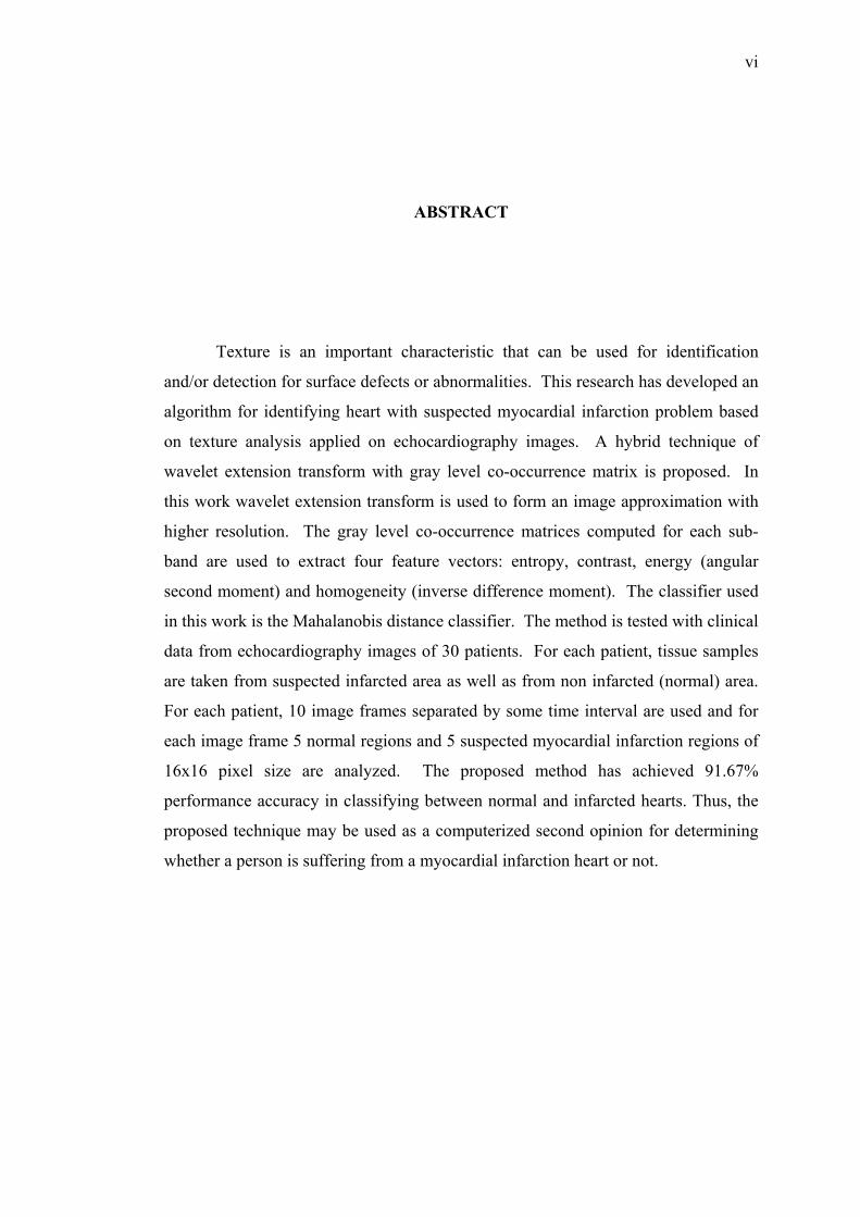
ABSTRACT
Texture is an important characteristic that can be used for identification
and/or detection for surface defects or abnormalities. This research has developed an
algorithm for identifying heart with suspected myocardial infarction problem based
on texture analysis applied on echocardiography images. A hybrid technique of
wavelet extension transform with gray level co-occurrence matrix is proposed. In
this work wavelet extension transform is used to form an image approximation with
higher resolution. The gray level co-occurrence matrices computed for each sub-
band are used to extract four feature vectors: entropy, contrast, energy (angular
second moment) and homogeneity (inverse difference moment). The classifier used
in this work is the Mahalanobis distance classifier. The method is tested with clinical
data from echocardiography images of 30 patients. For each patient, tissue samples
are taken from suspected infarcted area as well as from non infarcted (normal) area.
For each patient, 10 image frames separated by some time interval are used and for
each image frame 5 normal regions and 5 suspected myocardial infarction regions of
16x16 pixel size are analyzed. The proposed method has achieved 91.67%
performance accuracy in classifying between normal and infarcted hearts. Thus, the
proposed technique may be used as a computerized second opinion for determining
whether a person is suffering from a myocardial infarction heart or not.

vii
ABSTRAK
Tekstur adalah ciri penting yang dapat digunakan untuk mengenalpasti
dan/atau pengesanan permukaan untuk kerosakan atau keanehan. Penyelidikan ini
telah membangunkan sebuah algoritma untuk mengenalpasti jantung yang disyaki
mengalami infarksi miokardium berdasarkan menganalisa tekstur dengan
menggunakan imej daripada ekokardiografi. Di sini campuran daripada teknik
jelmaan wavelet tambahan dan teknik matrik se-kejadian tahap kelabu adalah
dicadangkan. Di dalam penyelidikan ini jelmaan wavelet tambahan digunakan untuk
menghasilkan sebuah imej hampiran yang mempunyai resolusi yang lebih besar.
Matrik se-kejadian tahap kelabu yang dihitung untuk setiap sub-jalur digunakan
untuk mencirikan empat sifat vektor: entropi, kontras, tenaga (sudut momen kedua)
dan kehomogenan (momen bezaan songsang). Pengklasifikasian yang digunakan di
dalam penyelidikan ini adalah pengklasifikasian jarak Mahalanobis. Kaedah yang
telah dicadangkan diuji dengan data klinikal daripada imej ekokardiografi untuk 30
orang pesakit. Untuk setiap pesakit, contoh tisu diambil daripada kawasan yang
disyaki infark dan kawasan bukan infark (normal). Untuk setiap pesakit, 10 bingkai
imej yang dipisahkan oleh sela waktu tertentu di mana 5 kawasan normal dan 5
kawasan disyaki infarksi miokardium berukuran 16x16 piksel akan dianalisa.
Kaedah yang dicadangkan ini telah mencapai prestasi ketepatan sebanyak 91.67%
dalam mengkelaskan antara jantung yang normal dan yang infark. Justeru itu, teknik
yang dicadangkan ini boleh digunakan sebagai pandangan kedua yang
dikomputerkan bagi menentukan sama ada seseorang itu mengalami infarksi
miokardium atau tidak.
viii
TABLE OF CONTENTS
CHAPTER TITLE PAGE
TITLE PAGE i
DECLARATION ii
DEDICATION iii
ACKNOWLEDMENT iv
ABSTRACT vi
ABSTRAK vii
TABLE OF CONTENTS viii
LIST OF TABLES xii
LIST OF FIGURES xiii
LIST OF ABBREVIATIONS AND SYMBOLS xvii
LIST OF TERMINOLOGY xix
LIST OF APPENDICES xx
1 INTRODUCTION
1.1. Background 1
1.2. Objectives 4
1.3. Scopes 4
1.4. Hypothesis 6
1.5. Contributions 6
1.6. Organization of this Thesis 7
ix
2 RELATED WORK ON TEXTURE ANALYSIS
2.1. Introduction 9
2.2. Texture Analysis 11
2.3. Mathematical Modeling of Texture Analysis 16
2.3.1. Statistical Methods 17
2.3.2. Structural Methods 18
2.3.3. Model Based Methods 19
2.3.4. Transform Methods 20
2.4. Applications 21
2.4.1. Texture Analysis and Classification Based on
WT and GLCM 21
2.4.2. Texture Analysis of Echocardiography Images 23
2.4.3. Other Technique Based on Echocardiography 24
2.5. Proposed Methods 26
2.6. Summary 27
3 WAVELET TRANSFORM FOR TEXTURE ANALYSIS
3.1. Introduction 29
3.2. One-Dimensional Wavelet Transform 30
3.3. Two-Dimensional Wavelet Transform 32
3.4. Daubechies Wavelets Family 36
3.5. Wavelet Image Extension Transform 41
3.6. Summary 45
4 CO-OCCURRENCE MATRIX FOR TEXTURE ANALYSIS
4.1. Introduction 46
4.2. Co-occurrence Matrix 47
4.3. Summary 52
x
5 THE PROPOSED ALGORITHM FOR IDENTIFICATION OF
MYOCARDIAL INFARCTION TISSUE
5.1. Introduction 53
5.2. Data Acquisition and Software Tools 54
5.3. Preprocessing Steps 58
5.4. Wavelet Image Extension Procedure 60
5.5. Distance Measure for Classification Phase 64
5.6. Texture Analysis for Identification
Myocardial Infarction Tissue 67
5.6.1. Feature Extraction Phase 67
5.6.2. Texture Classification Phase 76
5.7. Testing software for Similarity Problem 78
5.8. Summary 79
6 EXPERIMENTAL RESULTS AND DISCUSSIONS
6.1. Introduction 80
6.2. Classification Results 80
6.3. Comparison with wavelet extension based on energy 85
6.4. Average of Feature Extraction 90
6.5. Summary 93
7 CONCLUSIONS AND SUGGESTIONS FOR FUTURE
RESEARCH
7.1. Conclusions 95
7.2. Limitations 96
7.3. Future Work 97

xi
REFERENCES 98
Appendices A – C

xii
LIST OF TABLE
TABLE NO. TITLE PAGE
3.1 The low pass filter coefficient h(n) of the Daubechies
4, 6, 8 and 16-tap wavelet bases with the support of
the filter N = 2M. 39
4.1 Feature extracting value from matrix Figure 4.2c and
Figure 4.2d. 51
5.1 GLCM of tissue sample taken from a normal area for d = 1 73
5.2 GLCM of tissue sample taken from an infarcted area for d = 1 74
5.3 The result of feature extraction from eq. (5-8) and (5-9). 75
5.4 Texture measure result for similarity problem 79
6.1 Texture measure and classification result for proposed
method (Diastance value, α = 4.00). 82
6.2 Classification accuracy, numbers of samples data are 10
frame images each patient respectively with α = 4.00. 83
6.3 Texture measure and classification result based on energy,
a review method (Diastance value, α = 14.00). 86
6.4 Classification accuracy for wavelet extension based on
energy method, numbers of samples data are 10 frame
images each patient respectively with α = 14.00. 87
6.5 Performance of the classification rate for proposed method
and WET based on energy. 89
6.6 Feature extraction from normal area 91
6.7 Feature extraction from patient indicated infarcted area 92
xiii
LIST OF FIGURES
FIGURE NO. TITLE PAGE
1.1 (a) echocardiography image, a typical ultrasound image
of a human heart (b) example of natural texture collection. 3
1.2 Block diagram of the data acquisition system:
(a) Echocardiography, (b) Personal Computer
(Data acquisition, storage, and display) 5
1.3 a) A typical ultrasound image of a human heart. The white
square corresponds to texture sample of Region of interest
(ROI), b). A 16 x 16 pixel region of interest (ROI) has been
extracted from the ultrasound image figure 1.3 a. 6
2.1 Diagram of heart (Bianco, 2003) 10
2.2 Example of computer generated texture (a)
Example of statistical texture and, (b) structural texture 12
2.3 Some example of Brodatz texture: (a) Grass, D9. (b) Bark,
D12. (c) Straw, D15. (d) Herring bone, D15. (e) Woolen Cloth,
D19. (f) Wood grain, D68. (g) Brick wall, D94. (h) Raffia, D84
and (i) Plastic bubbles. 13
2.4 Different operation in texture analysis case 14
2.5 (a) An image consisting of five different textured regions:
cotton canvas (D77), straw matting (D55), raffia (D84),
herringbone weave (D17), and pressed calf leather.
(b) texture classification and (c). texture segmentation. 14
2.6 (a) original image and (b) synthesis image. 15
2.7 Shape from texture can extract orientation of the surface from
the variation of texture (defined by the bricks) in this image. 16
2.8 Illustrated of texture analysis approach. 28
3.1 Two-Band Analysis Bank (Decomposition step of

xiv
one-dimensional). 31
3.2 Two-Band Synthesis Bank. 31
3.3 The result of single-level discrete 1-D wavelet transform.
(a) Original signal (pure sinusoid with high-frequency
noise added to it)
(b) Coefficient approximations: high scale, low-frequency
components
(c) Coefficient details: low-scale, high-frequency components. 32
3.4 Decomposition step of two-dimensional DWT. 34
3.5 Reconstruction step of two-dimensional DWT. 34
3.6 The arrangement of the four sub-bands, (a) one level of
DWT and, (b) two level of DWT. 35
3.7 Example of single-level and second-level wavelet
decomposition where the ‘db8’ wavelet is chosen.
(a) Original image of sinsin, (b) Single-level wavelet
decomposition, (c) Second-level wavelet decomposition. 35
3.8 16-tap Daubechies wavelet : (a) Scaling function,
(b) Wavelet function, (c) Decomposition low-pass filter,
(d) Decomposition high-pass filter, (e) Reconstruction
low-pass filter, and (f) Reconstruction high-pass filter. 38
3.9 H and G filters of 8 and 16-tap Daubechies wavelets in
frequency domain are shown in a and b respectively. 40
3.10 Block diagram illustrating the complete wavelet decomposition
extension procedure: (a) decomposition part and
(b) the extension (synthesis) part. 43
3.11 The result of complete decomposition extension procedure
for one representative ultrasound image of a human heart:
(a) the original ultrasound image, (b) they are images
obtained after the decomposition, (c) Synthesized
(reconstruction) image with two times higher resolution. 44
4.1 Co-occurrence matrices as a function of angle. 49
4.2 The spatial co-occurrence calculation, a). Image example,
b). Construction of co-occurrence matrix,
c) Corresponding co-occurrence matrix for d =1 and θ = 0º,

xv
and d) Correspondence co-occurrence matrix for d = 1
and θ = 90º. 49
5.1 General flow process in identifying myocardial infarcted heart 53
5.2 Examples of data collection used the experiment from one
patient with contain 10 frames image every patient.
The data acquisitions are arranged from t0, t1, t2, t3, t4, t5,
t6, t7, t8, and t9 respectively. 56
5.3 Illustration of sample data taken from normal area (a)
and suspected an infarcted area of the myocardium (b). 57
5.4 Tissue samples are taken from ROI in figure 6.2 patient_27_t0.
(a) five tissue samples taken from a normal area, and
(b) five tissue samples taken from an suspected infarcted area. 57
5.5 Schematic representation for data collection (patient_15 frame
p_15_t0 data is shown in detail as an example) 58
5.6 Display indexed image matrix (a), and colormap matrix (b) 59
5.7 Display indexed image matrix after gray scale (a), and colormap
matrix (b) 60
5.8 A ultrasound image of a human heart. The white squares
correspond to tissue samples taken from suspected an infarcted
area (a) and a normal area (b) of the myocardium. 61
5.9 Tissue sample taken from a normal area (a) one level of
wavelet extension procedure (b) and two level of wavelet
extension procedure (c) The placement of the decomposed
and synthesized images is identical as in Figure 3.6(a) Chapter 3. 62
5.10 Tissue sample taken from suspected an infarcted area (a),
one level of wavelet extension procedure (b), and two level
of wavelet extension procedure (c) The placement of
the decomposed and synthesized images is identical as
in Figure 3.6(a) Chapter 3. 63
5.11 llustrative example of co-occurrence matrix distribution
of intensity from tissue sample a normal area from Figure
5.9(c) for θ = 0º and d = 1. (a) LL domain, (b) HL domain,
(c) LH domain, (d) HH domain 71
5.12 Illustrative example of co-occurrence matrix distribution of
xvi
intensity from tissue sample an infracted area from figure
5.10(c) for θ = 0º and d = 1. (a) LL domain, (b) HL domain,
(c) LH domain, (d) HH domain 72
5.13 Example of texture images used in this experiment. 78
6.1 Performance of distance value from sample data t_9 Table 6.1
for each patient. 84
6.2 Performance of distance value from sample data t_5 Table 6.3
for each patient. 88
6.3 Performance of each method in term of classification rates,
method 1, proposed method and method 2 review method
(wavelet extension based on energy method). 94

xvii
LIST OF ABBREVIATIONS AND SYMBOLS
jc - Space of approximation
φ - Scaling function
jd - Space of detail
↑2 - Up sampling
↓2 - Down sampling
1↑2 - Up sample rows: insert zeros at odd-indexed rows
1↓2 - Down sample rows: keep the even indexed rows
2↑1 - Up sample columns: insert zeros at odd-indexed columns
2↓1 - Down sample columns: keep the even indexed columns
ASM - Angular second moment
C - Covariance matrix
d - Distance measure
dc - Displacement in columns
dr - Displacement in rows
DWT - Discrete wavelet transform
E - Energy
FIR - Finite impulse response
g(-m) - Decomposition high pass filter
GLCM - Gray level co-occurrence matrix
h(-m) - Decomposition low pass filter
HH - High frequency (diagonal edges)
HL - Vertical high frequency (horizontal edges)
IDM - Inverse difference moment
LH - Horizontal high frequency (vertical edges)
LL - Low pass filter (low frequency component)
ROI - Region of interest
xviii
α - Threshold value
θ - Angle
μ - Mean vector
ψ - Mother wavelet
xix
LIST OF TERMINOLOGY
Algorithm: A set of instruction, especially ones that can be implemented on a
computer, for a procedure that can manipulate data.
Asynergy: Lack of coordination among various muscle groups during the
performance of complex movements, resulting in loss of skill and
speed.
Coronary Artery: The vessels that supply the heart muscle with blood rich in oxygen
Echocardiography: A diagnostic test which uses ultrasound waves to form images
of the heart chambers, valves and surrounding structures. It can
measure cardiac output and is a sensitive test for inflammation
around the heart (pericarditis). It can also be used to detect
abnormal anatomy or infections of the heart valves.
Infarction: Death of tissue from lack of oxygen.
Myocardial Infarction: Also called heart attacks occur when one or more of the
coronary arteries that supply blood to the heart completely
blocked and blood to the heart muscle is cut off
Myocardial refers to heart’s muscle mass.
Region-of-interest: A selected portion of the image whose individual or average
pixels value can be displayed numerically
Thrombosis: Formation or presence of a thrombus clotting within a blood vessel
which may cause infarction of tissue supplied to the vessel.
Tissue: A group of similar cells united to perform a specific function.
Ultrasound: A type of imaging technique which uses high-frequency sound waves.
xx
LIST OF APPENDICES
APPENDIX TITLE PAGE
A Flow Chart of the System 108
B Threshold Value 114
C Computer Program Listing in MATLAB 117
CHAPTER 1
INTRODUCTION
1.1. Background
Textures provide important role for automatic visual inspection. Their
analysis is fundamental to many applications such as industrial monitoring of product
quality control, remote sensing of earth resources, and medical diagnosis. Much
research work has been done on texture, such as classification, compression, retrieval
and segmentation for last three decades. Despite the effort, texture analysis is still
considered an interesting but difficult problem in image processing. Texture analysis
can be defined as an attribute representing the spatial arrangement of the gray levels
of the pixels in a region (Chang and Kuo, 1993).
Echocardiography is a diagnostic test that uses ultrasound waves to create an
image of the heart muscle. Echocardiography can provide a wealth of helpful
information, including the size and shape of the heart, its pumping strength, and the
location and extent of any damage of its tissue. It is especially useful for assessing
diseases of the heart valves. It not only allows doctors to evaluate the heart valves,
but it can detect abnormalities in the pattern of blood flow, such as the backward
flow of blood through partly closed heart valves, known as regurgitation.
Echocardiography can also help to detect the thickness of the heart’s wall in an
attempt to compensate for heart muscle weakness. Another advantage to
echocardiography is that it is noninvasive and has no known risks or side effects
(Bianco, 2003).
2
Smith (2004) has presented that a normal echocardiogram shows a normal
heart structure and the normal flow of blood through the heart chambers and heart
valves. However, a normal echocardiogram does not rule out the possibility of the
heart disease. An echocardiogram may show a number of abnormalities in the
structure and function of the heart, such as:
• Thickening of the wall of the heart muscle (especially the left
ventricle).
• Abnormal motion of the heart muscle.
• Blood leaking backward through the heart valves (regurgitation).
• Decrease blood flow through a heart valve.
Early detection and quantitative assessment of tissue alteration in a disease is
a challenge for noninvasive imaging techniques. Direct histologic assessment is
limited by a requirement for obtaining tissue for examination. Therefore, to better
characterize the onset and progression of myocardial infarction, a noninvasive
imaging technique for distinguishing normal from abnormal tissue would be of
particular importance (Kerut et al., 2003). Myocardial infarction is also called heart
attacks occur when one or more of the coronary arteries that supply blood to the heart
completely blocked and blood to the heart muscle is cut off (Smith, 2004).
Texture analysis of echocardiography images in this research are used for
diagnosis of myocardial infarction tissue. The approach is to characterize tissue
based on the spatial distribution of ultrasound amplitude signal within a region of
interest (ROI). Skorton et al., (1983) defined echocardiography image texture as:
Two-dimensional spatial distribution of echocardiography amplitudes or gray levels.
Most of the texture defect detection applications are on textile, paper, steel
and wood inspection. There have been a number of applications of texture processing
for inspection problem. Many of these approaches have provided goods results in
different fields of application, but a large number of them have shown very low
classification rate or could not be implemented at all when texture sample are of
small dimensions. However texture characterization of 2-D echocardiography image
3
is not an easy task to perform, because it is well known that ultrasound images have
very poor quality (Mojssilovié et al., 1997).
(a)
(b)
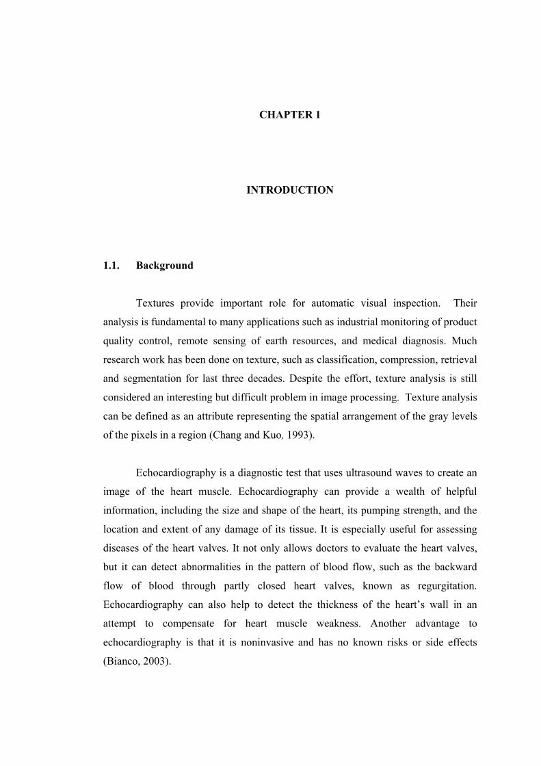
Figure 1.1: (a) echocardiography image, a typical ultrasound image of a human heart
(b) example of natural texture collection.
Figure 1.1 gives some example of natural texture (Fig.1.1b) and a typical
ultrasound image of a human heart (Fig.1.1a). In most cases, they are degraded by
speckle noise, acoustic shadowing, and system distortions present in all
instrumentation. The main disadvantage of 2-D echocardiography for the purpose of
texture description and classification in this application is caused by the structure of
the heart muscle.

4
1.2. Objectives
The objectives of this research are:
• To design and develop an algorithm for identifying myocardial infarction
tissue using texture analysis techniques.
• To evaluate the relationship between texture properties of myocardial
infarction using quantitative computer analysis on 2-dimensional
echocardiogram based on texture analysis techniques.
1.3. Scopes
The main focus of this research is based on texture analysis for identification
of myocardial infarction tissue. Some limitations are applied to the research activity
in order to keep the observation on its track. To achieve this goal, the scope of the
current research has been defined as follows:
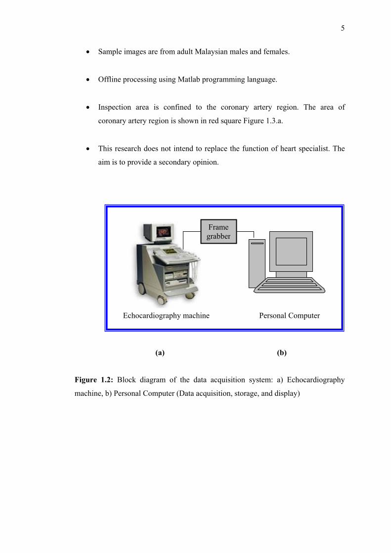
• The medical images are captured directly from the echocardiography machine
using PC via frame grabber card. Block diagram of the data acquisition
system is shown in Figure 1.2. All ultrasound images are captured from a HP
SONOS 5500 imaging system with a 3.5 MHz transducer probe with a depth
setting of 16 cm. Images were digitized with 512 x 512 pixels and 256 gray
level resolutions.
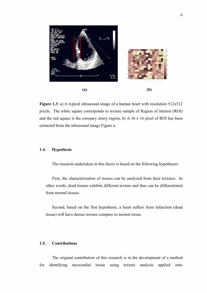
• Region of Interest (ROI) used for distinguishing a textural normal area and
infarcted area is set to 16x16 pixels. The sample data were taken by
supervised technician who is an experienced echocardiographer. The white
square corresponds to texture sample of Region of interest (ROI) and the red
square is the coronary artery region. Illustration of a typical ultrasound image
of human heart and 16x16 ROI is shown in Figure 1.3.
5
• Sample images are from adult Malaysian males and females.
• Offline processing using Matlab programming language.
• Inspection area is confined to the coronary artery region. The area of
coronary artery region is shown in red square Figure 1.3.a.
• This research does not intend to replace the function of heart specialist. The
aim is to provide a secondary opinion.
(a) (b)
Figure 1.2: Block diagram of the data acquisition system: a) Echocardiography
machine, b) Personal Computer (Data acquisition, storage, and display)
Frame grabber
Echocardiography machine Personal Computer
6
2 4 6 8 10 12 14 16
2
4
6
8
10
12
14
16
10
20
30
40
50
60
(a) (b) Figure 1.3: a) A typical ultrasound image of a human heart with resolution 512x512
pixels. The white square corresponds to texture sample of Region of interest (ROI)
and the red square is the coronary artery region, b) A 16 x 16 pixel of ROI has been
extracted from the ultrasound image Figure a.
1.4. Hypothesis
The research undertaken in this thesis is based on the following hypotheses:
First, the characterization of tissues can be analyzed from their textures. In
other words, dead tissues exhibits different texture and thus can be differentiated
from normal tissues.
Second, based on the first hypothesis, a heart suffers from infarction (dead
tissue) will have denser texture compare to normal tissue.
1.5. Contributions
The original contribution of this research is in the development of a method
for identifying myocardial tissue using texture analysis applied onto

7
echocardiography image. Much research work has been done on texture analysis for
defect detection or similarity measured problem. Most of them used texture samples
obtained from good quality and standard image size such as: textile, paper, steel,
rock and wood inspection. The proposed technique in this research performed
texture analysis based on poor quality images.
The proposed method is a hybrid between wavelet extension transform and
gray level co-occurrence matrix (GLCM). The developed algorithm has been trained
and tested with image data of size 16x16 pixels.
1.6. Organization of this Thesis
This thesis is divided into seven chapters. An overview for each chapter is
given in this section.
Chapter 1: Introduction to problems and brief overview of applications, where
objectives, scopes, contributions and structure of the thesis are explained.
Chapter 2: Literature review of previous works in texture analysis is discussed. In
this chapter proposed method for this work is also discussed after considering several
advantages and disadvantages of the existing techniques.
Chapter 3: This chapter describes a brief introduction to the wavelet transform and
wavelet extension transform as they will be the basis techniques in texture analysis.
Chapter 4: The concept for gray level co-occurrence matrix (GLCM) is given in this
chapter. This GLCM is used to perform feature extraction which is then used in the
texture analysis problem.
Chapter 5: Development of the algorithm for identification of myocardial infarction
tissue from echocardiography images is explained in this chapter. The processing are
described step by step and the developed algorithm are illustrated in this chapter.
8
Chapter 6: Results obtained from experiments based on the proposed approach are
given in this chapter. An application of texture analysis for medical image especially
echocardiography image is demonstrated.
Chapter 7: Conclusions of the thesis are given in this chapter. A summary of the
finding, followed by a list of area where further investigation may lead to
improvement in the proposed texture analysis algorithms are also discussed.
98
REFERENCES
Abhayaratne, G. C. K., Jermyn, L. H., and Zerubia Ariana, J (2005). Texture-
Adaptive Mother Wavelet Selection for Texture Analysis. Image Processing,
2005, ICIP, 2005, IEEE International Conference on Vol.2, pp. 1290-1293.
Agani, N., Abu-Bakar, S. A. R., and Sheikh Salleh, S. H. (2004a). Texture Analysis
and Classification Using Wavelet Extension and Gray Level Co-occurrence
Matrix for Defect Detection Small Dimension Images. International
Conference on Control Automation and systems, Bangkok, Thailand.
Agani, N., Abu-Bakar, S. A. R., and Sheikh Salleh, S. H. (2004b). Texture Defect
Detection in Low Quality Images Using Wavelet Extension Transform and
Gray Level Co-occurrence Matrix. The 1st International Conference on
Telematics System, Service, and Applications , Bandung, Indonesia.
Agani, N., Abu-Bakar, S. A. R., and Sheikh Salleh, S. H. (2005a). Texture Analysis
of Echocardiography Images for Diagnosis of Myocardial Tissue. International
Conference on Instrumentation, Communication and Information Technology,
Bandung, Indonesia.
Agani, N., Abu-Bakar, S. A. R., and Sheikh Salleh, S. H. (2005b). Wavelet
Extension and Gray Level Co-occurrence Matrix for Texture Defect Detection
and Texture Retrieval in Small Dimension Images. The 8th International
Conference on Quality in Research (QIR), Depok, Indonesia.
Aldasoro, R. C. C. (2004). A Guide to Co-occurrence Matrix Analysis. Departement
of Computer Science, The University of Warwick.
Amet, A. L., Ertüzün, A., and Erçil, A. (2000). An Efficient Method for Texture
Defect Detection: Subband domain Co-Occurrence Matrices. Image and
Vision Computing, Vol.18/6-7, pp.543-553.

99
Antonini, M., Barlaud, M., Mathieu, P., and Daubechies, I. (1992). Image Coding
Using Wavelet Transform. IEEE Transaction on Image Processing, vol. 1, no.
2, pp.205-220.
Baraldi, A., and Parmiggiani, F. (1995). An Investigation of the Textural
Characteristics Associated with Gray Level Co-occurrence Matrix Statistical
parameters. IEEE Transaction on Geoscience and Remote Sensing, Vol.33,
No.2, pp. 293-304
Bianco, C. (2003). Echocardiography. North American Society of Pacing and
Electrophysiology (NASPE).
Blostein.D., and Alhuja, N. (1989). Shape from Texture: Integrating Texture-
Element Extraction and Surface Estimation. IEEE Transaction on Pattern
Analysis and Machine Intelligence, PAMI-11, pp. 1233-1251.
Bose, T. (2004). Digital Signal and Image Processing. John Wiley and Sons, Inc.
Copyright 2004.
Boukerroui, D., Noble, J. A., and Brady, J. M. (2000). Feature Enhancement in Low
Quality Images with Application to Echocardiography. Medical Vision
Laboratory, Department of Engineering Science, University of Oxford.
Burill, J. H. P. (2003). Texture Mapping of Neorological Magnetic resonance
Images. Technical Report, Department of Electrical Engineering, Imperial
College of Science Technology & Medicine Exhibition Road, London SW7.
Burrus, C. S., Gopinath, R. A., and Guo, H. (1998). Introduction to Wavelets and
Wavelet Transforms. Prentice-Hall International, Inc. Houston, Texas.
Campisi, P., Neri, A., Panci, G., and Scarano, G. (2004). Robust Rotation-Invariant
Texture Classification Using a Model Based Approach. IEEE Transactions on
Image Processing, Vol.13, No.6, pp. 782-791
Chang, T., and Kuo, J. C. C. (1993). Texture Analysis and Classification with Tree-
Structured Wavelet Transform. IEEE Transaction on Image Processing, vol. 2,
no. 4, pp.429-441.
Chapple, P. B.,.Bertilone, D. C., Caprari, R. S., and Newsam, G. N. (2001).
Stochastic Model-Based Processing for Detection of Small Targets in Non-
Gaussian Natural Imagery. IEEE Transactions on Image Processing, Vol.10,
No.4, pp. 554-564.
Charalampidis, D. (2006). Texture Synthesis: Textons Revisited. IEEE Transactions
on Image Processing, Vol.15, No.3, pp. 777-787.
100
Chasal, P., Flynn, J., and Relly, R. B. (2005). Automated Processing of Shoeprint
Images Based on the Fourier Transform for Use in Forensic Science. IEEE
Transaction on Pattern Analysis and Machine Intelligence, Vol.27, No.3, pp.
341-350.
Chiu, E., Vaisey, J., and Atkins, M. S. (2001). Wavelet Based Space-Frequency
Compression of Ultrasound Images. IEEE Transaction on Information
Technology in Biomedicine, Vol.5, No.4, pp.300-310.
Christmas, W. J., .Kittler, J and., Petrou, M. (1995). Structural Matching in
Computer Vision Using Probabilistic Relaxation. IEEE Transaction on Pattern
Analysis and Machine Intelligence, Vol.17, No.8, pp. 749-764.
Clausi, D. A., and Deng, H. (2005). Design-Based Texture Feature Fusion Using
Gabor Filters and Co-occurrence Probabilities. IEEE Transactions on Image
Processing, Vol.14, No.7, pp. 925-935.
Cohen, F. S., Fan, Z., and Attali, S. (1991). Automated Inspection of textile Fabric
using Textural Models. IEEE Transactions on Pattern Analysis and Machine
Intelligence, Vol.13, No.8.
Daubechies, I. (1990). The Wavelet Transform, Time-Frequency Localization and
Signal Analysis. IEEE Transaction on Information Theory, Vol. 36, No.5,
pp.961-1004.
Deng, H., and Clausi, D. A. (2004). Gaussian MRF Rotation-Invariant Features for
Image Classification. IEEE Transaction on Pattern Analysis and Machine
Inteligence, Vol.26, No.7, pp. 951-955
Do, M. N. (2001). Directional Multiresolution Image Representations. Department
of Communication Systems, Swiss Federal Institute of Technology (EPEL),
Lausanne, Switzerland: Ph.D. Thesis.
Dunn, D., and Higgins, W. E. (1995). Optimal Gabor Filters for Texture
Segmentation. IEEE Trans. on Image Processing, vol. 4, no. 7, pp.947-964.
Epifanio, I., and Ayala, G. (2002). A random Set View of Texture Classification.
IEEE Transactions on Image Processing, Vol.11, No.8, pp. 859-867
Fajar Bakti Sdn. Bhd. (2004). Kamus Jururawat Bahasa Inggeris-Bahasa Melayu.
Shah Alam, Selangor Darul Ehsan.
Fan, G., and Xia, X. G. (1998). Texture Analysis and Synthesis using Wavelet-
Domain Hidden Markov Models. Research report, Department of Electrical
and Computer Engineering University of Delaware, Newark, DE 19716.
101
Fan, G., and Xia, X-G. (2003). Wavelet-Based Texture Analysis and Synthesis Using
Hidden Markov Models. IEEE Transaction on Circuits and System-I:
Fundamental Theory and Application, Vol.50, No.1, pp.106-120.
Fatemi, G. N. (1997). Performance Measures for Wavelet-based Segmentation
Algorithm. University of Surrey, Guildford: Ph.D. Thesis.
Garcia,.R., Xevi, C., and Battle, J. (2001). Detection of Matching in a Sequence of
Underwater Images Through Texture Analysis. IEEE Image Processing,
International Conference, Vol.1, pp.361-364.
Gerard, O., Billon, A. C., Rouet, J. M., Jacob, M., Fradkin, M., and Allouche, C.
(2002). Efficient Model-Based Quantification of Left Ventricular Function in
3-D Echocardiography. IEEE Transaction on Medical Imaging, Vol.21, No.9,
pp. 1059-1068.
Ghazel, M., Freeman, G.H., and Vrscay, E.R. (2003). Fractal Image Denoising.
IEEE Transactions on Image Processing, Vol.12, No.12, pp. 1560-1578.
Gonzales, R. C., Woods, R. E., and Eddins, S. L. (2004). Digital Image Processing
using with Matlab. Pearson Prentice Hall, Upper Saddle River, New Jersey,
Copyright 2004 by Pearson Education, Inc.
Graps, A. (1995). An Introduction to Wavelet. IEEE Computational Sciene and
Engineering, vol. 2, no. 2.
Guan, N., Yashiro, K., and Ohkawa, S. (2000). On Choice of Wavelet Bases in the
Wavelet Transform Approach. IEEE Transaction on Antennas and Propagation
Vol.4, No.8, pp. 1186-1191.
Hamarneh, G., and Gustavsson, T. (2000). Combining Snakes and Active Shape
Models for Segmentation the Human Left Ventricle in Echocardiographic
Images. IEEE Computing in Cardiology, Vol.27.
Haralick, R. (1979). Statistical and Structural Approaches to Texture. Proceeding
IEEE, Vol.67, No.5, pp.786-804.
He, P., and Zheng, J. (2001). Segmentation of Tibia Bone in Ultrasound Images
using Active Shape Models. In Proceeding 23rd Annual Conference-
IEEE/EMBS, Istanbul, Turkey .
Hewlett-Packard Company. (1998). System Basics HP SONOS 5500 Ultrasound
System. Massachusetts, USA
Hsin, H. (2000). Texture Segmentation Using Modulated Wavelet Transform. IEEE
Transactions on Image Processing, Vol.9, No.7, pp. 1299-1302
102
Johnson, R. A., and Wichern, D. W. (1998). Applied Multivariate Statistical
Analysis. Prentice Hall, Upper Saddle River, New Jersey, Fourth Edition.
Kandaswamy, U., Adjeroh, D. A., and Lee, M. C. (2005). Efficient Texture Analysis
of SAR Imagery. IEEE Transaction on Geoscience and Remote Sensing,
Vol.43, No.9, pp. 2075-2083.
Kaplan, L. M. (1999). Extended Fractal Analysis for Texture Classification and
Segmentation. IEEE Transactions on Image Processing, Vol.8, No.11, pp.
1572-1584.
Kathlkeyani,.V., Duraiswamy, K., and Kamalakkannan,.P. (2005). Texture Analysis
and Synthesis for Near-Reguler Textures. Inteligent Sensing and Information
Processing, Proceeding of 2005 International Conference, pp. 134-139.
Kerut, E. K., Given, M., and Thomas,.D. G. (2003). Review of Method for Texture
Analysis of Myocardium from Echocardiographic Images: A Mean of Tissue
Characterization. Echocardiography. A Journal of CV Ultrasound & Allied
Tech. Vol.20, No.8, pp.727-736.
Khauzani, J. K., and Zadeh, S. H. (2005). Rotation-Invariant Multiresolution Texture
Analysis Using Radon and Wavelet Transforms. IEEE Transactions on Image
Processing, Vol.14, No.6, pp. 783-795.
Kim, J.K., and Park, H. W. (1999). Statistical Textural Features for Detection of
Microcalcifications in Digitized. IEEE Transaction on Medical Imaging,
Vol.18, No.3, pp. 231-238.
Kim, N. D. (2000). Texture Representation Using Wavelet Filterbanks. Iowa State
University: Ph.D. Thesis.
Kim, N. D., Amin, V., Wilson, D., Rouse, G., and Udpa S. (1998). Ultrasound
Image Texture Analysis for Characterizing Intramuscular Fat Content of Live
Beef Cattle. Ultrasonic Imaging 20, pp. 191-205.
Kyriacou, E., Pavlopoulus, S., and Koutsouris, D. (1997). Computert Assisted
Characterization of Liver Tissue Using Image Texture Analysis Techniques on
B-Scan Images. IEEE Proceeding, Proceeding-19th International Conference
IEEE/EMBS, Chicago, IL, USA..
Lotfallah, O. A. (2002). Image Texture Feature Extraction Based on Wavelet
Decomposition. College of Engineering and Applied sciences Department of
Electrical Engineering.

103
Malal, K., and Sadasivam, V. (2005). Automatic Segmentation and Classification of
Diffused Liver Disease Using Wavelet Based Texture Analysis and Neural
Network. IEEE Indicon 2005 Conference, Chennai, India, pp.216-219.
Mallat, S. G. (1989a). A Theory for Multiresolution Signal Decomposition: The
Wavelet Representation. IEEE Transaction on Pattern Analysis and Machine
Inteligence, Vol. 11, No. 7, pp.674-693.
Mallat, S. G. (1989b). Multifrequency Channel Decomposition of Images and
Wavelet Models. IEEE Transaction on Acoustics, Speech and Signal
Processing, Vol.37, No.12, pp.2091-2110.
Manjunath, B. S., and Ma, W. Y. (1996). Texture Features for Browsing and
Retrieval of Image Data. IEEE Transaction on Pattern Analysis and Machine
Intelligence, Vol.18, No.8, pp.837-842.
Materka, A., and Strzelecki, M. (1998). Tecture Analysis Methods - A Review.
Technical University of Lodz, Institute of Electronics, COST B11 report,
Brussels.
Mery, D., and Filbert, D. (2002). Classification of Potensial Defects in the
Automatic Inspection of Aluminium Castings using Statistical Pattern
Recognition. European Conference on Non-Destructive Testing (ECNDT
2002), Barcelona, Spain.
Misiti, M., Misiti, Y., Oppenheim, G., and Poggi, J. M. (2002a). Image Processing
Toolbox for Use with Matlab. version 3.2, release 13, by the Mathworks, Inc.
Misiti, M., Misiti, Y., Oppenheim, G., and Poggi, J. M. (2002b). Wavelet Toolbox
for Use with Matlab. version 2.2, release 13, by the Mathworks, Inc.
Mitchell, S. C., Lelieveldt, B. P. F., Geest, R. J., Bosch, H. G., Reiber, J. H. C., and
Sonka, M. (2001). Multistage Hybrid Active Appearance Model Matching:
Segmentation of Left and Right Ventricles in Cardiac MR Images. IEEE
Transaction on Medical Imaging, Vol.20, No.5, pp. 415-423.
Mojsilović, A., Popović, M. V., and Rackov, D. M. (2000). On the Selection of an
Optimal Wavelet Basis for Texture Characterization. IEEE Transactions on
Image Processing, Vol.9, No.12.
Mojssilović, A., Popović, M. V., Nešković, A. N., and Popović, A. D. (1997).
Wavelet Image Extension for Analysis and Classification of Infarcted
Myocardial Tissue. IEEE Trans. on Biomedical Engineering, vol. 44, no. 9,
pp.856-866.
104
Neškoviĉ, A. N., Mojssilović, A., Jovanoviċ, T., Vasijlević, J., Popopić, M.,
Marinković, J., Bojić, M., and Popović, A. D. (1998). Myocardial Tissue
Characterization After Acute Myocardial Infarction with Wavelet Image
Decomposition. Cardiovascular research centre, Belgrade University Faculty
of Electrical Engineering; Institutes of Physiology and Pathology Belgrade
University Medical School.
Niessen, W. J., Bemmel, C. M., Frangi, A. F., Siers, M. J. A., and Wink, O. (2002).
Model-Based Segmentation of Cardiac and Vascular Images. In Proc 2002
IEEE International Symposium on Biomedical Imaging, pp.22-25, Washington
DC, USA.
Ojala, T., Pietikäinen, M., and Mäenpää, T. (2002). Multiresolution Gray-Scale and
Rotation Invariant Texture Classification with Local Binary Patterns. IEEE
Transaction on Pattern Analysis and Machine Intelligence, Vol.24, No.7.
Partio, M., Cramarius, B., Gabbaouj, M., and Visa, A. (2000). Rock Texture
Retrieval Using Gray Level Co-occurrence Matrix. Tampere University of
Technology, Finland.
Pavlopoulo, S., Kyriatou., Koutsouris, D., and Zoumpoulis, P. (2000). Fuzzy Neural
Network Based Texture Analysis of Ultrasonic Images. IEEE Engineering in
Medicine and Biology, Vol.19, No.1, pp. 39-47.
Perkins, C., and Fricke, T. (2000). Wavelets. Department of Electrical Engineering
University of California at Berkeley.
Qiang Ji., Engel, J., and Craine, E. (2000). Texture Analysis for Classification of
Cervix Lesions. IEEE Transactions on Medical Imaging, Vol 19, No.11,
pp.1144-1149.
Rellier, G., Descomber, X., Falzon, F., and Zerubia, J. (2004). Texture Feature
Analysis Using a Gauss-Markov in Hyperspectral Image Classification. IEEE
Transaction on Geoscience and Remote Sensing, Vol.42, No.7, pp.1543-1551.
Rezai, F. R., and Kinsner, W (1999). Texture Analysis and Segmentation of Images
Using Fractals. Proceeding of the 1999 IEEE Canadian Conference on
Electrical and Computer Engineering, pp. 786-791.
Rioul, O., and Vetterli, M. (1991). Wavelets and Signal Processing. IEEE SP
Magazine, pp.14-38.
105
Shaohua, Z. (2000). Wavelet-Based Texture Retrieval and Modeling Visual Texture
Perception. Thesis, Department of Electrical Engineering, National University
of Singapore.
Sharma, M., and Singh.S. (2001). Evaluation of Texture Methods for Image
Analysis. IEEE Intelligent Information Systems Conference, The seventh
Australian and New Zealand, pp. 117-121
Sheng, Y. (2000). The Transform and Applications Handbook: Second Edition,
A.D.Poularikas-chief Editor. ACRC Handbook Publisher in Cooperation with
IEEE Press, Chapter 10.
Sheppard, M. A., and Shih, L. (2005). Efficient Image Texture Analysis and
Classification for Prostate Ultrasound Diagnosis. IEEE Proceeding,
Computational Systems Bioinformatics Conference Workshops (CSBW’05).
Siew, L. H., and Hodgson, R. M. (1998). Texture Measures for Carpet Wear
Assessment. IEEE Transaction on Pattern Analysis and Machine Intelligence,
Vol.10, No.1.
Signal and Image Processing Institute. (1977). Brodatz texture database. Research
Programs, Unibersity of Southern California, Los Angeles.
Skorton, D. J., Collins, S.M., and Woskoff, S.D. (1983). Range and Azimuth-
dependent Variability of Image Texture in Two Dimensional Echocardiograms.
Circulation, Vol.68, pp.834-840.
Smith, J. F. (2004). Heart attack. Medical Library. The Thomson Corporation. All
right reserved. MyDiseaseDex ™ is a trademark of micromedex, Inc.
Sonka, M., Hlavac, V., and Boyle, R. (1999). Image Processing, Analysis, and
Machine Vision. PWS Publishing an Imprint of Brooks/Cole Publishing
company. An International Thomson Publishing Company, Second Edition,
Copyright 1999
Tao, I. H., Calway, A. D., and Wilson, R. (1993). Texture Analysis Using the
Multiresolution Fourier Transform. The 8th Scandinavian Conference on
Image Analysis, Tromso.
Tripathy, S. S. (2005). System for Diagnosing Valvular Heart Disease using Heart
Sounds. Master Thesis, Department of Computer Science & Engineering,
Indian Institute of Technology, Kanpur-208016, India.
Tsai, D. M., and Hsiao,B. O. (2001). Automatic surface inspection using wavelet
reconstruction. Pattern Recognition 34, 1285-1305.
106
Tuceryan, M., and Jain, A.K. (1998). Handbook of Pattern Recognition and
Computer Vision (2nd Edition), Chapter 2: Texture Analysis. World Scientific
Publishing Co, Michigan St. Indianapolis.
Űnsalan, C. (1998). Pattern Recognition Methods for Texture Analysis Case Study:
Steel Surface Classification. Master Thesis, Electrical and Electronics
Engineering, Boğaziçi University, Istanbul, Turkey.
Unser, M., Aldroubi, A., and Laine, A. (2003). Wavelets in Medical Imaging. IEEE
Transaction on Medical Imaging, Vol.22, No.3, pp. 285-288.
Walker, R. F., Jackway, P., and Longstaff, I. D. (1995). Improving Co-occurrence
Matrix Feature Discrimination. Proceedings of DICTA, The 3rd Conference on
Digital Image Computing: Techniques and Application, 6-8th , Brisbane,
Australia, pp.643-648.
Wan, Y. Y., Du, J. X., Huang, D. S., Chi, Z., Cheung, Y. M., Wang, X. F., and
Zhang, G. J. (2004). Bark Texture Feature Extraction Based on Statistical
Texture Analysis. Inteligent Multimedia, Video and Speech Processing, 2004,
Proceeding of 2004 International Symposium, Hongkong, pp. 482-485.
Wang, Z., Bovik, A. C., Sheikh, H. R., and Simoncell, E. P. (2004). Image Quality
Assessment: From Error Visibility to Structural Similarity. IEEE Transactions
on Image Processing, Vol.13, No.4, pp. 600-612
Wong, W. C. K., and Chung, A. C. S. (2005). Bayesian Image Segmentation Using
Local Iso-Intensity Structural Orientation. IEEE Transactions on Image
Processing, Vol.14, No.10, pp. 1512-1523.
Wu, C. M., Chen, Y. C., and Hsieh, K. S. (1992). Texture Features for
Classification of Ultrasonic Liver Images. IEEE Transaction on Medical
Imaging, Vol.11, No.2, pp. 141-152.
Xia, Y., Feng, D., and Zhao, R. (2006). Morphology-Based Multifractal Estimation
for Texture Segmentation. IEEE Transactions on Image Processing, Vol.15,
No.3, pp. 614-623.
Xu, J. (2003). A Generalized Discrete Morphological Skeleton Transform with
Multiple Structuring Elements for the Extraction of Structural Shape
Components. IEEE Transactions on Image Processing, Vol.12, No.12, pp.
1677-1686.
Yao, J. (1993). Complete Gabor transformation for Signal Representation. IEEE
Transactions on Image Processing, Vol.2, No.2, pp. 152-159.
107
Ye, X., Noble, J. A., and Atkinson, D. (2003). 3-D Freehand Echocardiography for
Automatic Left Ventricle Reconstruction and Analysis Based on Multiple
Acoustic Windows. IEEE Transaction on Medical Imaging, Vol.21, No.9, pp.
1051-1058.
Yfantis, E. A., Popovich, A., Angelopoulos, A., and Bebis, G. (2000). On Cancer
Recognition of Ultrasound Images. IEEE Proceeding, Computer Vision
Beyond the Visible Spectrum: Methods and Applications, pp. 44-49.
Zayed, N. M., Badwi, A. M., Elsayed, A., Elsherif, M. S., and Youssef, A. B. M.
(2001). Wavelet Segmentation for Fetal Ultrasound Images. IEEE Proceeding,
Circuit and System, MWSCAS, Vol.1, pp.501-504.
Zhou, S., Venkatesh, Y. Y., and Ko, C. C. (2000). Role of Phase in Visual Texture
Perception. Center for Automation Research, University of Maryland, USA,
Department of Electrical Engineering, Indian Institute of Science, Department
of Electrical Engineering, National University of Singapore.