identification of inhibitory mechanisms preventing cenp-a...
TRANSCRIPT

UNIVERSIDADE DE LISBOA
FACULDADE DE CIÊNCIAS
DEPARTAMENTO DE BIOLOGIA ANIMAL
Identification of inhibitory mechanisms preventing CENP-A
assembly in the S phase of the cell cycle
Mestrado em Biologia Evolutiva e do Desenvolvimento
Rúben Lopes Pereira Abreu
Dissertação orientada por:
Doutor Lars E. T. Jansen e Professora Doutora Solveig Thorsteinsdóttir
2016


i
Abstract
The centromere is the chromosomal locus that directs kinetochore formation in
order to secure faithful segregation of sister chromatids during mitosis. Nucleosomes that
contain CENP-A (centromere protein A), an H3-histone variant, are thought to be the
epigenetic mark indicating active centromeres, and its assembly into chromatin requires
the Mis18 complex and its dedicated chaperone HJURP. This process is restricted in the
cell cycle to telophase/early G1 phase and is a consequence of low Cdk1 and Cdk2 activity.
Outside G1 phase, these kinases inhibit CENP-A assembly by acting on assembly factors
Mis18 complex and HJURP. This model is derived from the observation that inhibition of
Cdk activity can induce assembly before mitosis in G2 phase. However, induction of
assembly following Cdk inhibition is not observed in S phase. In this study, I sought to
investigate what are the mechanisms of inhibition in this cell cycle stage. I pursued several
hypotheses, namely whether protein levels of HJURP and Mis18BP1, a subunit of Mis18
complex, were here diminished, which was not observed. Furthermore, I investigated a
possible role of DNA damage in the prevention of CENP-A assembly. DNA replication
occurs in S phase, which constitutes an important source of DNA damage. In addition,
some studies postulated the participation of CENP-A and HJURP in the DNA damage
response, which supported the hypothesis that DNA damage could constitute an inhibitory
mechanism. This was tested in two different ways. First, I confirmed a negative correlation
between the presence of DNA damage and CENP-A assembly, but inhibition of upstream
kinases involved in DNA damage signaling did not lead to CENP-A assembly in S phase.
Second, induction of DNA damage by two different means was not inhibitory in G1 and G2
phase. Unexpectedly, instead of blocking assembly, DNA damage induction was sufficient
to induce CENP-A assembly in G2 phase. A possible explanation may be the alleviation of
Cdk inhibition by activation of G2/M checkpoint. Together, these results suggest that the
refractory nature of S phase is not a consequence of lack of key components, nor is it due
to the existence of DNA damage. I will discuss further hypothesis and future experiments
that may shed light on this phenomenon.
Key-words: centromere, CENP-A, epigenetic inheritance, cell cycle, DNA damage

ii
Resumo
O centrómero é um domínio cromossómico distinto no qual se forma o cinetócoro.
Este por sua vez tem um papel fundamental na divisão celular, na medida em que permite
a associação dos microtúbulos do fuso mitótico com os cromossomas, e a sua segregação
para as células filhas. Como tal, o centrómero deve ser restrito a apenas um locus por
cromossoma, de modo a não comprometer a integridade do genoma.
Embora algumas características do DNA centromérico sejam partilhadas por várias
espécies, nomeadamente a natureza repetitiva, não existe uma sequência necessária ou
suficiente para a determinação da posição do centrómero. Em organismos eucariotas, o
principal candidato para tal é CENP-A, uma histona variante de H3 que se encontra
substancialmente enriquecida nos nucleossomas dos centrómeros em comparação com o
resto do genoma. Este papel de CENP-A constitui um exemplo paradigmático de
hereditariedade epigenética, pois manutenção da identidade do centrómero é mantida ao
longo das gerações.
Os nucleossomas possuidores de CENP-A são extremamente estáveis, e o único
fenómeno biológico que os faz ser movidos está relacionado com a redistribuição destes
na fase S para cada uma das recém-replicadas cadeias filhas de DNA. Isto faz com que cada
um dos centrómeros recém-formados em cada uma das cadeias filhas possua no máximo
metade dos nucleossomas possuidores de CENP-A presentes antes da replicação. De forma
a propagar o centrómero, estes têm de ser repostos posteriormente. Temporalmente, em
humanos, o recrutamento de CENP-A está restrito à telófase/início da fase G1. Isto difere
das janelas temporais de incorporação dos nucleossomas possuidores de H3.1, que ocorre
estritamente na fase S durante a replicação, e de H3.3, que ocorre ao longo da interfase.
O recrutamento de CENP-A é dependente de dois passos chave. Inicialmente, o
complexo Mis18, composto pelas subunidades Mis18α, Mis18β e Mis18BP1, é recrutado
para os centrómeros no fim da anáfase, induzindo possivelmente um estado de
permissibilidade da cromatina. O recrutamento de CENP-A em si é subsequentemente
efetuado por HJURP em telófase/início da fase G1. Este processo é concomitante com baixa
actividade de Cdk1 e Cdk2, duas cinases importantes na progressão do ciclo celular que
aumentam a sua actividade à medida que a célula progride na interfase, mas com uma
descida drástica em mitose. Inibição destas cinases na fase G2 através de um inibidor geral
de Cdks, Roscovitine, permite o recrutamento precoce de CENP-A. Foi então proposto que
a via de recrutamento de CENP-A está sob controlo do ciclo celular, isto é, Cdk1/Cdk2
regulam-na negativamente à medida que a sua actividade aumenta. De facto, Mis18BP1 e
HJURP são alvos de fosforilação destas cinases. No entanto, recrutamento de CENP-A não é

iii
observado na fase S de células humanas aquando da utilização do mesmo inibidor de Cdks.
Isto sugere a existência de mecanismos de inibição adicionais desta via, para além do
controlo exercido pelo ciclo celular.
A fase S do ciclo celular é caracterizada pela existência de replicação do DNA. O
período em que determinada região é replicada está relacionado com as características do
DNA e da cromatina aí presente. Um exemplo é o centrómero, que é replicado tardiamente
na fase S dada a sua natureza repetitiva e heterocromática, tendo problemas na
progressão do garfo de replicação. O encontro de um garfo de replicação com uma lesão do
DNA ou a indução de dano através de agentes exógenos pode levar à formação de DSBs
(quebras na cadeia dupla). A reparação deste tipo de dano é mediada por ATM e ATR, a
partir da acumulação de γH2AX perto dos DSBs. Existem dois estudos que relacionam a
reparação de DNA e a via de recrutamento de CENP-A; foi proposto que HJURP tem papel
activo na reparação do DNA, sendo um alvo de ATM, e os seus níveis proteicos aumentam
com a indução de dano. Também foi demonstrado que CENP-A é temporariamente
recrutada para DSBs, sem ser perdido nos centrómeros.
Este estudo tem como objectivo investigar mecanismos de inibição da via de
recrutamento de CENP-A na fase S, para além do já descrito controlo exercido pelo ciclo
celular. Como tal, foram formuladas duas hipóteses que levaram à elaboração de
abordagens distintas. A primeira e mais simples é que na fase S componentes essenciais ao
recrutamento de CENP-A, HJURP e Mis18BP1, estão ausentes. Para abordar esta hipótese,
um protocolo de sincronização celular foi implementado em células HeLa, seguido de um
western blot que permitiu averiguar os níveis proteicos destes componentes em diferentes
estádios do ciclo celular, inclusive a fase S. Em paralelo, os perfis de sincronização foram
verificados por FACS.
Tendo em consideração uma possível conexão descrita anteriormente entre as
duas vias, a segunda hipótese é que dano no DNA está a inibir o recrutamento de CENP-A
nos centrómeros. Pata testá-la, a abordagem consistiu inicialmente na caracterização do
recrutamento e dano na interfase em condições inibitórias de Cdks em células HeLa. Esta
caracterização foi parcialmente alargada a outras duas linhas celulares, U2OS e RPE, de
modo a testar a especificidade da ausência de recrutamento de CENP-A na fase S em
diversas linhas celulares. Para além disso, as células RPE são diploides e não-
transformadas, e portanto possuem um nível menor de instabilidade genómica. Isto
permitiu testar se células não cancerígenas também são refractárias ao recrutamento de
CENP-A na fase S, pois é expectável que possuam menos dano no DNA comparado com
células cancerígenas. De forma a dissecar o papel do dano no DNA propriamente dito, duas
experiências foram elaboradas. Primeiro, sinalização proveniente de dano no DNA foi

iv
inibida e complementada com inibição de Cdks, o que conduziria ao recrutamento de
CENP-A de acordo com a hipótese inicial. Segundo, foi induzido dano na fase G1 e na fase
G2, o que espectavelmente levaria à abrogação do recrutamento de CENP-A.
De forma a analisar o recrutamento de CENP-A nos centrómeros, todas as linhas
celulares utilizadas possuem CENP-A-SNAP. SNAP é uma enzima suicida que catalisa a
reacção de ligação irreversível de si próprio com dois substratos utilizados, BTP e TMR,
não-fluorescente e fluorescente respectivamente. Resumidamente, todas as moléculas de
CENP-A possuidoras do tag SNAP são marcadas no início da experiência com BTP,
tornando-as indetectáveis. Após síntese de novas moléculas de CENP-A, existe uma
marcação destas com TMR, tornando-as fluorescentes. O recrutamento de CENP-A é então
possível de ser detectado nos centrómeros por microscopia, se for existente.
Em relação à sincronização celular em diversos estádios do ciclo, esta mostrou ter
sido amplamente conseguida. A quantificação proteica de HJURP e Mis18BP1 mostrou que
estes não estão ausentes na fase S, o que indica que a natureza refractária desta ao
recrutamento de CENP-A está mais provavelmente relacionada com regulação pós-
traducional.
A caracterização do recrutamento de CENP-A em diferentes fases do ciclo celular
confirmou que este está ausente na fase S, com um aumento substancial de focos de
γH2AX, utilizados como marcador de dano no DNA, em relação à fase G1 e G2. Também foi
observado que o recrutamento em G2, após tratamento com Roscovitine, é acompanhado
por níveis de γH2AX semelhantes ao que é observado em G1, o que sugere a existência de
uma correlação negativa entre recrutamento de CENP-A e dano no DNA. A ausência de
recrutamento na fase S também foi observada em RPE, mas não em U2OS, o que sugere
que o fenómeno inibitório não está associado à instabilidade do genoma que é observado
em células HeLa.
A identificação de possíveis locais de fosforilação por ATM/ATR em HJURP e o
aumento da sua concentração após tratamento com hidroxiureia indiciou que dano no
DNA pode regular negativamente o recrutamento de CENP-A. Como tal, foram utilizados
três inibidores diferentes de ATM/ATR: CGK773 e cafeína, específicos para ATM/ATR, e
Wortmannin, um inibidor genérico das cinases da família PI3K. Tratamento de células
HeLa com estes inibidores, com e sem a presença de Roscovitine, não induziu
recrutamento de CENP-A em nenhum dos casos. No entanto, apenas Wortmannin foi capaz
de inibir ATM e ATR, confirmado pela redução de níveis de γH2AX. Este resultado sugere
que dano no DNA nestas células não é a causa da inibição de CENP-A na fase S.
A indução de dano no DNA em G1 e G2 com Etoposide, com e sem a presença de
Roscovitine, teve como consequência um aumento elevado dos níveis de γH2AX. Na fase

v
G1, a existência de dano não foi inibitória. Inesperadamente, o tratamento apenas com
Etoposide foi suficiente para induzir recrutamento de CENP-A na fase G2. Isto sugere que a
indução de dano fora da fase S para além de não ser inibitório, é indutor do recrutamento.
De forma a testar se este fenómeno é consequente do tratamento com Etoposide ou
genérico, dano no DNA foi induzido através da exposição de células a radiação-γ, e os
resultados obtidos foram semelhantes aquando do tratamento com o primeiro.
Importantemente, não foram observadas células na fase S com recrutamento de CENP-A
nos dois casos. Em suma, os resultados sugerem que a natureza refractária da fase S ao
recrutamento de CENP-A não está relacionada com a ausência de componentes
importantes, nem com dano no DNA, tendo-se inclusive demonstrado que este é suficiente
para induzir recrutamento na fase G2.

vi
Table of Contents
Abstract ..................................................................................................................................................................... i
Resumo .................................................................................................................................................................... ii
Table of Contents ............................................................................................................................................... vi
Introduction .......................................................................................................................................................... 1
The epigenetic specification of the centromere .................................................................................. 1
CENP-A assembly pathway is dependent on the Mis18 complex and HJURP ......................... 3
The Cell Cycle and its role in regulation of CENP-A assembly ....................................................... 4
The relationship between S phase and the CENP-A assembly pathway ................................... 6
Aims and strategy .............................................................................................................................................. 9
Results .................................................................................................................................................................... 12
Measuring levels of CENP-A assembly components HJURP and Mis18BP1 in different cell
cycle stages ....................................................................................................................................................... 12
Characterization of CENP-A assembly in G1, S and G2 phase and possible role of DNA
damage in its regulation .............................................................................................................................. 14
CENP-A assembly upon inhibition of DNA damage signaling and Cdk inhibition in S
phase ................................................................................................................................................................... 19
CENP-A assembly upon induction of damage .................................................................................... 23
Discussion ............................................................................................................................................................ 29
Materials and methods ................................................................................................................................. 35
References ............................................................................................................................................................ 38

1
Introduction
Propagation of life is a function of faithful DNA replication and accurate
distribution of the replicated genome to daughter cells, assuring cell viability and
organismal development. In mitosis, proper chromosome orientation and segregation
requires the existence of the kinetochore, a transient protein complex that provides the
surface for spindle microtubule binding, restricted to a single locus per chromosome in
order to avoid multiple spindle attachment points that could result in missegregation
(reviewed in Cheeseman and Desai, 2008; Figure 1A, B,). Functionally, the kinetochore is
also involved in reporting microtubule-binding status to the cell cycle machinery through
the spindle assembly checkpoint (SAC), a mitotic signaling mechanism that ensures
accurate chromosome segregation (reviewed in Foley and Kapoor, 2013). Structurally, the
outer kinetochore provides the interaction surface for spindle microtubules, and the inner
kinetochore forms the interface with the chromatin (Figure 1B).
The centromere is a distinct chromatin locus that is directly involved in
kinetochore formation, encompassing several kbs in size in higher eukaryotes.
Centromeric DNA is known to be fast evolving and is distinctive because of its
arrangement in highly variable tandemly repeated arrays (Henikoff et al. 2001), composed
of 171 base pair A-T rich repeated units designated α-satellite DNA in humans (Karpen
and Allshire, 1997; Tyler-Smith and Willard, 1993). Associated with the centromere is a
protein complex commonly named CCAN – constitutive centromere-associated network,
establishing the platform on which components of the outer kinetochore are sequentially
assembled, as the cell progresses towards mitosis (Foltz et al., 2006; Izuta et al., 2006).
The epigenetic specification of the centromere
While centromeric DNA is an integral part of centromeric chromatin and its
general features are shared by many different species, a consensus DNA sequence that
determines the location of the centromere has not been identified. An exception is budding
yeast (Saccharomyces cerevisiae) and some close relatives where a “point centromere” of
only 125 base pairs in size exists (Cottarel et al., 1989). In other eukaryotes, there is
compelling evidence that the centromeres are maintained throughout generations
independently of a specific DNA sequence, that is epigenetically. Key evidence comes from
case studies of neocentromeres and pseudodicentric chromosomes. In neocentromeres, an
active centromere is made de novo in an initially non-centromeric locus that is devoid of α-
satellite DNA (Marshall et al., 2008), and no specific DNA sequence or chromatin status has
been linked to it so far (Figure 1C). Pseudodicentric chromosomes are a consequence of

2
DNA translocations or inverted duplications, resulting in a single chromosome with two α-
satellite centromere-containing regions, of which one is deactivated while the other is kept
as functional (Rivera et al., 1989).
Figure 1. Centromere provides a platform for kinetochore formation and is epigenetically defined by CENP-A
nucleosomes. (A) In metaphase, all chromosomes should be bi-oriented and aligned in the middle of the spindle.
Adapted from Cheeseman et al, 2008. (B) Zoom-in of the box in A). Centromeric chromatin is associated with the
CCAN (not shown) composing the inner kinetochore, on which the interaction surface for spindle microtubules is
built upon - outer kinetochore. Reproduced from Allshire and Karpen, 2008. (C) Fluorescent in situ hybridization
(FISH) performed on the DNA of a patient carrying a neocentromeres in chromosome 4 (chr.4), showing
hybridization to the inactive centromere (white arrow) of chr. 4 and the centromeres of all other chromosomes but
not the neocentromere (black arrow). (Inset) Combined immunofluorescence and FISH on chr.4 using anti-CENP-A
antibody (red) and FISH with the same centromeric probe (green) Reproduced from Amor et al., 2004. (D)
Octameric models of H3 and CENP-A nucleosomes. Both nucleosomes are thought to be composed by two copies of
each histones H2A, H2B, H4 and H3 or CENP-A, in which the presence of CENP-A allows a more flexible association
of DNA entering and exiting the nucleosomes. (E) Model of the three‑dimensional structure of mitotic centromeres
in which CENP‑A nucleosomes are presented on the chromosome surface, allowing kinetochore assembly and
association with spindle microtubules. Reproduced from Allshire and Karpen, 2008.
The primary candidate defining centromere identity is CENP-A (centromere
protein A), an H3-histone variant that incorporates nucleosomes that are substantially
enriched in this locus in comparison to the rest of the genome (Sullivan et al. 1994; Bodor
et al. 2014). Depletion of CENP-A leads to mislocalization of kinetochore proteins, whereas
the inverse is only observed upon depletion of CENP-C (Falk et al., 2015), which advocates

3
for it as the upstream-most factor on which kinetochore components are built (Blower and
Karpen 2001; Takahashi and Yanagida 2000). Furthermore, CENP-A is sufficient to specify
the centromere and distinguish it as the location for kinetochore formation in a non-
centromeric locus, as its tethering is capable of establishing a functional kinetochore
(Barnhart et al., 2011; Mendiburo et al., 2011). In a nutshell, CENP-A is essential for
viability in all organisms tested so far, and all centromeres rely on its presence.
Structurally, CENP-A nucleosomes appear to be composed of octamers of histones
(Figure 1D, Hasson et al. 2013), and are interspersed with H3 nucleosomes where CENP-A
is a small minority in an unknown distribution (~4% of centromeric nucleosomes; Bodor
et al., 2014). The post-translational modification pattern of H3 nucleosomes is distinct
either from euchromatin or heterochromatin (reviewed in Sullivan and Karpen, 2004),
which may provide a chromatin status prone to CENP-A assembly (Figure 1E). As the cells
divide, the centromeric architecture is maintained due to the maintenance of a minimum
number of CENP-A nucleosomes that is sufficient to prevent its stochastic loss (Bodor et
al., 2014).
CENP-A assembly pathway is dependent on the Mis18 complex and HJURP
If CENP-A is the element that defines centromere identity in an epigenetic manner,
it calls for a spatio-temporal regulation that allows its stable propagation. Supporting this
idea, CENP-A nucleosomes are extremely stable, turning over only by redistribution
between sister chromatids during DNA replication in a poorly understood process, leading
to the establishment of a new centromere on each newly synthesized strand (Dunleavy et
al. 2011; Jansen et al. 2007). This leaves the two centromeres with, at most, half of the
number of CENP-A nucleosomes, which have to be replenished in order to perpetuate the
centromere. Temporally, in humans, CENP-A assembly is restricted to late telophase/early
G1 phase (Jansen et al., 2007) as the cells exit mitosis, and this property appears to be
specific to CENP-A, as incorporation of H3.1 or H3.3 takes place in S phase or is uncoupled
from the cell cycle respectively (Bodor et al., 2013; Ray-Gallet et al., 2011).
The CENP-A assembly pathway can be decomposed in a step-wise process in which
each one of the steps is not completely understood. The earliest recognized event is
targeting of the Mis18 complex to the centromere in late anaphase by Plk1 (McKinley and
Cheeseman, 2015), which is often described as a licensing or priming step, perhaps by
generating a local permissive chromatin state. This complex is formed by three distinct
subunits – Mis18α, Mis18β and Mis18BP1, whose unit composition in the complex is
unknown, though depletion of any of the three by RNAi knockdown lead to centromere
absence and abolishment of CENP-A assembly (Fujita et al., 2007). Under such conditions,

4
treatment of cells with a HDAC (histone deacetylase) inhibitor is capable of partially
rescuing CENP-A assembly, hinting for possible chromatin modifying activity mediated by
the complex. The recruitment to the centromere of CENP-A itself is subsequently executed
in telophase/early G1 phase by HJURP (Holiday junction recognition protein), the bona
fide CENP-A chaperone that stabilizes pre-nucleosomal CENP-A/H4 complexes by
interacting directly with the CATD (centromere targeting domain) of CENP-A. Indeed,
HJURP is necessary for CENP-A assembly in vivo (Dunleavy et al., 2009; Foltz et al., 2009),
having also the capacity to do so onto plasmid DNA in vitro (Barnhart et al., 2011).
Although the interaction between Mis18 and HJURP is unclear, siRNA knockdowns of
either Mis18α or Mis18BP1 abolished centromeric localization of HJURP (Barnhart et al.,
2011; Foltz et al., 2009), suggesting a dependency between these factors that could be
related to the restrictiveness of CENP-A assembly to this cell cycle phase.
The Cell Cycle and its role in regulation of CENP-A assembly
The cell cycle is orchestrated by Cdks (cyclin-dependent kinases), more specifically
Cdk1, Cdk2, Cdk4 and Cdk6, and their binding to cyclin counterparts. Cyclins, along with
Cdk activation by the CAK kinase (Cdk-activating kinase), modulate their activity and
allow phosphorylation of targets that promote cell cycle progression (i.e. through G1
phase) and transitions (onset of DNA replication, the start of mitosis and metaphase to
anaphase transition). The specificity of each Cdk-cyclin pair present in a certain cell cycle
stage is not absolute, as there is promiscuity in cyclin binding by Cdk1 and Cdk2 (Figure
2A). In mice, no Cdk except Cdk1 is essential for cell proliferation, suggesting Cdk1 is the
key cell cycle regulator. However, mouse embryos with different combinatorial knockouts
of remaining Cdks show several developmental problems, which in the majority of the
cases result in lack of viability. These studies led to the emergence of two models of cell
cycle control that fundamentally share two features: Cdk1 and Cdk2 activities become
dominant as the cell progresses in interphase, with a sharp decrease in their activities
before mitotic exit (reviewed in Hochegger et al., 2008; Malumbres and Barbacid, 2009;
Figure 2B, C).

5
Figure 2. Cdks form pairs with different cyclins and their activity drives the cell cycle. (A) Cdk1 and Cdk2
both show promiscuity in their choice of cyclin partners and can bind cyclins A, B, D and E, whereas Cdk4 and
Cdk6 bind only one partner, D-type cyclins. Thick lines represent the preferred pairing for each kinase. (B)
According to the classical model of cell-cycle control, D-type cyclins and Cdk4 or Cdk6 regulate events in early
G1 phase (not shown), cyclin E–Cdk2 triggers S phase, cyclin A–Cdk2 and cyclin A–Cdk1 regulate the
completion of S phase, and Cdk1–cyclin B is responsible for mitosis. (C) Based on the results of the cyclin and
Cdk-knockout studies, a minimal threshold model of cell cycle control has emerged. Accordingly, either Cdk1
or Cdk2 bound to cyclin A is sufficient to control interphase, whereas cyclin B–Cdk1 is essential to take cells
into mitosis. Adapted from Hochegger et al., 2008.
CENP-A assembly is restricted to G1 phase and is concomitant with the lowest
Cdk1/2 activity in the cell cycle. It has therefore been proposed that the proteins involved
in its assembly pathway are under cell cycle regulation, and indeed, it has been shown that
Mis18BP1 and HJURP are direct phosphorylation targets of Cdk1/2, unable to associate
with centromeric chromatin in G2 phase when these kinases are highly active (Müller et
al., 2014; Silva et al., 2012; Figure 3). This is further evidenced in human cells by
artificially inhibiting Cdks in G2 phase, as this leads to precocious CENP-A assembly.
Intriguingly, this is not observed in S phase under the same inhibitory conditions (Silva
and Stankovic 2012, unpublished results), which suggests the existence of additional
regulatory events besides the one mediated by these kinases. Assessment of why the S
phase is refractory to CENP-A assembly under Cdk inhibition is ultimately the aim of this
study.

6
Figure 3. Overview of the cell cycle regulation of CENP-A assembly. During S phase, CENP-A nucleosomes
(red) are redistributed to each daughter centromere, and by the end of mitosis, each daughter centromere
possesses one-half of the full complement of CENP-A nucleosomes. Upon mitotic exit, Cdk activity drops
dramatically below a certain threshold, rendering the assembly machinery composed of Mis18 complex and
HJURP active (Stankovic et al, unpublished), as opposite to the rest of the cell cycle where these factors are
kept phosphorylated (inactive). In anaphase Mis18 complex primes the centromere for CENP-A assembly, and
subsequently in late telophase/early G1 phase, the CENP-A-specific chaperone HJURP carries pre-nucleosomal
CENP-A/H4 complexes to the centromeres, where they are assembled onto chromatin. CENP-A protein levels
also vary in the cell cycle, with its expression starting in mid-S phase and peaking in G2 (not shown). Adapted
from Silva et al, 2012.
The relationship between S phase and the CENP-A assembly pathway
The major event in S phase is DNA replication and its accurateness is fundamental
to the maintenance of genomic integrity. The importance of this stage is reflected in its
tight cell cycle regulation, mediated by Cdk2-cyclin E at the early stages and Cdk1/2-cyclin
A in its progression and completion (reviewed in Hochegger et al., 2008; Figure 2). As the
daughter strands are being synthesized, there is incorporation of histone H3.1 throughout
the genome by the CAF complex (Smith and Stillman, 1989), which is responsible for
packaging of DNA and propagation of epigenetic marks. The process of replication,
however, is not continuous and unidirectional, as the activation of replication origins and
overall progression must be tightly regulated in order to avoid over-replication of some
parts of the genome. The timing and rate of DNA replication can be influenced by local
DNA sequence and chromatin features. A good example are the centromeres; these loci are
known to replicate in late S phase in humans (Ten Hagen et al., 1990), most likely due to
repetitive organization of DNA and their heterochromatic nature. This may delay firing of
replication origins or induce replication fork stalling. Another example is the existence of a

7
double strand break in an unreplicated region of the genome, on which replication will not
start until repair is complete (reviewed in Bartek et al., 2004).
DNA damage events impose constraints on where and when DNA replication
occurs. Besides repair events that are associated with replication itself, such as the
encounter of a replication fork with a DNA lesion, cells have to deal with genotoxic stress
such as ionizing radiation (IR) or reactive cellular metabolites. Of the many types of DNA
lesions that may be present in a specific timeframe, DNA double-strand breaks (DSBs) are
considered the most harmful, because if unrepaired they are sufficient to trigger
permanent growth arrest and cell death (Jackson, 2002). In S and G2 phase, DSBs are
mainly repaired through homologous recombination (HR), as this mechanism allows a
more error-free repair since it uses the recently-duplicated sister chromatid. The initial
step of HR occurs immediately after DSB formation and involves activation of the ATM
(Ataxia telangiectasia mutated) or ATR (Ataxia telangiectasia and Rad3-related) kinases,
dependent on whether the source is IR or replication-linked respectively. ATM or ATR, in
turn, phosphorylate the histone H2A variant H2AX as well as many other DNA repair and
checkpoint proteins. The phosphorylated form of H2AX, γH2AX, subsequently accumulates
near the site of DSB formation, creating a focus where proteins involved in DNA repair and
chromatin remodeling are recruited (reviewed in Bonner et al., 2008). Accumulation of
excessive DNA damage can trigger a cascade of events, initiated by ATM and ATR, which
result in the activation of checkpoints. These are specific to the cell cycle stage where it
takes place, but they share fundamental principles in terms of function: they rapidly
induce cell cycle delay through Cdk inactivation, help activate DNA repair, maintain the
cell cycle arrest until repair is complete, and then re-initiate cell cycle progression
(reviewed in Sancar et al., 2004).
There are two studies hinting at a relation between DNA damage response and
components involved CENP-A assembly pathway. First, HJURP is proposed to be a
downstream target of ATM, and its protein levels are increased upon induction of damage.
Furthermore, there is some evidence that HJURP co-immunoprecipitates and co-localizes
with members of the MRN complex, which is involved in the initial processing of DSBs in
HR, and binds Holliday junctions in vitro, an intermediate DNA structure in HR, which is
highly suggestive of a possible role in this repair pathway (Kato et al., 2007). Secondly, it
has been shown that CENP-A itself is transiently recruited to induced double-strand
breaks without being lost at the centromeres, and this recruitment is independent of
H2AX.
Altogether, these studies provide a tentative connection between the CENP-A
assembly pathway and the intrinsic characteristics of S phase. Particularly, its relation

8
with DNA damage signaling provides the entry point for the understanding of why S phase
is refractory to CENP-A assembly, which is the objective of this project.
9
Aims and strategy
This project starts from the premise that the CENP-A assembly pathway is under
cell cycle regulation. Canonical assembly occurs in G1 phase, while in other phases of the
cell cycle its components are negatively regulated by Cdk activity, providing a logic
framework for the mechanistic and temporal regulation of the assembly process. This
paradigm, however, is challenged in S phase of human cells, as CENP-A assembly is not
observed even upon abrogation of cell cycle control that is sufficient to induce assembly in
G2 phase. In this study I propose to investigate additional regulatory events of this
pathway in S phase, focusing on two hypotheses that led to the formulation of two
different sets of experimental approaches.
The first and simplest hypothesis is that in S phase essential components are
missing, preventing CENP-A assembly, even when Cdk activity is low. It has been reported
that protein levels of HJURP and Mis18 complex peak from late G2 until mitosis, fueling
the idea that these are already high enough for CENP-A assembly in G2 phase under Cdk
inhibition (Silva et al., 2012). A decrease in their levels upon canonical assembly in G1
phase could constitute a barrier in S phase. To address this hypothesis, the approach
consisted in assessing protein levels of HJURP and Mis18BP1 in different cell cycle stages.
A synchronization protocol was employed by using different drugs, followed by a western
blot for protein quantification, and in parallel, synchronization profiles were evaluated by
FACS (see Materials and methods).
The second hypothesis is based on potential links between the CENP-A assembly
machinery and DNA damage. Besides its role as an assembly factor, HJURP has been
proposed to be involved in DNA damage signaling. S phase may be seen as a critical cell
cycle window as DNA replication can be a source of DNA damage which may negatively
regulate CENP-A assembly, and this could be particularly important at the centromeres,
whose repetitive nature of DNA organization may constitute a barrier for replication fork
progression. Therefore, we hypothesize that DNA damage is inhibiting CENP-A assembly
at the centromeres.
To tackle this hypothesis, the approach consisted first in the characterization of
CENP-A assembly and DNA damage in interphase under Cdk inhibition in HeLa cells, in
order to determine to what extend CENP-A assembly can be induced in S and G2 phase.
This characterization was further extended to another cancer cell line, human
osteosarcoma-derived (U2OS), and a diploid stable cell line, retinal pigment epithelial-
derived (RPE). The use of these cell lines had the objective of assessing the cell specificity
of these phenomena. Besides, RPE cells are non-transformed cells with lower genomic

10
instability which is a hallmark of cancer cells. We predicted that such non-cancerous cells
would have lower levels of S phase associated damage and tested whether S phase was
still refractory. To dissect the role of DNA damage signaling itself, two complementary
experiments were devised. First, DNA damage signaling was inhibited in S phase by means
of different inhibitors and complemented by Cdk inhibition, which should lead to CENP-A
assembly if, indeed, assembly is blocked by DNA damage. Second, DNA damage was
induced under conditions where it was previously shown that assembly can happen, that
is G1 phase and G2 phase under Cdk inhibition, predictably resulting in its abrogation.
Figure 4. Strategy adopted to assess
possible regulation of CENP-A assembly
by DNA damage signaling. (A) Inhibition
of both Cdk activity and DNA damage
signaling in S phase. If the latter
constitutes and an inhibitory signal than
its inhibition would be expected to result
in CENP-A assembly. (B) Induction of DNA
damage combined with inhibition of Cdk
activity both in G1 and G2 phase. If CENP-
A assembly is inhibited by DNA damage
signaling, than introduction of damage is
expect to block assembly, even under low
Cdk activity.
In order to assess CENP-A assembly under previous conditions, a fluorescence
microscopy-based method of protein turnover analysis was employed, after which
analysis of signals was performed, either by counting foci visually or quantified by using
an automated algorithm - CrAQ (Bodor et al., 2012).
SNAP-labeling as a method to analyze CENP-A assembly
The three cell lines used, constitutively express CENP-A-SNAP to assay assembly of
new CENP-A at centromeres (see Materials and methods, Cell lines and culture
conditions). SNAP is a suicide enzyme that catalyzes its irreversible binding with two cell-
permeable substrates used throughout this study, BTP and TMR, non-fluorescent and
fluorescent respectively. In brief, the pool of SNAP-tagged CENP-A that is present at the
onset of the experiment is labeled by BTP (quench), rendering it undetectable. Following a
chase period, in which there is synthesis of new and unlabeled CENP-A-SNAP, this pool is
labeled with TMR (Figure 5). This standard protocol, named Quench-Chase-Pulse, as well

11
as its combination with different drug treatments, is discriminated in the subsequent
section.
Figure 5. Principle of SNAP-based imaging and Quench-Chase-Pulse strategy. A) SNAP is an
enzyme that catalyzes its own irreversible covalent binding to specific substrates, and in this case, is cloned as
an epitope tag to CENP-A (CENP-A-SNAP). Incubation with a SNAP substrate allows for the irreversible
labeling of the existing pool of CENP-A-SNAP, and subsequent removal of the substrate ensures that none of
the molecules synthesized right after will be labeled. Therefore, this population of labeled molecules can be
separately tracked. B) Cells that produce and turnover SNAP-tagged protein are incubated with a non-
fluorescent SNAP substrate BTP (Quench) at time T0, rendering the available cellular pool unavailable for
subsequent fluorescent labeling (blue). Following substrate washout (chase), cells continue to synthesize
SNAP protein (grey) that is not labeled. After a set chase time, nascent protein is specifically labeled with TMR-
Star (red). This nascent (new) fluorescent pool of SNAP can be visualized and quantified at various time points
(Tn) during the subsequent chase by microscopy.
12
Results
Measuring levels of CENP-A assembly components HJURP and Mis18BP1 in
different cell cycle stages
CENP-A assembly is dependent on two major components: the Mis18 complex and
HJURP (Foltz et al., 2009; Fujita et al., 2007). While they localize at centromeres in early G1
phase, their protein dynamics after triggering assembly have not been described. Although
this machinery is already poised for activation in G2 phase, supporting assembly of CENP-
A upon Cdk inhibition (Silva et al., 2012), we hypothesized that absence of such factors in S
phase could constitute an additional layer of regulation, in which one of these or both
could be possibly degraded after G1 phase. This could be particularly important for the
Mis18 complex, as it is composed of three subunits - Mis18α, Mis18β and Mis18BP1 - that
are equally important for execution of its function, and therefore constitute putative
individual regulatory targets (Fujita et al., 2007). Previous results, however, have
proposed Mis18BP1 as the centromeric determinant of the complex (Silva et al., 2012) and
mediator of chromatin priming (McKinley and Cheeseman, 2015), thus showcasing its
potential as a primary regulated component in the pathway. This prompted us to explore
the possibility that this subunit could also be regulated at the protein level, which logically
goes along with the hypothesis that was raised.
As described in materials in the Materials and methods (Synchronization and
western blot section), HeLa CENP-A-SNAP cells were synchronized in different cell cycle
stages – Mitosis, early G1, G1/S boundary, S and G2 phase - and their profiles were
monitored by FACS (Figure 6A). In brief, synchrony in S phase was achieved by treatment
with thymidine, which blocks DNA replication due to a halt in deoxynucleotide
biosyinthesis; for M, eG1 and G1/S synchrony, cells were treated with Eg5 inhibitor, which
blocks cells in mitosis due to impairment in formation of the bipolar spindle, with a
subsequent release in medium and thymidine for eG1 and G1/S respectively; for G2 phase
synchrony, cells were arrested in late G2 with RO-3316, a highly specific Cdk1 inhibitor.
Whole cells extracts were then prepared and analyzed for each of the conditions above by
Western blot to determine proteins levels of HJURP and Mis18BP1.
Cell cycle distribution was determined by propidium iodide (PI) staining followed
by fluorescence activated cell sorting (FACS). PI is an intercalating agent binding to DNA,
therefore giving rise to a signal that is proportional to the concentration of DNA. As such,
cells that have not replicated (2N) have half of the signal of cells that have undergone
replication (4N). The frequency of events is plotted as a function of signal intensity (Figure
6).

13
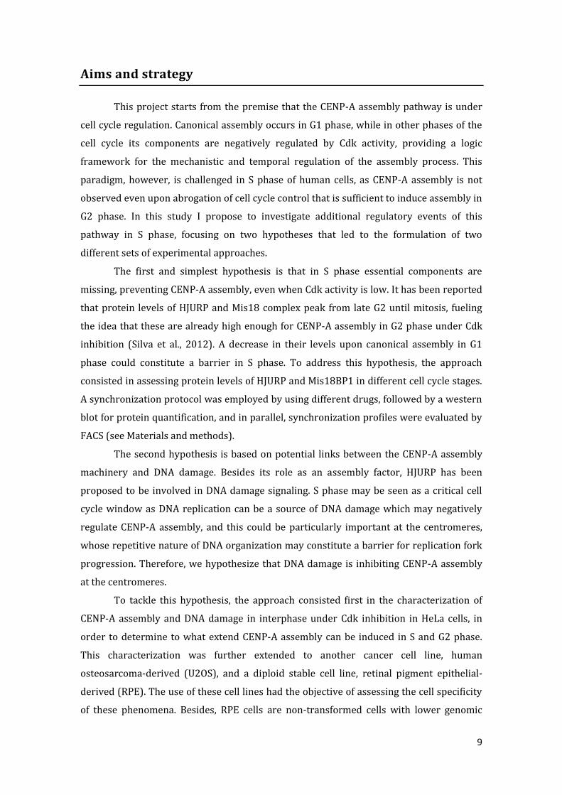
Figure 6. HJURP and Mis18BP1 are present in S phase. (A) FACS profiles of synchronized cells, obtained by
propdium iodide (PI) staining for two independent experiments (Exp 1 and Exp 2 respectively). (B)
Immunoblot for HJURP and Mis18BP1 for Exp 1 and Exp 2. L corresponds to protein ladder, Async. to
asynchronous population, and M, eG1, G1/S, S, G2 corresponds to mitosis, early G1, G1/S boundary, S phase
and G2 phase respectively. α-tubulin was used as the loading control. (C) Quantification of the bands in the two
experiments was performed using an Odyssey Scanner. Grey bars and black bars correspond to HJURP and
Mis18BP1, respectively. Intensity of the bands was normalized to α-tubulin in each case, and these values were
subsequently normalized to unsynchronized (Async.) cells. Error bars indicate SD.
As shown in Figure 6A, the majority of the events represent the quantity of DNA
that is roughly expected to be present in each cell cycle stage analyzed, although there is a
shift in the relative position of the peaks among treatments, most likely due to a technical
artifact of the FACS machine. The overall efficiency of synchronization gave confidence for
the analysis of the western blots, represented in Figure 6B. Quantification of the results
show that HJURP protein levels vary little across all cell cycle stages with a slight increase
in S phase, and Mis18BP1 levels rise before S, peak in S and drop in G2 phase (Figure 6C).
While it is difficult to determine whether these proteins have a specific cell cycle profile, it
14
is evident that neither HJURP nor M18BP1 are absent in S phase. These results indicate
that the inability of S phase cells to assemble CENP-A is not the result of the absence of
these key assembly factors, and regulation of this pathway most likely occurs at the post-
translational level.
Characterization of CENP-A assembly in G1, S and G2 phase and possible role
of DNA damage in its regulation
The presence of the key assembly factors in S phase indicates that the lack of
CENP-A assembly in S phase is perhaps controlled in a more elaborate manner. We then
focused on bridging previous reported results to some possible entry points in order to
elucidate this phenomenon. The restricted assembly of CENP-A to late telophase/early G1
phase (Jansen et al., 2007) suggests that it is subjected to cell cycle regulation.
Additionally, assembly can be induced artificially in G2 phase by inhibition of Cdk activity
(Silva et al., 2012), but not in S phase. This refractory nature of S phase, however, does not
imply insensitivity of assembly components to Cdk-mediated inhibition; instead, there
may be an additional uncharacterized mechanism that is dominant over it. Accumulation
of possible hints without proper mechanistic detail, namely the involvement of HJURP and
CENP-A in DNA damage response and other specificities of S phase, prompted us to
investigate a possible causal relation between CENP-A assembly and DNA damage, in
which we hypothesized an inhibition of the former by the latter.
In order to analyze in further detail this possibility, the first step consisted in a
characterization of both assembly and DNA damage levels in different cell cycle stages of a
HeLa cell line constitutively expressing CENP-A (CENP-A-SNAP), both in unperturbed cells
(assembly in G1 phase) or in the presence of Roscovitine, a pan-Cdk inhibitor, inducing
assembly in G2 phase. A qualitative analysis of CENP-A signal was performed by
employing the SNAP-based Quench-Chase-Pulse (QCP) strategy described earlier, allowing
for the detection of centromeric signals of nascent CENP-A by microscopy. For detection of
cells in S phase an additional step in QCP is required, an incubation with a modified
thymidine analog EdU (Buck et al., 2008) for 15 minutes prior to fixation. The
incorporation of EdU into newly synthesized DNA in proliferating cells was subsequently
revealed in a chemical reaction before immunofluorescence.
Quantification of the levels of DNA damage was approximated by using γH2AX as
a marker. This phosphorylated form of the histone H2A variant, H2AX, was chosen for two
reasons. First, it is a key response signal at double strand breaks. Second, a positive
feedback loop ensures that its signal is amplified from the focus of a double strand break,
allowing its detection and quantification by immunofluorescence (Bonner et al., 2008).

15
Altogether, the number of γH2AX foci should correlate with DNA damage and was
therefore used as a readout throughout this study.
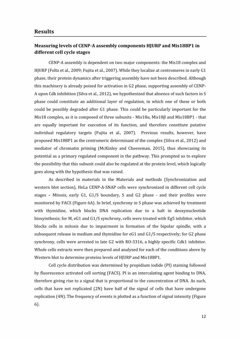
Figure 7. Characterization of CENP-A assembly profile in different cell cycle stages. (A) Schematic of the
protocol used for three experiments. HeLa CENP-A-SNAP cells were quenched and subsequently pulsed and
fixed after 6 and 7 hours respectively. Cells treated with Roscovitine (100 μM) were incubated with this drug
for 1.5h before fixation, and EdU was added 15 min prior to fixation. (B) Representative deconvolved images of
cells following each condition. Images were individually intensity scaled. (C) Fraction of CENP-A loading cells
in different stages. G1 phase cells were chosen based on absence of Cyclin-B signal as well as nuclear
morphology and proximity among pairs; S phase cells were identified using Click-iT EdU Alexa Fluor 647
imaging kit; G2 phase cells were chosen based on positive staining for Cyclin-B. Cells were considered positive
for CENP-A loading if visible CENP-A signal co-localized with centromeric marker CENP-T. Cells were
normalized to untreated G1 cells. 100 cells were analyzed in each condition, and the same criteria were
applied throughout this study. (D) Quantitative analysis of CENP-A centromere signal by CrAQ. For
quantification, random fields were imaged and a total of 20 cells positive for CENP-A were quantified. In the
case of S phase and non-treated G2 phase, no CENP-A positive cells were identified and random cells were
quantified instead (representing background signal indicated by dotted line). Error bars indicate SD.

16
The results, resumed in Figure 7, confirmed the absence of CENP-A loading cells in
S phase either with or without incubation with Roscovitine, while there is a substantial
proportion of CENP-A loading (~ 80%) in G2 phase cells that were subjected to the same
treatment (Figure 7C). Moreover, I observed an increase in the number of γH2AX foci in S
phase when compared to non-treated G1 and G2 cells (~30-fold and ~20-fold
respectively), and a decrease in the number of γH2AX foci in G2 Roscovitine-treated cells
to G1-like levels when compared to its control (Figure 8). This suggests the existence of a
negative correlation between DNA damage and CENP-A assembly, in which this only
occurs whenever levels of DNA damage are similar to those observed during canonical
loading in G1 phase.
Figure 8. DNA damage quantification of the experiment represented in Figure 7. (A) Representative
images of each one of the conditions (non-deconvolved). Images were intensity scaled independent from each
other. (B) Quantitative analysis of DNA damage. γH2AX foci were scored for each condition as described in
materials and methods and normalized to untreated G1 cells. 100 cells were analyzed per condition. Error bars
indicate SD.

17
Because the criteria to score CENP-A positive cells is based on visual observation,
which is somewhat subjective and may vary among different observers, additional
unbiased signal analysis was performed by using CrAQ (Bodor et al., 2012), an algorithm
that is extensively used for quantification of centromeric spots. Efficiency of CENP-A
assembly in G2 Roscovitine-treated cells was similar to non-treated G1 cells and
intriguingly, G1 Roscovitine-treated cells showed an increased efficiency of CENP-A
assembly (~1.4-fold) when compared to non-treated ones (Figure 7D). While this last
result can be explained in various ways, a simple interpretation is that residual Cdk1/2
activity remains capable of attenuating canonical G1 phase loading. Similar levels of CENP-
A assembly were observed in G1 and G2 Roscovitine-treated cells, and since Cdk1/2
activity increases as the cell progresses towards mitosis, it is reasonable to assume that
inhibition of these kinases was efficient in all cell cycle stages including S phase, as Cdk1/2
activity is lower at this stage. Most importantly, it highlights an additional non-Cdk1/2
based mechanism of CENP-A assembly inhibition in S phase which closely correlates with
the level of γH2AX foci. If the presence of DNA damage is inhibitory to CENP-A assembly,
this inhibition could be present in every cell cycle stage, but with an increased robustness
in this phase given the number of DNA damage events herein existent.
We also sought to evaluate to what extent the refractory nature of S phase to
CENP-A assembly is a general phenomenon and can be extended to other cell lines. Our
prediction was that since non-cancer cell lines have lower genomic instability relative to
cancer cell lines, they may present lower levels of DNA damage in S phase, with the
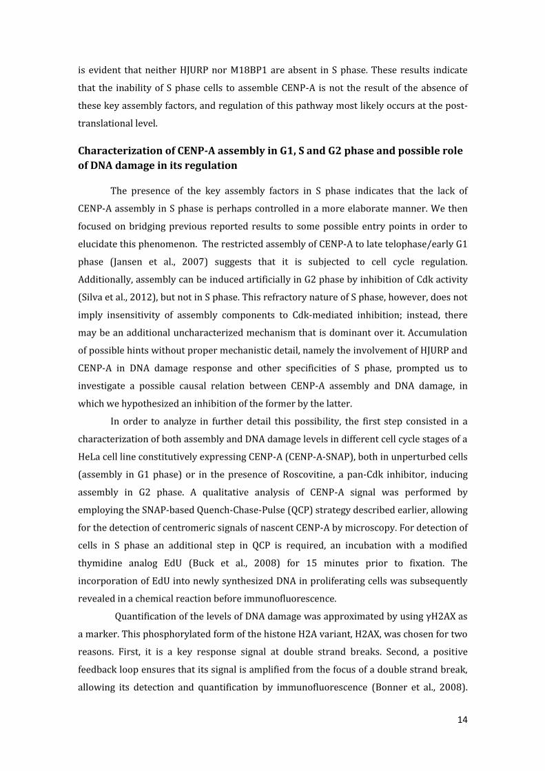
consequence of being permissive to CENP-A assembly. Preliminary results from a single
experiment in RPE (RPE CENP-A-SNAP) – a diploid stable cell line, and U2OS (U2OS CENP-
A-SNAP), another cancer cell line, suggest that in both cases there is CENP-A assembly in
G2 phase under Cdk inhibition (Figure 9C). Surprisingly, CENP-A assembly was observed
in a great number of U2OS cells in S phase, yet analysis by CrAQ showed that CENP-A
signals were highly diminished when compared to the ones observed in G2 phase (Figure
9D). One possible explanation of this low level but pervasive assembly in S phase may be
the high degree of CENP-A-SNAP overexpression in this constructed cell line. While the
refractory nature of S phase was still observed in this line, this result showed that CENP-A
can in principle be loaded at centromeres in S phase. In RPE cells, the number of CENP-A
loading cells under Roscovitine treatment was similar to what was observed previously in
HeLa. While I did not directly measure the intrinsic level of DNA damage in these cells,
these results show that RPE cells are, like HeLa cells, refractory to CENP-A loading in S
phase. This indicates that cells that have a stable genome maintain this S phase inhibited

18
state, suggesting the inhibition is not the result of the high genomic instability observed in
HeLa cells.
Figure 9. U2OS cells but not RPEs, are permissive to CENP-A assembly in S phase under Cdk inhibition.
(A) Schematic of the protocol used for one experiment. RPE or U2OS CENP-A-SNAP cells were quenched and
subsequently pulsed and fixed after 6 and 7 hours respectively. Cells treated with Roscovitine (100 μM) were
incubated with this drug for 1.5h before fixation. (B) Representative images (non-deconvolved) of each one of
the conditions. Images were independently intensity scaled. Left and right panels correspond to RPEs and
U2OS respectively. All the cell cycle stages were identified as described in Figure 7C, with the exception of p-
H3 that was used as a G2 marker in RPEs. (C) Number of CENP-A loading cells in RPE and U2OS in G2 and S
phase upon Roscovitine treatment. 100 cells were analyzed in each condition. (D) Quantitative analysis of
CENP-A centromere signal in U2OS by CrAQ. As described for Figure 7, 20 random cells from the pool that
scored positive for assembly in (C) were quantified. Error bars indicate SD.
19
Overall, the results showed that the refractory nature of S phase is transversal to
different cell lines, and most importantly, and that there appears to be a negative
correlation between CENP-A assembly and DNA damage. Further experiments were
performed in order to determine what is the effect of the latter in CENP-A assembly in
each specific cell cycle stage.
CENP-A assembly upon inhibition of DNA damage signaling and Cdk
inhibition in S phase
Our previous set of results suggested a negative correlation between the presence
of DNA damage and CENP-A assembly. Next, we then sought to investigate if that could be
translated into a mechanism of inhibition in HeLa cells. In S phase, homologous
recombination is the preferential method adopted by the cell for the repair of double
strand breaks (Branzei and Foiani, 2008), in which rapid recruitment of γH2AX to these
lesions provides a platform to downstream components that are mostly activated by ATM.
Previous studies hinted at the possibility of involvement of CENP-A and HJURP in the
process of repair (Kato et al., 2007; Zeitlin et al., 2009), but without mechanistic details
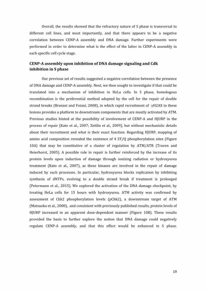
about their recruitment and what is their exact function. Regarding HJURP, mapping of
amino acid composition revealed the existence of 4 ST/Q phosphorylation sites (Figure
10A) that may be constitutive of a cluster of regulation by ATM/ATR (Traven and
Heierhorst, 2005). A possible role in repair is further reinforced by the increase of its
protein levels upon induction of damage through ionizing radiation or hydroxyurea
treatment (Kato et al., 2007), as these kinases are involved in the repair of damage
induced by such processes. In particular, hydroxyurea blocks replication by inhibiting
synthesis of dNTPs, evolving to a double strand break if treatment is prolonged
(Petermann et al., 2015). We explored the activation of the DNA damage checkpoint, by
treating HeLa cells for 15 hours with hydroxyurea. ATM activity was confirmed by
assessment of Chk2 phosphorylation levels (pChk2), a downstream target of ATM
(Matsuoka et al., 2000), and consistent with previously published results, protein levels of
HJURP increased in an apparent dose-dependent manner (Figure 10B). These results
provided the basis to further explore the notion that DNA damage could negatively
regulate CENP-A assembly, and that this effect would be enhanced in S phase.

20
Figure 10. HJURP has several putative ATM/ATR phospho-sites and its protein levels increase upon
incubation with hydroxyurea (A) Map of identified HJURP domains and putative phosphorylation sites by
ATM and ATR (red bars). (B) Quantification of the immunoblot of HJURP and (C) pChk2 of cells treated for 15-
hours with 0,5 or 1 mM hydroxyurea (HU) using an Odyssey Scanner (single experiment).
If DNA damage is indeed inhibiting this pathway, we would expect to induce CENP-
A assembly in S phase by inhibiting upstream damage signaling kinases, such ATM and
ATR, in conjunction with Cdk inhibition. In order to test this hypothesis, three different
inhibitors of DNA damage signaling with a broad range of action, i.e. low specificity were
independently incorporated in the Quench-Chase-Pulse strategy used before (Figure 11).
Wortmannin, a pan-PI3K family inhibitor, is capable of inhibiting both ATM and ATR at
high concentrations (Sarkaria et al., 1998). CGK773 and Caffeine are reported as specific
ATM/ATR inhibitors, although their true effect is controversial (Choi et al., 2011; Cortez,
2003). These drugs were added one hour prior to Roscovitine treatment in order to
distinguish eventual effects of their inhibition alone.
Given the known role of ATM and ATR in DNA damage response, I was able to
measure their effectiveness by directly scoring the number of γH2AX foci visible in the
cells. The results, described in Figure 12, showed a reduction of foci only upon treatment
with Wortmannin or Wortmannin combined with Roscovitine (~5-fold and ~10-fold
respectively) relative to untreated cells. Importantly, this latter result recapitulates the
amount of damage present in G2 phase Roscovitine-treated cells (Figure 12, red bar),
where CENP-A assembly is observed. Reduction of γH2AX foci was not observed in any of
the other treatments, perhaps due to insufficient concentration or length of the drug
incubation. Treatment with Wortmannin, however, proved to be highly toxic for the cells.
In addition, a strong and constant background signal hindered the analysis, resulting in a
possible underestimation of γH2AX foci. This is probably a consequence of a pre-apoptotic
state.

21
Figure 11. Characterization of CENP-A assembly upon treatment with different DNA damage inhibitors
in S phase of HeLa cells. (A) Schematic of the protocol used for two experiments. Cells were pre-incubated
with either CGK773 20uM, Wortmannin 100uM or Caffeine 8mM 1 hour before treatment with Roscovitine.
Drugs treatments were maintained until fixation. (B) Representative images (deconvolved) of each one of the
conditions. Images were independently intensity scaled. –R or +R denotes no treatment or treatment with
Roscovitine respectively.

22
Figure 12. DNA damage quantification of the experiment represented in Figure 11. (A) Representative
images of each of the conditions (non-deconvolved). Images were independently intensity scaled. (B)
Quantitative analysis of DNA damage. γH2AX foci were scored for each condition as described in materials and
methods and normalized to untreated non-treated S phase cells (No treatment). Red bar (G2 Roscovitine)
corresponds to the number of foci observed in Figure 8B. 100 cells were analyzed per condition. Error bars
indicate SD.
Importantly, irrespective of the condition or reduction in γH2AX foci, no CENP-A
positive cells were observed in S phase. Given our observation that Wortmannin treatment
brings down the level of γH2AX foci to similar levels of G2 Roscovitine-treated cells, where

23
assembly is observed, it suggests that the high levels of DNA damage in these cells is not
the primary reason for inhibition of CENP-A assembly.
CENP-A assembly upon induction of damage
In the previous section, I described our attempts to abrogate the putative
inhibitory regulation by DNA damage signaling preventing CENP-A assembly in S phase.
Here, I present reciprocal experiments in which rather than alleviating inhibition, DNA
damage was induced to determine whether it could block CENP-A assembly. This was
tested this both in G1 phase, the canonical cell cycle stage for assembly, as well as in G2
phase, where we can induce it by Cdk inhibition. If our hypothesis is correct, we would
expect a decrease in CENP-A assembly in G1 and G2 phase-treated cells upon induction of
DNA damage. From the multiple ways to induce damage in the cells, we decided to utilize
Etoposide, a DNA Toposisomerase-II inhibitor that stabilizes covalent enzyme-DNA
complexes, generating permanent DNA strand breaks (Nitiss, 2009) as well as γ-radiation
(see below).
Relying on the Quench-Chase-Pulse strategy, cells were treated with Etoposide 1
hour prior to incubation with Roscovitine until fixation (Figure 13A). As predicted, this
drug resulted in elevated numbers of γH2AX foci both in G1 and G2 phase (Figure 14). The
key set of conditions that allowed to test our hypothesis were the induction of damage and
subsequent alleviation of Cdk inhibition both in G1 and G2 phase, which proved to be
conclusive since pervasive CENP-A assembly was observed in both cases even under high
levels of DNA damage compared to the controls (~10-fold and ~20-fold respectively,
Figure 13D and E).
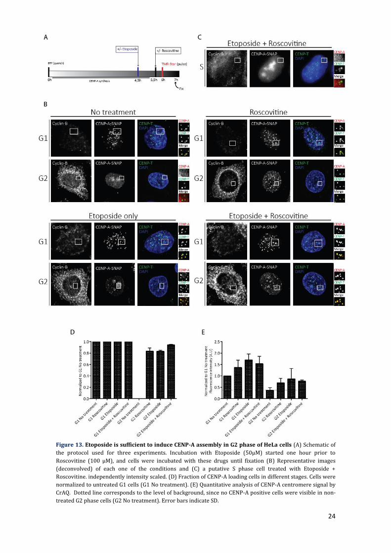
Unexpectedly, treatment with Etoposide alone was sufficient to induce CENP-A
assembly, with the number and efficiency of CENP-A loading cells in G2 phase being
similar to what is observed in G2 cells treated only with Roscovitine, but with an increase
(~5-fold) in γH2AX foci compared to this condition. In G1 phase, although I observed an
increase (~10-fold) in γH2AX foci, efficiency of CENP-A loading (~1,5-fold) also increased
relative to the control, similar to what is observed upon Roscovitine-only treatment
(Figure 7D).
24
Figure 13. Etoposide is sufficient to induce CENP-A assembly in G2 phase of HeLa cells (A) Schematic of
the protocol used for three experiments. Incubation with Etoposide (50μM) started one hour prior to
Roscovitine (100 μM), and cells were incubated with these drugs until fixation (B) Representative images
(deconvolved) of each one of the conditions and (C) a putative S phase cell treated with Etoposide +
Roscovitine. independently intensity scaled. (D) Fraction of CENP-A loading cells in different stages. Cells were
normalized to untreated G1 cells (G1 No treatment). (E) Quantitative analysis of CENP-A centromere signal by
CrAQ. Dotted line corresponds to the level of background, since no CENP-A positive cells were visible in non-
treated G2 phase cells (G2 No treatment). Error bars indicate SD.

25
Figure 14. DNA damage quantification of the experiment represented in Figure 13. (A) Representative
images of each one of the conditions (non-deconvolved). Images were independently intensity scaled. (B)
Quantitative analysis of DNA damage. γH2AX foci were scored for each condition as described in materials and
methods and normalized to untreated G1 cells. Error bars indicate SD.

26
These results suggest that under such conditions, presence of DNA damage does
not inhibit CENP-A assembly outside S phase, but on the contrary, it induces it. We then
ask ourselves if this unforeseen phenomenon could be observed in S phase, despite not
having used an S phase marker in this experiment. Analogous to the set of experiments
described in Figure 7, cells in G1 phase were chosen from an asynchronous population
stained for Cyclin-B (G2 cells) by applying previously described criteria. The remaining
cells, which were neither in G1 nor G2 phase, were assumed to be either in late G1 or S
phase; in this subset of cells, no CENP-A assembly was observed in around 50% of the cells
(Figure 13C, quantification not shown). The presence of a population of cells in which no
assembly occurs strongly suggests that S phase is still refractory to it, even though this is
efficient in other phases of the cell cycle. However, to address any doubts that may arise
from the difficulty of distinguishing a late G1 from an S phase cell, this experiment should
be repeated with an S phase marker.
In order to determine if these results were specific to Etoposide treatment or
represent a broader phenomenon following induction of DNA damage, we complemented
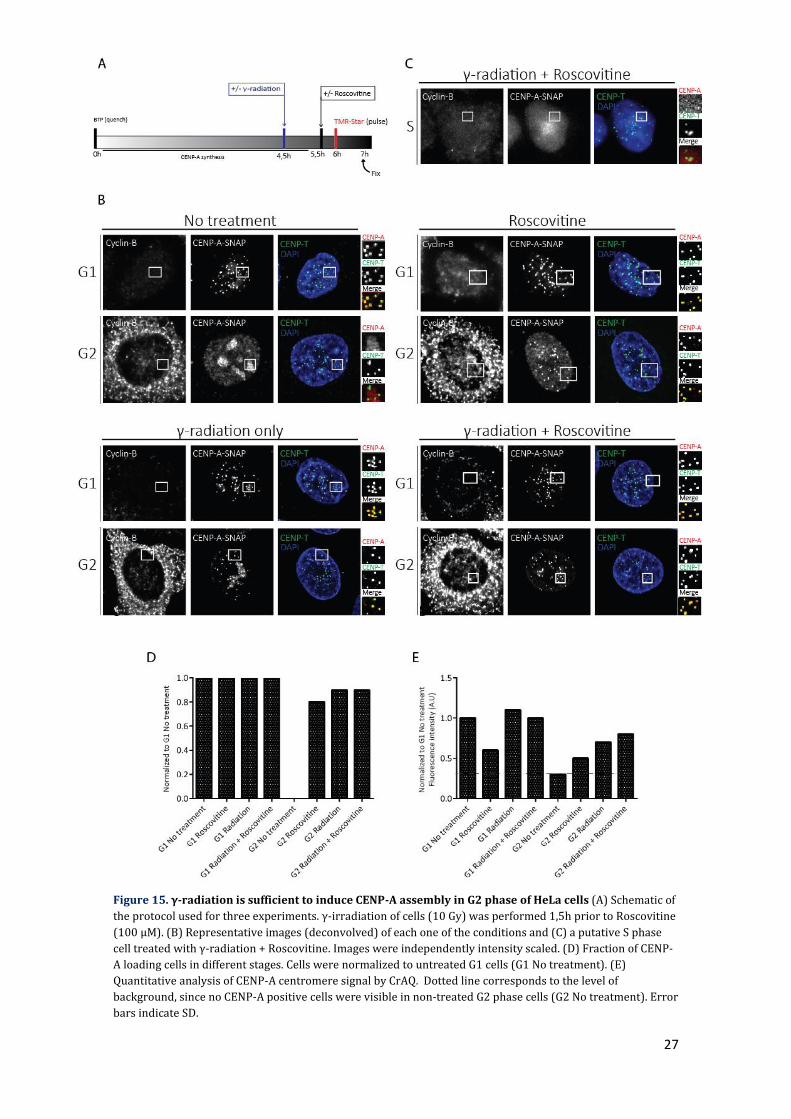
the previous experiments with a preliminary one by using γ-radiation. Irradiation of cells
with 10 Gy (gray) of gamma rays mostly recapitulated the results of the Etoposide
treatment, in which induction of damage was sufficient to induce CENP-A assembly in G2
phase both in the number of cells and efficiency (Figure 15). Since the number of γH2AX
foci obtained upon damage induction is also similar (Figure 16), it hints to the possibility
that the undisclosed mechanism that allowed assembly reached its maximum capacity.
However, by comparison of these images to the ones obtained by Etoposide (Figure 14), it
is visually discernible that in the latter case the number of foci was underestimated due to
saturation in the quantification method that was used. Taken together, the results show
that the hypothesis raised is inconsistent with our observations, and in fact DNA damage
had an unexpected positive effect in G1 and G2, but not in S phase.
27
Figure 15. γ-radiation is sufficient to induce CENP-A assembly in G2 phase of HeLa cells (A) Schematic of
the protocol used for three experiments. γ-irradiation of cells (10 Gy) was performed 1,5h prior to Roscovitine
(100 μM). (B) Representative images (deconvolved) of each one of the conditions and (C) a putative S phase
cell treated with γ-radiation + Roscovitine. Images were independently intensity scaled. (D) Fraction of CENP-
A loading cells in different stages. Cells were normalized to untreated G1 cells (G1 No treatment). (E)
Quantitative analysis of CENP-A centromere signal by CrAQ. Dotted line corresponds to the level of
background, since no CENP-A positive cells were visible in non-treated G2 phase cells (G2 No treatment). Error
bars indicate SD.

28
Figure 16. DNA damage quantification of the experiment represented in Figure 15. (A) Representative
images of each one of the conditions (non-deconvolved). Images were independently intensity scaled. (B)
Quantitative analysis of DNA damage. γH2AX foci were scored for each condition as described in materials and
methods and normalized to untreated G1 cells. Error bars indicate SD.

29
Discussion
The centromere is fundamental to cell division, and its continued maintenance is
dependent on the tight control that is exerted in the assembly and redistribution of CENP-
A nucleosomes at centromeric chromatin throughout cell generations. The cell cycle
regulation of this process appears primarily mediated by Cdk1 and Cdk2 activity, and it
has been established previously in the lab that these kinases are necessary for restricting
canonical CENP-A assembly to G1 phase. Cdk activities are low in G1 phase, while in S, G2
and M phase their activity increases inhibiting the assembly machinery. Support for this
idea arises from the fact that abrogation of Cdk activity prior to mitosis is sufficient to
induce CENP-A assembly in G2 phase. This negative regulation acts upon the Mis18
complex and HJURP (Ana Stankovic, unpublished). However, strikingly, such inhibition
does not lead to assembly in S phase in HeLa cells (Mariana Silva unpublished, this thesis).
The only known exception are DT40 cells (chicken cell line) where S phase assembly has
been observed (Silva et al., 2012), although the degree of penetrance and efficiency is
unclear. There hasn’t been any published study about a comprehensive understanding of
how this phase is refractory to CENP-A assembly to date, which is still a major blank space
in the full characterization of the pathway. In this project, I have tried to address this
question through the exploration of two possibilities.
As CENP-A assembly is dependent on the Mis18 complex and HJURP, it is to be
expected that their protein levels should not go below a certain threshold in order to
support assembly. Downregulation or degradation of one or both after canonical assembly
could account for the refractory nature of S phase to this phenomenon. By assessing
protein levels of Mis18BP1, a key component of the Mis18 complex, and HJURP, I managed
to observe that protein levels of these components were not diminished in S phase
compared to the rest of the cell cycle. Therefore, it is unlikely that S phase assembly is
absent due to the lack of components. Nevertheless, it is possible that there are other
untested components such as Mis18α and Mis18β that will prove to be rate-limiting to this
process. Rather than depletion, I observed an increase in protein levels of HJURP in S
phase (Figure 6). M18BP1 showed a similar peak in S phase albeit less pronounced.
Although this increase is harder to explain, the peak in HJURP may be explained in two
ways. My observation of an increase in HJURP (Figure 10) is reminiscent of a previous
report demonstrating increased levels of HJURP upon induction of damage (Kato et al.,
2007). Second, synchronization in this stage was achieved due to treatment with
Thymidine, a drug that by blocking DNA replication due to a halt in deoxynucleotide
biosyinthesis (Harper, 2005), induces damage and a repair response that is mediated by

30
ATM (Bolderson et al., 2004). Therefore, DNA damage induction due to Thymidine
treatment may lead to HJURP stabilization.
The general method to induce CENP-A assembly in G2 phase involves treatment
with a Cdk inhibitor. According to the minimalist model presented in Figure 2C, the cell
cycle in S, G2 and mitosis phases is mainly driven by Cdk1 and Cdk2 activities. Due to
rising cyclin levels, the corresponding Cdk activities rise as cells progress from S through
G2 (Cdk1/2) and peak in mitosis (Cdk1). As mentioned before, there is no reason to doubt
that Cdk inhibition is not achievable specifically in S phase. Even if this specific aspect of
Cdk1/2 regulation which is phosphorylation of Mis18BP1 and HJURP could have a higher
degree of redundancy in this cell cycle stage compared to others, that is compensatory
Cdk4 and Cdk6 activity, the usage of a pan-Cdk inhibitor such as Roscovitine would predict
full Cdk inhibition with regard to the concentration and length of incubation time used.
The explanation of why CENP-A assembly in S phase was not observed in HeLa and RPE
cell lines, but to some extend in U20S, is probably not due to non-existent Cdk regulation
in U2OS, but related to strong overexpression of CENP-A-SNAP. Either way, abrogation of
cell cycle control by means of Cdk inhibition can be bypassed, with transfection of both
Mis18BP1 and HJURP without putative Cdk phospho-sites. One possibility that has not yet
been explored is to verify how much of Mis18BP1 and HJURP is still phosphorylated after
Roscovitine treatment, as the phosphatases themselves could be under strict regulation in
S phase.
I also explored the possible role of DNA damage as an additional regulatory
mechanism besides the Cdk-mediated one. This idea came from the realization that S
phase has an increased number of DNA damage events due to replication. In addition,
previous observations have linked CENP-A assembly and DNA damage. These include
increased protein levels of HJURP upon DNA damage (Figure 10B; Kato et al., 2007),
possibly through ATM-regulation, as well as transient accumulation of CENP-A at DNA
damage sites upon induction of double-strand breaks (Zeitlin et al., 2009). Based on these
assumptions, although lacking specific details of how that would happen, we hypothesized
that endogenous DNA damage in S phase would be sufficient for inhibition of CENP-A
assembly. This was tested by inhibition of DNA damage signaling through the use of three
different ATM/ATR inhibitors. I found that only Wortmannin was capable of blocking DNA
damage signaling as measured by a reduction of γH2AX foci to G2 phase-like levels.
However, in none of the conditions was premature CENP-A assembly was observed, either
in the presence or absence of Cdk inhibition.
The results obtained with Wortmannin (Figure 12), may have two different
possible interpretations: either residual DNA damage signaling might continue to block

31
CENP-A assembly, or DNA signaling does not inhibit CENP-A assembly. This same
interpretation can be extended to RPE cell line, which is assumed to have less genomic
instability compared to cancer cell lines. Nevertheless, CENP-A assembly was not observed
in S phase in this cell line either (Figure 9C). The use of Wortmannin, however, has
pleiotropic effects that may introduce confounding variables in the interpretation of the
results. As HJURP possesses putative ATM/ATR phosphorylation sites (Figure 10A), a
crucial future experiment to be done is to express HJURP in which these putative phospho-
sites are mutated possibly uncoupling HJURP from regulation by ATM/ATR. This may lead
to two possible outcomes. On the one hand these kinases are negatively regulating CENP-A
assembly, which according to the hypothesis, mutation of phospho-sites would induce
CENP-A assembly. On the other, DNA damage somehow makes HJURP increase its protein
levels (Figure 10; Kato et al., 2007), perhaps through stabilization by ATM/ATR. As DNA
damage is always observed, even in G1 phase during canonical assembly (Figure 8),
insensitivity to its stabilization could lead to its degradation, and consequently absence of
CENP-A assembly not only in S phase, but in every cell cycle stage.
Another key aspect to be explored in the future, which was part of the initial plan
of this project, is to distinguish between DNA damage at the centromeres and the rest of
the genome. This means independently of the existence of global damage, a restricted
number of damage events at the centromeres could be sufficient to exert a local inhibitory
effect. Centromeres are known to replicate late in S phase. Due to their repetitive nature,
replication fork stalling can be a source of damage. Taking advantage of the characteristics
of EdU incorporation, which allow identification of cells in different phases of the S phase
due to active incorporation of nucleotides in replication regions, it would be possible to
observe if there is co-localization between γH2AX foci and centromeres somewhere across
S phase. Although not thoroughly explored, this does not seem to be the case, which argues
against the existence of local inhibition. To test this idea directly, we could make use of
CRISPR/Cas9, to selectively induce double-strand breaks specifically at the centromeres in
G1 and G2 phase, while assessing CENP-A assembly in these stages.
A complementary set of experiments to our initial hypothesis was also tested: If
DNA damage signaling blocks assembly we would expect to not observe CENP-A assembly
in G1 phase and G2 phase upon induction of DNA damage, even in the absence of Cdk
regulation. We induced DNA damage by two different means, chemically by Etoposide and
physically by γ-radiation in G2 phase. To our surprise either of these treatments was
sufficient to induce CENP-A assembly. This result strongly suggests that DNA damage and
its resulting signaling is not responsible for blocking CENP-A assembly; on the contrary, it
enhances it.

32
One explanation for this observation is the activation of the G2 DNA damage
checkpoint, which monitors the structural stability of the chromosomes prior to the entry
in mitosis (Figure 17). The main objective of this checkpoint, just like any other, is to delay
or arrest the cell cycle. For that it relies on the inactivation of Cdk1-Cyclin B, the principal
orchestrator of mitosis entry. Upon induction of damage, ATM and ATR are activated,
triggering a signaling cascade that maintains Cdk1 phosphorylated through the negative
regulation of its phosphatase CDC25 by CHK1 and CHK2. Inactivation of Cdk1 may then
lead to alleviation of the CENP-A assembly machinery, enabling CENP-A assembly at this
stage. Relative to Cdk2, it has been reported that is involved in the G2 checkpoint in p53-
independent cell lines, such as HeLa, in order to keep Cdk1 inactivated (Chung and Bunz,
2010). Therefore, participation in such a process could have led to alleviation of regulation
key components, similar to what happens in Cdk1. In order to confirm the direct
involvement of DNA damage in CENP-A assembly, a DNA damage signaling inhibitor could
be introduced before cell treatment with Etoposide or exposure to γ-radiation, expectedly
leading to absence of assembly. To test if indeed Cdks are being inactivated through the
induction of DNA damage, it is important to assess the phosphorylation status of known
Cdk targets. For example, this could be achieved by an immunofluorescence or western
blot with phospho-specific antibodies against HJURP and Mis18BP1 or any other known
Cdk target.
Despite the induction of CENP-A assembly by DNA damage it is still possible that it
can inhibit it when present at lower levels, and titration of different levels should be
performed to rule out any negative role of damage signaling on CENP-A assembly.
Importantly, CENP-A assembly was not observed in all cells; while G1 and G2 cells showed
a very high degree of assembly ~50% of the population did not assemble CENP-A at all.
Most of these are presumable in S phase (Figure 13C and 15C), indicating that even under
these conditions of premature assembly in G2 phase, S phase remains refractory. In S
phase, induction of damage leads to a delay in cell cycle progression that is dependent on a
mechanism similar to G2 checkpoint, but with inactivation of Cdk2 instead of Cdk1
(Lobrich and Jeggo, 2007), which may have not being sufficiently robust to induce
assembly: as this was still not observed even with the combination of DNA damage and
Roscovitine treatment, it highlights the robustness of the inhibitory mechanism taking
place.

33
Figure 17. Schematic of the G2/M checkpoint. DSBs in G2 can directly activate ATM (not shown), and
indirectly, via ATM-dependent strand resection, can lead to ATR activation. The G2/M checkpoint is initiated
by the phosphorylation of checkpoint kinases (CHK1 and CHK2) and phosphatases (probably CDC25C). This
prevents dephosporylation of Cdk1–cyclin B, which is required for progression into mitosis. Adapted from
Lobrich and Jeggo, 2007.
The initial hypotheses that were raised at the beginning turned out to be incorrect.
Another explanation may be related to the features of S phase. As mentioned, this stage is
characterized by DNA replication. Upon formation of newly-synthesized DNA, there is
incorporation of H3.1 specifically during this cell cycle stage, and also H3.3 to a lesser
extent (Ray-Gallet et al., 2011). Given the genomic architecture of the centromere, where
CENP-A nucleosomes are a small minority (1 in 25) compared to H3 nucleosomes, perhaps
there is a need for a temporal distinction of histone incorporation between the two events,
with assembly and re-distribution of CENP-A occurring in G1 and S phase respectively,
ensuring its maintenance in stable numbers across cell divisions. Indeed, there’s a high
level of histone turnover during S phase in comparison to CENP-A, and its assembly into
chromatin at this period could lead to potential mistargeting with severe consequences to

34
the cell. This is not probably the case in the U2OS, as CENP-A assembly was observed in
this phase at the centromeres without apparent consequences for viability. A possible
mechanism of inhibition could be simply related to competition with H3.1 and H3.3
incorporation; with H3.1, it would be a matter of a superior number of these nucleosomes
being assembled at this stage compared to CENP-A. In contrast, assembly of H3.3
nucleosomes in S phase has been proposed as a placeholding step that marks the sites of
posterior CENP-A assembly in G1 phase (Dunleavy et al., 2011), and in this case the
competition would come up to the affinity between the two assembly components
involved in this process. Importantly, several proteins are shared between the assembly
complexes of H3.1, H3.3 and CENP-A. These include histone H4 as well as the histone
chaperones RbAb46 and 48 (Foltz et al., 2006; Furuyama et al., 2006). In brief, competition
between H3 variant assembly pathways may explain why CENP-A cannot be assembled in
S phase. Impairment of both H3.1 and H3.3 assembly components at the centromere could
constitute an important experiment in order to assess this possibility. Another way to
address this is by overexpression of limiting shared factors such as H4.
In this study I investigated the mechanisms underlying inhibition of CENP-A
assembly in S phase. I showed that some key assembly factors are present in this phase,
indicating that the capacity for assembly is not absent. I tested whether DNA damage and
its consequent signaling are involved, and found that instead of being inhibitory this
pathway induces premature assembly in G2, but not in S phase. Finally, I presented a
speculative model based on a possible competition between CENP-A and canonical
chromatin assembly that may explain the refractory nature of S phase to centromere
assembly. In conclusion, this study allowed us to explore several possibilities regarding
the refractory nature of S phase, serving as a starting point for the elaboration of further
experiments that will allow elucidating this phenomenon.
35
Materials and methods
Cell lines and culture conditions
Construction of the monoclonal HeLa cell line (CENP-A-SNAP-3xHA) was previously
described (Bodor et al. 2012, Support Protocol 1; Jansen et al. 2007). RPE-CENP-A-SNAP
was established by integrating CENPA-SNAP-BLAST using a retroviral vector (pBABE) in
an RPE-hTERT background. U2OS-CENPA-SNAP cell line was a gift by Sebastian Müller,
Institut Curie, France. HeLa and U2OS cells were grown in DMEM and media was
supplemented with 1mM sodium pyruvate (SP), 2mM glutamine and 100 U/mL penicillin;
HeLa cellswere also supplemented with 10% newborn calf serum (NCS) and maintained
with 1μg/mL of Blasticidin, while U2OS were supplemented with 10% fetal bovine serum
(FBS) and maintained with 100 μg/mL of Geneticin. RPE cells were grown in DMEM/F12,
media was supplemented with 2mM glutamine and 100 U/mL penicillin, and cells were
maintained in 5 μg/mL of Blasticidin. All the human cell lines used were grown at 37ᵒC,
5% CO2.
Drug treatment of cells
Throughout this study, several drugs were used, with the respective periods of incubation
defined in each experiment. Roscovitine (Sigma) was used at a concentration of 100 μM.
CGK773 (Calbiochem), Wortmannin (Sigma) and Caffeine (Sigma) were used in a
concentration of 20 μM, 100 μM and 4 mM respectively. Etoposide (Sigma) was used in a
concentration of 50 μM. γ-radiation (10 Gy) was induced by a Gamma cell 2000
instrument (Molsgaard Medical, Denmark).
Immunofluorescence
Cell fixation, immunofluorescence and DAPI staining were performed as described (Bodor
et al., 2012), with the exception of cells stained for S phase, in which a commercial Click-iT
EdU Alexa Fluor 647 imaging kit (ThermoFisher) was used prior to immunofluorescence.
Mouse monoclonals α-gH2AX (Merck Millipore) and α-Cyclin B (Santa Cruz Biotechnology)
were used at a dilution of 1:1000 and 1:50 respectively. Rabbit polyclonals α-CENP-T
(from D. Cleveland, Ludwig Institute for Cancer Research, San Diego, USA) and α-Cyclin B
(Santa Cruz Biotechnology) were used at a dilution of 1:1000 and 1:50 respectively.
Secondary antibodies Donkey α-Rabbit and α-mouse FITC (Jackson ImmunoResearch), as
well as Donkey α-Rabbit, α-mouse and α-Rat Dy680 (Rockland) were used at a dilution of
1:200.

36
CENP-A-SNAP pulse labeling
SNAP labeling was performed as previously described (Bodor et al., 2012; Jansen et al.,
2007) except when otherwise specified. Fluorescent substrate TMR-Star and the non-
fluorescent substrate BTP (New England Biolabs) were used at a concentration of 2 μM.
Microscopy and Image Analysis
Images were acquired on a commercial Leica High Content Screening microscope, based
on Leica DMI6000 equipped with a Hamamatsu Flash 4.0 LT sCMOS 4.0 Mpx camera,
using a 63x 1.4 NA objective with binning 2×2, 0.3-mm Z-section nuclear scanning, DAPI +
fluorescence filtersets and controlled with the Leica LAS X software. All of the image
analysis was performed using FIJI with maximum intensity projections: centromeric TMR-
Star fluorescence was quantified using CRaQ (Bodor et al., 2012); γH2AX foci were
counted automatically by using the function “Find Maxima” in FIJI, after establishing a
threshold for each experiment in non-treated cells. This threshold was defined in order to
have one pixel per focus, and only foci with pixel intensities above it were counted.
Cell synchronization
HeLa cells were synchronized in different cell cycle stages following a specific protocol for
each case. For mitosis, early G1 (eG1) and G1/S boundary (G1/S), cells were incubated
with Eg5 inhibitor III for 17 hours, with a subsequent 5 hour-release in medium for eG1
and an overnight release in thymidine for G1/S. For S phase (S), a double-thymidine block
was employed, in which cells were incubated with thymidine for 17 hours, released in
Deoxycytidine for 9 hours, incubated with thymidine for another 17 hours and released in
medium for 5 hours. For G2 phase (G2), cells were incubated with the Cdk1 inhibitor RO-
3306 for 20 hours. Eg5 inhibitor III (Santa Cruz Biotechnology), Thymidine (Sigma), RO-
3306 (Calbiochem) and Deoxycytidine (Sigma) were used at a concentration of 1 μM, 2
mM, 9 mM and 24 μM respectively.
SDS PAGE and Western Blotting
Whole cell extracts were prepared by direct lysis in Laemmli sample buffer, separated in a
12% SDS-PAGE gel and transferred to nitrocellulose membranes. Blots were probed with
Rabbit polyclonals α-HJURP (gift from Dan Foltz, University of Virginia, USA), α-Mis18BP1
(Bethyl Laboratories) and Mouse monoclonal α-Tubulin (Sigma) at a dilution of 1:1000,
1:2000 and 1:1000 respectively. Secondary antibodies Goat α-Rabbit and α-Mouse IRDye
(LI-COR Biosciences), as well as Goat α-Mouse Dy680 (Thermo Scientific) were used at a
dilution of 1:200.

37
Cell sorting
Cells trypsinized (TrypLE Express Enzyme 1X, Gibco), washed in PBS and fixed in 70%
Ethanol, were posteriorly treated with 200 μg/mL RNAse A (Invitrogen) and stained with
5 μg/mL Propidium Iodine (Sigma) at the same time for at least 2 hours. Flow cytometry
analysis was performed on a FACScan (Beckton Dickinson) using CellQuest software.

38
References
Barnhart, M.C., Kuich, P.H.J.L., Stellfox, M.E., Ward, J.A., Bassett, E.A., Black, B.E., Foltz, D.R., 2011. HJURP is a CENP-A chromatin assembly factor sufficient to form a functional de novo kinetochore. J. Cell Biol. 194 , 229–243.
Bartek, J., Lukas, C., Lukas, J., 2004. Checking on DNA damage in S phase. Nat. Rev. Mol. Cell Biol. 5, 792–804.
Blower, M.D., Karpen, G.H., 2001. The role of Drosophila CID in kinetochore formation, cell-cycle progression and heterochromatin interactions. Nat Cell Biol 3, 730–739.
Bodor, D.L., Mata, J.F., Sergeev, M., David, A.F., Salimian, K.J., Panchenko, T., Cleveland, D.W., Black, B.E., Shah, J. V., Jansen, L.E.T., 2014. The quantitative architecture of centromeric chromatin. Elife 2014.
Bodor, D.L., Rodríguez, M.G., Moreno, N., Jansen, L.E.T., 2012. Analysis of protein turnover by quantitative SNAP-based pulse-chase imaging. Curr. Protoc. Cell Biol.
Bodor, D.L., Valente, L.P., Mata, J.F., Black, B.E., Jansen, L.E.T., 2013. Assembly in G1 phase and long-term stability are unique intrinsic features of CENP-A nucleosomes. Mol. Biol. Cell 24, 923–932.
Bolderson, E., Scorah, J., Helleday, T., Smythe, C., Meuth, M., 2004. ATM is required for the cellular response to thymidine induced replication fork stress. Hum. Mol. Genet. 13 , 2937–2945.
Bonner, W.M., Redon, C.E., Dickey, J.S., Nakamura, A.J., Sedelnikova, O.A., Solier, S., Pommier, Y., 2008. [gamma]H2AX and cancer. Nat Rev Cancer 8, 957–967.
Branzei, D., Foiani, M., 2008. Regulation of DNA repair throughout the cell cycle. Nat. Rev. Mol. Cell Biol. 9, 297–308.
Buck, S.B., Bradford, J., Gee, K.R., Agnew, B.J., Clarke, S.T., Salic, A., 2008. Detection of S-phase cell cycle progression using 5-ethynyl-2’-deoxyuridine incorporation with click chemistry, an alternative to using 5-bromo-2'-deoxyuridine antibodies. Biotechniques 44, 927–9.
Cheeseman, I.M., Desai, A., 2008. Molecular architecture of the kinetochore-microtubule interface. Nat. Rev. Mol. Cell Biol. 9, 33–46.
Choi, S., Toledo, L.I., Fernandez-Capetillo, O., Bakkenist, C.J., 2011. CGK733 does not inhibit ATM or ATR kinase activity in H460 human lung cancer cells. DNA Repair (Amst). 10, 1000–1; author reply 1002.
Chung, J.H., Bunz, F., 2010. Cdk2 Is Required for p53-Independent G2/M Checkpoint Control. PLoS Genet 6, e1000863.
Cortez, D., 2003. Caffeine Inhibits Checkpoint Responses without Inhibiting the Ataxia-Telangiectasia-mutated (ATM) and ATM- and Rad3-related (ATR) Protein Kinases. J. Biol. Chem. 278 , 37139–37145.
Cottarel, G., Shero, J.H., Hieter, P., Hegemann, J.H., 1989. A 125-base-pair CEN6 DNA fragment is sufficient for complete meiotic and mitotic centromere functions in Saccharomyces cerevisiae. Mol. Cell. Biol. 9, 3342–3349.

39
Dunleavy, E.M., Almouzni, G., Karpen, G.H., 2011. H3.3 is deposited at centromeres in S phase as a placeholder for newly assembled CENP-A in G1 phase. Nucleus 2, 146–157.
Dunleavy, E.M., Roche, D., Tagami, H., Lacoste, N., Ray-Gallet, D., Nakamura, Y., Daigo, Y., Nakatani, Y., Almouzni-Pettinotti, G., 2009. HJURP Is a Cell-Cycle-Dependent Maintenance and Deposition Factor of CENP-A at Centromeres. Cell 137, 485–497.
Falk, S.J., Guo, L.Y., Sekulic, N., Smoak, E.M., Mani, T., Logsdon, G.A., Gupta, K., Jansen, L.E.T., Van Duyne, G.D., Vinogradov, S.A., Lampson, M.A., Black, B.E., 2015. CENP-C reshapes and stabilizes CENP-A nucleosomes at the centromere. Science. 348 , 699–703.
Foley, E.A., Kapoor, T.M., 2013. Microtubule attachment and spindle assembly checkpoint signalling at the kinetochore. Nat Rev Mol Cell Biol 14, 25–37.
Foltz, D.R., Jansen, L.E.T., Bailey, A.O., Yates, J.R., Bassett, E.A., Wood, S., Black, B.E., Cleveland, D.W., 2009. Centromere-specific assembly of CENP-a nucleosomes is mediated by HJURP. Cell 137, 472–84.
Foltz, D.R., Jansen, L.E.T., Black, B.E., Bailey, A.O., Yates, J.R., Cleveland, D.W., 2006. The human CENP-A centromeric nucleosome-associated complex. Nat. Cell Biol. 8, 458–469.
Fujita, Y., Hayashi, T., Kiyomitsu, T., Toyoda, Y., Kokubu, A., Obuse, C., Yanagida, M., 2007. Priming of centromere for CENP-A recruitment by human hMis18alpha, hMis18beta, and M18BP1. Dev. Cell 12, 17–30.
Furuyama, T., Dalal, Y., Henikoff, S., 2006. Chaperone-mediated assembly of centromeric chromatin in vitro. Proc. Natl. Acad. Sci. 103 , 6172–6177.
Harper, J., 2005. Synchronization of Cell Populations in G1/S and G2/M Phases of the Cell Cycle. In: Humphrey, T., Brooks, G. (Eds.), Cell Cycle Control SE - 7, Methods in Molecular BiologyTM. Humana Press, pp. 157–166.
Hasson, D., Panchenko, T., Salimian, K.J., Salman, M.U., Sekulic, N., Alonso, A., Warburton, P.E., Black, B.E., 2013. The octamer is the major form of CENP-A nucleosomes at human centromeres. Nat Struct Mol Biol 20, 687–695.
Henikoff, S., Ahmad, K., Malik, H.S., 2001. The Centromere Paradox: Stable Inheritance with Rapidly Evolving DNA. Science. 293 , 1098–1102.
Hochegger, H., Takeda, S., Hunt, T., 2008. Cyclin-dependent kinases and cell-cycle transitions: does one fit all? Nat Rev Mol Cell Biol 9, 910–916.
Izuta, H., Ikeno, M., Suzuki, N., Tomonaga, T., Nozaki, N., Obuse, C., Kisu, Y., Goshima, N., Nomura, F., Nomura, N., Yoda, K., 2006. Comprehensive analysis of the ICEN (Interphase Centromere Complex) components enriched in the CENP-A chromatin of human cells. Genes to Cells 11, 673–684.
Jackson, S.P., 2002. Sensing and repairing DNA double-strand breaks. Carcinog. 23 , 687–696.
Jansen, L.E.T., Black, B.E., Foltz, D.R., Cleveland, D.W., 2007. Propagation of centromeric chromatin requires exit from mitosis. J. Cell Biol. 176, 795–805.
Karpen, G.H., Allshire, R.C., 1997. The case for epigenetic effects on centromere identity and function. Trends Genet. 13, 489–496.

40
Kato, T., Sato, N., Hayama, S., Yamabuki, T., Ito, T., Miyamoto, M., Kondo, S., Nakamura, Y., Daigo, Y., 2007. Activation of Holliday junction-recognizing protein involved in the chromosomal stability and immortality of cancer cells. Cancer Res. 67, 8544–8553.
Lobrich, M., Jeggo, P.A., 2007. The impact of a negligent G2/M checkpoint on genomic instability and cancer induction. Nat Rev Cancer 7, 861–869.
Malumbres, M., Barbacid, M., 2009. Cell cycle, CDKs and cancer: a changing paradigm. Nat Rev Cancer 9, 153–166.
Marshall, O.J., Chueh, A.C., Wong, L.H., Choo, K.H.A., 2008. Neocentromeres: New Insights into Centromere Structure, Disease Development, and Karyotype Evolution. Am. J. Hum. Genet. 82, 261–282.
Matsuoka, S., Rotman, G., Ogawa, A., Shiloh, Y., Tamai, K., Elledge, S.J., 2000. Ataxia telangiectasia-mutated phosphorylates Chk2 in vivo and in vitro. Proc. Natl. Acad. Sci. 97 , 10389–10394.
McKinley, K.L., Cheeseman, I.M., 2015. Polo-like Kinase 1 Licenses CENP-A Deposition at Centromeres. Cell 158, 397–411.
Mendiburo, M.J., Padeken, J., Fülöp, S., Schepers, A., Heun, P., 2011. Drosophila CENH3 Is Sufficient for Centromere Formation. Science. 334 , 686–690.
Müller, S., Montes de Oca, R., Lacoste, N., Dingli, F., Loew, D., Almouzni, G., 2014. Phosphorylation and DNA binding of HJURP determine its centromeric recruitment and function in CenH3(CENP-A) loading. Cell Rep. 8, 190–203.
Nitiss, J.L., 2009. Targeting DNA topoisomerase II in cancer chemotherapy. Nat Rev Cancer 9, 338–350.
Petermann, E., Orta, M.L., Issaeva, N., Schultz, N., Helleday, T., 2015. Hydroxyurea-Stalled Replication Forks Become Progressively Inactivated and Require Two Different RAD51-Mediated Pathways for Restart and Repair. Mol. Cell 37, 492–502.
Ray-Gallet, D., Woolfe, A., Vassias, I., Pellentz, C., Lacoste, N., Puri, A., Schultz, D.C., Pchelintsev, N.A., Adams, P.D., Jansen, L.E.T., Almouzni, G., 2011. Dynamics of Histone H3 Deposition In Vivo Reveal a Nucleosome Gap-Filling Mechanism for H3.3 to Maintain Chromatin Integrity. Mol. Cell 44, 928–941.
Rivera, H., Zuffardi, O., Maraschio, P., Caiulo, A., Anichini, C., Scarinci, R., Vivarelli, R., 1989. Alternate centromere inactivation in a pseudodicentric (15;20)(pter;pter) associated with a progressive neurological disorder. J. Med. Genet. 26, 626–630.
Sancar, A., Lindsey-Boltz, L.A., Ünsal-Kaçmaz, K., Linn, S., 2004. Molecular Mechanisms of Mammalian DNA Repair and the DNA Damage Checkpoints. Annu. Rev. Biochem. 73, 39–85.
Sarkaria, J.N., Tibbetts, R.S., Busby, E.C., Kennedy, A.P., Hill, D.E., Abraham, R.T., 1998. Inhibition of Phosphoinositide 3-Kinase Related Kinases by the Radiosensitizing Agent Wortmannin. Cancer Res. 58 , 4375–4382.
Silva, M.C.C., Bodor, D.L., Stellfox, M.E., Martins, N.M.C., Hochegger, H., Foltz, D.R., Jansen, L.E.T., 2012. Cdk Activity Couples Epigenetic Centromere Inheritance to Cell Cycle Progression. Dev. Cell 22, 52–63.
Smith, S., Stillman, B., 1989. Purification and characterization of CAF-I, a human cell factor required for chromatin assembly during DNA replication in vitro. Cell 58, 15–25.
41
Sullivan, B.A., Karpen, G.H., 2004. Centromeric chromatin exhibits a histone modification pattern that is distinct from both euchromatin and heterochromatin. Nat Struct Mol Biol 11, 1076–1083.
Sullivan, K.F., Hechenberger, M., Masri, K., 1994. Human CENP-A contains a histone H3 related histone fold domain that is required for targeting to the centromere. J. Cell Biol. 127 , 581–592.
Takahashi, K., Chen, E.S., Yanagida, M., 2000. Requirement of Mis6 Centromere Connector for Localizing a CENP-A-Like Protein in Fission Yeast. Science. 288 , 2215–2219.
Ten Hagen, K.G., Gilbert, D.M., Willard, H.F., Cohen, S.N., 1990. Replication timing of DNA sequences associated with human centromeres and telomeres. Mol. Cell. Biol. 10, 6348–6355.
Traven, A., Heierhorst, J., 2005. SQ/TQ cluster domains: concentrated ATM/ATR kinase phosphorylation site regions in DNA-damage-response proteins. BioEssays 27, 397–407.
Tyler-Smith, C., Willard, H.F., 1993. Mammalian chromosome structure. Curr. Opin. Genet. Dev. 3, 390–397.
Zeitlin, S.G., Baker, N.M., Chapados, B.R., Soutoglou, E., Wang, J.Y.J., Berns, M.W., Cleveland, D.W., 2009. Double-strand DNA breaks recruit the centromeric histone CENP-A. Proc. Natl. Acad. Sci. U. S. A. 106, 15762–15767.