identification and characterization of proteins and...
TRANSCRIPT

ACTAUNIVERSITATIS
UPSALIENSISUPPSALA
2014
Digital Comprehensive Summaries of Uppsala Dissertationsfrom the Faculty of Medicine 961
Identification andCharacterization of Proteinsand MicroRNAs that ModulateReceptor Signaling, VesicularTrafficking and Cell Migration inVascular Cells
JOHAN HELDIN
ISSN 1651-6206ISBN 978-91-554-8833-8urn:nbn:se:uu:diva-212949

Dissertation presented at Uppsala University to be publicly examined in A1:111a, Husargatan3, BMC, Uppsala, Friday, 14 February 2014 at 13:15 for the degree of Doctor of Philosophy(Faculty of Medicine). The examination will be conducted in English. Faculty examiner: doc.Jonas Fuxe (Karolinska Institutet).
AbstractHeldin, J. 2014. Identification and Characterization of Proteins and MicroRNAs thatModulate Receptor Signaling, Vesicular Trafficking and Cell Migration in Vascular Cells.Digital Comprehensive Summaries of Uppsala Dissertations from the Faculty of Medicine961. 61 pp. Uppsala: Acta Universitatis Upsaliensis. ISBN 978-91-554-8833-8.
Blood vessels deliver nutrients and oxygen to tissues. Importantly, the functions and growth ofblood vessels are commonly altered in disease. The inside of all blood vessels are lined withendothelium, a thin specialized layer of endothelial cells that separate the blood from othertissues. This thesis deals with the identification and functional characterization of proteins andmicroRNAs that have key roles as modulators of growth factor signaling and directed cellmigration of endothelial cells and other vascular cells.
A previously uncharacterized protein of the exocyst complex, Exocyst complex component3-like 2 (ExoC3L2) was identified and shown to be highly expressed in endothelial cells ofsprouting vessels. Suppression of ExoC3L2 resulted in reduced VEGF-A signaling togetherwith reduced chemotaxis in response to VEGF-A gradients. VEGF-A-signaling via its receptorVEGFR-2 is thus modulated by the exocyst complex and ExoC3L2.
Expression profiling of highly vascularized tissues were used to identify several microRNAsselectively expressed in blood vessels. miR-145, targeting the transcription factor Fli1, wasshown to be expressed in pericytes and mural cells. Elevated levels of miR-145 reducedchemotaxis of both endothelial cells and fibroblasts in response to growth factor gradients.miR-145 depletion in fibroblasts was shown to inhibit chemotaxis in response to PDGF-BB.
The guanine nucleotide exchange factor FGD5 was shown to be selectively expressed inendothelial cells and to regulate Cdc42 activity. FGD5 was shown to regulate the turnoverof activated VEGF-receptors. Suppression of FGD5 impaired endothelial cell chemotaxis,suggesting that FGD5 is required for efficient and sustained VEGF-A signaling.
Inactivation of RhoD, a regulator of endosomal trafficking, resulted in an increased pool ofacetylated and stable microtubules. Knockdown of RhoD in human fibroblasts resulted in aloss of cell polarity. A link between PDGFR-β and RhoD was implicated by the finding thatPDGF-BB was shown to trigger formation of GTP-bound RhoD. Chemotaxis towards PDGF-BB was severely inhibited in cells with reduced RhoD expression, suggesting a role for RhoDin chemotaxis via its regulation of microtubule dynamics.
Keywords: Angiogenesis, cell migration, signaling, VEGF, FGD5, ExoC3L2, microRNA,RhoD
Johan Heldin, Department of Medical Biochemistry and Microbiology, Box 582, UppsalaUniversity, SE-75123 Uppsala, Sweden.
© Johan Heldin 2014
ISSN 1651-6206ISBN 978-91-554-8833-8urn:nbn:se:uu:diva-212949 (http://urn.kb.se/resolve?urn=urn:nbn:se:uu:diva-212949)

Make it work -Tim Gun, fashion guru


List of Papers
This thesis is based on the following papers, which are referred to in the text by their Roman numerals.
I Barkefors, I., Fredlund Fuchs, P., Heldin, J., Bergstrom, T., Forsberg-Nilsson, K and Kreuger, J. (2011) Exocyst Complex Com-ponent-3 like-2 (ExoC3L2) associates with the exocyst and mediates directional migration of endothelial cells. Journal of Biological Chemistry, 286, 24189–24199.
II Larsson, E., Fredlund Fuchs, P., Heldin, J., Barkefors, I., Bondjers, C., Genove, G., Arrondel, C., Gerwins, P., Kurschat, C., Schermer, B., Benzing, T., Harvey, S. J., Kreuger, J., Lindahl, P. (2009) Dis-covery of microvascular miRNAs using public gene expression data: miR-145 is expressed in pericytes and is a regulator of Fli1. Genome Medicine, 1,108.
III Heldin J., Fredlund Fuchs P.*, O’Callaghan P.*, Kasza Z., Le Jan S., Kampf C., Aspenström P., Gerwins P. and Kreuger J. FGD5 promotes VEGF-A signaling via inhibition of VEGF-receptor ubiquitination and degradation. Manuscript.
IV Almeida F*, Heldin J*, Reis K, Kreuger J. and Aspenström P. A RhoD-regulated pathway is needed for PDGF-BB-induced chemo-taxis. Manuscript.
* These authors contributed equally to the study
Reprints were made with permission from the publishers.


Contents
Introduction ................................................................................................... 11
Background ................................................................................................... 12 The vascular system .................................................................................. 12 Angiogenesis ............................................................................................ 15
Sprouting angiogenesis ........................................................................ 15 Important regulators of sprouting angiogenesis ................................... 18
Vascular endothelial growth factors ......................................................... 19 VEGFR-2 signaling ............................................................................. 20 VEGFR-2 recycling and degradation ................................................... 22
GTPases .................................................................................................... 23 Rho GTPases ........................................................................................ 25 Rho GTPases and actin dynamics ........................................................ 26 RhoD .................................................................................................... 26 Rho GTPases and cell polarity ............................................................. 27 Rho GTPases regulate adherens junctions and tight junctions ............ 28
FGD family of RhoGEFs .......................................................................... 28 FGD5 .................................................................................................... 29
Ubiquitination ........................................................................................... 30 Rho GTPases and Ubiquitination ......................................................... 31
The exocyst complex and vesicular trafficking ........................................ 32 The exocyst complex ........................................................................... 32 Vesicular trafficking ............................................................................ 34
Cell migration ........................................................................................... 37 MicroRNA ................................................................................................ 38
Present investigation ..................................................................................... 40 Aims .......................................................................................................... 40 Results and conclusions ............................................................................ 40 Discussion and future perspectives ........................................................... 43
Populärvetenskaplig sammanfattning ........................................................... 46
Acknowledgements ....................................................................................... 48
References ..................................................................................................... 51

Abbreviations
ALK Ang1 Arf Arp BM Clip DH Dll4 DUB EB EEA-1 EGF Erk FAK FGD FGF FILIP1 FRG GAP GPCR GEF Grb2 HECT HMVEC HUVEC IGFR JNK LAMP-1 LIF MAPK miRNA MRCK MVB Nck
Activin-receptor-like kinase Angiopoietin 1 ADP-ribosylation factor Actin-related protein Basement membrane CAP-GLY domain containing linker protein DBL homology domain Delta-like ligand 4 De-ubiquitinating enzyme Embryoid body Early endosome antigen 1 Epidermal growth factor Extracellular-signal-regulated kinase Focal adhesion kinase FYVE, RhoGEF and PH domain-containing protein Fibroblast growth factor Filamin A-binding protein 1 FGD1 related Cdc42-GEF GTPase-activating protein G-protein coupled receptor Guanine nucleotide exchange factor Growth factor receptor-bound protein 2 Homologous to the E6-AP carboxyl terminus Human dermal microvascular endothelial cells Human umbilical vein endothelial cells Insulin growth factor receptor c-Jun N-terminal kinase Lysosome associated membrane protein 1 Leukemia inhibitory factor Mitogen-activated protein kinase MicroRNA Myotonic dystrophy kinase–related Cdc42-binding kinase Multi-vesicular bodies Non-catalytic region of tyrosine kinase adaptor pro-tein

NPF PAK Par PDGF PH PI3K PIP PLC PlGF PKC PRK1 PROX-1 PTB1B RhoGDI RING RISC ROCK RTK Smad SMURF1 SNAP TGF-β TGFβR-II Tie2 TSAd Tyr VEGF VEGFR VRAP vSMC WASP WAVE WHAMM ZIPK
Actin nucleation promoting factors p21-activated kinase Protease activated receptor Platelet-derived growth factor Pleckstrin homology domain Phosphoinositide 3-kinase Phosphatidylinositolphosphate Phospholipase C Placenta growth factor Protein kinase C Protein kinase N1 Prospero homeobox protein 1 Protein-tyrosine phosphatase 1B Rho GDP-dissociation inhibitors Really interesting new gene RNA-induced silencing complex Rho-associated coiled-coil containing kinase Receptor tyrosine kinase Sma and mothers against decapentaplegic SMAD specific E3 ubiquitin protein ligase 1 Soluble N-ethylmaleimide-sensitive factor attachment protein Transforming growth factor β Transforming growth factor β receptor type II Tunica intima endothelial kinase 2 T cell–specific adapter Tyrosine Vascular endothelial growth factor Vascular endothelial growth factor receptor VEGF receptor-associated protein Vascular smooth muscle cell Wiskott-Aldrich syndrome protein Wiskott-Aldrich syndrome protein family member WAS homolog associated with actin Golgi mem-branes and microtubules Zipper-interacting protein kinase


11
Introduction
All organisms that are too large to depend on passive diffusion of nutrients and oxygen for their survival are dependent on a functional vascular system. Formation of new blood vessels from preexisting vessels is called angiogen-esis. It is important during embryogenesis, wound healing and the menstrual cycle. However, the vasculature is also essential for many pathological func-tions. Fore example, cancer cells also depend on it to grow and spread throughout the body. Due to that, angiogenesis and the vasculature have attracted a lot of attention the last decades. It is believed that if one could inhibit angiogenesis one could also prevent the growth of cancer tumors. However, this task has proven more challenging than expected, and currently there are no effective angiogenesis inhibitors at the market.
In this thesis I present findings that will increase the understanding of how the vasculature is regulated and hopefully help find good anti-angiogenesis drug targets.

12
Background
The vascular system The vascular system has been known for a long time. As early as in the 16th century Andreas Vesalius was able to depict the vascular tree in some detail (Fig. 1), and since then the vasculature has been meticulously investigated. A functional vasculature is essential for the development of the embryo to a fetus and thereafter for continued growth and tissue homeostasis. The vascu-lar network plays important roles in physiological processes such as wound healing and the menstrual cycle, as well as in several pathological conditions including tumor growth and retinopathy.1
The vasculature consists of two circulatory systems, the small pulmonary circulation and the large systemic circulation. The pulmonary circulation transports oxygen-poor blood in a loop from the heart to the lungs. In the lungs the blood becomes oxygenated and subsequently transported back to the heart. The systemic circulation transports oxygen- and nutrient-rich blood and nutrients to the many tissues and organs of the body. The systemic circulation begins with the aorta, the biggest artery in the body, which branches into arteries and smaller vessels called arterioles. The arterioles branch further forming the capillary plexus where most of the exchange of nutrients and oxygen occurs. The capillaries thereafter merge into venules followed by larger veins that transport the nutrient- and oxygen-depleted blood along with cellular waste products. On its way back to the heart, the blood passes by the intestines to refuel nutrients and the liver to take care of the waste products.
Blood vessels are mainly built up by three cell types: endothelial cells, pericytes and vascular smooth muscle cells (vSMC). The endothelial cells form the inner lining of the vascular tube. A basement membrane (BM) con-sisting of collagen IV and laminin is deposited on the abluminal side of the endothelial cells. Mural cells such as pericytes and vascular smooth muscle cells support and stabilize the endothelial cells and subsequently the whole vessel. There are presently no molecular markers that allow for a confident identification of pericytes and vSMCs; instead these cell types are distin-guished by differences in their morphology and localization.2 Pericytes have gap junctions3 towards endothelial cells and are embedded in, and contribute to, the BM by producing its components. vSMCs on the other hand are more or less separated from the BM. However, these definitions are not absolute

13
and several intermediate cell phenotypes may occur.2 These two cell types in part have different functions. Pericytes appear to be responsible for cell-cell communication as well as the coordination of endothelial cell activity in the capillaries. vSMCs on the other hand give mechanical support to larger ves-sels and regulates vessel contractility. Furthermore, it has been suggested that pericytes may act as sensors for hypoglycemia and hypoxia, which could be reflected by the high pericyte-coverage of vessels in the retina and the brain where the metabolic demands are high.2
The vascular system is mirrored by the lymphatic system. Its main task is to drain excess fluid from the extracellular matrix and transport it back to the blood system. The lymphatic vascular system originates from the cardinal vein and starts to form primary lymph sacs at embryonic day 10.5.4 The dif-ferentiation from vein to lymph sacs, and subsequently the formation of the lymph vasculature is induced by vascular endothelial growth factor C (VEGF-C) binding to vascular endothelial growth factor receptor 3 (VEGFR-3). This induces the expression of the transcription factor prospero homeobox protein 1 (PROX-1). Without PROX-1 the endothelial cells in the vein fails to express lymph vessel markers like VEGFR-3 and lymphatic vessel endothelial hyaluronan receptor-1 (LYVE-1), which makes them una-ble to sustain the lymphatic phenotype.4
There are similarities between the blood vessels and lymph vessels, but there are also differences. Like blood vessels, the lymph vessels consist of an inner lining of (lymph) endothelial cells with supporting mural cells sur-rounding the larger lymph vessels. However, the lymph vessel walls lack smooth muscle cells. Instead they are dependent on nearby skeletal muscles for pushing the lymphatic fluid forward. Where blood vessels are construct-ed to retain fluid, lymph vessels on the other hand, are constructed to drain excess fluid from the extracellular matrix. Due to this the junctions between the lymph endothelial cells are fenestrated, compared to the dense junctions that are present between blood endothelial cells. The lymphatic system is blunt-ended and contains lymph nodes. These lymph nodes are used by the immune system as checkpoints for pathogens. The lymph system is also used by metastatic cancer cells for spreading. The lymphatic fluid is returned to the blood stream by large lymph vessels emptying the lymphatic fluid in the internal jugular and the subclavian veins.

14
Figure 1. The human vascular tree as described by Andreas Vesalius in De humani corporis fabrica libri septem, published in 1543. Image reproduced by the courtesy of the U.S. National Library of Medicine (http://www.nlm.nih.gov).

15
Angiogenesis Angiogenesis is defined as the formation of new blood vessels from pre-existing vasculature. In contrast, vasculogenesis is defined as blood vessel formation from progenitor cells (also known as angioblasts). Vasculogenesis most likely only occurs during embryogenesis.5
Three main mechanisms of angiogenesis have been described. Intussus-ceptive angiogenesis is when a vessel splits longitudinally into two vessels, which is mainly seen during embryogenesis. This is a fast and efficient way of doubling the amount of vessels in a tissue that is expanding rapidly. The vessel is divided by a mural cell pushing into the vessel. This creates an in-vagination in the vessel that will eventually push the endothelial cells on each side of the invagination together. Once the endothelial cells comes in contact with each other, they will reorganize and form two new parallel blood vessels. One great advantage with this mechanism is that the vessels are fully functional throughout the whole process and can maintain a blood flow.6 The second type of angiogenesis is called looping angiogenesis, which is believed to occur mainly during adult wound healing. It involves myofibroblasts contracting the newly formed extracellular matrix and there-by pulling the vessel towards the site of injury. This mechanism is initially independent of proliferation. Instead the endothelial cells and pericytes are redistributed, which allows the vessel to elongate without losing time on proliferation and migration. Since the blood vessels are fully perfused this is a fast way of resupplying the wounded area with blood vessels.7 The third type of angiogenesis is sprouting angiogenesis, which occurs when an endo-thelial cell breaks free from a blood vessel, and proliferates and migrates towards the source of a chemoattractant. The sprouts are blunt and non-perfused, which means that the sprout needs to find another sprout or blood vessel to fuse with (anastomosis) before the vessel is fully functional. This process is very time consuming compared to the other two mechanisms.
Sprouting angiogenesis During sprouting angiogenesis the sprout is led by a tip cell that is followed by stalk cells.8 For a long time it was unknown if the same tip cell guided the sprout throughout the whole angiogenic process. However, recent publica-tions indicate that the cells in the front of the sprout compete for the tip cell position, thus making the tip cell phenotype dynamic.9 The tip cell is charac-terized by high expressions of Delta-like ligand 4 (Dll4), platelet-derived growth factor B (PDGF-B), unc5b, vascular endothelial growth factor recep-tor 2 (VEGFR-2) and a high number of finger-like projections called filopo-dia. The filopodia carry receptors for guidance. They respond to gradients of both chemoattractants and repellants, thus making it possible for the tip cell to guide the sprout (Fig. 2).8,10-12 In support of the theory regarding the dy-

16
namic competition for the tip cell position, it has been shown that intracellu-lar Dll4 accumulates in the tip cells as well as to a lower degree in adjacent stalk cells.10,13 Stalk cells on the other hand are characterized by being hyper-proliferative and have a high vascular endothelial growth factor receptor 1 (VEGFR-1) expression but few filopodia. They also have established ad-herens and tight junctions that stabilize the newly formed blood vessel.12
Sprouting angiogenesis is initiated by the release of vascular endothelial growth factor A (VEGF-A) in response to e.g. hypoxia. This starts a polari-zation of endothelial cells in neighboring blood vessels and subsequently a competition between endothelial cells for the tip cell position. The cell with the highest levels of activated VEGFR-2 will be able to up-regulate Dll4 to a higher degree than the neighboring cells.9,11-13 Dll4 then activates Notch1 in neighboring cells, leading to a down-regulation of VEGFR-211 and thereby to a down-regulation of Dll49 in these cells. This secures that the tip cell has the highest levels of both VEGFR-2 and Dll4. In addition, upon the Dll4-mediated activation of Notch1 the expression of VEGFR-1 is up-regulated.14 It has been suggested that VEGFR-1 in this setting acts as a decoy receptor that competes with VEGFR-2 for binding to VEGF-A and reduces VEGFR-2 signaling in the stalk cells, thereby stabilizing the stalk cell phenotype (Fig. 2).12,14 While migrating towards the source of VEGF-A, the tip cell secretes PDGF-BB. This recruits pericytes to the stalk cells, which further stabilize the newly formed sprout.

17
Figure 2. Illustration of the interplay between endothelial tip and stalk cells dur-ing blood vessel sprouting. Upon release of VEGF-A, VEGFR-2 on the tip cell is activated. This initiates a positive autocrine feedback loop, which further induce the expression of VEGFR-2, PDGF-B and Dll4 in the tip cell. The tip cell also signals to the adjacent stalk cells through Dll4, which activates Notch1 in these cells. Notch1 activation induces the expression of VEGFR-1 and decreases the expression of Dll4 and VEGFR-2, thus securing the stalk cell phenotype. During the elongation of the sprout the secreted PDGF-BB recruits pericytes to the grow-ing vessel and thus stabilizes it. Eventually the tip cell may be replaced by anoth-er endothelial cell, which then will resume a tip cell phenotype.

18
Important regulators of sprouting angiogenesis Delta-like ligand 4 has been implicated as a negative regulator of sprouting, since it has been shown that knockdown of Dll4 or inhibition of Notch1 re-sults in hypersprouting.9-11,13 It has also been shown that endothelial cells with decreased VEGFR-2 or Dll4 expression are more prone to become stalk cells. It seems that they are equally important for the tip cell phenotype since there are no difference between cells with a low expression of Dll4 or VEGFR-2. Endothelial cells with decreased VEGFR-1 expression on the other hand have an increased probability of becoming tip cells.9
The Notch ligand Jagged-1 is expressed by endothelial cells and antago-nizes Dll4-mediated Notch signaling. In contrast to Dll4, Jagged-1 has been proposed to activate Notch3 in adjacent mural cells such as pericytes. Notch3 activation is believed to be important for pericyte/smooth muscle cell maturation.15
Pericytes and endothelial cells interact and are functionally dependent on each other. For example, transforming growth factor β (TGF-β) acts on en-dothelial cells via the type I serine/threonine kinase receptor, activin-receptor-like kinase 1 (ALK1) and -5 (ALK5), and on pericytes via ALK5. Both ALK1 and -5 form complexes with TGF-β receptor type II (TGFβR-II) and signal through Smad1, -5 and -8, and Smad2 and -3, respectively.2,16 Endothelial cells also express endoglin that acts as a co-receptor, switching from ALK5 to ALK1 signaling.17 It has been shown that lack of endoglin-dependent TGF-β signaling in endothelial cells disturbs vSMC recruitment to the vessel by lowering of endothelial-cell-secreted TGF-β.18 Endothelial cells respond differently to TGF-β depending on which receptor is activated. ALK1 is activated by a high dose of TGF-β and promotes migration and proliferation via Smad1 and -5, whereas a lower and prolonged dose of TGF-β activates ALK5 that promotes differentiation/maturation via Smad2 and -3.2,16,19
Another example of the tight communication between endothelial cells and pericytes is the Ang1-Tie2 signaling axis, which promotes maturation of the microvascular endothelium20,21. The Tie2 receptor20 is considered to be endothelial cell specific and its ligand angiopoietin-1 (Ang1)21,22 is produced by pericytes. Ang2 on the other hand is expressed by endothelial cells during angiogenesis and is an antagonist of the Tie2 receptor, thus destabilizing endothelial cells to promote sprouting in the presence of VEGF-A.23
Another important regulator of angiogenesis is PDGF-BB, which is se-creted by the tip cell. PDGF-BB plays an important role during angiogenesis by promoting migration and proliferation of pericytes via binding to PDGFR-β, thereby recruiting them to the growing vascular sprout.2,8

19
Vascular endothelial growth factors The VEGF24-26 family of ligands along with signaling proteins of the PDGF27,28, EGF29,30 and FGF31 families bind to tyrosine kinase receptors. VEGF receptors (VEGFR) are composed of seven immunoglobulin (Ig)-like folds in the extracellular domain, a single transmembrane domain and a tyro-sine-kinase domain that is divided into two segments by a 70-amino-acid kinase insert.32
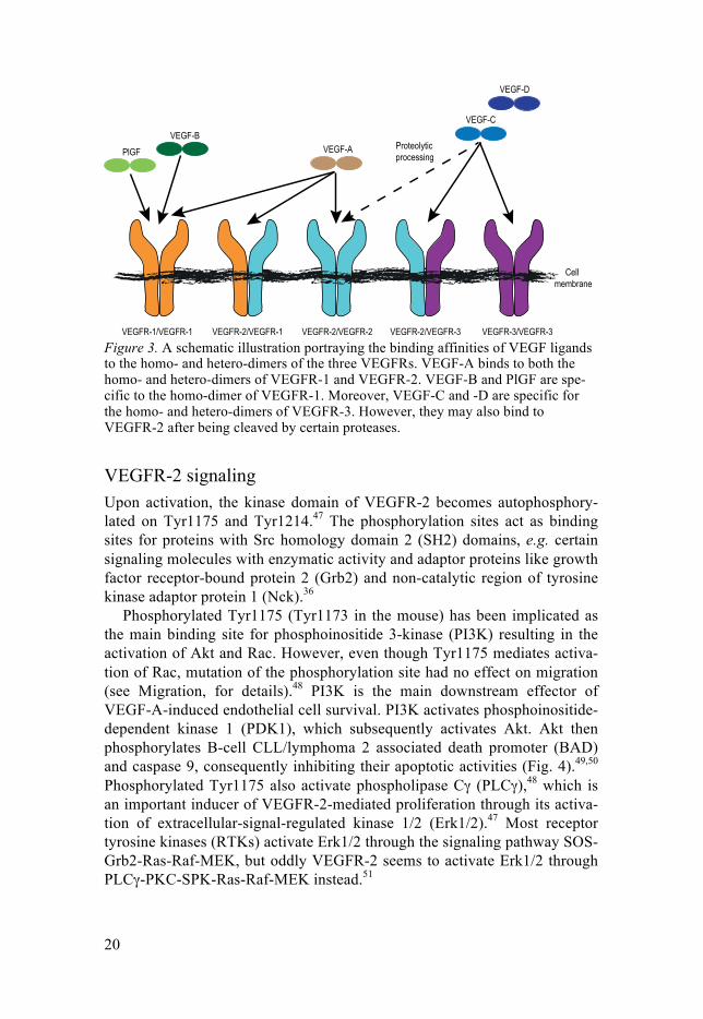
VEGFR-1, -2 and -3 form homo- or heterodimers upon activation by the homodimeric ligands VEGF-A, -B, -C and -D, as well as placenta growth factor (PlGF).32 The receptors show an overlapping expression pattern. However, VEGFR-1 is mainly expressed in endothelial cells, monocytes and macrophages, whereas VEGFR-2 and VEGFR-3 are predominantly ex-pressed in endothelial cells and lymphendothelial cells, respectively.
VEGF-A is the ligand that is mainly involved in angiogenesis and vascu-logenesis through its binding to VEGFR-2.33-35 VEGF-A is produced and secreted by endothelial cells and the resulting autocrine signaling has been shown to be essential for endothelial cell survival. Most parenchymal cells can also produce and secrete VEGF-A, which results in a paracrine activa-tion of VEGFR-2.36 This activation promotes migration and in some cases survival of endothelial cells.33-35
VEGF-C4 has been associated with lymphangiogenesis through VEGFR-3-mediated activation of the transcription factor PROX-1 that is a main regu-lator of lymphatic endothelial cell differentiation.4,37,38
VEGF-A, -B and PlGF39 all bind to VEGFR-1 (Fig. 3). VEGF-A has a higher affinity for VEGFR-1 than for VEGFR-2,40 but VEGFR-1 has a weaker kinase activity.40 Mice with a kinase-dead VEGFR-1 develop normal vessels,41 however, if VEGFR-1 is completely knocked-out the mice develop massive blood vessel malformations.42 This has raised the question whether VEGFR-1 in endothelial cells function as a negative regulator of VEGFR-2 signaling, possibly in part through an alternatively spliced soluble isoform that acts as a ligand trap.43
VEGF-C and -D both bind to VEGFR-3. However, after proteolytic pro-cessing they can also bind VEGFR-2, although with lower affinity than for VEGFR-3.32
Most cells can produce several isoforms of VEGF-A. The two most common isoforms are VEGF-A121 and VEGF-A165, where the short isoform lacks the heparin/heparan sulfate44 and neuropilin-145 binding domains. Hep-aran sulfate and neuropilin-1 may act as co-receptors and are believed to increase the intensity and half-life of VEGFR-2 activation.45,46
20
Figure 3. A schematic illustration portraying the binding affinities of VEGF ligands to the homo- and hetero-dimers of the three VEGFRs. VEGF-A binds to both the homo- and hetero-dimers of VEGFR-1 and VEGFR-2. VEGF-B and PlGF are spe-cific to the homo-dimer of VEGFR-1. Moreover, VEGF-C and -D are specific for the homo- and hetero-dimers of VEGFR-3. However, they may also bind to VEGFR-2 after being cleaved by certain proteases.
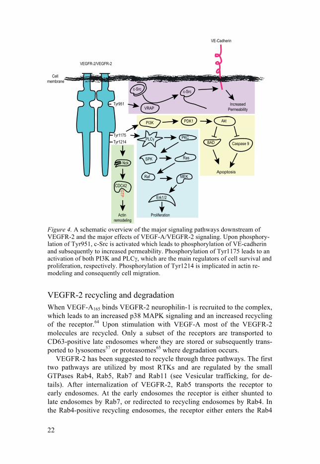
VEGFR-2 signaling Upon activation, the kinase domain of VEGFR-2 becomes autophosphory-lated on Tyr1175 and Tyr1214.47 The phosphorylation sites act as binding sites for proteins with Src homology domain 2 (SH2) domains, e.g. certain signaling molecules with enzymatic activity and adaptor proteins like growth factor receptor-bound protein 2 (Grb2) and non-catalytic region of tyrosine kinase adaptor protein 1 (Nck).36
Phosphorylated Tyr1175 (Tyr1173 in the mouse) has been implicated as the main binding site for phosphoinositide 3-kinase (PI3K) resulting in the activation of Akt and Rac. However, even though Tyr1175 mediates activa-tion of Rac, mutation of the phosphorylation site had no effect on migration (see Migration, for details).48 PI3K is the main downstream effector of VEGF-A-induced endothelial cell survival. PI3K activates phosphoinositide-dependent kinase 1 (PDK1), which subsequently activates Akt. Akt then phosphorylates B-cell CLL/lymphoma 2 associated death promoter (BAD) and caspase 9, consequently inhibiting their apoptotic activities (Fig. 4).49,50 Phosphorylated Tyr1175 also activate phospholipase Cγ (PLCγ),48 which is an important inducer of VEGFR-2-mediated proliferation through its activa-tion of extracellular-signal-regulated kinase 1/2 (Erk1/2).47 Most receptor tyrosine kinases (RTKs) activate Erk1/2 through the signaling pathway SOS- Grb2-Ras-Raf-MEK, but oddly VEGFR-2 seems to activate Erk1/2 through PLCγ-PKC-SPK-Ras-Raf-MEK instead.51

21
Tyr95152 and Tyr121453 (corresponding to Tyr949 and Tyr1212 in mice) are strongly involved in migration and permeability. Phosphorylation of Tyr951 enables binding of VEGF receptor-associated protein (VRAP, also called TSAd).54 This recruits c-Src, which has been implicated in cell migra-tion and vascular permeability. VE-cadherin situated in adherens junctions forms complexes with VEGFR-2. Upon activation of Tyr951, c-Src becomes activated and phosphorylates VE-cadherin. This disrupts the homophilic VE-Cadherin-mediated bonds between cells in the adherens junctions, which results in increased vascular permeability.55,56
Phosphorylation of Tyr1214 regulates VEGF-induced remodeling of the actin cytoskeleton through activation of Nck and Cdc42, which is important for an accurate actin remodeling during cell migration (Fig. 4).53
In contrast to most RTKs, that are situated in the cell membrane when in-active, half of the total VEGFR-2 pool has been suggested to be stored in microtubule-associated vesicles. These vesicles are in close proximity to the plasma membrane ready to be released upon stimulation of VEGF-A.57,58 Binding of VEGF-A causes internalization of VEGFR-2 through clathrin-mediated endocytosis.36 Notably, signaling by VEGFR-2 is not terminated after internalization. Instead it continues at least during the early stages of the endosome pathway, and the signaling is dampened first by degradation or dephosphorylation by phosphatases, e.g. protein-tyrosine phosphatase 1B (PTP1B) that dephosphorylates Tyr1175.59,60 It has been shown that different signaling pathways are activated at different stages of internalization. Akt signaling only occurs at the cell membrane, but a full Erk and Rac activation is achieved first in the early endosomes.61
Importantly, VEGFR-2 has been shown to interact with integrins. The vit-ronectin-binding integrin αVβ3 in complex with VEGFR-2 is essential for an active angiogenesis and a full activation of the p38 mitogen-activated protein kinase (MAPK) and focal adhesion kinases FAK signaling pathways during cell migration.62 Collagen I-binding integrins on the other hand reduce VEGFR-2 signaling by recruiting the protein-tyrosine phosphatase SHP2.63
22
Figure 4. A schematic overview of the major signaling pathways downstream of VEGFR-2 and the major effects of VEGF-A/VEGFR-2 signaling. Upon phosphory-lation of Tyr951, c-Src is activated which leads to phosphorylation of VE-cadherin and subsequently to increased permeability. Phosphorylation of Tyr1175 leads to an activation of both PI3K and PLCγ, which are the main regulators of cell survival and proliferation, respectively. Phosphorylation of Tyr1214 is implicated in actin re-modeling and consequently cell migration.
VEGFR-2 recycling and degradation When VEGF-A165 binds VEGFR-2 neurophilin-1 is recruited to the complex, which leads to an increased p38 MAPK signaling and an increased recycling of the receptor.64 Upon stimulation with VEGF-A most of the VEGFR-2 molecules are recycled. Only a subset of the receptors are transported to CD63-positive late endosomes where they are stored or subsequently trans-ported to lysosomes57 or proteasomes65 where degradation occurs.
VEGFR-2 has been suggested to recycle through three pathways. The first two pathways are utilized by most RTKs and are regulated by the small GTPases Rab4, Rab5, Rab7 and Rab11 (see Vesicular trafficking, for de-tails). After internalization of VEGFR-2, Rab5 transports the receptor to early endosomes. At the early endosomes the receptor is either shunted to late endosomes by Rab7, or redirected to recycling endosomes by Rab4. In the Rab4-positive recycling endosomes, the receptor either enters the Rab4

23
regulated pathway, which quickly recycles the receptor back to the cell membrane, or enters a second pathway involving a detour through the juxta-nuclear Rab11-regulated long loop-recycling pathway. However, this is less commonly used than the first pathway.57 The third pathway is VEGFR-specific and Src-dependent. It involves vesicles that are close to the cell membrane and is separated from the Rab4 pathway.57
The C-terminal tail in VEGR-2 is believed to be important for a rapid in-ternalization and degradation of the receptor by a protein kinase C (PKC)-dependent phosphorylation of Ser1188/1191.66 These amino acid (aa) resi-dues are situated in a PEST motif (aa 1171-1209), which is rich in proline [P], glutamic acid [E], serine [S] and threonine [T] residues. Phosphorylation of Ser1188/1191 recruits β-Trcp1, which is an F-box containing E3 ubiquitin ligase. This causes degradation of VEGFR-2 through Lys48-linked ubiquiti-nation.65 Casein kinase I-δ (CKI-δ) has also been proposed to be able to re-cruit β-Trcp1 to the VEGFR-2 through phosphorylation of the C-terminal, however, the phosphorylation sites involved remain to be identified.67 Fine-tuned receptor degradation is important for an accurate and sensitive re-sponse to VEGF gradients, e.g. during cell migration, and it has been shown that endothelial cell filopodia retracts upon VEGF-A blockade.11
GTPases The superfamily of Ras-related small GTPases are divided into seven fami-lies: Rho, Ras, Rab, ARF, Sar1, Ran and Rad/Gem.68 Small GTPases may be considered as molecular switches that regulate many different processes in a cell. They often function as linkers that coordinate downstream signaling from different receptor systems. GTPases cycle between an active GTP-bound form and an inactive GDP-bound form. Guanine nucleotide exchange factors (GEF) catalyze the release of GDP from the GTPases. Thus enabling binding of GTP to the GTPase, since GTP is present at a higher concentra-tion in the cell. The GTPase is inactivated by GTPase activating protein (GAP), which accelerates the intrinsic GTPase activity.69
Rho GTPases can also be inactivated by Rho GDP-dissociation inhibitors (RhoGDI) that inhibit the nucleotide exchange and relocalize the inactive GTPase from the cell membrane to the cytoplasm (Fig. 5).68 Some Rho GTPases can also be stabilized by scaffold proteins such as IQGAP1, which lowers the intrinsic GTPase activity.70
GTPases are usually ubiquitously expressed in all cells of the body, but can have different functions in different cell types. The context-dependent function of a GTPase comes from the expression of its GEFs and their bind-ing affinity to structural membranes within the cell.71
The Ras GTPases have a crucial role in cell survival, differentiation and proliferation, and are often activated down-stream of RTKs.72

24
The large family of Rab GTPases has mainly been associated with differ-ent steps of membrane trafficking, such as assembly of transport vesicles and membrane fusion between vesicles and target compartment.72 The Rab GTPases achieve this in part by the activation of vesicular SNAP receptors (vSNAREs) at the right vesicles.73 Rab GTPases are also involved in guiding and promoting movement of vesicles. They do so by interacting with and recruiting different acceptor/effector proteins, thus guiding the content of the vesicle in the right direction.72
Arf GTPases also have key roles in membrane trafficking by recruiting li-pid modifying enzymes and coat proteins, predominately at the Golgi net-work, which mediates sorting of the vesicle cargo.72,74 In addition, Arf GTPases regulate organelle structure.72
Rho GTPases are the main masters of actin cytoskeleton remodeling and cell surface dynamics.72
Figure 5. A schematic illustration of activation and inactivation of GTPases. The GTPase is activated by GEFs catalyzing the release of GDP, thus allowing a GTP to bind. The GTP is removed by GAPs that increase the intrinsic GTPase activity, thus returning the GTPase to its inactive GDP-bound form. GTPases can also be embar-goed by for example RhoGDI, which will hinder activation by GEFs by mediating translocation of inactive GTPases to the cytoplasm.

25
Rho GTPases To date, 22 Rho GTPases have been identified in humans and the best char-acterized are the Rho-like GTPases (RhoA-C), the Rac-like GTPases (Rac1-3 and RhoG) and Cdc42.68 The less characterized Rho GTPases are RhoD, Rif, Rnd1, Rnd2, Rnd3/RhoE, Miro-1, Miro-2, TC10, TCL/RhoJ, RhoBTB1, RhoBTB2, RhoH/TTF, CHP and finally Wrch-1.75,76 Oddly, the Rnd proteins and RhoH lack detectible GTPase activity, raising the question if they should be considered as GTPases at all.76
RhoA promotes the formation of stress fibers and membrane ruffles through actin polymerization. Moreover, RhoA also regulates assembly of focal adhesion complexes.77-81 Rac1 is usually activated by growth factors, such as PDGF-BB, and promotes lamellipodia formation through actin polymerization. Rac1 may also induce stress fibers and membrane ruffles since it sometimes act upstream of RhoA.77,79-81 Activation of Cdc42 pro-motes formation of thin actin-rich projections (filopodia formation). In addi-tion, Cdc42 may also induce lamellipodia formation and membrane ruffles, since Cdc42 also acts upstream of Rac.80-82 Cdc42 is also involved in other cellular functions, e.g. the regulation of secretory membrane trafficking, cell polarization and G1 transition during cell cycle.83,84
It has become evident that the Rho, Rac and Cdc42 pathways are con-nected and influence each other. This means that affecting the activity of one of these GTPases also could affect the functions of other Rho GTPases.80 Apart from regulation of the actin cytoskeleton, Rho GTPases have also been implicated in cell growth.81
It is not uncommon that Rho GTPases cooperate with GTPases of other families to achieve specific cellular responses; e.g. during cell migration where they collaborate foremost with Ras GTPases, Arf GTPases and Rab GTPases.72
Moreover, RhoA as well as the RhoA-specific GEF Syx are redirected from disassembling tight junctions to the leading edge in migrating endothe-lial cells by Rab11.85 Also, during rapid remodeling of the cytoskeleton due to phagocytosis, Rac1 and Cdc42 are recruited to the cell membrane by Rab35-mediated transportation on microtubule tracks.86
Rab1 regulates ER-to-Golgi transport through its interactions with the membrane proteins p11587 and gm13088, which helps to mediate vessel dock-ing. Cdc42 activation inhibits retrograde transport from the Golgi compart-ment to the ER by activation of the neuronal Wiskott-Aldrich syndrome protein (N-WASP) and actin-related protein 2/3 (Arp2/3). This is dependent on Arf1 (ADP-ribosylation factor 1) activation, since Arf1 recruitment of membrane docking proteins is required to exit the Golgi.89

26
Rho GTPases and actin dynamics Cdc42 and Rac1 facilitate nucleation of actin filaments by recruiting actin nucleation promoting factors (NPFs), e.g. N-WASP and WASP family member (WAVE), respectively. Through this mechanism they affect the activity of the Arp2/3 complex that facilitates nucleation and elongation of branched actin networks.76 Other NPFs, including formins, are instead downstream of Cdc42 and RhoA and stimulate nucleation and elongation of actin bundles by activation of the diaphanous-related formin mDia1-3.72,76 mDia1-3 stimulates extension of actin filaments by binding to the plus end of the actin filament and thereby preventing binding of capping proteins that would normally terminate the elongation.90
Additionally, Cdc42 and Rac1-3, have been shown to bind to and activate proteins of the p21-activated kinase (PAK) family of serine/threonine kinas-es. The PAK family consists of six members of which three (PAK1-3) are effector proteins of Cdc42 and Rac1-3. However, PAKs can also be activat-ed by tyrosine kinase receptors by binding to scaffold proteins with SH3 domains, such as Nck and Grb2. PAKs, have been implicated in a plethora of cellular functions, e.g. cytoskeletal dynamics that is crucial for cell motili-ty and cell polarity.91 Activated PAK1-3 can activate LIM kinases by phos-phorylation. Subsequently, the LIM kinases phosphorylate and inactivate the F-actin depolymerization factor cofilin90,91
RhoD One of the least studied Rho GTPases so far is RhoD. No GEFs or GAPs have been shown to regulate RhoD. RhoD occurs predominantly in the GTP-loaded form and is presumably regulated at the transcriptional level and/or via interactions with proteins that are able to sequester RhoD.92
Activation of the zipper-interacting protein kinase (ZIPK) is associated with cell death responses including membrane blebbing and formation of autophagic vesicles. RhoD has been shown to suppress ZIPK-induced cell contraction, focal adhesion size and stress fiber formation in a GTP-dependent manner.92
The actin nucleation-promoting factor WHAMM (WAS homolog associ-ated with actin Golgi membranes and microtubules) and filamin A-binding protein 1 (FILIP1) can bind to and regulate Arp2/3 and filamin A, respec-tively. In addition to ZIPK, RhoD has also been shown to regulate cytoskele-tal dynamics through interactions with WHAMM and FILIP1. Suppression of RhoD resulted in increased cell adhesion and subsequently decreased migration, most likely in part due to increased formation of focal adhesions together with disintegration of stress fibers.93
Moreover, RhoD has been suggested to induce very thin filopodia-like protrusions through the diaphanous-related formin mDia3C. RhoD also has
27
the capacity to align endosomes with actin filaments in a c-Src- and hDia2C-dependent manner.94,95
RhoD can regulate internalization and recycling of early endosomes through its interaction with the Rab5 effector protein Rabankyrin-5. Rab5 is situated on early endosomes. Depletion of RhoD inhibits internalization and stalls endosomal recycling, which results in decreased PDGFR-β phosphory-lation. RhoD and Rab5 share the same binding site on Rabankyrin-5, and thus compete for binding. In contrast to Rab5, RhoD’s binding to Rabankyr-in-5 is GTP-independent.96 Finally, RhoD has also been implicated as a regulator of centrosome integrity as well as centrosome duplication during cell division.97
Rho GTPases and cell polarity Cdc42 has been described as a master regulator of cell polarity. Polarization of cells can be initiated by; loss of physical constrains like in a scratch wound, cell-cell contact through adhesion molecules like cadherins or by soluble ligands like VEGF, PDGF and EGF.98
PI3K activity is important for the formation and maintenance of the lead-ing edge in migrating cells due to its activation of Cdc42 and Rac1.99 The leading edge is sustained by a positive feedback loop of PI3K,100 which is Rac1-dependent and results in accumulation of PIP3.98-101 When polarized, the cell aligns the microtubules and centrosome/Golgi towards the leading edge in a Cdc42 dependent manner.98
The small GTPase Arf6 is crucial for Cdc42, Rac1 and Cdc42/Rac1-GEF β-PIX accumulation at the leading edge as Arf6 recruits and activates type I PIP5 kinases.74,102 At the leading edge, Cdc42 recruits and binds to the Par3/Par6 complex, which induces a conformational change in Par6 leading to its activation. The complex between Par6 and Cdc42 activates the atypical protein kinase, PKCζ.76,98 The activated Cdc42/Par6/PKCζ complex is im-portant for the ability of Cdc42 to regulate cell polarity. Cdc42 mediates reorientation of the Golgi/centrosome towards the leading edge, thus ena-bling the aligning of the microtubules so that the plus end is at the leading edge and the minus end is connected to the Golgi/centrosome.98,103 Further-more, Cdc42 and Rac1 also promote polarization through microtubule cap-ture. This is facilitated by interaction with IQGAP1, which interacts with the microtubule-plus-end-associated protein Clip170.104 Additionally, Cdc42 also regulates localization and activity of Rac1 by activating the Ser/Thr kinase PAK1, which subsequently governs the activity of Cdc42/Rac1-GEF β-PIX.103 Rac1, and to a lesser extent Cdc42, can control microtubule stabil-ity by activating PAK, which can phosphorylate and thereby inactivate the microtubule-destabilizing protein stathmin.105
28
In conclusion, Rac1 is crucial for the formation of the leading edge, whereas Cdc42 is crucial for the localization and maintenance of a single leading edge.99
Rho GTPases regulate adherens junctions and tight junctions RhoA, Rac1 and Cdc42 are all important for the maintenance of adherens junctions and tight junctions. However, depending on cell type and other regulatory signals, their effects can be either stimulatory or inhibitory.76 Fur-thermore, RhoA frequently antagonizes the effects of both Cdc42 and Rac1.72
The Cdc42/Par6/PKCζ complex is important for the formation of cell-cell junctions, cell polarity and establishment of the baso-lateral membranes in epithelial cells, but the specific molecular mechanisms remain to be eluci-dated.98
RhoA-mediated ROCK (Rho-associated coiled-coil containing kinase) ac-tivation loosens tight junctions in endothelial cell by inducing actomyosin- mediated cytoskeleton contraction leading to decreased tight junction integ-rity and subsequently increased permeability.106,107 Subsequently, Rac1 activ-ity stabilizes tight junctions in endothelial cells and thereby decreases per-meability.106,107 However, in epithelial cells ROCK activation leads to an increased integrity of tight junctions and decreased permeability.108
FGD family of RhoGEFs All RhoGEFs consist of two functional domains. The first one is the ~250 aa residues long DBL homology domain (DH) that is responsible for the RhoGEF catalytical function. The second one is the ~100 aa residues long pleckstrin homology (PH) domain109 in the C-terminal part, which is respon-sible for the binding affinity of the RhoGEF. The PH domain is important for the association of RhoGEFs with other proteins110 and also interactions with lipid membranes by its affinity to phosphatidylinositol-4,5-bis-phosphate (PI(4,5)P2) generated by class II PI3Ks.1,109
The first RhoGEF to be identified was DBL, which activates RhoA111 and Cdc42112, and since then numerous RhoGEFs have been identified and char-acterized. The FYVE, RhoGEF and PH domain-containing (FGD) protein family consists of FRG, FGD1113, -2114, -3115, -4 (Frabin)116, -5 and -6. Just like other RhoGEFs, members of the FGD family have a DH and PH do-main, however, they also have an additional PH domain with a zinc-finger domain (FYVE) in between the PH domains. The FYVE domain adds a se-cond layer of binding specificity as it mediates interactions with phosphati-dylinositol-3´-phosphate.1

29
FGD1 is the best characterized FGD protein and has been found to acti-vate Cdc42. A mutation in FGD1 is responsible for the X-linked develop-mental faciogenital dysplasia, also known as the Aarskog-Scott syndrome. Patients with Aarskog-Scott syndrome are characterized by short height and skeletal, facial and urogenital anomalies.117 This probably reflect that FGD1 is highly expressed during bone growth and mineralization, where it is be-lieved to be involved in membrane transport from the Golgi network.84 FGD1 is highly expressed in cells that originate from mesenchymal stem cells, including fibroblasts, osteoblasts, chondrocytes and adipocytes.118 Fur-thermore, it has been shown that FGD1 can activate the c-Jun N-terminal kinase (JNK) MAPK pathway and promote G1 cell cycle progression in a Cdc42-independent manner.119
FGD2 is expressed by antigen presenting cells and is believed to be spe-cific for Cdc42. Furthermore, its cellular localization has been associated with EEA-1 positive early endosomes through binding via the FYVE do-main.120 FGD2 has also been associated with membrane ruffles in the lead-ing edge of migrating cells, which normally is controlled by Rac1; possibly the activation of Rac1 is Cdc42 dependent.120
FGD3 has also been hypothesized to activate Cdc42, however, instead of long finger-like protrusions that is normally seen after FGD1 activation, FGD3 induces short microspikes or lamellipodia.121 It has been speculated that FGD3 has an inhibitory effect on migration, but this has not been thor-oughly investigated.121
FGD4 has been found to activate Cdc42 and a mutation in the DH domain has been associated with Charcot-Marie-Tooth Type 4H disorder, which is characterized by muscle weakness and foot and hand abnormalies.122 More-over, FGD4 has an F-actin binding domain that promotes FGD4 binding to F-actin.116
FGD5 To date there are only a few articles about FGD5. All these articles show that FGD5 is selectively expressed in endothelial cells123-125 and support the no-tion that FGD5 can regulate Cdc42123,124,126. However, several of the pub-lished papers disagree when it comes to which processes that are regulated by FGD5.
Kurogane et al124 and Nakhaei-Nejad et al125 propose that treatment with FGD5 siRNA decreases endothelial cell sprouting and migration124. Cheng et al123 on the other hand propose that treatment with FGD5 siRNA increases endothelial cell sprouting. Cheng et al123 also overexpressed FGD5 and found that an increased amount of FGD5 decreased sprouting in the retina. One possible explanation for the different results and conclusions regarding the effects of FGD5 could be that Cheng et al123 did most experiments in vivo, whereas Kurogane et al124 and Nakhaei-Nejad et al125 did all analyses

30
in vitro or ex vivo. Another reason could be that Cheng et al123 seem to have ubiquitously overexpressed FGD5 in all cell types of the retina, including pericytes and astrocytes. Even though the cells are in a proper biological context, the interpretation is clouded by the fact that all cells express FGD5, and effects of FGD5 in non-endothelial cells can have contributed to the phenotype. It should also be noted that dysregulation (due to overexpression) could be enough to disturb FGD5’s function in vivo and that input from sys-tems than VEGFR123-125, such as other RTKs, GPCRs or Notch, could be affected directly or indirectly by FGD5.
Cheng et al123 proposed that FGD5 inhibits neovascularization by induc-ing apoptosis through the HEY1-p53 pathway; this is contradictory to the findings by Nakhaei-Nejad et al125 who proposed that FGD5-deficiency sen-sitizes cells to apoptosis. Kurogane et al124 were unable to se any effects on apoptosis.
However, Kurogane et al124 detected a decreased VEGF-A- and FGF-2- mediated proliferation in cells depleted for FGD5. Nakhaei-Nejad et al125 showed that FGD5 deficiency decreased both integrin-mediated cell spread-ing as well as Akt signaling downstream of VEGF-A and insulin receptors. Erk signaling was, however, unaffected. This contradicts Kurogane et al124 who showed the opposite effects due to FGD5 deficiency, i.e. unaffected Akt signaling and decreased Erk signaling that was suggested to be Cdc42-independent.
A fourth paper by Ando et al126 did not investigate the role of FGD5 in cell migration, but proposed that FGD5 is downstream of the small GTPase Rap1 to promote formation of circumferential actin bundles and resulting in increased cell-cell barrier function. FGD5 was suggested to via Cdc42 acti-vate the myotonic dystrophy kinase–related Cdc42-binding kinase (MRCK), which activates non-muscle myosin II.126
Ubiquitination Ubiquitination of proteins is a three-step process, which is initiated by the ATP-dependent ubiquitin-activating enzyme (E1) that transfers ubiquitin to an ubiquitin-conjugating enzyme (E2). The ubiquitin is thereafter covalently attached to its target by a collaboration between the ubiquitin-conjugating enzyme and an ubiquitin protein (E3) ligase.127 There are hundreds of pro-teins that could potentially act as an E3 ligase and they can be divided into RING (really interesting new gene) and HECT (homologous to the E6-AP carboxyl terminus) superfamilies of E3 ligases.127,128
The ubiquitination of a protein will result in different fates, depending on whether the protein is mono- or poly-ubiquitinated and which of the seven lysine residues (K6, K11, K27, K29, K33, K48 and K63) or the ubiquitin N-terminus that are used to build the ubiquitin chain.127-130 Mono-ubiquitination

31
is usually sufficient but not necessary for receptor internalization and poly-ubiquitination is associated with degradation.127,129-131 K48-linked ubiquitina-tion is generally associated with proteasomal degradation.129,130 K63-linked ubiquitination has been connected to protein trafficking and DNA repair,130 however, K63-linked poly-ubiquitination has also been associated with a more efficient internalization and lysosomal degradation.132
The conjugation of ubiquitin affects the subcellular transport and fate of the protein and enables for new interactions with proteins that has ubiquitin binding motifs, such as the endocytic proteins Eps15 and Hrs.133 Moreover, a subset of SH3 domains has also been shown to bind ubiquitin.134 The com-plete role of mono-ubiquitination has not been fully elucidated. It has been suggested that mono-ubiquitination could function as a “grip” for proteins with ubiquitin binding motifs, which could lead to increased and prolonged signaling of receptors and downstream kinases. On the other hand, it has been suggested that in some cases it also has an auto-inhibitory effect and cause dissociation of the signaling complex.129,135 Ubiquitination of proteins are nevertheless reversible and can be undone by de-ubiquitinating enzymes (DUBs).136 Interestingly, some of the DUBs are unable to de-ubiquitinate degradation-associated Lys48-linked ubiquitin chains.129
Rho GTPases and Ubiquitination Rho GTPases like RhoA and Rac1 are susceptible to ubiquitination. The detailed mechanism whereby Cdc42 is degraded, is however, unknown.137 The activity of Cdc42 seems to be mainly regulated via ubiquitination and degradation of its GEFs, e.g. FGD1138, FGD3121 and β-PIX139.
Even though several E3 ubiquitin ligases have been shown to bind to Rac1, none of them seems to ubiquitinate the GTPase itself,127 implying that the E3 ligase that targets Rac1 is still unknown. RhoA, however, have been shown to be ubiquitinated by several E3 ligases, including SMURF1140 and Cullin-3141. SMURF1 ubiquitinates active RhoA, but not Rac1 and Cdc42 in response to growth factor signaling and is regulated by PKCζ,140 which is a key component in the Par6-Cdc42 complex at the leading edge of migrating cells. This implies that SMURF1 facilitates migration at the leading edge by down-regulation of RhoA. Cullin-3 only ubiquitinates inactive RhoA and therefore does not regulate the same pool of RhoA as SMURF1.141
It has been shown that activation of Cdc42 inhibits EGFR degradation by impounding the RING E3 ligase Cbl,142 which in order to escape its jailor, starts to ubiquitinate the Cdc42/Rac1 GEF β-PIX.139

32
The exocyst complex and vesicular trafficking Exocytosis and vesicular trafficking is important during a lot of cellular pro-cesses, such as secretion of growth factors or cytokines, transport of newly synthesized membrane proteins from the Golgi network and recycling of membrane proteins including receptors. The exocyst complex is also im-portant for the ability of cells to polarize, however, the mechanism by which the exocyst complex regulates polarization is poorly understood.143,144
Exocytosis occurs in three different manners, it can be constitutive, polar-ized or regulated. Constitutive exocytosis occurs constantly at the cell mem-brane to maintain the composition of the plasma membranes with regard to membrane-bound proteins. Polarized exocytosis is utilized by the cell when it needs to transport a large amount of proteins to a special location on the cell membrane, e.g. during cell migration. Regulated exocytosis is utilized when the cell needs to empty the content of prefilled vesicles to the outside of the cell, e.g. neurotransmitter release in the synaptic cleft in response to a specific stimuli.145
The exocyst complex The exocyst complex is built up by two subunit complexes, one localized at the cell membrane and one localized at vesicles. Exocyst complex compo-nents ExoC1 and ExoC7 (called Sec3 and Exo70 in yeast) constitutes the first complex and are present at the cell membrane where they act as docking sites for incoming vesicles.146 It has been shown that both ExoC1147 and Ex-oC7148,149 are recruited to the plasma membrane by interactions with PI(4,5)P2 that is produced by PIP-kinases, e.g. at the migratory front. Inter-estingly, ExoC1 and ExoC7 do not seem to interact with PI(3,5)P2, PI(3)P or PI(4)P, which are localized to membranes of other cellular compartments.148 The other exocyst subunit complex, ExoC2-6 (called Sec5, Sec6, Sec8, Sec10 and Sec15 in yeast) and ExoC8 (called Exo84 in yeast)144 are predom-inantly enriched at the surface of secretory vesicles and is referred to as the vesicle-tethered exocyst complex. Docking of the vesicle-tethered exocyst complex to the acceptor exocyst complex components at the plasma mem-brane fulfills the formation of the complete octameric exocyst complex, and this docking step precedes membrane fusion and exocytosis.
In order for a vesicle to fuse with the cell membrane, they have to over-come their mutual repulsion of each other. They do so with membrane dock-ing proteins that assist the vesicle and cell membrane to come close enough for the fusion to be able to occur. These membrane-docking proteins are called SNAREs (SNAP receptors). SNAREs mediate fusion between secre-tory vesicles and the cell membrane. In this process, vSNAREs on vesicles and tSNAREs located at the target membrane forms a complex and thus catalyzing membrane fusion.143,150 With more than 30 different SNAREs,

33
they add a certain specificity to the vesicle trafficking, i.e. only vesicles with the right activated vSNAREs will be able to bind and initiate fusion in the intended spot. However, it is unlikely that the SNAREs provide the only basis for specificity.73,150 VEGFR-2 trafficking from Golgi to the cell mem-brane requires the Golgi-localized tSNARE syntaxin 6.151
An important part of exocytosis is the initial reorganization of the actin cytoskeleton, which makes Rho GTPases important regulators of exocytosis. Depending on cell type, different Rho GTPases have different effects. For example, in PC12 cells Rac1 and Cdc42 support the neurosecretory re-sponse,145 however, the same response in neutrophils is Rac2-dependent.152 Cdc42 is also important for ejection of the vesicle content by regulating con-traction of actin filaments via activation of N-WASP and Arp2/3. Without the Cdc42-mediated ejection, a large portion of the vesicle content would be recycled back to the cytoplasm.146
In yeast, Cdc42 has an important role in regulating Sec3 (ExoC1) locali-zation at the bud tip.147 However, in mammals the binding of Cdc42 to the exocyst complex has been hard to verify, suggesting that Cdc42 might bind to the exocyst complex through other proteins, or that other GTPases have acquired the role of Cdc42 in mammals.145 There are nevertheless sugges-tions that Cdc42 can regulate the exocyst complex by activating the small Ras GTPase RalA.153 Rho3 also regulates exocytosis, through binding to Exo70 (ExoC7). However, the localization of Exo70 or the exocyst complex was unaffected by knockdown of Rho3, indicating that Rho3 regulates exo-cytosis via other yet unknown mechanisms.143
In PC12 cells it has been shown that the interaction between ExoC7 and Cdc42-related GTPase TC10 is crucial for the localization of ExoC7 at the cell membrane in growth cones, recruitment of insulin growth factor receptor 1 (IGFR-1) and elongation of the neuron.154 Furthermore, it has been sug-gested that local TC10 activation counteracts Cdc42-mediated activation of N-WASP. Hence, the balance between TC10 and Cdc42 seems to be im-portant for a correct regulation of exocytosis.155
The exocyst complex component ExoC6 interacts with the small GTPase Rab11; this interaction is crucial for the ability of Rab11 to guide recycling endosomes back to the cell membrane.156,157 Another GTPase that is im-portant for recycling endosome function is Arf6, which has been shown to promote and specify the relocalization and fusion of perinuclear ExoC5-coated endosomes to the plasma membrane.158 Furthermore, plasma mem-brane localized RalA interacts with ExoC2 and ExoC8 and is crucial for the correct sorting of membrane proteins from Golgi-derived vesicles to the basolateral membrane in epithelial cells.159
Besides exocytosis, the exocyst complex also regulates actin polymeriza-tion. For example, ExoC7 interacts with Arp2/3 and is crucial for Arp2/3 recruitment to the cell membrane. Therefore, ExoC7 may have an impact on the ability of cells to migrate.160

34
Vesicular trafficking The cell can be divided into “compartments”, e.g. the plasma membrane, the Golgi, the endoplasmic reticulum (ER), the nucleus and the vesi-cles/endosomes. Additionally, the Golgi can further be divided into trans-, medial- and cis-Golgi. Proteins are constantly transported between these compartments. In order for the cell to be able to shunt different proteins to their correct compartments there have to be mechanisms for sorting and pro-teins that functions as “switches” along the way, i.e. proteins that can change the direction of a vesicle. Both Rab and Arf GTPases have been proposed to function as switches even though they have different functions (see GTPases, for details).161,162
Rab conversion, i.e. how a vesicle switches from one Rab GTPase to an-other is still not completely understood. However, a cascade chain of events has been proposed. The activated Rab GTPase recruits the GEF for the Rab GTPase next in line, which results in a feedback loop that recruits the Rab GAP of the preceding Rab GTPase and inactivates it.163-165
Vesicular trafficking is guided by the involvement of several Rab GTPases. However, in this short summary I will mainly focus on GTPases that are implicated in receptor internalization, recycling and degradation (for more comprehensive reviews on these topics see references 90,162,163,166,167).
Initiation of the formation of endocytic vesicle requires Arf6 to recruit PIP5 kinases and clatrin.167 Furthermore, Rab5 regulates internalization168, transportation162 and fusion168 of early endosomes by assembling clathrin coated pits,169 recruiting SNAREs73 and other effector proteins, e.g. phos-phoinositide (PI) kinases (PI3Kβ and Vps34)170,171 or phosphoinositide phosphatases (PI5‑phosphatase and PI4‑phosphatase)171 (Fig. 6). By utiliz-ing both phosphatases and kinases phosphatidylinositol-3´-phosphate (PI(3)P) is formed, which is an effector molecule for both Rab5 and Rab7. PI(3)P recruits and binds FYVE-domain-containing proteins, like EEA1172 and Rabenosyn-5173, to Rab5- and Rab7-decorated endosomes. Early endo-some antigen 1 (EEA1) is a dimeric Rab5 effector protein, which facilitates fusion between Rab5-positive early endosomes.172,174,175 Growth factors, such as EGF, can activate Rab5. This is required for growth factor-induced activa-tion of Rac1, which takes place on early endosomes instead of the cell mem-brane.176
At the early endosomes the proteins are at a crossroad. From there they can either be recycled back to the cell membrane through the short loop re-cycling pathway mediated by Rab4, or be recycled through the longer juxta-nuclear pathway facilitated by Rab11 (Fig. 6).57,162,177 It is unclear why the cell needs two recycling pathways and if the Rab4- and Rab11-mediated pathways have different purposes in the cell.177 It has been suggested that these two pathways actually could represent different steps of a single path-way.61

35
If the protein is destined for degradation, the early endosome will start to recruit Rab7 effector proteins by replacement of Rab5 with Rab7. When Rab7 outnumbers Rab5, the vesicle will be transported and fused with CD63-decorated late endosomes, multi-vesicular bodies (MVB) and finally to LAMP-1-decorated lysosomes (Fig. 6).164,165
Different Rab GTPases can co-exist together on vesicle membranes, for example Rab4, Rab5 and Rab11 have been shown to be simultaneously pre-sent on recycling endosomes, however, they do not seem to interact but in-stead occupy different “Rab microdomains”.162
RhoB is located on early endosomes and aligns the endosomes with actin filaments.178,179 Upon activation, RhoB delays the trafficking of membrane receptors to late endosomes through mDia1-dependent recruitment of F-actin coats of the early endosomes. The actin coat sequesters the endosomes to the actin filaments, thus inhibiting the endosomes from being transported on microtubules to late endosomes.178 RhoB has also been shown to bind and activate protein kinase PRK1, however, its function in RhoB signaling is still not completely elucidated.179
Like RhoB, RhoD has also been shown to be a major player in endosomal trafficking through its binding to Rabankyrin-5 in competition with Rab5 (see RhoD, for details).96

36
Figure 6. A schematic illustration showing the important steps of protein internaliza-tion, fusion, recycling and degradation. Firstly, proteins may be internalized in a Rab5-dependent manner. Rab5 recruits EEA-1, which enables fusion of early endo-somes to larger endosomes. If recycled, Rab4 and Rab11 are recruited and the pro-teins can be recycled through either a short loop (Rab4-mediated pathway) or by a longer juxtanuclear pathway (Rab11-mediated). However, if destined for degrada-tion, Rab7 is recruited to the vesicle to mediate transport of the protein cargo to late endosomes, which merges with other late endosomes creating larger late endosomes. The late endosomes turns into multi-vesicular bodies, which over time become transformed into lysosomes.

37
Cell migration Regulated cell migration is of crucial importance during embryogenesis.180,181 Moreover, cell migration continues to be of importance during adulthood. For example, immune cells migrate in response to a wide variety of guidance cues in order to reach sites of infection and defeat patho-gens. Cell migration is also central during wound healing, physiological and pathological angiogenesis, tumor growth and tumor spread (metastasis).182 Importantly, the ability of cells to polarize and focus their vesicular transport (of integrins and other plasma membrane molecules) to the cell migratory front (known as the leading edge) is of utmost importance for the ability to move in response to guidance cues.
The detailed mechanisms in control of cell migration are still subject to intense investigation. Two main mechanistic models describing how cells move have been proposed; the actin-remodeling model and the membrane flow model. Different cell types use different methods and the majority of them probably use a combination of both.
In the actin-remodeling model, the cell is pulled forward via mechanical forces generated by the cytoskeleton. In this mode, actin is continuously polymerized at the leading edge, and at the same time depolymerized at the rear end, creating a treadmill motion.183 Cdc42 and Rac1 have been shown to induce actin polymerization at the leading edge through activation of Arp2/3. Furthermore, Cdc42 has been suggested to predominantly promote actin polymerization in filopodia, whereas Rac1 promotes formation of the actin cytoskeleton just behind the leading edge.184-186 There are reports suggesting that active Rac1 is localized at the rear end of migratory cells; its exact func-tion remains unclear but it seems that Rac1 is important for “tail retraction”.185 While Rac1 is crucial for migration per se, Cdc42 activity also seems to be required for cell polarization by restricting Rac1 activation (see Rho GTPases and cell polarity, for details).103
RhoA acts by activating ROCK on the lateral sides of the cell and in the rear end, which mediates actomyocin-induced contraction of the cytoskele-ton. This decreases integrin adhesion and further prevents unsuitable lamel-lipodia formation at these sites.187,188 However, a basal Rho activity is needed for the assembly of focal adhesions, yet, too much Rho activity decreases migration.187 This is probably due to, for a cell to be able to migrate it needs to hold on to the surface. However, if the cell is unable to let go, it will not be able to move.
In the membrane flow model of cell movement, vesicles are continuously transported from the rear to the front, thus shrinking the rear and moving the leading edge forward.189
For directed migration the cell needs a cue. If the cue comes from a solu-ble chemo-attractant it is referred to as chemotaxis,190 but if the chemo-attractant is immobilized, e.g. by being bound to the extracellular matrix, the

38
process is called haptotaxis.191 Chemokinetic factors on the other hand are signaling molecules that induce increased motility, but do not induce direc-tional cell migration.
MicroRNA MicroRNAs (miRNAs) are small non-coding RNAs that fine-tune gene ex-pression by predominately inhibiting translation of proteins from mRNA. They have been proposed to be important during the embryonic develop-ment, where genes need to be turned on or silenced with a high precision over time. In animals, miRNAs normally bind to the 3´UTR in the mRNA and thereby inhibit its translation (Fig. 7), or in rare cases even cause cleav-age of the mRNA. The miRNAs can be found between genes expressed by their own promoter or intragenically within introns in which case they may be expressed together with their “host” gene.192 In the case of intergenic miRNA, a single promoter usually controls the transcription of a whole clus-ter of miRNAs.
During the transcription by DNA polymerase II, the microRNA is excised by the enzyme Drosha to a ~70 nucleotide long hairpin (pre-miRNA) that is exported to the cytoplasm.193 In the cytoplasm the hairpin is further truncated by the enzyme Dicer194,195 into a ~20 nucleotide double stranded RNA of which one strand is incorporated into the argonaute protein containing RNA-induced silencing complex (RISC); the other strand is usually degraded but may also be incorporated into RISC.196 It is not fully established by which mechanism the translation is inhibited, but in contrast to siRNAs, miRNAs binds to their mRNA targets with poor complementary, which is believed to be the reason why they inhibit the translation rather than cleave the mRNA like siRNAs.197

39
Figure 7. miRNA biogenesis and maturation begins by the miRNA being tran-scribed by DNA polymerase II. The immature miRNA (pri-miRNA) is excised by the enzyme Drosha resulting in a ~70 nucleotide long hairpin structure called pre-miRNA. The pre-miRNA is transported from the nucleus to the cytoplasm and fur-ther truncated by Dicer, thus forming the ~20 nucleotide long mature double strand-ed miRNA. One of the strands is incorporated in to the RNA-induced silencing complex (RISC), thereby enabling RISC to silence mRNA expression by binding to the 3´UTR of the target mRNA. The picture was kindly provided by Peder Fredlund Fuchs.

40
Present investigation
Aims The aim of this thesis was to identify and characterize proteins and small RNAs that regulate cellular functions with emphasis on VEGF-receptor sig-naling and cell migration.
Results and conclusions
Paper I This paper addresses the roles of the exocyst complex in endothelial cells. We show that exocyst complex component 3-like 2 (ExoC3L2) is expressed in endothelial cells of actively growing blood vessels. ExoC3L2 protein ex-pression was shown to be induced by VEGF-A and collagen I. ExoC3L2 was further shown to co-localize with the known exocyst component ExoC4 sug-gesting that ExoC3L2 is an integral component of the exocyst complex. This was validated by co-immunoprecipitation of ExoC4 and ExoC3L2. To ad-dress the roles of ExoC3L2 and the exocyst complex in chemotaxis, migra-tion of endothelial cells treated or not with siRNA targeting ExoC3L2 was analyzed using a novel microfluidic chemotaxis assay, where the endothelial cells migrated in stable gradients of VEGF-A. This assay allows for the analysis of migrating cells by time-lapse microscopy followed by tracking of individual cells. Cells treated with siRNA targeting ExoC3L2 lost their abil-ity to sense the VEGF-A gradient, which could be explained by impaired phosphorylation of the VEGFR-2 autophosphorylation site Y1175.
The data suggests that the VEGF-A/VEGFR-2 signaling is modulated by or even dependent on the functions of the exocyst complex and ExoC3L2. Taken together, this study for the first time describes the role of ExoC3L2 and the exocyst complex in endothelial cells, showing that VEGFR-2 signal-ing and endothelial cell chemotaxis is negatively affected by loss of Ex-oC3L2 expression.
Paper II In this study, publicly available datasets containing information about mi-croRNA (miRNA) expression in different tissues were analyzed. By compar-

41
ing the miRNA-expression profiles of highly vascularized tissues, such as the lung and the kidney glomerulus, several miRNAs with selective expres-sion in blood vessels could be identified, including miR-145, miR-23a and miR-24. The vascular expression of the different miRNAs was validated by the analysis of miRNA expression in microvascular fragments isolated from different mouse organs by RT-qPCR. We choose to study miR-145 in more detail due to its high expression in the vascular fragments. miR-145 was predicted to target the endothelial-cell-specific Ets transcription factor friend leukemia virus integration 1 (Fli1). It has previously been shown that Fli1 is an early marker of hemangioblast differentiation, central to the initial devel-opment of the vascular system. Using luciferase assays, we could confirm that Fli1 mRNA indeed is a target for miR-145. The expression of miR-145 was further examined by the analysis of vascular fragments isolated from embryoid bodies, where the vascular fragments had been separated into CD31+ (endothelial) and NG2+ (pericyte) fractions. This showed somewhat surprisingly that miR-145 was expressed predominantly in pericytes and mural cells, and not in endothelial cells. miR-145 expression in pericytes was confirmed using in situ hybridization on tissue sections as well as by qPCR analysis of vascular fragments isolated from tissues of the PDGF-B-retention motif mutant mice with reduced pericyte coverage. Finally, we could show that elevated levels of miR-145 reduced directed cell migration of both endothelial cells and fibroblasts cells in response to growth factor gradients. Loss-of-function experiments where the miR-145 levels were suppressed in fibroblasts also had an inhibitory effect on chemotaxis in re-sponse to PDGF-BB, possibly illustrating that the machinery regulating di-rected cell migration is carefully titrated.
Thus, in addition to identifying a range of miRNAs expressed in mi-crovessels, this study demonstrates that pericytes express miR-145 that tar-get Fli1, and that elevated levels of miR-145 can suppress migration of mi-crovascular cells in response to growth factor gradients in vitro.
Paper III FYVE, RhoGEF and PH domain containing protein 5 (FGD5) was first iden-tified using a microarray approach and analysis of sprouting vessels. To con-firm endothelial cell expression of FGD5, vascular fragments (endothelial cells and pericytes) as well as the surrounding cells were isolated from em-bryoid bodies; we could show that FGD5 was enriched in the vascular frag-ments fraction together with VEGFR-2. This finding was also confirmed by immunohistochemical analyses of a large set of human tissues, as well as by qPCR analysis comparing FGD5 expression in primary endothelial cells, cancer epithelial cells and fibroblasts. FGD5 was shown to localize together with the tight junction marker ZO-1 at the plasma membrane; however, FGD5 protein also decorated a subset of EEA-1 positive vesicles. FGD5 was further shown to specifically activate the small Rho GTPase Cdc42. The
42
effects of FGD5 depletion on endothelial cell behavior and VEGFR-2 signal-ing was analyzed using transfection of human microvascular endothelial cells (HMVECs) with siRNA targeting FGD5. HMVECs with suppressed levels of FGD5 showed an increased amount of multi-lamellar bodies, sug-gesting that FGD5 could be involved in trafficking of endosomes. Further-more, whereas FGD5-suppression had no significant effect on endothelial cell proliferation, apoptosis, polarization or ATP production, loss of FGD5 was shown to impair endothelial cell chemotaxis in response to VEGF-A gradients. This led us to investigate the role of FGD5 in VEGF-A signaling and VEGFR-2 degradation. VEGFR-2 signaling was indeed impaired in cells depleted for FGD5, both at the receptor level, and at the levels of the downstream signaling effectors Erk1/2 and p38 MAPK. Also the absolute VEGFR-2 levels in endothelial cells where affected by siRNA-mediated suppression of FGD5. This led us to investigate how FGD5 suppression affected degradation of VEGFR-2. Interestingly, reduced FGD5 expression was shown to result in increased VEGF-A-induced ubiquitination of VEGFR-2. Finally, suppression of Cdc42 expression in endothelial cells also led to increased degradation of VEGFR-2.
Our data thus suggest a novel mechanism whereby FGD5, through Cdc42, dampen ubiquitin-dependent degradation of VEGFR-2 leading to increased signal transduction.
Paper IV RhoD has been suggested to influence the organization of actin filaments and is expressed in most cell types throughout the body. It has previously been demonstrated that RhoD regulates internalization of PDGFR-β. RhoD was in the present study shown to be enriched in the perinuclear space, simi-lar to the distribution of the Golgi apparatus. However, co-staining with RhoD and the Golgi protein GM130 showed just a minor spatial overlap, instead suggesting that RhoD protein might be enriched in the microtubule-organizing center. By expression of siRNA targeting of RhoD, constitutively active RhoD and dominant negative RhoD, it could be concluded that RhoD activation decreases acetylation of microtubules. Acetylation of microtu-bules is known to reduce the dynamics of the microtubules, which is im-portant for the ability of cells to polarize. To investigate cell polarization we employed a wound-closure assay. Here, it could be shown that BJ-hTert cells in which RhoD was knocked-down by siRNA had a lower capacity to close the wound compared to control cells. It has previously been shown that RhoD depletion decreases internalization of PDGFR-β, resulting in de-creased down-stream signaling. We therefore investigated if PDGFR-β could regulate the activation of RhoD. Indeed, a strong, transient increase of GTP-bound RhoD was detected after PDGF-BB stimulation with a peak around 15 minutes. Finally, we investigated the ability of fibroblasts to sense and migrate towards a gradient of PDGF-BB. Knockdown of RhoD in fibroblasts
43
resulted in a significant decrease of their ability to chemotax towards a stable PDGF-BB gradient.
Taken together, the data presented in this study suggest that RhoD is re-quired for efficient chemotaxis of fibroblasts in response to PDGF-BB gra-dients.
Discussion and future perspectives
Paper I One hypothesis is that ExoC3L2 competes with the closely related ExoC3 protein for incorporation into the exocyst complex. It would be of utmost interest to investigate if there are any functional differences between Ex-oC3L2 and ExoC3. Since ExoC3L2 is upregulated during endothelial cell activation by VEGF-A, it would also be interesting to see how the cellular context (e.g. the precise location in vessels) and interactions with other cell types such as pericytes affect the expression of the exocyst proteins, and if the different exocyst components may be active in different sub-cellular compartments within endothelial cells. Furthermore, we have unpublished data suggesting that splice variants of ExoC3L2 exist. A thorough character-ization of the function and expression of the different splice variants could help to elucidate the functions of ExoC3L2 in more detail. Finally, tissue-selective ExoC3L2 knock-out mice will undoubtedly shed light on the in vivo functions of this gene/protein. As ExoC3L2 seems to be expressed not only by endothelial cells, it will be of great interest also to examine loss-of-function phenotypes in for example secretory cells with high ExoC3L2 ex-pression.
Paper II The enigma with miRNAs is that since they, at least in theory, has the capac-ity to regulate the expression of many proteins. Phenotypes obtained after miRNA depletion most likely reflects a combination of phenotypes depend-ent on the inhibition of multiple targets. Nevertheless, miR-145 has been reported by several studies to inhibit cell migration. It would be of great interest to investigate other miR-145 targets than Fli-1, and to see if suppres-sion of these other targets alone or in combination also may result in a cell migration phenotype. Since Fli-1 predominately is expressed in endothelial cells, it is unlikely that Fli-1 is the main contributor to the chemotaxis defect observed in vascular smooth muscle cells and fibroblasts as a result of miR-145 depletion or overexpression in these perivascular cell types.
44
Paper III To date there are only a few articles published on the biological functions of FGD5 in endothelial cells, and all the published papers draw partially differ-ent conclusions considering the potential roles of FGD5 in angiogenesis. In paper III of this thesis, we propose a molecular mechanism that may explain how FGD5 modulates VEGF-A signaling. In our model, FGD5 activates Cdc42 to dampen the ubiquitination of VEGFR-2, resulting in prolonged and increased VEGF-A/VEGFR-2 signaling. Interestingly, it has been suggested that the EGF-receptor (EGFR) is ubiquitinated by the E3 ubiquitin ligase Cbl.142 Activation of Cdc42 led to decreased ubiquitination of the EGFR by a mechanism whereby Cdc42 binds to and retains Cbl, thereby making it im-possible for Cbl to ubiquitinate EGFR.
Interestingly, although the Rho GTPases Rac1 and RhoA may be ubiqui-tinated leading to degradation, Cdc42 itself seems not to be ubiquitinated. Instead it appears as if the activity of Cdc42 is regulated only by its GEFs, and that it is these Cdc42 GEFs that instead are ubiquitinated and thereby degraded. It would be of great interest to investigate if FGD5 is targeted by the same E3 ligase as VEGFR-2, which has been suggested to be β-Trcp1.65 Moreover, it would also be very interesting to investigate if FGD5 depletion, leading to increased VEGFR-2 ubiquitination and degradation, could be “rescued” by siRNA-mediated knockdown of ubiquitin ligases, such as β-Trcp1.
RhoGEFs are activated by phosphorylation of tyrosine or serine residues, which unfolds the GEF from an inactive “jackknife” conformation to an active linear conformation in which the GEF catalytic domain is exposed and fully active. It would be of interest to investigate if FGD5 also is activated in this fashion, and if so, which signaling pathways that promote FGD5 activa-tion.
Finally, in two recent articles, insulin growth factor (IGF) and fibroblast growth factor (FGF) signaling were shown to be affected by siRNA against FGD5. It would therefore be of interest to explore if FGD5 regulates other tyrosine kinase receptors in endothelial cells, and if it does so in the same manner as VEGFR-2. It would also be of interest to expand the search for pathways regulated by FGD5 beyond tyrosine kinase receptors, and to also investigate if FGD5 could affect other receptor classes. However, prelimi-nary experiments performed in our group seem to suggest that, e.g. TGF-β signaling is unaffected in cells with altered FGD5 expression.
Paper IV RhoD has been shown to regulate several different effector proteins, such as ZIPK, WHAMM and FILIP1. It would be intresting to perform chemotaxis experiments where the expression levels of these targets have been sup-pressed in perivascular cells, either individually or in combination using siRNA. Such experiments would help to elucidate the different roles of the

45
RhoD effector proteins in chemotaxis. It would also be of interest to test knockdown of RhoD, ZIPK, WHAMM and FILIP1 in endothelial cell and to study the consequences for chemotaxis, since Rho GTPases can have differ-ent roles depending on cell type. For example, RhoA activity induces perme-ability in endothelial cells while it inhibits permeability in epithelial cells.

46
Populärvetenskaplig sammanfattning
En fungerande blodcirkulation är viktig för kroppens förmåga att transpor-tera ut syre och näringsämnen till alla kroppens celler, samt för att transpor-tera bort slaggprodukter från cellerna i kroppen. Blodcirkulationen är också viktig för vårt immunförsvar, då exempelvis vita blodkroppar färdas genom cirkulationen på jakt efter infektioner pga. till exempel bakterier eller virus. I vissa fall sprids även cancerceller via blodkärlen, och på så sätt kan en tumör som sitter på ett ställe i kroppen sprida sig till andra vävnader i en process som kallas metastasering. Blodkärlsbildning, s.k. angiogenes, sker normalt i kroppen under embryonalutvecklingen samt till exempel vid sårläkning. Angiogenes sker också vid en rad sjukdomstillstånd, exempelvis cancer.
Det kardiovaskulära systemet består av hjärtat, vars uppgift är att pumpa runt blodet i kroppen, och blodkärlen i vilka blodet transporteras. Det finns flera olika typer av blodkärl i kroppen. Beroende på om det är en artär, en kapillär eller en ven så ser blodkärlen olika ut med avseende på vilka celler som bygger upp kärlet och hur de är organiserade. Olika typer av kärl har också olika funktioner. Ett blodkärl är primärt uppbyggt av två olika sorters celler, endotelceller och stödjeceller. Endotelcellerna utgör det innersta lag-ret i blodkärlet och har till uppgift att utgöra en barriär för att förhindra blo-det från att läcka ut till omgivande vävnader. Endotelceller reglerar också upptag och transport av näringsämnen från blodet till omliggande vävnad. De andra cellerna i kärlväggen, stödjecellerna, kan delas in i pericyter eller glattmuskelceller. Dessa har bland annat till uppgift att mekaniskt stödja blodkärlet samt hjälpa till med att bibehålla blodtrycket i kärlet.
Glattmuskelcellerna finns i artärers och veners väggar och kan dra ihop sig och på så sätt påverka blodtryck och blodgenomströmning. Pericyterna som finns runt de allra minsta kärlen, kapillärerna, har utöver stödjefunkt-ionen även till uppgift att styra blodflödet och kärlens genomsläpplighet i kapillärnätverket, där den största delen av syreupptag och näringsupptag till vävnad sker. Pericyterna är också viktiga för att stabilisera blodkärlet vid angiogenes.
I denna avhandling har jag identifierat proteiner och så kallade mikroRNA (som påverkar bildandet av proteiner) som har stor betydelse för endotelcellernas och stödjecellernas förmåga att kommunicera med varandra.

47
Jag har också undersökt hur dessa nya proteiner och mikroRNA påverkar cellernas förmåga att röra sig mot signaler som i kroppen styr blodkärlstill-växt och i vilken riktning blodkärl växer.
Vi har identifierat proteinet ExoC3L2, som finns särskilt uttryckt i en-dotelceller och som är involverat i hur dessa celler kan skicka ut så kallade receptorer till sin egen cellyta; dessa receptorer är viktiga för att cellen ska kunna ta emot signaler från andra celler. Vi har kunnat visa att ExoC3L2 är nödvändigt för att endotelceller ska kunna orientera sig när den blir aktiverad av ett yttre stimuli.
En stor del av mitt arbete handlar om FGD5, som vi också har kunnat visa är viktig för att endotelceller ska kunna röra sig mot riktade signaler. Vi har för första gången kunnat visa att FGD5 är med och reglerar nedbrytningen av den endotelcellsspecifika receptorn för tillväxtfaktorn VEGF-A, som heter VEGFR-2. FGD5 skyddar VEGFR-2 från nedbrytning, vilket gör att endotelcellerna ökar sina möjligheter att känna av koncentrationsgradienter av VEGF-A. Det är bland annat koncentrationsgradienter av VEGF-A som talar om för endotelcellen vart den skall röra sig vid blodkärlsnybildning.
miR-145 är ett mikroRNA som genom att binda till proteinbildande RNA (mRNA) kan förhindra att proteinet bildas. miR-145 finns i stödjeceller, paradoxalt nog hindrade både för mycket och för lite miR-145 cellernas förmåga att orientera sig när de blivit aktiverade av yttre stimulin.
Vi har upptäckt att proteinet RhoD underlättar för endotelceller att ori-entera sig när de känner av yttre stimuli. RhoD styr orienteringen av cellens fram och baksida vilket är nödvändigt för att celler ska kunna vandra.
Sammantaget så har mitt arbete lett fram till att vi har identifierat en rad nya gener och mikroRNA som är mycket viktiga för att celler i blodkärl ska kunna fungera, och som är viktiga för att dessa celler ska kunna röra på sig för att kunna bilda nya blodkärl. Min förhoppning är att denna nya kunskap i framtiden ska kunna leda fram till nya behandlingsmetoder av patienter med patologisk kärlbildning.

48
Acknowledgements
This work has been conducted at the Department of Medical Biochemistry and Microbiology (IMBIM), Faculty of Medicine, Uppsala University. A lot of people have had a huge impact on my Ph.D. years and subsequently also on this thesis. I would especially like to thank: My main supervisor, Johan Kreuger, for creating an excellent scientific atmosphere, which has lead to interesting, fruitful and sometime very intense discussions. I would also like to thank you for giving me a lot of good advise as well as freedom to shape my line of research, especially during the FGD5 project. My co-supervisor, Pär Gerwins, for words of wisdom and also for keeping me company at BMC when I was working late and at weekends.
My other co-supervisor, Sebastien le Jan, for invaluable advice and support, especially during my first year as a Ph.D. student. Salute! I would also like to thank my examiner, Staffan Johansson, for always keeping your door open for questions, even though they were sometimes unrelated to your line of research. Furthermore, I would like to extend my thanks to the faculty opponent Jonas Fuxe and the members of the committee; Maria Ringvall, Magnus Åbrink and Mats Hellström for taking time to read and scrutinize my thesis. All past and present members of the vascular constellation (the Kreuger and Gerwins laboratories). Peder for always being “young and optimistic”, your view of the world has forever changed mine. Irmeli for technical and scien-tific help, but especially for your bacon ice-cream. Zsolt for your awesome sense of humor and for introducing rock music in the cell room. Ludvig for very interesting discussions on unrelated subjects. Katarina for introducing zebrafish as a model and for fruitful discussions. Paul for interesting discus-sions during lunch, your solid scientific input in my projects and for showing me what a perfect fist should look like. HRH Ewa for always keeping my mood up and providing help when needed. Femke for entertaining life sto-

49
ries and scientific support. Francois, the new French-speaking guy, your knowledge of neutrophils and ice hockey are invaluable. I would also like to thank; Anna for being such an energy package and for always laughing when I joke (and sometimes when I don’t). Christoffer for a lot of help, support and interesting discussions especially during beer club. Pijuanita for enlightening me that every thing started in Catalonia and for so generously sharing your south European “aura”. Vahid for providing funny life stories and for giving me motivation to go to the gym. EvaG, my alcohol fairy; who will now be your favorite Ph.D. student? All co-authors and collaborators for your contribution to this thesis Past and present members of the D9:4 and B9:3 corridors for all your help and the nice working environment. Past and present members at the Ludwig Institute for Cancer Research for a lot of help during my project works as a young student and also later during my Ph.D. studies. I would especially like to thank; Carina for your excellent teaching skills, when trying to teach me everything I needed to know about conducting signal transduction experiments. Aris for always (even though sometimes in time constraints) taking time to answer questions and give input. Eυχαριστω! The administration at IMBIM, especially Barbro L, Erika E, Susanne L, Rehné Å, Marianne W, Olav N och Kerstin L. A special thanks goes also out to all my fellow coffee and beer drinkers at work. Thank you for letting a Greek guy be in charge of the beer club fi-nances. Every one that should have been included but, due to size constrains in my memory, where not. I would also like to give a special thank you to my friends and family: All my Childhood friends, especially Pelle, Erik, Martin, Jonas, Markus, Ravi, Magnus and Filip, for always being supportive and for all the good times.
All my newer friends, ingen nämnd och ingen glömd, for introducing glitter och glamour, traktor-races, schlagerfester and play-dates into my life.

50
My Mom, for always giving me good advice, even though sometimes not asked for and for enlightening me that everything started in Greece! Σαγαπω!
My Dad, for a lot of advice and support. It feels safe knowing that I can always find you at the patio on Saturdays, reading articles while using a seat cover as a blanket. I would also like to thank you for always making sure that I’m fully stocked with äppelmos and svartvinbärssylt. Yammy! My brothers and their families, Storebror, Eleni, little Christos, Angelos, and Helena for always being supportive and for putting up with science as the topic of every Sunday dinner conversation. My parents in-law Bie and Bosse, for helping out with baby sitting during the writing of this thesis and for letting me build on your house. I really ap-preciate your faith in me. My sister in law Åsa, for outstanding help with grafical matters (especially but not exclusively, the cover of this thesis) as well as for excellent baby sitting. I would also like to congratulate you on soon having a spiritual advisor, mentor and god with a Ph.D. Akilles, Hermes and Herakles for always believing in me. Min dotter Astrid, för all uppbackning under skrivandet av denna avhan-dling. Utan din hjälp att begränsa mej hade avhandlingen sett mycket an-nolunda ut. Du är det finaste som finns och ditt leende och ibland smått fanatiska skratt värmer mitt hjärta alla dagar på året! Jag älskar dej, bit-attackerna till trots!!!♥♥♥ Sist men inte minst min underbara fru Lena för mycket tålamod, stöd och skratt genom åren, och för min årliga födelsedagstårta (den måste upp-märksammas!). Du och Astrid är det bästa jag vet. Jag älskar dej, φεγγάρι µου!!!♥♥♥
51
References
1. Graupera, M. & Potente, M. Regulation of angiogenesis by PI3K signaling networks. Exp Cell Res 319, 1348-1355 (2013).
2. Armulik, A., Abramsson, A. & Betsholtz, C. Endothelial/pericyte interactions. Circ Res 97, 512-523 (2005).
3. Hirschi, K.K., Burt, J.M., Hirschi, K.D. & Dai, C. Gap junction communication mediates transforming growth factor-beta activation and endothelial-induced mural cell differentiation. Circ Res 93, 429-437 (2003).
4. Karkkainen, M.J., et al. Vascular endothelial growth factor C is required for sprouting of the first lymphatic vessels from embryonic veins. Nat Immunol 5, 74-80 (2004).
5. Risau, W. Mechanisms of angiogenesis. Nature 386, 671-674 (1997). 6. Burri, P.H. & Djonov, V. Intussusceptive angiogenesis--the alternative to
capillary sprouting. Mol Aspects Med 23, S1-27 (2002). 7. Kilarski, W.W., Samolov, B., Petersson, L., Kvanta, A. & Gerwins, P.
Biomechanical regulation of blood vessel growth during tissue vascularization. Nat Med 15, 657-664 (2009).
8. Gerhardt, H., et al. VEGF guides angiogenic sprouting utilizing endothelial tip cell filopodia. J Cell Biol 161, 1163-1177 (2003).
9. Jakobsson, L., et al. Endothelial cells dynamically compete for the tip cell position during angiogenic sprouting. Nature cell biology 12, 943-953 (2010).
10. Hellstrom, M., et al. Dll4 signalling through Notch1 regulates formation of tip cells during angiogenesis. Nature 445, 776-780 (2007).
11. Suchting, S., et al. The Notch ligand Delta-like 4 negatively regulates endothelial tip cell formation and vessel branching. Proc Natl Acad Sci U S A 104, 3225-3230 (2007).
12. Blanco, R. & Gerhardt, H. VEGF and Notch in tip and stalk cell selection. Cold Spring Harbor perspectives in medicine 3, a006569 (2013).
13. Lobov, I.B., et al. Delta-like ligand 4 (Dll4) is induced by VEGF as a negative regulator of angiogenic sprouting. Proc Natl Acad Sci U S A 104, 3219-3224 (2007).
14. Funahashi, Y., et al. Notch regulates the angiogenic response via induction of VEGFR-1. J Angiogenes Res 2, 3 (2010).
15. Liu, H., Kennard, S. & Lilly, B. NOTCH3 expression is induced in mural cells through an autoregulatory loop that requires endothelial-expressed JAGGED1. Circ Res 104, 466-475 (2009).
16. Goumans, M.J., Lebrin, F. & Valdimarsdottir, G. Controlling the angiogenic switch: a balance between two distinct TGF-b receptor signaling pathways. Trends Cardiovasc Med 13, 301-307 (2003).
17. Lebrin, F., et al. Endoglin promotes endothelial cell proliferation and TGF-beta/ALK1 signal transduction. EMBO J 23, 4018-4028 (2004).
52
18. Carvalho, R.L., et al. Defective paracrine signalling by TGFbeta in yolk sac vasculature of endoglin mutant mice: a paradigm for hereditary haemorrhagic telangiectasia. Development 131, 6237-6247 (2004).
19. Goumans, M.J., et al. Balancing the activation state of the endothelium via two distinct TGF-beta type I receptors. EMBO J 21, 1743-1753 (2002).
20. Davis, S., et al. Isolation of angiopoietin-1, a ligand for the TIE2 receptor, by secretion-trap expression cloning. Cell 87, 1161-1169 (1996).
21. Wakui, S., et al. Localization of Ang-1, -2, Tie-2, and VEGF expression at endothelial-pericyte interdigitation in rat angiogenesis. Lab Invest 86, 1172-1184 (2006).
22. Sundberg, C., Kowanetz, M., Brown, L.F., Detmar, M. & Dvorak, H.F. Stable expression of angiopoietin-1 and other markers by cultured pericytes: phenotypic similarities to a subpopulation of cells in maturing vessels during later stages of angiogenesis in vivo. Lab Invest 82, 387-401 (2002).
23. Maisonpierre, P.C., et al. Angiopoietin-2, a natural antagonist for Tie2 that disrupts in vivo angiogenesis. Science 277, 55-60 (1997).
24. Ferrara, N. & Henzel, W.J. Pituitary follicular cells secrete a novel heparin-binding growth factor specific for vascular endothelial cells. Biochem Biophys Res Commun 161, 851-858 (1989).
25. Connolly, D.T., et al. Human vascular permeability factor. Isolation from U937 cells. The Journal of biological chemistry 264, 20017-20024 (1989).
26. Senger, D.R., et al. Tumor cells secrete a vascular permeability factor that promotes accumulation of ascites fluid. Science 219, 983-985 (1983).
27. Heldin, C.H., Westermark, B. & Wasteson, A. Platelet-derived growth factor: purification and partial characterization. Proc Natl Acad Sci U S A 76, 3722-3726 (1979).
28. Heldin, C.H., Westermark, B. & Wasteson, A. Specific receptors for platelet-derived growth factor on cells derived from connective tissue and glia. Proc Natl Acad Sci U S A 78, 3664-3668 (1981).
29. Cohen, S. Isolation of a mouse submaxillary gland protein accelerating incisor eruption and eyelid opening in the new-born animal. The Journal of biological chemistry 237, 1555-1562 (1962).
30. Carpenter, G., Lembach, K.J., Morrison, M.M. & Cohen, S. Characterization of the binding of 125-I-labeled epidermal growth factor to human fibroblasts. The Journal of biological chemistry 250, 4297-4304 (1975).
31. Armelin, H.A. Pituitary extracts and steroid hormones in the control of 3T3 cell growth. Proc Natl Acad Sci U S A 70, 2702-2706 (1973).
32. Olsson, A.K., Dimberg, A., Kreuger, J. & Claesson-Welsh, L. VEGF receptor signalling - in control of vascular function. Nat Rev Mol Cell Biol 7, 359-371 (2006).
33. Carmeliet, P., et al. Abnormal blood vessel development and lethality in embryos lacking a single VEGF allele. Nature 380, 435-439 (1996).
34. Ferrara, N., et al. Heterozygous embryonic lethality induced by targeted inactivation of the VEGF gene. Nature 380, 439-442 (1996).
35. Shalaby, F., et al. Failure of blood-island formation and vasculogenesis in Flk-1-deficient mice. Nature 376, 62-66 (1995).
36. Koch, S. & Claesson-Welsh, L. Signal transduction by vascular endothelial growth factor receptors. Cold Spring Harbor perspectives in medicine 2, a006502 (2012).
37. Wigle, J.T. & Oliver, G. Prox1 function is required for the development of the murine lymphatic system. Cell 98, 769-778 (1999).

53
38. Petrova, T.V., et al. Lymphatic endothelial reprogramming of vascular endothelial cells by the Prox-1 homeobox transcription factor. EMBO J 21, 4593-4599 (2002).
39. Park, J.E., Chen, H.H., Winer, J., Houck, K.A. & Ferrara, N. Placenta growth factor. Potentiation of vascular endothelial growth factor bioactivity, in vitro and in vivo, and high affinity binding to Flt-1 but not to Flk-1/KDR. The Journal of biological chemistry 269, 25646-25654 (1994).
40. Waltenberger, J., Claesson-Welsh, L., Siegbahn, A., Shibuya, M. & Heldin, C.H. Different signal transduction properties of KDR and Flt1, two receptors for vascular endothelial growth factor. The Journal of biological chemistry 269, 26988-26995 (1994).
41. Hiratsuka, S., Minowa, O., Kuno, J., Noda, T. & Shibuya, M. Flt-1 lacking the tyrosine kinase domain is sufficient for normal development and angiogenesis in mice. Proc Natl Acad Sci U S A 95, 9349-9354 (1998).
42. Fong, G.H., Rossant, J., Gertsenstein, M. & Breitman, M.L. Role of the Flt-1 receptor tyrosine kinase in regulating the assembly of vascular endothelium. Nature 376, 66-70 (1995).
43. Kendall, R.L. & Thomas, K.A. Inhibition of vascular endothelial cell growth factor activity by an endogenously encoded soluble receptor. Proc Natl Acad Sci U S A 90, 10705-10709 (1993).
44. Ruhrberg, C., et al. Spatially restricted patterning cues provided by heparin-binding VEGF-A control blood vessel branching morphogenesis. Genes Dev 16, 2684-2698 (2002).
45. Soker, S., Takashima, S., Miao, H.Q., Neufeld, G. & Klagsbrun, M. Neuropilin-1 is expressed by endothelial and tumor cells as an isoform-specific receptor for vascular endothelial growth factor. Cell 92, 735-745 (1998).
46. Ashikari-Hada, S., Habuchi, H., Kariya, Y. & Kimata, K. Heparin regulates vascular endothelial growth factor165-dependent mitogenic activity, tube formation, and its receptor phosphorylation of human endothelial cells. Comparison of the effects of heparin and modified heparins. The Journal of biological chemistry 280, 31508-31515 (2005).
47. Takahashi, T., Yamaguchi, S., Chida, K. & Shibuya, M. A single autophosphorylation site on KDR/Flk-1 is essential for VEGF-A-dependent activation of PLC-gamma and DNA synthesis in vascular endothelial cells. EMBO J 20, 2768-2778 (2001).
48. Dayanir, V., Meyer, R.D., Lashkari, K. & Rahimi, N. Identification of tyrosine residues in vascular endothelial growth factor receptor-2/FLK-1 involved in activation of phosphatidylinositol 3-kinase and cell proliferation. The Journal of biological chemistry 276, 17686-17692 (2001).
49. Cardone, M.H., et al. Regulation of cell death protease caspase-9 by phosphorylation. Science 282, 1318-1321 (1998).
50. Cantley, L.C. The phosphoinositide 3-kinase pathway. Science 296, 1655-1657 (2002).
51. Shu, X., Wu, W., Mosteller, R.D. & Broek, D. Sphingosine kinase mediates vascular endothelial growth factor-induced activation of ras and mitogen-activated protein kinases. Mol Cell Biol 22, 7758-7768 (2002).
52. Zeng, H., Sanyal, S. & Mukhopadhyay, D. Tyrosine residues 951 and 1059 of vascular endothelial growth factor receptor-2 (KDR) are essential for vascular permeability factor/vascular endothelial growth factor-induced endothelium migration and proliferation, respectively. The Journal of biological chemistry 276, 32714-32719 (2001).
54
53. Lamalice, L., Houle, F., Jourdan, G. & Huot, J. Phosphorylation of tyrosine 1214 on VEGFR2 is required for VEGF-induced activation of Cdc42 upstream of SAPK2/p38. Oncogene 23, 434-445 (2004).
54. Matsumoto, T., et al. VEGF receptor-2 Y951 signaling and a role for the adapter molecule TSAd in tumor angiogenesis. EMBO J 24, 2342-2353 (2005).
55. Bazzoni, G. & Dejana, E. Endothelial cell-to-cell junctions: molecular organization and role in vascular homeostasis. Physiol Rev 84, 869-901 (2004).
56. Weis, S.M. & Cheresh, D.A. Pathophysiological consequences of VEGF-induced vascular permeability. Nature 437, 497-504 (2005).
57. Gampel, A., et al. VEGF regulates the mobilization of VEGFR2/KDR from an intracellular endothelial storage compartment. Blood 108, 2624-2631 (2006).
58. Nakayama, M. & Berger, P. Coordination of VEGF receptor trafficking and signaling by coreceptors. Exp Cell Res 319, 1340-1347 (2013).
59. Lanahan, A.A., et al. VEGF receptor 2 endocytic trafficking regulates arterial morphogenesis. Dev Cell 18, 713-724 (2010).
60. Lampugnani, M.G., Orsenigo, F., Gagliani, M.C., Tacchetti, C. & Dejana, E. Vascular endothelial cadherin controls VEGFR-2 internalization and signaling from intracellular compartments. J Cell Biol 174, 593-604 (2006).
61. Gaengel, K. & Betsholtz, C. Endocytosis regulates VEGF signalling during angiogenesis. Nature cell biology 15, 233-235 (2013).
62. Masson-Gadais, B., Houle, F., Laferriere, J. & Huot, J. Integrin alphavbeta3, requirement for VEGFR2-mediated activation of SAPK2/p38 and for Hsp90-dependent phosphorylation of focal adhesion kinase in endothelial cells activated by VEGF. Cell stress & chaperones 8, 37-52 (2003).
63. Mitola, S., et al. Type I collagen limits VEGFR-2 signaling by a SHP2 protein-tyrosine phosphatase-dependent mechanism 1. Circ Res 98, 45-54 (2006).
64. Kawamura, H., et al. Neuropilin-1 in regulation of VEGF-induced activation of p38MAPK and endothelial cell organization. Blood 112, 3638-3649 (2008).
65. Meyer, R.D., et al. PEST motif serine and tyrosine phosphorylation controls vascular endothelial growth factor receptor 2 stability and downregulation. Mol Cell Biol 31, 2010-2025 (2011).
66. Singh, A.J., Meyer, R.D., Band, H. & Rahimi, N. The carboxyl terminus of VEGFR-2 is required for PKC-mediated down-regulation. Mol Biol Cell 16, 2106-2118 (2005).
67. Shaik, S., et al. SCF(beta-TRCP) suppresses angiogenesis and thyroid cancer cell migration by promoting ubiquitination and destruction of VEGF receptor 2. The Journal of experimental medicine 209, 1289-1307 (2012).
68. Rossman, K.L., Der, C.J. & Sondek, J. GEF means go: turning on RHO GTPases with guanine nucleotide-exchange factors. Nat Rev Mol Cell Biol 6, 167-180 (2005).
69. Sinha, S. & Yang, W. Cellular signaling for activation of Rho GTPase Cdc42. Cell Signal 20, 1927-1934 (2008).
70. Yamaoka-Tojo, M., et al. IQGAP1, a novel vascular endothelial growth factor receptor binding protein, is involved in reactive oxygen species--dependent endothelial migration and proliferation. Circ Res 95, 276-283 (2004).
71. Kather, J.N. & Kroll, J. Rho guanine exchange factors in blood vessels: fine-tuners of angiogenesis and vascular function. Exp Cell Res 319, 1289-1297 (2013).
72. de Curtis, I. & Meldolesi, J. Cell surface dynamics - how Rho GTPases orchestrate the interplay between the plasma membrane and the cortical cytoskeleton. J Cell Sci 125, 4435-4444 (2012).
55
73. Lian, J.P., Stone, S., Jiang, Y., Lyons, P. & Ferro-Novick, S. Ypt1p implicated in v-SNARE activation. Nature 372, 698-701 (1994).
74. Myers, K.R. & Casanova, J.E. Regulation of actin cytoskeleton dynamics by Arf-family GTPases. Trends Cell Biol 18, 184-192 (2008).
75. Aspenstrom, P., Fransson, A. & Saras, J. Rho GTPases have diverse effects on the organization of the actin filament system. Biochem J 377, 327-337 (2004).
76. Jaffe, A.B. & Hall, A. Rho GTPases: biochemistry and biology. Annual review of cell and developmental biology 21, 247-269 (2005).
77. Ridley, A.J. & Hall, A. The small GTP-binding protein rho regulates the assembly of focal adhesions and actin stress fibers in response to growth factors. Cell 70, 389-399 (1992).
78. Paterson, H.F., et al. Microinjection of recombinant p21rho induces rapid changes in cell morphology. J Cell Biol 111, 1001-1007 (1990).
79. Ridley, A.J., Paterson, H.F., Johnston, C.L., Diekmann, D. & Hall, A. The small GTP-binding protein rac regulates growth factor-induced membrane ruffling. Cell 70, 401-410 (1992).
80. Nobes, C.D. & Hall, A. Rho, rac, and cdc42 GTPases regulate the assembly of multimolecular focal complexes associated with actin stress fibers, lamellipodia, and filopodia. Cell 81, 53-62 (1995).
81. Olson, M.F., Ashworth, A. & Hall, A. An essential role for Rho, Rac, and Cdc42 GTPases in cell cycle progression through G1. Science 269, 1270-1272 (1995).
82. Kozma, R., Ahmed, S., Best, A. & Lim, L. The Ras-related protein Cdc42Hs and bradykinin promote formation of peripheral actin microspikes and filopodia in Swiss 3T3 fibroblasts. Mol Cell Biol 15, 1942-1952 (1995).
83. Genot, E., Daubon, T., Sorrentino, V. & Buccione, R. FGD1 as a central regulator of extracellular matrix remodelling - lessons from faciogenital dysplasia. J Cell Sci (2012).
84. Egorov, M.V., et al. Faciogenital dysplasia protein (FGD1) regulates export of cargo proteins from the golgi complex via Cdc42 activation. Mol Biol Cell 20, 2413-2427 (2009).
85. Wu, C., et al. Rab13-dependent trafficking of RhoA is required for directional migration and angiogenesis. The Journal of biological chemistry 286, 23511-23520 (2011).
86. Shim, J., et al. Rab35 mediates transport of Cdc42 and Rac1 to the plasma membrane during phagocytosis. Mol Cell Biol 30, 1421-1433 (2010).
87. Allan, B.B., Moyer, B.D. & Balch, W.E. Rab1 recruitment of p115 into a cis-SNARE complex: programming budding COPII vesicles for fusion. Science 289, 444-448 (2000).
88. Moyer, B.D., Allan, B.B. & Balch, W.E. Rab1 interaction with a GM130 effector complex regulates COPII vesicle cis--Golgi tethering. Traffic 2, 268-276 (2001).
89. Luna, A., et al. Regulation of protein transport from the Golgi complex to the endoplasmic reticulum by CDC42 and N-WASP. Mol Biol Cell 13, 866-879 (2002).
90. Ridley, A.J. Rho GTPases and actin dynamics in membrane protrusions and vesicle trafficking. Trends Cell Biol 16, 522-529 (2006).
91. Bokoch, G.M. Biology of the p21-activated kinases. Annual review of biochemistry 72, 743-781 (2003).
92. Nehru, V., Almeida, F.N. & Aspenstrom, P. Interaction of RhoD and ZIP kinase modulates actin filament assembly and focal adhesion dynamics. Biochem Biophys Res Commun 433, 163-169 (2013).
56
93. Gad, A.K., Nehru, V., Ruusala, A. & Aspenstrom, P. RhoD regulates cytoskeletal dynamics via the actin nucleation-promoting factor WASp homologue associated with actin Golgi membranes and microtubules. Mol Biol Cell 23, 4807-4819 (2012).
94. Koizumi, K., et al. RhoD activated by fibroblast growth factor induces cytoneme-like cellular protrusions through mDia3C. Mol Biol Cell 23, 4647-4661 (2012).
95. Gasman, S., Kalaidzidis, Y. & Zerial, M. RhoD regulates endosome dynamics through Diaphanous-related Formin and Src tyrosine kinase. Nature cell biology 5, 195-204 (2003).
96. Nehru, V., Voytyuk, O., Lennartsson, J. & Aspenstrom, P. RhoD Binds the Rab5 Effector Rabankyrin-5 and has a Role in Trafficking of the Platelet-derived Growth Factor Receptor. Traffic 14, 1242-1254 (2013).
97. Kyrkou, A., et al. RhoD participates in the regulation of cell-cycle progression and centrosome duplication. Oncogene 32, 1831-1842 (2013).
98. Etienne-Manneville, S. Cdc42--the centre of polarity. J Cell Sci 117, 1291-1300 (2004).
99. Srinivasan, S., et al. Rac and Cdc42 play distinct roles in regulating PI(3,4,5)P3 and polarity during neutrophil chemotaxis. J Cell Biol 160, 375-385 (2003).
100. Weiner, O.D., et al. A PtdInsP(3)- and Rho GTPase-mediated positive feedback loop regulates neutrophil polarity. Nature cell biology 4, 509-513 (2002).
101. Li, Z., et al. Directional sensing requires G beta gamma-mediated PAK1 and PIX alpha-dependent activation of Cdc42. Cell 114, 215-227 (2003).
102. Osmani, N., Peglion, F., Chavrier, P. & Etienne-Manneville, S. Cdc42 localization and cell polarity depend on membrane traffic. J Cell Biol 191, 1261-1269 (2010).
103. Cau, J. & Hall, A. Cdc42 controls the polarity of the actin and microtubule cytoskeletons through two distinct signal transduction pathways. J Cell Sci 118, 2579-2587 (2005).
104. Fukata, M., et al. Rac1 and Cdc42 capture microtubules through IQGAP1 and CLIP-170. Cell 109, 873-885 (2002).
105. Daub, H., Gevaert, K., Vandekerckhove, J., Sobel, A. & Hall, A. Rac/Cdc42 and p65PAK regulate the microtubule-destabilizing protein stathmin through phosphorylation at serine 16. The Journal of biological chemistry 276, 1677-1680 (2001).
106. Wojciak-Stothard, B., Potempa, S., Eichholtz, T. & Ridley, A.J. Rho and Rac but not Cdc42 regulate endothelial cell permeability. J Cell Sci 114, 1343-1355 (2001).
107. Wojciak-Stothard, B. & Ridley, A.J. Rho GTPases and the regulation of endothelial permeability. Vascular pharmacology 39, 187-199 (2002).
108. Walsh, S.V., et al. Rho kinase regulates tight junction function and is necessary for tight junction assembly in polarized intestinal epithelia. Gastroenterology 121, 566-579 (2001).
109. Harlan, J.E., Hajduk, P.J., Yoon, H.S. & Fesik, S.W. Pleckstrin homology domains bind to phosphatidylinositol-4,5-bisphosphate. Nature 371, 168-170 (1994).
110. Mayer, B.J., Ren, R., Clark, K.L. & Baltimore, D. A putative modular domain present in diverse signaling proteins. Cell 73, 629-630 (1993).

57
111. Hart, M.J., et al. Cellular transformation and guanine nucleotide exchange activity are catalyzed by a common domain on the dbl oncogene product. The Journal of biological chemistry 269, 62-65 (1994).
112. Hart, M.J., Eva, A., Evans, T., Aaronson, S.A. & Cerione, R.A. Catalysis of guanine nucleotide exchange on the CDC42Hs protein by the dbl oncogene product. Nature 354, 311-314 (1991).
113. Olson, M.F., Pasteris, N.G., Gorski, J.L. & Hall, A. Faciogenital dysplasia protein (FGD1) and Vav, two related proteins required for normal embryonic development, are upstream regulators of Rho GTPases. Curr Biol 6, 1628-1633 (1996).
114. Pasteris, N.G. & Gorski, J.L. Isolation, characterization, and mapping of the mouse and human Fgd2 genes, faciogenital dysplasia (FGD1; Aarskog syndrome) gene homologues. Genomics 60, 57-66 (1999).
115. Pasteris, N.G., Nagata, K., Hall, A. & Gorski, J.L. Isolation, characterization, and mapping of the mouse Fgd3 gene, a new Faciogenital Dysplasia (FGD1; Aarskog Syndrome) gene homologue. Gene 242, 237-247 (2000).
116. Obaishi, H., et al. Frabin, a novel FGD1-related actin filament-binding protein capable of changing cell shape and activating c-Jun N-terminal kinase. The Journal of biological chemistry 273, 18697-18700 (1998).
117. Pasteris, N.G., et al. Isolation and characterization of the faciogenital dysplasia (Aarskog-Scott syndrome) gene: a putative Rho/Rac guanine nucleotide exchange factor. Cell 79, 669-678 (1994).
118. Gao, L., Gorski, J.L. & Chen, C.S. The Cdc42 Guanine Nucleotide Exchange Factor FGD1 Regulates Osteogenesis in Human Mesenchymal Stem Cells. Am J Pathol 178, 969-974 (2011).
119. Nagata, K., Driessens, M., Lamarche, N., Gorski, J.L. & Hall, A. Activation of G1 progression, JNK mitogen-activated protein kinase, and actin filament assembly by the exchange factor FGD1. The Journal of biological chemistry 273, 15453-15457 (1998).
120. Huber, C., Martensson, A., Bokoch, G.M., Nemazee, D. & Gavin, A.L. FGD2, a CDC42-specific exchange factor expressed by antigen-presenting cells, localizes to early endosomes and active membrane ruffles. The Journal of biological chemistry 283, 34002-34012 (2008).
121. Hayakawa, M., et al. Novel insights into FGD3, a putative GEF for Cdc42, that undergoes SCF(FWD1/beta-TrCP)-mediated proteasomal degradation analogous to that of its homologue FGD1 but regulates cell morphology and motility differently from FGD1. Genes Cells 13, 329-342 (2008).
122. Delague, V., et al. Mutations in FGD4 encoding the Rho GDP/GTP exchange factor FRABIN cause autosomal recessive Charcot-Marie-Tooth type 4H. Am J Hum Genet 81, 1-16 (2007).
123. Cheng, C., et al. Endothelial Cell Specific FGD5 Involvement in Vascular Pruning Defines Neovessel Fate in Mice. Circulation (2012).
124. Kurogane, Y., et al. FGD5 Mediates Proangiogenic Action of Vascular Endothelial Growth Factor in Human Vascular Endothelial Cells. Arterioscler Thromb Vasc Biol (2012).
125. Nakhaei-Nejad, M., Haddad, G., Zhang, Q.X. & Murray, A.G. Facio-Genital Dysplasia-5 Regulates Matrix Adhesion and Survival of Human Endothelial Cells. Arterioscler Thromb Vasc Biol (2012).
126. Ando, K., et al. Rap1 potentiates endothelial cell junctions by spatially controlling myosin II activity and actin organization. J Cell Biol 202, 901-916 (2013).

58
127. Nethe, M. & Hordijk, P.L. The role of ubiquitylation and degradation in RhoGTPase signalling. J Cell Sci 123, 4011-4018 (2010).
128. Rotin, D. & Kumar, S. Physiological functions of the HECT family of ubiquitin ligases. Nat Rev Mol Cell Biol 10, 398-409 (2009).
129. Acconcia, F., Sigismund, S. & Polo, S. Ubiquitin in trafficking: the network at work. Exp Cell Res 315, 1610-1618 (2009).
130. Welchman, R.L., Gordon, C. & Mayer, R.J. Ubiquitin and ubiquitin-like proteins as multifunctional signals. Nat Rev Mol Cell Biol 6, 599-609 (2005).
131. Haglund, K., et al. Multiple monoubiquitination of RTKs is sufficient for their endocytosis and degradation. Nature cell biology 5, 461-466 (2003).
132. Huang, F., Kirkpatrick, D., Jiang, X., Gygi, S. & Sorkin, A. Differential regulation of EGF receptor internalization and degradation by multiubiquitination within the kinase domain. Mol Cell 21, 737-748 (2006).
133. Polo, S., et al. A single motif responsible for ubiquitin recognition and monoubiquitination in endocytic proteins. Nature 416, 451-455 (2002).
134. Stamenova, S.D., et al. Ubiquitin binds to and regulates a subset of SH3 domains. Mol Cell 25, 273-284 (2007).
135. Pfleger, C.M. Ubiquitin on ras: warden or partner in crime? Science signaling 4, pe12 (2011).
136. Sowa, M.E., Bennett, E.J., Gygi, S.P. & Harper, J.W. Defining the human deubiquitinating enzyme interaction landscape. Cell 138, 389-403 (2009).
137. Doye, A., et al. CNF1 exploits the ubiquitin-proteasome machinery to restrict Rho GTPase activation for bacterial host cell invasion. Cell 111, 553-564 (2002).
138. Hayakawa, M., Kitagawa, H., Miyazawa, K., Kitagawa, M. & Kikugawa, K. The FWD1/beta-TrCP-mediated degradation pathway establishes a 'turning off switch' of a Cdc42 guanine nucleotide exchange factor, FGD1. Genes Cells 10, 241-251 (2005).
139. Schmidt, M.H., Husnjak, K., Szymkiewicz, I., Haglund, K. & Dikic, I. Cbl escapes Cdc42-mediated inhibition by downregulation of the adaptor molecule betaPix. Oncogene 25, 3071-3078 (2006).
140. Wang, H.R., et al. Regulation of cell polarity and protrusion formation by targeting RhoA for degradation. Science 302, 1775-1779 (2003).
141. Chen, Y., et al. Cullin mediates degradation of RhoA through evolutionarily conserved BTB adaptors to control actin cytoskeleton structure and cell movement. Mol Cell 35, 841-855 (2009).
142. Wu, W.J., Tu, S. & Cerione, R.A. Activated Cdc42 sequesters c-Cbl and prevents EGF receptor degradation. Cell 114, 715-725 (2003).
143. Wu, H., Rossi, G. & Brennwald, P. The ghost in the machine: small GTPases as spatial regulators of exocytosis. Trends Cell Biol 18, 397-404 (2008).
144. He, B. & Guo, W. The exocyst complex in polarized exocytosis. Curr Opin Cell Biol 21, 537-542 (2009).
145. Ory, S. & Gasman, S. Rho GTPases and exocytosis: what are the molecular links? Semin Cell Dev Biol 22, 27-32 (2011).
146. Nightingale, T.D., Cutler, D.F. & Cramer, L.P. Actin coats and rings promote regulated exocytosis. Trends Cell Biol 22, 329-337 (2012).
147. Zhang, X., et al. Membrane association and functional regulation of Sec3 by phospholipids and Cdc42. J Cell Biol 180, 145-158 (2008).
148. He, B., Xi, F., Zhang, X., Zhang, J. & Guo, W. Exo70 interacts with phospholipids and mediates the targeting of the exocyst to the plasma membrane. EMBO J 26, 4053-4065 (2007).

59
149. Liu, J., Zuo, X., Yue, P. & Guo, W. Phosphatidylinositol 4,5-bisphosphate mediates the targeting of the exocyst to the plasma membrane for exocytosis in mammalian cells. Mol Biol Cell 18, 4483-4492 (2007).
150. Sztul, E. & Lupashin, V. Role of tethering factors in secretory membrane traffic. American journal of physiology. Cell physiology 290, C11-26 (2006).
151. Manickam, V., et al. Regulation of vascular endothelial growth factor receptor 2 trafficking and angiogenesis by Golgi localized t-SNARE syntaxin 6. Blood 117, 1425-1435 (2011).
152. Abdel-Latif, D., Steward, M. & Lacy, P. Neutrophil primary granule release and maximal superoxide generation depend on Rac2 in a common signalling pathway. Canadian journal of physiology and pharmacology 83, 69-75 (2005).
153. Sugihara, K., et al. The exocyst complex binds the small GTPase RalA to mediate filopodia formation. Nature cell biology 4, 73-78 (2002).
154. Dupraz, S., et al. The TC10-Exo70 complex is essential for membrane expansion and axonal specification in developing neurons. J Neurosci 29, 13292-13301 (2009).
155. Pommereit, D. & Wouters, F.S. An NGF-induced Exo70-TC10 complex locally antagonises Cdc42-mediated activation of N-WASP to modulate neurite outgrowth. J Cell Sci 120, 2694-2705 (2007).
156. Zhang, X.M., Ellis, S., Sriratana, A., Mitchell, C.A. & Rowe, T. Sec15 is an effector for the Rab11 GTPase in mammalian cells. The Journal of biological chemistry 279, 43027-43034 (2004).
157. Wu, S., Mehta, S.Q., Pichaud, F., Bellen, H.J. & Quiocho, F.A. Sec15 interacts with Rab11 via a novel domain and affects Rab11 localization in vivo. Nature structural & molecular biology 12, 879-885 (2005).
158. Prigent, M., et al. ARF6 controls post-endocytic recycling through its downstream exocyst complex effector. J Cell Biol 163, 1111-1121 (2003).
159. Moskalenko, S., et al. Ral GTPases regulate exocyst assembly through dual subunit interactions. The Journal of biological chemistry 278, 51743-51748 (2003).
160. Zuo, X., et al. Exo70 interacts with the Arp2/3 complex and regulates cell migration. Nature cell biology 8, 1383-1388 (2006).
161. Segev, N. Coordination of intracellular transport steps by GTPases. Semin Cell Dev Biol 22, 33-38 (2011).
162. Stenmark, H. Rab GTPases as coordinators of vesicle traffic. Nat Rev Mol Cell Biol 10, 513-525 (2009).
163. Barr, F.A. Review series: Rab GTPases and membrane identity: causal or inconsequential? J Cell Biol 202, 191-199 (2013).
164. Rink, J., Ghigo, E., Kalaidzidis, Y. & Zerial, M. Rab conversion as a mechanism of progression from early to late endosomes. Cell 122, 735-749 (2005).
165. Del Conte-Zerial, P., et al. Membrane identity and GTPase cascades regulated by toggle and cut-out switches. Molecular systems biology 4, 206 (2008).
166. Mizuno-Yamasaki, E., Rivera-Molina, F. & Novick, P. GTPase networks in membrane traffic. Annual review of biochemistry 81, 637-659 (2012).
167. D'Souza-Schorey, C. & Chavrier, P. ARF proteins: roles in membrane traffic and beyond. Nat Rev Mol Cell Biol 7, 347-358 (2006).
168. Stenmark, H., et al. Inhibition of rab5 GTPase activity stimulates membrane fusion in endocytosis. EMBO J 13, 1287-1296 (1994).
169. McLauchlan, H., et al. A novel role for Rab5-GDI in ligand sequestration into clathrin-coated pits. Curr Biol 8, 34-45 (1998).

60
170. Christoforidis, S., et al. Phosphatidylinositol-3-OH kinases are Rab5 effectors. Nature cell biology 1, 249-252 (1999).
171. Shin, H.W., et al. An enzymatic cascade of Rab5 effectors regulates phosphoinositide turnover in the endocytic pathway. J Cell Biol 170, 607-618 (2005).
172. Simonsen, A., et al. EEA1 links PI(3)K function to Rab5 regulation of endosome fusion. Nature 394, 494-498 (1998).
173. Nielsen, E., et al. Rabenosyn-5, a novel Rab5 effector, is complexed with hVPS45 and recruited to endosomes through a FYVE finger domain. J Cell Biol 151, 601-612 (2000).
174. Christoforidis, S., McBride, H.M., Burgoyne, R.D. & Zerial, M. The Rab5 effector EEA1 is a core component of endosome docking. Nature 397, 621-625 (1999).
175. Callaghan, J., Simonsen, A., Gaullier, J.M., Toh, B.H. & Stenmark, H. The endosome fusion regulator early-endosomal autoantigen 1 (EEA1) is a dimer. Biochem J 338 ( Pt 2), 539-543 (1999).
176. Palamidessi, A., et al. Endocytic trafficking of Rac is required for the spatial restriction of signaling in cell migration. Cell 134, 135-147 (2008).
177. Sonnichsen, B., De Renzis, S., Nielsen, E., Rietdorf, J. & Zerial, M. Distinct membrane domains on endosomes in the recycling pathway visualized by multicolor imaging of Rab4, Rab5, and Rab11. J Cell Biol 149, 901-914 (2000).
178. Fernandez-Borja, M., Janssen, L., Verwoerd, D., Hordijk, P. & Neefjes, J. RhoB regulates endosome transport by promoting actin assembly on endosomal membranes through Dia1. J Cell Sci 118, 2661-2670 (2005).
179. Gampel, A., Parker, P.J. & Mellor, H. Regulation of epidermal growth factor receptor traffic by the small GTPase rhoB. Curr Biol 9, 955-958 (1999).
180. Kulesa, P.M. & Fraser, S.E. In ovo time-lapse analysis of chick hindbrain neural crest cell migration shows cell interactions during migration to the branchial arches. Development 127, 1161-1172 (2000).
181. Young, H.M., et al. Dynamics of neural crest-derived cell migration in the embryonic mouse gut. Dev Biol 270, 455-473 (2004).
182. Scheel, C., Onder, T., Karnoub, A. & Weinberg, R.A. Adaptation versus selection: the origins of metastatic behavior. Cancer Res 67, 11476-11479; discussion 11479-11480 (2007).
183. Wang, Y.L. Exchange of actin subunits at the leading edge of living fibroblasts: possible role of treadmilling. J Cell Biol 101, 597-602 (1985).
184. Itoh, R.E., et al. Activation of rac and cdc42 video imaged by fluorescent resonance energy transfer-based single-molecule probes in the membrane of living cells. Mol Cell Biol 22, 6582-6591 (2002).
185. Gardiner, E.M., et al. Spatial and temporal analysis of Rac activation during live neutrophil chemotaxis. Curr Biol 12, 2029-2034 (2002).
186. Kraynov, V.S., et al. Localized Rac activation dynamics visualized in living cells. Science 290, 333-337 (2000).
187. Riento, K. & Ridley, A.J. Rocks: multifunctional kinases in cell behaviour. Nat Rev Mol Cell Biol 4, 446-456 (2003).
188. Worthylake, R.A. & Burridge, K. RhoA and ROCK promote migration by limiting membrane protrusions. The Journal of biological chemistry 278, 13578-13584 (2003).
189. Bretscher, M.S. Directed lipid flow in cell membranes. Nature 260, 21-23 (1976).

61
190. Keller, H.U., Wissler, J.H. & Ploem, J. Chemotaxis is not a special case of haptotaxis. Experientia 35, 1669-1671 (1979).
191. Carter, S.B. Haptotaxis and the mechanism of cell motility. Nature 213, 256-260 (1967).
192. Kim, Y.K. & Kim, V.N. Processing of intronic microRNAs. EMBO J 26, 775-783 (2007).
193. Lee, Y., et al. The nuclear RNase III Drosha initiates microRNA processing. Nature 425, 415-419 (2003).
194. Bernstein, E., Caudy, A.A., Hammond, S.M. & Hannon, G.J. Role for a bidentate ribonuclease in the initiation step of RNA interference. Nature 409, 363-366 (2001).
195. Grishok, A., et al. Genes and mechanisms related to RNA interference regulate expression of the small temporal RNAs that control C. elegans developmental timing. Cell 106, 23-34 (2001).
196. Hutvagner, G. & Zamore, P.D. A microRNA in a multiple-turnover RNAi enzyme complex. Science 297, 2056-2060 (2002).
197. Yekta, S., Shih, I.H. & Bartel, D.P. MicroRNA-directed cleavage of HOXB8 mRNA. Science 304, 594-596 (2004).

Acta Universitatis UpsaliensisDigital Comprehensive Summaries of Uppsala Dissertationsfrom the Faculty of Medicine 961
Editor: The Dean of the Faculty of Medicine
A doctoral dissertation from the Faculty of Medicine, UppsalaUniversity, is usually a summary of a number of papers. A fewcopies of the complete dissertation are kept at major Swedishresearch libraries, while the summary alone is distributedinternationally through the series Digital ComprehensiveSummaries of Uppsala Dissertations from the Faculty ofMedicine.
Distribution: publications.uu.seurn:nbn:se:uu:diva-212949
ACTAUNIVERSITATIS
UPSALIENSISUPPSALA
2014