identification and characterization of high affinity antisense pnas for
TRANSCRIPT
Identification and characterization of highaffinity antisense PNAs for the human unr(upstream of N-ras) mRNA which is uniquelyoverexpressed in MCF-7 breast cancer cellsHuafeng Fang, Xuan Yue, Xiaoxu Li and John-Stephen Taylor*
Department of Chemistry, Washington University, St Louis, MO 63130, USA
Received as resubmission October 12, 2005; Revised and Accepted November 2, 2005
ABSTRACT
We have recently shown that an MCF-7 tumor can beimaged in a mouse by PET with 64Cu-labeled Peptidenucleic acids (PNAs) tethered to the permeation pep-tide Lys4 that recognize the uniquely overexpressedand very abundant upstream of N-ras or N-ras relatedgene (unr mRNA) expressed in these cells. Herein wedescribe how the high affinity antisense PNAs tothe unr mRNA were identified and characterized.First, antisense binding sites on the unr mRNA weremapped by an reverse transcriptase random oligo-nucleotide library (RT-ROL) method that we haveimproved, and by a serial analysis of antisense bind-ing sites (SAABS) method that we have developedwhich is similar to another recently describedmethod. The relative binding affinities of oligodeoxy-nucleotides (ODNs) complementary to the antisensebinding sites were then qualitatively ranked by anew Dynabead-based dot blot assay. Dissociationconstants for a subset of the ODNs were determinedby a new Dynabead-based solution assay and werefound to be 300 pM for the best binders in 1 M salt.PNAs corresponding to the ODNs with the highestaffinities were synthesized with an N-terminalCysTyr and C-terminal Lys4 sequence. Dissociationconstants of these hybrid PNAs were determined bythe Dynabead-based solution assay to be about 10 pMfor the highest affinity binders.
INTRODUCTION
One general approach to targeting cancer cells with drugs andprobes make use of a cancer cell-specific or overexpressed
surface receptor that can bind to a ligand and internalize theappended drug or probe (1–6). Another approach makes useof antibody drug or probe conjugates to recognize a cancercell-specific or overexpressed surface protein (7,8). Otherapproaches involve the use of enzymes overexpressed by acancer cell to selectively activate a prodrug or probe (9). In allthese approaches a cancer cell-specific or overexpressed pro-tein or enzyme has to be identified and an appropriate ligand,antibody or substrate developed to recognize it, necessitating agreat deal of research time and expense. Furthermore, patient-specific variability and spontaneous mutations of the targetproteins during treatment could compromise the effectivenessof a given probe or drug conjugate.
Cancer cells that have unique or overexpressed proteins,also have unique or overexpressed mRNA precursors. Unlikedesigning a ligand for a protein, designing ligands that canrecognize an overexpressed mRNA is straightforward in prin-ciple, requiring nothing more than a Watson–Crick comple-mentary antisense oligodeoxynucleotide (ODN) or analog.Such antisense-based agents could be readily be madepatient-specific and could easily be adapted to deal with spon-taneous mutations during the course of treatment. There are,however, a number of considerations that must still be takeninto account in order to make use of an antisense strategy fortargeting probes and drugs to a cell.
The first consideration is that messenger RNA molecules areinside a cell, and before an antisense ODN or analog canrecognize and bind to the mRNA it must be able to enterthe cell. Recently, a class of peptide sequences, known asprotein transduction domains, or permeation peptides havebeen identified that enable a wide variety of attachedmolecules to enter cells, including ODNs and analogs(10–12). As a result, there have been a number of reportsof attempts to image cells by way of antisense ODNs or ana-logs attached to permeation peptides, but often the selectivityis poor and/or the signal is weak (13–18). The limited successof this approach to date may be due to a number of factors,
*To whom correspondence should be addressed. Tel: +1 314 935 6721; Fax: +1 314 935 4481; Email: [email protected]
� The Author 2005. Published by Oxford University Press. All rights reserved.
The online version of this article has been published under an open access model. Users are entitled to use, reproduce, disseminate, or display the open accessversion of this article for non-commercial purposes provided that: the original authorship is properly and fully attributed; the Journal and Oxford University Pressare attributed as the original place of publication with the correct citation details given; if an article is subsequently reproduced or disseminated not in its entirety butonly in part or as a derivative work this must be clearly indicated. For commercial re-use, please contact [email protected]
6700–6711 Nucleic Acids Research, 2005, Vol. 33, No. 21doi:10.1093/nar/gki968
Published online November 27, 2005
Downloaded from https://academic.oup.com/nar/article-abstract/33/21/6700/2401128by gueston 15 March 2018

among which are: targeting an mRNA of low abundance, lowbinding affinity of the antisense agent to the target sequencedue to its inaccessibility and/or conformation (19), and the useof cleavable linkers between the antisense agent and the per-meation peptide which would prevent the antisense agent fromexiting cells deficient in the targeting mRNA.
In principle, much better cell discrimination with antisensePeptide nucleic acids (PNAs) could be achieved by targetinghigh affinity binding sites on a uniquely overexpressed mRNAthat is also very abundant. In addition, the antisense agentshould be irreversibly linked to the permeation peptide toallow it to equilibrate with the extracellular media and select-ively concentrate in cells having high concentrations of themRNA. Recently, we showed that we could image an MCF-7tumor in a mouse by PET with 64Cu-labeled PNAs bearing aLys4 permeation peptide, and that the best image was obtainedwith an antisense PNA for the upstream of N-ras or N-rasrelated gene (unr mRNA) that is highly abundant and uniquelyoverexpressed in that tumor type (20). Antisense PNAs for theunr mRNA could also be used as part of a nucleic acid trig-gered prodrug and probe activation (NATPA) system that wehave been developing (21–25). In this paper, we describe thegeneral methodologies that we used to map and characterizehigh affinity antisense ODN and PNA binding sites on the unrmRNA that were used in the design of the PET MCF-7imaging agents.
MATERIALS AND METHODS
General
Image clone 5285557 containing the unr mRNA sequence(D1S155E, NM_007158.2, GI: 20070240 and ATTC clone#7020864) was obtained from ATTC. pZErO-1 vector usedfor serial analysis of antisense binding sites (SAABS) analysiswas from Invitrogen (Carlsbad, CA). Streptavidin coatedDynabeads were Dynabead M-280 Streptavidin fromDynal Biotech (Brown Deer, WI). Diisopropylethylamine(DIPEA), trifluoroacetic acid (TFA), m-cresol and diethylether (anhydride) were purchased from Aldrich. a-N-9-Fluorenylmethoxycarbonyl (Fmoc) protected amino acids(Cys(Trt)-OH, Tyr(tBu)-OH and Lys(Boc)-OH) were pur-chased from NovaBiochem. O-(7-Azabenzo-triazol-1-yl)-1,1,3,3-tetramethyluronium hexafluorophosphate (HATU),Fmoc-XAL PEG-PS resin, PNA building blocks (Fmoc-A-(Bhoc)-OH, Fmoc-C-(Bhoc)-OH, Fmoc-G-(Bhoc)-OH andFmoc-T-OH) and other reagents and solvents for PNA andpeptide synthesis were purchased from PerSeptive Biosys-tems. UV spectral data were acquired on a Bausch andLomb Spectronic 1001 spectrophotometer or Varian Cary100 Bio UV-Visible Spectrophotometer. Matrix-assistedlaser desorption ionization (MALDI) mass spectra of PNA-peptide conjugates were measured on a PerSeptive VoyagerRP MALDI-time of flight (TOF) mass spectrometer usingsinapinic acid as a matrix and calibrated versus insulin (aver-age [M + H+] ¼ 5734.5) that was present as an internal stand-ard. High-pressure liquid chromatography (HPLC) of PNAswas carried out on Beckman Coulter System Gold 126 HPLCsystem. ODN and PNA synthesis was carried out on an auto-mated ABI 8909 synthesizer (Applied Biosystems) with aPNA option.
Substrate preparation
Human unr mRNA. Transcription reactions were carried outusing the Promega RiboMAX Large Scale RNA productionsystem utilizing the T7 promoter at the 50 end of the unr gene ina pBluescriptR plasmid (IMAGE clone 5285557) and linear-izing the plasmid at the unique NdeI site at the 30 end of thegene before transcription. Transcription was allowed to pro-ceed with T7 RNA polymerase and NTPs for 3 h at 37�C, andwas followed by DNase I treatment, phenol:chloroform:isoamylalcohol (25:24:1) extraction and ethanol precipitation.NTPs were then removed with a NucAway spin column(Ambion, Austin TX). The final purified unr RNA wasquite homogeneous and corresponded in size to the expected3.4 Kb transcript. For the SAABS and Dynabead-based-assays, the unr mRNA was transcribed in the presence ofBio-UTP.
ODNs. ODNs were either obtained from Integrated DNATechnologies (IDT, Coralville, IA) or were synthesized bystandard solid phase phosphoramidite synthesis. Randomsequences were synthesized using a mixture of phosphoramid-ites in a ratio of 1.5:1.25:1.15:1.0 (A:C:G:T). All ODNs werepurified by 15 or 20% denaturing polyacrylamide gels beforeand after any 32P-radiolabeling. The reverse transcriptaseprimer used in the reverse transcriptase random oligo-deoxynucleotide library (RT-ROL) assay (XN9-tag) was:d(GGATTTGCTGGTGCAGTACANNNNNNNNNX). Theprimers used in the PCR amplification step of the RT-ROLassaywere:unr1:d(GCTGAGCTGTTGGGTATGAAG),unr2:d(TCATCCTTTGAAACGTGTGC), unr3: d(ACGAACGT-AATGGGGAAGTG); unr4: d(AAATCCAAGGTGACCCT-GCT); unr5: d(TGACTGTGGGGTGAAACTGA); unr6:d(GAGGGCGATATGAAAGGTGA); unr7: d(AACCA-CATCCACAAAGCACA). The ODNs used in the SAABSassay were: S1: d(GGATTTGCTGGTGCAACATG); CS1:d(CATGTTGCACCAGCAAATCC); CS2: d(CCTCGAATT-CAAGCTTCATG); S1-ROL-S2: d(GGATTTGCTGGTG-CAACATGN8CATGAAGCTTGAATTCGAGG).
PNA-peptide conjugates. The hybrid PNA-peptides were syn-thesized with a capping step in 2 mmol scale using Fmocchemistry and the manufacturer’s protocols, reagents andPNA monomers. After completion of automated synthesis,PNAs were cleaved from the solid support and the baseswere deprotected using trifluoroacetic acid:m-cresol (4:1)for 2 h and then precipitated with diethyl ether. The hybridPNA-peptides were purified by reverse phase chromatographyon a Microsorb-MV 300-5 C-18 column, (250 · 4.6 mm col-umn, 300 s pore size, Varian Inc.) with a 0–5% B/5 min,5–60% B/30 min, 60–95% B/5 min, 95% B for 5 min, 95–0%/10 min, where A ¼ 0.1% TFA in water, B ¼ 0.08% TFA inCH3CN. The fractions containing the products were evapor-ated to dryness in a Savant Speedvac, and redissolved in purewater. Peptide-PNA conjugates were quantified by spectro-photometric A260 values and characterized by MALDI-TOF (Table 2).
Radioiodination of PNA-peptide conjugates. The PNA-peptides were radioiodinated by a procedure described foriodination of PNAs with 125I (13). Thus Na131I (12 ml,5 mCi/120 ml solution from Amersham) was added to 2 ml
Nucleic Acids Research, 2005, Vol. 33, No. 21 6701
Downloaded from https://academic.oup.com/nar/article-abstract/33/21/6700/2401128by gueston 15 March 2018

of water and 2 ml of 1 M sodium phosphate (pH 7), then 2 ml of100 mM NaI was added, followed by 4 ml of 1 mM chloramine-T and 4 ml of 100 mM PNA. The reaction was mixed bymicropipetting, and allowed to stand for 5 min at room tem-perature and quenched with 4 ml of 10 mM sodium metabi-sulfite. The reaction mixture was then diluted with 80 ml ofwater and placed on to a Waters Sep-Pak cartridge (Vac C18,6 cc, Part #WAT036905) that had been prewashed with 5 ml of0.1% TFA in water. The unreacted Na131I (average of 27% ofthe total radioactivity) was washed out with 10 ml of 0.1%TFA in water. This was followed by 5–10 ml of 5% acetoni-trile in 0.1% TFA/water, which contained little radioactivity.The radiolabeled PNA-peptide (average of 42% of the radio-activity) was eluted with 5 ml of 40% CH3CN in 0.1% TFA/water, leaving an average of 31% of the total radioactivity onthe column. The average recovery of the labeled PNA-peptidewas 57%. An aliquot of the fraction containing the labeledPNA-peptide was then diluted down to make a 100 mM stocksolution. The actual concentration was then determined basedon the activity of the solution in comparison to the 5 ml stocksolution.
Mapping assays
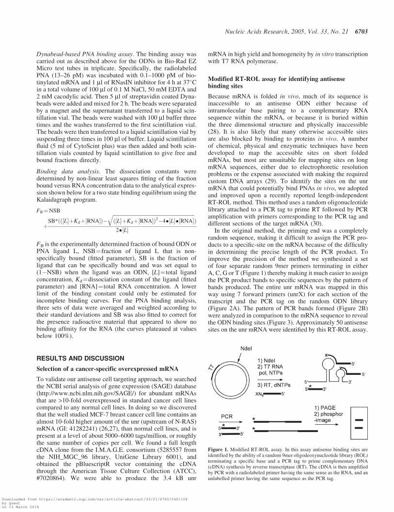
Modified RT-ROL mapping assay. The ODNs used for map-ping the antisense binding sites on the unr mRNA by themodified RT-ROL assay are shown in Figure 2. The mappingwas carried out by heating the RNA at 65�C for 5 min withdNTPs followed by a quickly chilling on ice for 1 min and thenadding the random ODN library primer XN9-tag, RNasIn(Promega, Madison WI) RNase inhibitor and SuperScript IIreverse transcriptase (Invitrogen Life Technologies) foranother 5 min. The reaction was incubated at 42�Cfor 50 min, and the enzyme inactivated by heating at 70�Cfor 15 min. Then 50-endlabeled PCR primer, dNTPs and Taqpolymerase were added and subjected to an initial incubationat 94�C for 3 min, followed by 30 cycles of PCR (1 min 94�C,1 min 55�C and 1 min 72�C). The products were then separ-ated on 6% denaturing polyacrylamide gels.
SAABS assay. Streptavidin coated Dynabeads were firstwashed with solution A (DEPC-treated 0.1 M NaOH andDEPC-treated 0.05 M NaCl), followed by three washeswith solution B (DEPC-treated 0.1 M NaCl), and then washedtwo times with 100 ml of hybridization solution [1M NaCl andTris–HCl (pH 7.4)]. The beads were then resuspended in 50 mlof hybridization solution. The biotinylated RNA (3 ml of3 mg/ml) was incubated with 45 ml of hybridization solutionat 65�C for 5 min, and then cooled at room temperature for10 min. The ODN containing the random 8mer (S1-ROL-S2)was then annealed with the complementary ODNs CS1 andCS2 in hybridization buffer (50 ml) by heating at 65�C for5 min, and then cooled to room temperature for 10 min. TheRNA solution was then mixed with S1-ROL-S2�CS1�CS2together with 2 ml RNasIN, and incubated at 27�C for 1 h.The solution of beads was then added and resuspended every5 min by pipetting for a total of 15 min. Following the incuba-tion, the beads were separated with a magnet and washed6 times with the hybridization solution and then resuspendedin 100 ml of H2O. Thirty cycles of PCR were then carried outon the beads with Taq DNA polymerase, and S1 and CS2primers. After amplification, the reaction mix was directly
loaded on to a 12% denaturing PAGE gel and electrophoresedto separate the 48 bp PCR product. The 48 bp PCR band wasexcised and the eluted product digested with NlaIII at 37�Covernight to release the 12mer product from the tags whichwas purified by 20% denaturing PAGE gel. The 12mer pro-ducts were ligated with T4 DNA ligase and ATP at 16�C for2 h. Concatemers were isolated using the Qiaquick Gel Extrac-tion Kit (Qiagen, Valencia, CA), following the manufacturer’smanual. The mixture was then electrophoresed on a 1% agar-ose gel (TAE) and fractions with 300–500 bp length wereexcised. Concatemers were ligated into the SphI site ofpZErO-1 (Invitrogen) with T4 DNA ligase and ATP for 4 hat 16�C and then transfected into Escherichia coli, followingthe manufacturer’s manual (Oneshot� Top10, Invitrogen).The transfectants were plated on low salt Luria–Bertani(LB) plates containing 50 mg/ml Zeocin� (Invivogen, SanDiego, CA) and incubated for about 18 h at 37�C. Zeocin�resistant transformants were picked using pipette tips, incub-ated in 5 ml SOB containing 50 ml/ml Zeocin� and grownovernight at 37�C. Plasmid DNA was prepared by the Plasmidmini prep kit (Qiagen). Plasmids containing sizable insertswere then forwarded for sequence analysis. The sequenceswere analyzed by a computer program written expressly forthis purpose. The program first extracts the 8mer sequencesfrom between Nla III sites, and then scans for a perfect matchbetween either the sense or antisense 8mer sequences andthe mRNA sequence. If either the sense or antisense 8mersequence matches uniquely, each matching base in themRNA is assigned one point. If the sequence matches n dif-ferent sites, each matching base at each site is assigned 1/npoints. This sequence of steps is repeated for sequences onlymatching 6–7 bases.
Binding assays
Dynabead-based dot blot assay. Strepavidin-coated Dyna-beads were washed following the manufacturer’s protocol,and then washed two times with hybridization buffer [1 MNaCl, 0.5 mM EDTA and 20 mM sodium cacodylate (pH 7.0)].After the last wash, the beads were resuspended in hybridiza-tion buffer in the presence of biotinylated unr mRNA, incub-ated for 30 min at 37�C, and mixed occasionally by flickingthe tube with a finger. The tube containing the beads was thenplaced on a magnetic stand for 1 min, following which thesupernatant was removed and discarded. The beads were thenwashed three times with hybridization buffer andthen resuspended in hybridization buffer in the presence of1 pmol of 32P-labeled ODN, incubated for 30 min at 37�Cwith periodic agitation by flicking. After washing three timesas described above, the beads were resuspended in 20 mlhybridization buffer, and 2 ml was spotted on a Nylonmembrane.
Dynabead-based ODN binding assay. The radiolabeled ODN(100 pM) was incubated with the 0.003–10 nM of biotinylatedmRNA and 1 ml of RNasIN inhibitor for 4 h at 37�C in atotal volume of 100 ml. Then streptavidin coated Dynabeadswere added and mixed for 30 min. The beads were thenseparated by a magnet and the solution removed. The beadswere resuspended in 100 ml of water and both solutions coun-ted by liquid scintillation to give free and bound fractionsdirectly.
6702 Nucleic Acids Research, 2005, Vol. 33, No. 21
Downloaded from https://academic.oup.com/nar/article-abstract/33/21/6700/2401128by gueston 15 March 2018
Dynabead-based PNA binding assay. The binding assay wascarried out as described above for the ODNs in Bio-Rad EZMicro test tubes in triplicate. Specifically, the radiolabeledPNA (13–26 pM) was incubated with 0.1–1000 pM of bio-tinylated mRNA and 1 ml of RNasIN inhibitor for 4 h at 37�Cin a total volume of 100 ml of 0.1 M NaCl, 50 mM EDTA and2 mM cacodylic acid. Then 5 ml of streptavidin coated Dyna-beads were added and mixed for 2 h. The beads were separatedby a magnet and the supernatant transferred to a liquid scin-tillation vial. The beads were washed with 100 ml buffer threetimes and the washes transferred to the first scintillation vial.The beads were then transferred to a liquid scintillation vial bysuspending three times in 100 ml of buffer. Liquid scintillationfluid (5 ml of CytoScint plus) was then added and both scin-tillation vials counted by liquid scintillation to give free andbound fractions directly.
Binding data analysis. The dissociation constants weredetermined by non-linear least squares fitting of the fractionbound versus RNA concentration data to the analytical expres-sion shown below for a two state binding equilibrium using theKalaidagraph program.
FB¼NSB
þSB*ðð½L�þKdþ½RNA�Þ
ffiffiffiffiffiffiffiffiffiffiffiffiffiffiffiffiffiffiffiffiffiffiffiffiffiffiffiffiffiffiffiffiffiffiffiffiffiffiffiffiffiffiffiffiffiffiffiffiffiffiffiffiffiffiffiffiffiffiffiffiffiffiffiffiffiffiffiffið½L�þKdþ½RNA�Þ24�½L��½RNA�
qÞ
2�½L�
FB is the experimentally determined fraction of bound ODN orPNA ligand L, NSB¼fraction of ligand L that is non-specifically bound (fitted parameter), SB is the fraction ofligand that can be specifically bound and was set equal to(1NSB) when the ligand was an ODN, [L]¼ total ligandconcentration, Kd¼dissociation constant of the ligand (fittedparameter) and [RNA]¼ total RNA concentration. A lowerlimit of the binding constant could only be estimated forincomplete binding curves. For the PNA binding analysis,three sets of data were averaged and weighted according totheir standard deviations and SB was also fitted to correct forthe presence radioactive material that appeared to show nobinding affinity for the RNA (the curves plateaued at valuesbelow 100%).
RESULTS AND DISCUSSION
Selection of a cancer-specific overexpressed mRNA
To validate our antisense cell targeting approach, we searchedthe NCBI serial analysis of gene expression (SAGE) database(http://www.ncbi.nlm.nih.gov/SAGE/) for abundant mRNAsthat are >10-fold overexpressed in standard cancer cell linescompared to any normal cell lines. In doing so we discoveredthat the well studied MCF-7 breast cancer cell line contains analmost 10-fold higher amount of the unr (upstream of N-RAS)mRNA (GI: 41282241) (26,27), than normal cell lines, and ispresent at a level of about 5000–6000 tags/million, or roughlythe same number of copies per cell. We found a full lengthcDNA clone from the I.M.A.G.E. consortium (5285557 fromthe NIH_MGC_96 library, UniGene Library 6001), andobtained the pBluescriptR vector containing the cDNAthrough the American Tissue Culture Collection (ATCC),#7020864). We were able to produce the 3.4 kB unr
mRNA in high yield and homogeneity by in vitro transcriptionwith T7 RNA polymerase.
Modified RT-ROL assay for identifying antisensebinding sites
Because mRNA is folded in vivo, much of its sequence isinaccessible to an antisense ODN either because ofintramolecular base pairing to a complementary RNAsequence within the mRNA, or because it is buried withinthe three dimensional structure and physically inaccessible(28). It is also likely that many otherwise accessible sitesare also blocked by binding to proteins in vivo. A numberof chemical, physical and enzymatic techniques have beendeveloped to map the accessible sites on short foldedmRNAs, but most are unsuitable for mapping sites on longmRNA sequences, either due to electrophoretic resolutionproblems or the expense associated with making the requiredcustom DNA arrays (29). To identify the sites on the unrmRNA that could potentially bind PNAs in vivo, we adoptedand improved upon a recently reported length-independentRT-ROL method. This method uses a random oligonucleotidelibrary attached to a PCR tag to prime RT followed by PCRamplification with primers corresponding to the PCR tag anddifferent sections of the target mRNA (30).
In the original method, the priming end was a completelyrandom sequence, making it difficult to assign the PCR pro-ducts to a specific-site on the mRNA because of the difficultyin determining the precise length of the PCR product. Toimprove the precision of the method we synthesized a setof four separate random 9mer primers terminating in eitherA, C, G or T (Figure 1) thereby making it much easier to assignthe PCR product bands to specific sequences by the pattern ofbands produced. The entire unr mRNA was mapped in thisway using 7 forward primers (unrX) for each section of thetranscript and the PCR tag on the random ODN library(Figure 2A). The pattern of PCR bands formed (Figure 2B)were analyzed in comparison to the mRNA sequence to revealthe ODN binding sites (Figure 3). Approximately 50 antisensesites on the unr mRNA were identified by this RT-ROL assay.
Figure 1. Modified RT-ROL assay. In this assay antisense binding sites areidentified by the ability of a random 9mer oligodeoxynucleotide library (ROL)terminating a specific base and a PCR tag to prime complementary DNA(cDNA) synthesis by reverse transcriptase (RT). The cDNA is then amplifiedby PCR with a radiolabeled primer having the same sense as the RNA, and anunlabelled primer having the same sequence as the PCR tag.
Nucleic Acids Research, 2005, Vol. 33, No. 21 6703
Downloaded from https://academic.oup.com/nar/article-abstract/33/21/6700/2401128by gueston 15 March 2018

Serial analysis of antisense binding sites
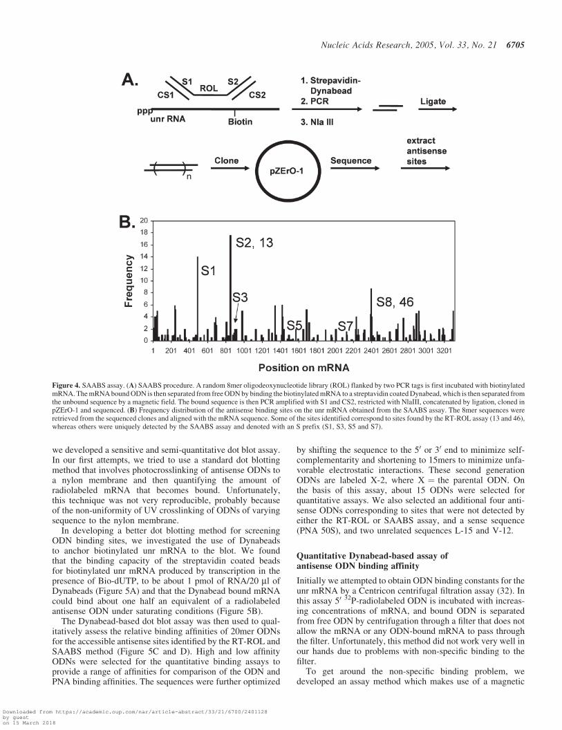
A potential problem with the RT-ROL assay is that thepriming of RT might be sensitive to secondary and tertiarystructure, and as a result, the intensity of the PCR bands pro-duced in the RT-ROL assay might not correspond directly tothe actual binding affinity of the ODN. To circumvent thisproblem we developed an enzyme independent method formapping antisense binding sites that adapts methodologyused in SAGE to determine which members of a randomlibrary of ODNs bind to an mRNA molecule and with whatfrequency. The method, which we call SAABS for serial ana-lysis of antisense binding sites is shown in Figure 4A. In thefirst step, members of a random oligonucleotide library thatbind to the mRNA are separated from those that do not. In thesecond step, the selected sequences are PCR amplified, restric-ted, concatenated by ligation and then cloned. In the third step,the clones are sequenced and the sequences retrieved frombetween the restriction sites and then aligned with the RNAsequence by a computer program.
Recently, another group has reported a similar strategy foridentifying antisense binding sites, which they called MASTfor mRNA accessible site tagging (31). Our method is verysimilar to theirs, which also uses streptavidin coated Dyna-beads, except that we use an 8mer random nucleotide libraryinstead of an 18mer library, add the Dynabeads after bindingof the ODNs to the biotinylated mRNA, and concatenate theantisense sequence tags before sequencing, which they do not.
In our procedure (Figure 4A) a random 8mer ODN librarylinked to two PCR tags is incubated with a biotinylated mRNAmolecule that is bound to a strepavidin-coated Dynabead. ThePCR tags are prevented from hybridizing to the mRNA bybinding to the complementary ODNs CS1 and CS2. Followingincubation with the mRNA, the Dynabeads are separated bya magnet and washed to remove unbound ODNs. The boundODNs are then PCR amplified with primers S1 and CS2,restricted with NlaIII to give 12mers that are isolated by dena-turing 20% PAGE, concatenated by ligation, cloned into thepZErO-1 vector and transfected into E.coli. Plasmids contain-ing inserts are then sequenced, and the 8mer sequence betweenthe NlaIII restriction sites were extracted by a computer pro-gram and both strands matched to complementary sites on theRNA sequence.
The program scored each matching nucleotide in the mRNAby the number of times that it was matched to an 8mersequence divided by the number of other sites that alsomatched that same antisense sequence. If a complete matchwas not found, the process was repeated for all possible 7merand then 6mer sequences. The relative frequency of thesequences retrieved by the SAABS method are plotted againsttheir position as shown in Figure 4B. Some of these sites werenot detected by the RT-ROL assay and are labeled with aS. Presumably, these sites correspond to binding sites thatreverse transcriptase may not have been able to extend. Fifteenof the highest frequency sites uniquely determined by thismethod were chosen for further analysis.
Affinity screening of potential antisense ODNs by aDynabead-based dot blot assay
To determine the relative affinity of ODNs for the antisensebinding sites determined by the RT-ROL and SAABS method
Figure 2. (A) Primers used to map ODN accessible sites on unr transcript.Sequences of unrX is given in the experimental section. (B) Phosphorimage ofpolyacrylamide gels of the PCR products for each section (X ¼ 1–7) of the unrtranscript. Boxed site is analyzed in Figure 3.
Figure 3. Interpretation of the RT-ROL gel. (A) Blowup of boxed section of gelin Figure 2 showing the approximate alignment of the bands with standardlength markers. (B) Alignment of the observed bands with the mRNA sequence,taking into account the sequence of the mRNA and the terminal nucleotide onthe primer, along with the fact that the PCR tag adds an additional 20 nt to theextra 9 random nucleotides in the PCR amplified product band.
6704 Nucleic Acids Research, 2005, Vol. 33, No. 21
Downloaded from https://academic.oup.com/nar/article-abstract/33/21/6700/2401128by gueston 15 March 2018
we developed a sensitive and semi-quantitative dot blot assay.In our first attempts, we tried to use a standard dot blottingmethod that involves photocrosslinking of antisense ODNs toa nylon membrane and then quantifying the amount ofradiolabeled mRNA that becomes bound. Unfortunately,this technique was not very reproducible, probably becauseof the non-uniformity of UV crosslinking of ODNs of varyingsequence to the nylon membrane.
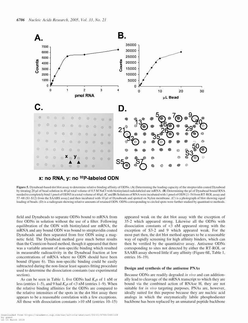
In developing a better dot blotting method for screeningODN binding sites, we investigated the use of Dynabeadsto anchor biotinylated unr mRNA to the blot. We foundthat the binding capacity of the streptavidin coated beadsfor biotinylated unr mRNA produced by transcription in thepresence of Bio-dUTP, to be about 1 pmol of RNA/20 ml ofDynabeads (Figure 5A) and that the Dynabead bound mRNAcould bind about one half an equivalent of a radiolabeledantisense ODN under saturating conditions (Figure 5B).
The Dynabead-based dot blot assay was then used to qual-itatively assess the relative binding affinities of 20mer ODNsfor the accessible antisense sites identified by the RT-ROL andSAABS method (Figure 5C and D). High and low affinityODNs were selected for the quantitative binding assays toprovide a range of affinities for comparison of the ODN andPNA binding affinities. The sequences were further optimized
by shifting the sequence to the 50 or 30 end to minimize self-complementarity and shortening to 15mers to minimize unfa-vorable electrostatic interactions. These second generationODNs are labeled X-2, where X ¼ the parental ODN. Onthe basis of this assay, about 15 ODNs were selected forquantitative assays. We also selected an additional four anti-sense ODNs corresponding to sites that were not detected byeither the RT-ROL or SAABS assay, and a sense sequence(PNA 50S), and two unrelated sequences L-15 and V-12.
Quantitative Dynabead-based assay ofantisense ODN binding affinity
Initially we attempted to obtain ODN binding constants for theunr mRNA by a Centricon centrifugal filtration assay (32). Inthis assay 50 32P-radiolabeled ODN is incubated with increas-ing concentrations of mRNA, and bound ODN is separatedfrom free ODN by centrifugation through a filter that does notallow the mRNA or any ODN-bound mRNA to pass throughthe filter. Unfortunately, this method did not work very well inour hands due to problems with non-specific binding to thefilter.
To get around the non-specific binding problem, wedeveloped an assay method which makes use of a magnetic
Figure 4. SAABS assay. (A) SAABS procedure. A random 8mer oligodeoxynucleotide library (ROL) flanked by two PCR tags is first incubated with biotinylatedmRNA. The mRNA bound ODN is then separated from free ODN by binding the biotinylated mRNA to a streptavidin coated Dynabead, which is then separated fromthe unbound sequence by a magnetic field. The bound sequence is then PCR amplified with S1 and CS2, restricted with NlaIII, concatenated by ligation, cloned inpZErO-1 and sequenced. (B) Frequency distribution of the antisense binding sites on the unr mRNA obtained from the SAABS assay. The 8mer sequences wereretrieved from the sequenced clones and aligned with the mRNA sequence. Some of the sites identified correspond to sites found by the RT-ROL assay (13 and 46),whereas others were uniquely detected by the SAABS assay and denoted with an S prefix (S1, S3, S5 and S7).
Nucleic Acids Research, 2005, Vol. 33, No. 21 6705
Downloaded from https://academic.oup.com/nar/article-abstract/33/21/6700/2401128by gueston 15 March 2018
field and Dynabeads to separate ODNs bound to mRNA fromfree ODNs in solution without the use of a filter. Followingequilibration of the ODN with biotinylated unr mRNA, themRNA and any bound ODN was bound to streptavidin coatedDynabeads and then separated from free ODN using a mag-netic field. The Dynabead method gave much better resultsthan the Centricon-based method, though it appeared that therewas a variable amount of non-specific binding which resultedin measurable radioactivity in the Dynabead fraction at lowconcentrations of mRNA where no ODN should have beenbound (Figure 6). This non-specific binding could be easilysubtracted during the non-linear least squares fitting procedureused to determine the dissociation constants (see experimentalsection).
As can be seen in Table 1, five ODNs had Kds of 1 nM orless (entries 1–5), and 9 had Kds of <3 nM (entries 1–9). Whenthe relative binding affinities for the ODNs are compared tothe relative intensities of the spots in the dot blot assay thereappears to be a reasonable correlation with a few exceptions.All those with dissociation constants >10 nM (entries 10–15)
appeared weak on the dot blot assay with the exception of35-2 which appeared strong. Likewise all the ODNs withdissociation constants of <3 nM appeared strong with theexception of S3-2 and 9 which appeared weak. For themost part then, the dot blot method appears to be a reasonableway of rapidly screening for high affinity binders, which canthen be verified by the quantitative assay. Antisense ODNscorresponding to sites not detected by either the RT-ROL orSAABS assay showed little if any affinity (Figure 6E, Table 1,entries 16–19).
Design and synthesis of the antisense PNAs
Because ODNs are readily degraded in vivo and can addition-ally lead to cleavage of the mRNA transcript to which they arebound via the combined action of RNAse H, they are notsuitable for in vivo targeting purposes. PNAs are, however,ideally suited for this purpose because they are nucleic acidanalogs in which the enzymatically labile phosphodiesterbackbone has been replaced by an unnatural peptide backbone
Figure 5. Dynabead-based dot blot assay to determine relative binding affinity of ODNs. (A) Determining the loading capacity of the streptavidin coated Dynabeadby titrating 20 ml of bead solution in 40 ml total volume of 0.5 M NaCl with biotinylated radiolabeled unr mRNA. (B) Determining the ml of Dynabead bound RNAneeded to completely bind 1 pmol of ODN5 in a total volume of 40ml. (C and D) Solutions of RNA were incubated with 1 pmol of ODN [1–54 from RT-ROL assay and57–68 (S1-S12) from the SAABS assay] and then incubated with 10 ml of Dynabeads and spotted on Nylon membrane. (C) is a photograph of blot showing equalloading of beads. (D) is a radiogram showing relative amounts of retained ODN. ODNs corresponding to circled spots were further studied by quantitative methods.
6706 Nucleic Acids Research, 2005, Vol. 33, No. 21
Downloaded from https://academic.oup.com/nar/article-abstract/33/21/6700/2401128by gueston 15 March 2018

Figure 6. Curve fits of the data from the Dynabead ODN binding assay. (A–C) antisense ODNs for sites identified by the RT-ROL assay, (D) antisense ODNs for sitesidentified by the SAABS assay, and two unrelated ODNs, L-15 and V-12, (E) antisense ODNs for sites not identified by either the RT-ROL or SAABS assay.
Nucleic Acids Research, 2005, Vol. 33, No. 21 6707
Downloaded from https://academic.oup.com/nar/article-abstract/33/21/6700/2401128by gueston 15 March 2018

(33,34). As a result, PNAs are highly resistant to enzymaticdegradation (35), do not activate RNAse H activity (36), havea high affinity for complementary mRNA and can invadeduplex regions (37).
Therefore, we designed hybrid PNAs corresponding to thefour ODNs that showed the highest binding affinity, and ODN5-2 which showed slightly less affinity. We also synthesizedPNAs corresponding to four ODNs showing low affinity and tothe four ODNs corresponding to sites not detected by eithermethod. The sense sequence, PNA50-2S, corresponding to thePNA50-2 binding site was also made as a control. The PNAswere synthesized with a recently reported four lysine permea-tion peptide (38,39) at the C-terminus, which was used forour PET imaging studies in mice (20), and to increase water
solubility and mRNA binding affinity. A cysteine-tyrosinesequence was added to the N-terminal end to enable sub-sequent attachment of reporter groups to the cysteine andradioiodination of the tyrosine. The PNAs were synthesizedby standard solid phase Fmoc chemistry on an ABI Expedite8909 automated synthesizer, purified by reverse phase HPLC,and characterized by molecular weight determination byMALDI-TOF (Table 2).
Quantitative Dynabead-based assayof antisense PNA binding affinity
The binding affinity of the PNAs for the unr mRNA weredetermined by monitoring bound and free 131I-labeled PNAas a function of mRNA concentration using the Dynabeadassay method that we developed to quantify ODN bindingto the mRNA (Figure 7). We chose 131I to label the PNAsbecause of the high specific activity in which it can be obtainedand its short half-life of 8 days. The PNAs were radiolabeledby a procedure previously used to label PNAs with 125I andChloramine-T (13).
We also checked the integrity of the PNA produced underthese conditions by repeating the labeling experiment with ashort PNA test sequence, DOTA-Tyr-ATGC-Lys (20), withnon-radioactive iodine by the chloramine-T method and alsoby IODO-beads and analyzed the products by MALDI. Wefound that at a 5 min reaction time, both methods lead prim-arily to the mono-iodinated compound and some of thediiodinated compound (tyrosine can react twice), but that atlonger times, other products are produced (data not shown).Thus the 5 min time period was used for radiolabeling theCys-Try-PNA-Lys4 by 131I.
In the first set of PNA binding experiments we discoveredthat we were not recovering all the radioactivity and were ableto trace the loss to non-specific binding of the PNA to themicrofuge tubes that we were using for the incubations. Afterordering and testing a number of tubes and microtiter platesadvertised as minimizing binding of peptides and nucleicacids, we found that Corning NBS microtiter plates and themore convenient Bio-Rad EZ Micro 1.5 ml test tubes had thelowest non-specific binding affinity for the PNAs.
Table 1. Binding affinity of the antisense ODNs by the Dynabead assay
Entry Site Code ODN sequence Kd (nM)
1 2851 50-2 TGGTGTGCTTTGTGG 0.3 ± 0.12 676 7-2 TTTCCCAGTCCGTCG 0.3 ± 0.13 1414 S5-2 TTTGTCACGTCGGTC 0.7 ± 0.24 901 S3-2 ATCTCCAGTTTCCAG 0.8 ± 0.25 1145 16-2 CATTTCTGTCCTTGA 1.0 ± 0.36 1802 26-2 CATCCTCAGCCTCCT 1.1 ± 0.37 727 9 ATTCGTTCTTCAGGGAGGAT 1.2 ± 0.68 839 13-2 CACTTCCCCATTACG 1.4 ± 0.69 476 5-2 TATGTCCATTGTTGT 1.5 ± 0.7
10 2020 32 CCAAAATTATCCTTCAGAGT >1011 1396 21 TCTGTTGAAATATTAAACCT >1012 1927 29-2 CCTCTGTTTGTCACT >1013 2114 35-2 TGTCCCCCAGTTCCA >1014 1389 20 AATATTAAACCTAACATGGT >1015 2115 S7 ATGTCCCCCAGTTCC >10016 1012 1012 TCCTTCATGGCACAAACTAC >10017 1263 1263 GGTTTTTACTGGGTACTTTT >10018 1526 1526 CACACACTTGATGAAACCAA >10019 1653 1653 TAATAGCATGATTTCTTTGA >10020 na V-12 CGATTGGAGCGC >10021 na L-15 AGATCGCAACTCATA >100
The antisense site refers to the position in the mRNA (start codon at 448) that iscomplementary to the 30 end of the corresponding ODN. The code number X-Yrefers to ODN X used in the dot blot experiments, and if followed by a 2 refers toan optimized second generation sequence that overlaps the original sequence.The S prefix refers to a site uniquely identified by the SAABS assay.
Table 2. Binding affinity of Cys-Tyr-PNA-Lys4 by the Dynabead-based solution binding assay
Entry Site Code Calcd M + 1a Obsvd M + 1a PNA sequence Kd (pM)
1 899 S3-2 5571 5576 TATCTCCAGTTTCCAGCT 6 ± 32 673 7-2 5588 5594 TTTCCCAGTCCGTCGGTC 12 ± 43 476 5-2 5641 5646 CATTATGTCCATTGTTGT 17 ± 64 2848 50-2 5779 5784 TGGTGTGCTTTGTGGATG 20 ± 75 1414 S5-2 5651 5659 TAATTTGTCACGTCGGTC 49 ± 66 1927 29 5592 5594 CCTCTGTTTGTCACTAAT 300 ± 307 2020 32 5603 5604 CCAAAATTATCCTTCAGA 380 ± 608 2114 35 5587 5591 TGTCCCCCAGTTCCAGGC 680 ± 1009 1389 20 5658 5660 TATTAAACCTAACATGGT 820 ± 190
10 1263 1263 5653 5655 TTTTTACTGGGTACTTTT 340 ± 6011 1012 1012 5604 5608 CTTCATGGCACAAACTAC 650 ± 17012 1526 1526 5637 5640 CACACTTGATGAAACCAA 1000 ± 31013 1653 1653 5680 5683 ATAGCATGATTTCTTTGA 1000 ± 19014 nab 50-2S 5562 5568 CATCCACAAAGCACACCA >10 000
The site refers to the position in the mRNA that is complementary to the 30 end of the PNA sequence shown (the start codon is at 448). The code number X-Y is the sameas that for the corresponding ODN listed in Table 1.aCalculated MW is for the average M + 1 ion, observed MW is for the M + 1 ion detected by MALDI-TOF.bnot applicable.
6708 Nucleic Acids Research, 2005, Vol. 33, No. 21
Downloaded from https://academic.oup.com/nar/article-abstract/33/21/6700/2401128by gueston 15 March 2018

Figure 7. Curve fits to the PNA binding data from the Dynabead assay. (A–C) antisense PNAs corresponding to high affinity antisense ODNs identified by the RT-ROL assay, (D) sense PNA corresponding to PNA50-2, (E and F) antisense PNAs corresponding to high affinity antisense ODNs identified by the SAABS assay,(G) antisense PNAs corresponding to low affinity antisense ODNs identified by the RT-ROL assay, (H) antisense PNAs corresponding to sites not identified by eitherassay. The error bars represent the standard deviation of the average of three experiments and are not shown for (G) and (H) for clarity.
Nucleic Acids Research, 2005, Vol. 33, No. 21 6709
Downloaded from https://academic.oup.com/nar/article-abstract/33/21/6700/2401128by gueston 15 March 2018

Triplicate sets of data were obtained and a plot of % boundas a function of RNA concentration was fit to a simple twostate binding equilibrium as we had done for the ODNs(Figure 7). The Kds for the PNAs are tabulated in Table 2.As expected, the PNAs corresponding to the high affinityODNs show very high binding affinity (low Kds) for theunr mRNA with Kds that range from 7 to 50 pM at 0.1 Msalt (entries 1–5). The PNAs corresponding to the low affinityODNs, showed higher Kds of 300–820 pM (entries 6–9). Sur-prisingly, the PNAs corresponding to sites not detected byeither the RT-ROL or SAABS assays also bound with similarKds of 340–1000 pM (entries 10–13) found for the low affinitysites. The sense PNA 50-2S, appeared to have essentially noaffinity for the RNA (entry 14).
The binding affinities of the PNAs for the unr mRNA aremuch greater (lower Kds) than those of the correspondingODNs. The difference in binding affinity between the PNAsand ODNs is likely to be much greater in vivo because the PNAdissociation constants were obtained at the physiological con-centration of 0.1 M NaCl, whereas the ODN dissociation con-stants were acquired at 1 M salt. At 0.1 M salt the Kds for theODNs are expected to be greater due to electrostatic repul-sions, whereas the PNAs are expected to have lower Kds due tofavorable electrostatic attraction with the Lys4 tail. PNA50-2S, which is identical in sequence to the mRNA target,showed no significant binding in the range of RNA concen-trations that bound tightly to the antisense sequences.
The significant binding affinity of the PNAs to sites notdetected by either the RT-ROL or SAABS assay (Kds of340–1000 pM) might be attributable to the ability of PNAto invade and bind to regions of folded RNA that might notbind ODNs because of electrostatic repulsion. Even though,the best of these PNAs (PNA 1927) has >50-fold less affinitythan the highest affinity PNA identified by the SAABS assay(PNA S3-2), and >25-fold less than the highest affinity PNAfound by the RT-ROL assay (PNA 7-2). Likewise, PNAscorresponding to low affinity antisense ODNs bind withless affinity (300–820 pM) than those PNAs correspondingto the high affinity antisense ODNs. These results suggestthat accessibility of a site to an ODN may be a prerequisitefor high affinity PNA binding. Thus it would appear that theRT-ROL and SAABS assay offer a more direct and efficientway to find high affinity antisense PNAs than might be foundby simply screening PNAs to randomly selected sites.
CONCLUSION
We have identified high affinity antisense binding sites on unrmRNA produced in vitro through an improved RT-ROL andSAABS mapping methods, and have quantified their bindingaffinity by a new Dynabead-based solution assay. The twomapping methods identified many of the same antisense bind-ing sites, but there were also a few high affinity sites identifiedby the SAABS method that were not detected by the RT-ROLmethod, presumably because reverse transcriptase was unableto bind or extend an ODN at that site. The affinity of PNAs forthese sites on the unr mRNA in vivo remains to be determined,although our recent results with PET imaging MCF-7 tumorsin mice with derivatives of PNA50-2, PNA5-2 and PNA7-2and PNA50-2S are consistent with a contribution from an
antisense mechanism. The methods that we improved uponand developed for mapping and quantifying the antisensebinding sites might also be useful for designing siRNA agents,and for guiding RNA folding calculations.
ACKNOWLEDGEMENTS
We want to thank Demetrios Sarantites for help with the radio-iodination experiments. This research was supported by anNCI/NASA Biomolecular Sensors contract (N01-CO-27103)and by the National Institutes of Health Program of Excellencein Nanotechnology (1 U01 HL080729-01). Funding to pay theOpen Access publication charges for this article was providedby NIH HL080729.
Conflict of interest statement. None declared.
REFERENCES
1. Dyba,M., Tarasova,N.I. and Michejda,C.J. (2004) Small molecule toxinstargeting tumor receptors. Curr. Pharm. Des., 10, 2311–2334.
2. Brannon-Peppas,L. and Blanchette,J.O. (2004) Nanoparticle and targetedsystems for cancer therapy. Adv. Drug Deliv. Rev., 56, 1649–1659.
3. Tanaka,T., Shiramoto,S., Miyashita,M., Fujishima,Y. and Kaneo,Y.(2004) Tumor targeting based on the effect of enhanced permeability andretention (EPR) and the mechanism of receptor-mediated endocytosis(RME). Int. J. Pharm., 277, 39–61.
4. Bennasroune,A., Gardin,A., Aunis,D., Cremel,G. and Hubert,P. (2004)Tyrosine kinase receptors as attractive targets of cancer therapy.Crit. Rev. Oncol. Hematol., 50, 23–38.
5. Zhao,X.B. and Lee,R.J. (2004) Tumor-selective targeted delivery ofgenes and antisense oligodeoxyribonucleotides via the folate receptor.Adv. Drug Deliv. Rev., 56, 1193–1204.
6. Shadidi,M. and Sioud,M. (2003) Selective targeting of cancer cells usingsynthetic peptides. Drug Resist. Updat., 6, 363–371.
7. van Dongen,G.A., Visser,G.W. and Vrouenraets,M.B. (2004)Photosensitizer-antibody conjugates for detection and therapy of cancer.Adv. Drug Deliv. Rev., 56, 31–52.
8. Goodwin,D.A. and Meares,C.F. (2001) Advances in pretargetingbiotechnology. Biotechnol. Adv., 19, 435–450.
9. Rooseboom,M., Commandeur,J.N. and Vermeulen,N.P. (2004)Enzyme-catalyzed activation of anticancer prodrugs. Pharmacol.Rev., 56, 53–102.
10. Wadia,J.S. and Dowdy,S.F. (2002) Protein transduction technology.Curr. Opin. Biotechnol., 13, 52–56.
11. Moulton,H.M. and Moulton,J.D. (2003) Peptide-assisted delivery ofsteric-blocking antisense oligomers. Curr. Opin. Mol. Ther., 5,123–132.
12. Kabouridis,P.S. (2003) Biological applications of protein transductiontechnology. Trends Biotechnol., 21, 498–503.
13. Lee,H.J., Boado,R.J., Braasch,D.A., Corey,D.R. and Pardridge,W.M.(2002) Imaging gene expression in the brain in vivo in a transgenic mousemodel of Huntington’s disease with an antisense radiopharmaceutical anddrug-targeting technology. J. Nucl. Med, 43, 948–956.
14. Lewis,M.R., Jia,F., Gallazzi,F., Wang,Y., Zhang,J., Shenoy,N.,Lever,S.Z. and Hannink,M. (2002) Radiometal-labeled peptide-PNAconjugates for targeting bcl-2 expression: preparation, characterization,and in vitro mRNA binding. Bioconjug. Chem., 13, 1176–1180.
15. Rao,P.S., Tian,X., Qin,W., Aruva,M.R., Sauter,E.R., Thakur,M.L. andWickstrom,E. (2003) 99mTc-peptide-peptide nucleic acid probes forimaging oncogene mRNAs in tumours. Nucl. Med. Commun., 24,857–863.
16. Tian,X., Aruva,M.R., Rao,P.S., Qin,W., Read,P., Sauter,E.R.,Thakur,M.L. and Wickstrom,E. (2003) Imaging oncogene expression.Ann. N. Y. Acad. Sci., 1002, 165–188.
17. Gallazzi,F., Wang,Y., Jia,F., Shenoy,N., Landon,L.A., Hannink,M.,Lever,S.Z. and Lewis,M.R. (2003) Synthesis of radiometal-labeled andfluorescent cell-permeating peptide-PNA conjugates for targeting thebcl-2 proto-oncogene. Bioconjug. Chem., 14, 1083–1095.
6710 Nucleic Acids Research, 2005, Vol. 33, No. 21
Downloaded from https://academic.oup.com/nar/article-abstract/33/21/6700/2401128by gueston 15 March 2018

18. Shi,N., Boado,R.J. and Pardridge,W.M. (2000) Antisense imaging of geneexpression in the brain in vivo. Proc. Natl Acad. Sci. USA, 97,14709–14714.
19. Sohail,M. and Southern,E.M. (2000) Selecting optimal antisensereagents. Adv. Drug Deliv. Rev., 44, 23–34.
20. Sun,X., Fang,H., Li,X., Rossin,R., Welch,M.J. and Taylor,J.S. (2005)MicroPET imaging of MCF-7 tumors in mice via unr mRNA-targetedpeptide nucleic acids. Bioconj. Chem., 16, 294–305.
21. Ma,Z. and Taylor,J.S. (2003) PNA-based RNA-triggered drug-releasingsystem. Bioconjug. Chem., 14, 679–683.
22. Ma,Z. and Taylor,J.S. (2000) Nucleic acid triggered catalytic drug release.Proc. Natl Acad. Sci. USA, 97, 11159–11163.
23. Ma,Z. and Taylor,J.S. (2001) Nucleic acid triggered catalytic drug andprobe release: a new concept for the design of chemotherapeutic anddiagnostic agents. Bioorg. Med. Chem., 9, 2501–2510.
24. Cai,J., Li,X., Yue,X. and Taylor,J.S. (2004) Nucleic acid-triggeredfluorescent probe activation by the Staudinger reaction.J. Am. Chem. Soc., 126, 16324–16325.
25. Cai,J., Li,X. and Taylor,J.S. (2005) Improved nucleic acid triggered probeactivation through the use of a 5-thiomethyluracil peptide nucleic acidbuilding block. Org. Lett., 7, 751–754.
26. Jeffers,M., Paciucci,R. and Pellicer,A. (1990) Characterizationof unr; a gene closely linked to N-ras. Nucleic Acids Res., 18, 4891–4899.
27. Ferrer,N., Garcia-Espana,A., Jeffers,M. and Pellicer,A. (1999) Theunr gene: evolutionary considerations and nucleic acid-bindingproperties of its long isoform product. DNA Cell. Biol., 18, 209–218.
28. Sohail,M. and Southern,E.M. (2000) Hybridization of antisense reagentsto RNA. Curr. Opin. Mol. Ther., 2, 264–271.
29. Smith,L., Andersen,K.B., Hovgaard,L. and Jaroszewski,J.W. (2000)Rational selection of antisense oligonucleotide sequences. Eur. J. Pharm.Sci., 11, 191–198.
30. Allawi,H.T., Dong,F., Ip,H.S., Neri,B.P. and Lyamichev,V.I. (2001)Mapping of RNA accessible sites by extension of random oligonucleotidelibraries with reverse transcriptase. RNA, 7, 314–327.
31. Zhang,H.Y., Mao,J., Zhou,D., Xu,Y., Thonberg,H., Liang,Z. andWahlestedt,C. (2003) mRNA accessible site tagging (MAST): a novelhigh throughput method for selecting effective antisenseoligonucleotides. Nucleic Acids Res., 31, e72.
32. Walton,S.P.,Stephanopoulos,G.N., Yarmush,M.L. and Roth,C.M. (2002)Thermodynamic and kinetic characterization of antisenseoligodeoxynucleotide binding to a structured mRNA. Biophys. J., 82,366–377.
33. Uhlmann,E., Peyman,A., Breipohl,G. and Will,D.W. (1998) PNA:synthetic polyamide nucleic acids with unusual binding properties.Angew. Chem. Int. Ed, 37, 2796–2823.
34. Ray,A. and Norden,B. (2000) Peptide nucleic acid (PNA): its medical andbiotechnical applications and promise for the future. FASEB. J., 14,1041–1060.
35. Uhlmann,E. and Peyman,A. (1990) Antisense oligonucleotides: a newtherapeutic principle. Chem. Rev., 90, 543–584.
36. Gee,J.E., Robbins,I., van der Laan,A.C., van Boom,J.H., Colombier,C.,Leng,M., Raible,A.M., Nelson,J.S. and Lebleu,B. (1998)Assessment of high-affinity hybridization, RNase H cleavage, andcovalent linkage in translation arrest by antisenseoligonucleotides. Antisense Nucleic Acid Drug Dev., 8,103–111.
37. Peffer,N.J., Hanvey,J.C., Bisi,J.E., Thomson,S.A., Hassman,C.F.,Noble,S.A. and Babiss,L.E. (1993) Strand-invasion of duplex DNA bypeptide nucleic acid oligomers. Proc. Natl Acad. Sci. USA, 90,10648–10652.
38. Sazani,P., Kang,S.H., Maier,M.A., Wei,C., Dillman,J., Summerton,J.,Manoharan,M. and Kole,R. (2001) Nuclear antisense effects of neutral,anionic and cationic oligonucleotide analogs. Nucleic Acids Res., 29,3965–3974.
39. Sazani,P., Gemignani,F., Kang,S.H., Maier,M.A., Manoharan,M.,Persmark,M., Bortner,D. and Kole,R. (2002) Systemically deliveredantisense oligomers upregulate gene expression in mouse tissues.Nat. Biotechnol., 20, 1228–1233.
Nucleic Acids Research, 2005, Vol. 33, No. 21 6711
Downloaded from https://academic.oup.com/nar/article-abstract/33/21/6700/2401128by gueston 15 March 2018