identification and characterization of cardiac troponin t ... · pdf filefirst validated using...
TRANSCRIPT

Identification and Characterization of CardiacTroponin T Fragments in Serum of Patients Suffering
from Acute Myocardial InfarctionAlexander S. Streng,1 Douwe de Boer,1 William P.T.M. van Doorn,1 Freek G. Bouwman,2
Edwin C.M. Mariman,2 Otto Bekers,1 Marja P. van Dieijen-Visser,1 and Will K.W.H. Wodzig1*
BACKGROUND: Cardiac troponin T (cTnT) is the pre-ferred biomarker for the diagnosis of acute myocardialinfarction (AMI). It has been suggested that cTnT ispresent predominantly in fragmented forms in humanserum following AMI. In this study, we have used a tar-geted mass spectrometry assay and epitope mapping us-ing Western blotting to confirm this hypothesis.
METHODS: cTnT was captured from the serum of 12 pa-tients diagnosed with AMI using an immunoprecipita-tion technique employing the M11.7 catcher antibodyand fractionated with SDS-PAGE. Coomassie-stainedbands of 4 patients at 37, 29, and 16 kDa were excisedfrom the gel, digested with trypsin, and analyzed on a QExactive instrument set on targeted Selected Ion Moni-toring mode with data-dependent tandem mass spec-trometry (MS/MS) for identification. Western blottingemploying 3 different antibodies was used for epitopemapping.
RESULTS: Ten cTnT peptides of interest were targeted.By using MS/MS, all of these peptides were identified inthe 37-kDa, intact, cTnT band. In the 29- and 16-kDafragment bands, 8 and 4 cTnT-specific peptides wereidentified, respectively. Some of these peptides were“semitryptic,” meaning that their C-termini were notformed by trypsin cleavage. The C-termini of these semi-tryptic peptides represent the C-terminal end of thecTnT molecules present in these bands. These resultswere confirmed independently by epitope mapping.
CONCLUSIONS: Using LC-MS, we have succeeded in pos-itively identifying the 29- and 16-kDa fragment bands ascTnT-derived products. The amino acid sequences of the
29- and 16-kDa fragments are Ser79-Trp297 and Ser79-Gln199, respectively.© 2016 American Association for Clinical Chemistry
Cardiac troponin T (cTnT)3 and I (cTnI) have beenfirmly established as the gold standard biomarkers for thediagnosis of acute myocardial infarction (AMI) (1–3 ).Owing to patent restrictions, the commercially availablecTnT assay by Roche Diagnostics is still the only clinicalcTnT assay on the market and is actively being used in51% of hospitals in Europe (4 ). The assay works accord-ing to the sandwich principle, in which a detector (M7)and catcher (M11.7) antibody are allowed to bind tocTnT, forming a sandwich complex. Since both antibod-ies bind closely to one another in the center of the cTnTmolecule, the assay is able to detect all cTnT isoforms,protein complexes, protein fragments, and peptides con-taining the epitope sequences corresponding to these an-tibodies. However, the affinity of the Roche antibodiesfor the different cTnT molecules may vary. Conse-quently, any molecular heterogeneity of cTnT may havean impact on the performance of the cTnT assay; there-fore, it is important to know whether or not differentimmunoreactive molecules exist in the serum or plasmaof patients suffering from AMI.
In previously published papers, Michielsen et al. (5 )and Cardinaels et al. (6 ) used Western blotting and gelfiltration chromatography (GFC) to show that cTnT wasprimarily present in fragmented forms in human serum.These fragments were detected using the antibodies fromthe Roche assay (6 ). A consistent pattern frequently ob-served in these studies was the appearance of intact cTnTat 37 kDa, a primary cTnT fragment band at 29 kDa,and several smaller secondary fragments between 15 and
1 Department of Clinical Chemistry, Central Diagnostic Laboratory, Maastricht UniversityMedical Centre, Maastricht, the Netherlands; 2 Department of Human Biology, Maas-tricht University, Maastricht, the Netherlands.
* Address correspondence to this author at: Central Diagnostic Laboratory, Maastricht Uni-versity Medical Centre, P. Debyelaan 25, P.O. Box 5800, 6202 AZ Maastricht, the Neth-erlands. Fax +31-(0)43-3874692; e-mail [email protected].
Received May 30, 2016; accepted September 6, 2016.Previously published online at DOI: 10.1373/clinchem.2016.261511
© 2016 American Association for Clinical Chemistry3 Nonstandard abbreviations: cTnT, cardiac troponin T; cTnI, cardiac troponin I; AMI, acute
myocardial infarction; GFC, gel filtration chromatography; STEMI, ST-segment elevationmyocardial infarction; LOD, limit of detection; UHPLC, ultra-high-performance liquidchromatography; AUC, area under the curve; XIC, extracted ion chromatogram; P/R,precursor/reference; skTnT, skeletal troponin T; SIM, selected ion monitoring; ESRD, end-stage renal disease.
Clinical Chemistry 63:2563–572 (2017)
Proteomics and Protein Markers
563

20 kDa. Intact cTnT was only observed in a small per-centage (approximately 20%) of patients in these studies.This is in contrast to the findings of Fahie-Wilson andBates et al., who were unable to identify cTnT fragmentsand argued that cTnT circulates only in the intact andcomplexed form (7, 8 ). The primary detection methodof all of these different studies was based without excep-tion on immunoreactivity with the Roche M7 detectorantibody in combination with a separation technique. Ithas been suggested that the appearance of cTnT frag-ments in these patients should be verified with mass spec-trometry (9 ).
Recently, we have developed a gel-based targetedmass spectrometry assay to identify molecular changes incTnT (10 ). We validated the assay with an in vitro ex-periment where purified human intact cTnT was spikedin a human serum matrix. Incubation at 37 °C resulted infragmentation similar to that seen in AMI-patients. Withtandem mass spectrometry (MS/MS), these fragmentswere identified as cTnT-derived products. In addition,we showed using relative quantification that it is possibleto pinpoint specific cTnT peptides that are differentiallypresent within the different protein bands.
In this study, this targeted mass spectrometry assay isused to identify the observed immunoreactive bands inthe serum of patients diagnosed with AMI. Regions ofcTnT that are cleaved off are identified using a combina-tion of Western blotting and mass spectrometry. Theidentification of semitryptic peptides allowed for the di-rect pinpointing of cleavage sites of cTnT within thesesamples.
Materials and Methods
An expanded materials and methods section is availablein the online Supplemental Material (see the Data Sup-plement that accompanies the online version of this arti-cle at http://www.clinchem.org/content/vol63/issue2).
PATIENT SERUM SAMPLES AND QUALITY CONTROL
Patients diagnosed with ST-segment elevation myocar-dial infarction (STEMI) were included over a period of 1year when at least 1 routinely analyzed serum sample hada cTnT concentration of �8000 ng/L. According tothese criteria, 14 patients were included and all relatedserum samples were collected and stored at �80 °C untilanalysis. Two patients were later excluded because fewerthan 3 serum samples were available. Pooled human se-rum ([cTnT] �14 ng/L) obtained from healthy volun-teers was used as a negative QC. As a positive QC, cTnTpurified from human heart tissue was purchased fromHytest. In addition, a synthetic peptide standard consist-ing of the targeted peptides (synthesized by Pepscan withan average purity of �95%) was used as an instrumentQC. Handling of all serum samples was in accordance
with the code for proper secondary use of human tissue inthe Netherlands (www.fmwv.nl).
Peptide and epitope sequence numbering through-out this article is based on the canonical human cTnTprotein species (cTnT-1, P45379). Peptide sequenceswith a bold type, lowercase m represent peptides with anoxidized methionine.
cTnT CONCENTRATION MEASUREMENTS
cTnT concentration was measured with the 5th-generation (high-sensitivity) cTnT-STAT assay on theCobas® 6000 instrument (Roche Diagnostics). The as-say has a limit of blank of 3 ng/L, a limit of detection(LOD) of 5 ng/L, and a limit of quantification of 13ng/L. The linear measuring range is 3.00–10000 ng/L.
IMMUNOPRECIPITATION AND WESTERN BLOTTING
Immunoprecipitation of cTnT, fractionation with SDS-PAGE, and the subsequent detection of cTnT with dif-ferent antibodies using Western blot was performed onpatient serum samples, and on the positive and negativeQCs as described previously (10, 11 ). All experimentswere done in duplicate, where one gel was used for West-ern blotting and the other for Coomassie staining andmass spectrometry.
MASS SPECTROMETRIC ANALYSIS
Sample work-up, mass spectrometry and data analysiswere performed as previously described (10 ). In brief,individual gel bands at 37, 29, and 16 kDa were carefullyexcised from the gel and digested with mass spectrometrygrade trypsin gold (Promega). Quantitative LC-MS/MSmeasurements were performed on a Q Exactive hybridquadrupole-Orbitrap mass spectrometer, connected to anultra-high-performance liquid chromatography (UHPLC)Dionex Ultimate 3000 (both by Thermo Fisher Scientific).Table 1 shows all specific settings, including target m/z, ofthe employed mass spectrometry method.
DATA ANALYSIS
Product ion spectra were searched against the humanUniProtKB/Swiss-Prot database (dated November 13,2013, with a total of 39 690 entries) using the searchengine SEQUEST with Proteome Discoverer, version1.8. Quantification was performed by calculating thearea under the curve (AUC) of the monoisotopic (M�0)precursor ions from the extracted ion chromatograms(XIC) using Skyline v.2.6 (12, 13 ). A precursor/refer-ence (P/R) ratio was calculated for each targeted ion ineach sample as described previously (10 ). The P/R ratiois defined as the AUC of a specific precursor ion (P)divided by the AUC of a reference ion (R) within thesame sample. Resulting P/R ratios were then normalizedwith respect to the highest ratio within each singleexperiment.
564 Clinical Chemistry 63:2 (2017)

DATA ACCESSIBILITY
The mass spectrometry data (.raw files and .msf files)associated with this research are accessible at the Pro-teomeXchange Consortium (http://proteomecentral.proteomexchange.org) via the PRIDE partner reposi-tory (14 ) with the data set identifier PXD004660.
Results
FRAGMENTATION PATTERN ANALYSIS USING WESTERN
BLOTTING
The specificity of the custom-designed antibodies wasfirst validated using purified skeletal (sk)TnT and thenegative QC. These antibodies are specific for cTnTand show no cross-reaction with skTnT (see Supple-mental Fig. 1 in the online Data Supplement). Fur-thermore, online Supplemental Fig. 2 in the onlineData Supplement shows that both antibodies do notnonspecifically bind to proteins present in serum andthat the LODs of the N- and C-terminal antibodies,expressed as absolute amount of cTnT, are 100 and 50pg, respectively.
The cTnT concentration profile and Western blotsof a patient showing all commonly observed bands isdepicted in Fig. 1. Fig. 1A shows the concentration pro-file of cTnT. Figs. 1B, 1C, and 1D show Western blotsof serum samples from this same patient probed withthe Roche M7, the Medimabs N-terminal, and theMedimabs C-terminal antibodies, respectively. It is ap-parent that the N-terminal antibody only stains the 37-kDa, intact, protein band (Fig. 1C). The C-terminal an-tibody stains the intact protein and the 29-kDa, primaryfragment, band (Fig. 1D). The secondary fragments at15–20 kDa are only visible with the M7 detector anti-body as employed in the clinical assay (Fig. 1B). Only the37-kDa, intact, band is present in the positive QC sam-ples, indicating that the degradation is independent fromthe methodology used. All Western blot images from theother 11 patients support these observations and areavailable in the online Supplemental Material.
VALIDATION OF THE MASS SPECTROMETRY ASSAY
The LC-MS assay used in this study was established pre-viously and was validated extensively (10, 15 ). Two pep-tides that were targeted in the validation study have beenomitted in this study because their CVs were deemed toohigh. The CVs of all targeted peptides in the validationstudy were �23% except for the elongated N-terminalpeptide 53AEED. . .PKPR78 which had a CV of 55%.This peptide was included because of its location on theN-terminus of cTnT which was considered crucial forthis study. All targets showed good linearity (r2 � 0.95),except for the 291AKVTGRW297 peptide (r2 � 0.60).The LOD of each peptide (expressed as serum equivalentof immunoreactive cTnT in ng/L) was also determinedin the validation study and was lower than the cTnTconcentration in our samples. Furthermore, we showedthat all targeted peptides were specific for cTnT (withthe notable exception of 95VDFDDIHR102, which is alsopresent in skTnT).
Table 1. Liquid chromatography and mass spectrometrysettings for the t-SIM/dd-MS/MSa assay.
Parameter Setting
Chromatography
Gradient 4%–55%, 30 min
Flow 300 nL/min
t-SIM
Ion source Electrospray
Polarity Positive
Target m/z and chargeb
53AEEDEEEEEAKEAEDGPMEESKPKPR78 747.8279, (4+)64EAEDGPMEESKPKPR78 567.2700, (3+)64EAEDGPmEESKPKPR78 572.6017, (3+)79SFMPNLVPPK88 565.3074, (2+)79SFmPNLVPPK88 573.3048, (2+)95VDFDDIHR102 339.4980, (3+)124KKEEEELVSLK134 444.5854, (3+)187ALSNMMHFGGYIQ199,c 734.8392, (2+)187ALSN(mM/Mm)HFGGYIQ199,c 742.8367, (2+)187ALSNmmHFGGYIQ199,c 750.8341, (2+)187ALSNMMHFGGYIQK200 532.9269, (3+)187ALSN(mM/Mm)HFGGYIQK200 538.2585, (3+)187ALSNmmHFGGYIQK200 543.5902, (3+)228VLAIDHLNEDQLR240 512.6107, (3+)269YEINVLR275 453.7558, (2+)291AKVTGRW297 409.2376, (2+)
Detection window 2.0 Th
Automated gain control 1e5
Maximum injection time 250 ms
Mass resolution 70 000 FWHMd
at m/z 200
dd-MS/MS
Triggering threshold 5e3
Isolation window 0.6 Th
Dissociation method HCDe
Normalized collision energy 27
Automated gain control 5e4
Maximum injection time 120 ms
Mass resolution 35 000 FWHMd
at m/z 200
Dynamic exclusion 10 s
a t-SIM/dd-MS/MS, targeted SIM with data-dependent MS/MS.b A lowercase, bold type m indicates an oxidized methionine.c This peptide was not included in the synthetic peptide standard.d FWHM, full width at half maximum.e HCD, higher-energy collisional dissociation.
Identification and Characterization of cTnT in Serum
Clinical Chemistry 63:2 (2017) 565

MASS SPECTROMETRIC ANALYSIS OF FRAGMENTS
Samples from patient nos. 6 (n � 3), 7 (n � 4), 10 (n �4), and 12 (n � 4) were selected for mass spectrometricidentification of the excised bands based on the cTnTconcentrations in the available samples. Since only a fewsamples had the intact cTnT band visible at the positionof 37 kDa on the Western blot, bands in gels at this positionwere considered to be identified as intact cTnT when thecharacteristic N-terminal peptide 64EADG. . .PKPR78 wasidentified. For patients in which no 37-kDa band couldbe included according to this criterion, comparisons weremade with a positive QC sample (purified human cTnTspiked in PBS).
Fig. 2 shows the relative abundance of each targetedpeptide (expressed as normalized P/R ratio) in the posi-
tive QC samples (top) and in each of the 4 patients. In thepositive QC samples, all targeted peptides are presentexcept for the 2 peptides 187ALSN. . .GYIQ199 and291AKVTGRW297, which are semitryptic. Semitrypticpeptides are peptides of which 1 of the 2 termini (in thesecases: the C-terminus) is not formed by trypsin cleavage,and can be used to identify proteolytic cleavage sites byproteases other than trypsin. Since all other targeted pep-tides are found in high abundance, in view of the Westernblot results (Fig. 1), the positive QC can be identified asintact human cTnT. Supplemental Fig. 3 in the onlineData Supplement shows the annotated chromatogram ofthe targeted peptides in the positive QC sample.
The 37-kDa protein band was detected as cTnT in 3of the 12 patients using Western blot, but could only be
Fig. 1. cTnT concentration curve and Western blots of an AMI patient.(A) Concentration curve of cTnT in a patient suffering from acute myocardial infarction (AMI). Time points are relative to the time of first serumsampling. (B–D) Western blots of immunoprecipitated serum samples using the Roche M7 antibody (B), the Medimabs N-terminal antibody(C), and the Medimabs C-terminal antibody (D). Samples were diluted to the same cTnT concentration prior to blotting. P indicates the positiveQC sample; N indicates the negative QC sample. Ab, antibody.
566 Clinical Chemistry 63:2 (2017)

identified as intact cTnT in 3 samples belonging to pa-tient No. 10 using MS. Although patient nos. 7 and 12also had a faint 37-kDa protein band visible on the West-ern blots (see the Supplemental Material in the onlineData Supplement), the amount of cTnT in those bandsproved to be insufficient to identify the N-terminal pep-
tides that are characteristic of intact cTnT. For this rea-son, only the intact, 37-kDa band measurements of pa-tient no. 10 are shown.
When considering the identification of the pri-mary (29 kDa) and secondary (16 kDa) fragmentbands, it was apparent that the 2 N-terminal peptides
Fig. 2. Normalized P/R ratios of each targeted peptide in different samples from 4 AMI patients.The top panel shows the normalized P/R ratios of all targeted peptides in the positive (Pos) QC samples (n = 4). The bottom 4 panels show thenormalized P/R ratios of all targeted peptides in 4 patients suffering from acute myocardial infarction (AMI). All n = 4, except for patient 6,where n = 3. Starred peptides (*) indicate semitryptic peptides. Pat., patient; term., terminus.
Identification and Characterization of cTnT in Serum
Clinical Chemistry 63:2 (2017) 567

(53AEED. . .PKPR78 and 64EADG. . .PKPR78) wereabsent in both of them (Fig. 2). This was consistentin all samples of all 4 patients. The peptide79SFMP. . .VPPK88, which lies directly adjacent to theabsent peptides, was identified in all samples. More-over, in the intact and primary fragment samples, allC-terminal peptides (up to and including the semi-tryptic 291AKVTGRW297 peptide) were detected. In-terestingly, this semitryptic peptide was not found inthe positive QC sample (Fig. 2). This may mean thatthe last amino acid of the cTnT protein, Lys-298,while present in the purified cTnT standard, was ab-sent in the intact and primary fragment bands inserum.
Only centrally located cTnT peptides could be de-tected in the secondary fragment. Interestingly, in these
samples, a double peak was observed in the extracted ionchromatogram (XIC) of the 187ALSN. . .YIQK200 pep-tide (Fig. 3A, right panel). This double peak was absentin the XIC of the intact cTnT bands (Fig. 3A, leftpanel) and in the primary fragment bands (not shown)of all analyzed samples. MS/MS measurements iden-tified the leftmost peak as the targeted peptide(187ALSN. . .YIQK200) itself and the rightmost peakas the isobaric peptide 186KALS. . .GYIQ199 (Fig. 3A,inserts), containing both a missed cleavage and a semi-tryptic end. The formation of a semitryptic peptide atthis abundance is a rare occurrence and allowed only inspecific cases (like in the C-terminal end of a protein)(16 ). Missed cleavages, however, are relatively commonand can be the result of incomplete digestion or reducedaffinity for the target substrate (17 ). We hypothesized
Fig. 3. Extracted ion chromatograms (XIC) of the ALSNMMHFGGYIQ(K) peptides of an AMI patient.(A) XIC at m/z 532.93. Left panel shows a single peak in the 37-kDa band, the right panel shows a double peak in the 16-kDa fragment band.(B) XIC at m/z 734.84. Left panel shows only noise in the 37-kDa band, the right panel shows a peak in the 16-kDa fragment band. Insets showMS/MS product ion spectra identifying each peptide.
568 Clinical Chemistry 63:2 (2017)

that this semitryptic peptide might represent theC-terminal cleavage site of the secondary fragment. Ifthis were the case, the semitryptic peptide without themissed cleavage (187ALSN. . .GYIQ199) should alsobe present in these samples. Fig. 3B shows that187ALSN. . .GYIQ199 is indeed present in the secondarycTnT fragment (right panel) and absent in intact cTnT(left panel) and the primary fragments (not shown). Thiswas confirmed in all analyzed samples, as well as thepositive QC (Fig. 2).
Discussion
While the fragmentation of cTnI in blood is well recog-nized (18 ) and likely influences assay harmonization(19 ), fragmentation of cTnT is subject to an ongoingdebate. However, it was reported in 1998, that cTnTmay also be degraded in the serum of AMI patients (20 ),which was subsequently confirmed by Western blottingin 2000 (21 ). Over the years, this effect has been repro-duced in various other studies using multiple antibodiesspecific to cTnT (5, 6 ). It has also been shown that thespiking of purified intact cTnT in the serum of healthyindividuals and incubation at 37 °C for prolonged pe-riods of time induces the fragmentation of cTnT in asimilar pattern (10, 22 ). Furthermore, in vitro studiesemploying neonatal rat cardiomyocytes (23 ) and im-mortalized mouse atrial cardiomyocytes (24 ) showthat cTnT fragmentation products can be formed in-tracellularly when subjected to metabolic inhibition orischemic conditions. Lastly, a recently performedstudy showed similar cTnT fragments in heart tissue ofdeceased individuals resulting from AMI and otherconditions (25 ). All of these studies repeatedly andconsistently show that cTnT can be present in theintact form (37 kDa), a primary fragment (29 kDa),and several secondary fragments (15–20 kDa) in theserum of patients suffering from AMI. In addition tothese forms of free cTnT, troponin was found to bepresent in its complex form (26 ).
In the present study, we have used a combination ofWestern blotting and mass spectrometry to identify thenature of these fragments. At least 4 proteotypic cTnTpeptides have been detected in all fragment samples. Aproteotypic peptide is a peptide that is observable by MSand is unique for a specific protein or protein isoform(27, 28 ). After the identification of these peptides withtandem-MS, we can now state with certainty that thebands visible on the Western blots are indeed cTnT-derived products. It is a common practice in discovery-based proteomics that at least 2 proteotypic peptides needto be identified in a sample to reliably identify a specificprotein (29 ). This is not necessarily the case with quan-titative proteomics, where often single “quantotypic”peptides are targeted and quantified absolutely for each
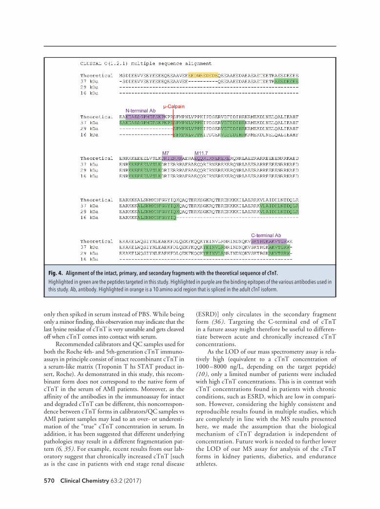
protein of interest (30 ). Great care should be taken wheninterpreting such quantification results as peptide selec-tion merely based on the optimal mass spectrometric re-sponse may not always be representative. We have shownhere, with relative quantification of the selected ion mon-itoring (SIM) measurements, that the 29-kDa fragmentis an N-terminally cleaved degradation product of cTnT.The 16-kDa secondary fragment is further degraded atthe C-terminus and is most likely cleaved between Gln-199 and Lys-200. This interpretation is summarized inFig. 4. With this knowledge, the absolute amount ofthese different fragments can now be determined by thedirect digestion of proteins in serum—for example, usinga strategy based on so-called “proteolytic signature pep-tides” (31 ).
The N-terminal cleavage of cTnT between Arg-78and Ser-79 has been described previously (6 ) and is sug-gested to be caused by �-calpain cleavage (32 ). However,it was recently shown that thrombin may also play a rolein this process (33 ). To test this hypothesis, the epitopeof the polyclonal N-terminal antibody designed for theWestern blots performed in this study was chosen to liedirectly adjacent to this hypothesized cleavage site (Fig.4). Our Western blotting and mass spectrometry resultsboth showed that this targeted region was removed in the29- and 16-kDa fragments. Another suggested cleavagesite, catalyzed by caspase-3, was claimed to lie betweenAsp-98 and Asp-99 (34 ). However, our results showedthat this site remains intact in all analyzed samples. Onthe C-terminal side, we had no prior indication on wherecleavage would occur (if at all). For this reason, theC-terminal antibody was designed to bind as far towardthe C-terminal end of cTnT as possible. Western blottingand mass spectrometry results were in agreement that theC-terminus of cTnT was being cleaved off in the second-ary fragments of AMI patients, but that it was still presentin the primary fragment band.
Similar results were found in an in vitro experi-ment where purified cTnT was spiked in a humanserum matrix and analyzed with this same MS assay(10 ). A few notable differences need to be discussed.First and foremost, the semitryptic peptide definingthe C-terminal end of the 16-kDa fragment(187ALSN. . .GYIQ199) was not detected in the invitro experiment. For this reason, the reference pep-tide chosen in this study was different from the oneused in the validation study, which covered this site.This may suggest that, while similar, the in vitro ex-periment performed previously (10 ) was not com-pletely comparable to the various processes occurringin vivo (such as the potential intracellular degradationof troponin). Lastly, in the in vitro experiment, thesemitryptic peptide 291AKVTGRW297 was also de-tected. This was an interesting observation because thesame positive QC was used as in the current study,
Identification and Characterization of cTnT in Serum
Clinical Chemistry 63:2 (2017) 569
only then spiked in serum instead of PBS. While beingonly a minor finding, this observation may indicate that thelast lysine residue of cTnT is very unstable and gets cleavedoff when cTnT comes into contact with serum.
Recommended calibrators and QC samples used forboth the Roche 4th- and 5th-generation cTnT immuno-assays in principle consist of intact recombinant cTnT ina serum-like matrix (Troponin T hs STAT product in-sert, Roche). As demonstrated in this study, this recom-binant form does not correspond to the native form ofcTnT in the serum of AMI patients. Moreover, as theaffinity of the antibodies in the immunoassay for intactand degraded cTnT can be different, this noncorrespon-dence between cTnT forms in calibrators/QC samples vsAMI patient samples may lead to an over- or underesti-mation of the “true” cTnT concentration in serum. Inaddition, it has been suggested that different underlyingpathologies may result in a different fragmentation pat-tern (6, 35 ). For example, recent results from our lab-oratory suggest that chronically increased cTnT [suchas is the case in patients with end stage renal disease
(ESRD)] only circulates in the secondary fragmentform (36 ). Targeting the C-terminal end of cTnTin a future assay might therefore be useful to differen-tiate between acute and chronically increased cTnTconcentrations.
As the LOD of our mass spectrometry assay is rela-tively high (equivalent to a cTnT concentration of1000–8000 ng/L, depending on the target peptide)(10 ), only a limited number of patients were includedwith high cTnT concentrations. This is in contrast withcTnT concentrations found in patients with chronicconditions, such as ESRD, which are low in compari-son. However, considering the highly consistent andreproducible results found in multiple studies, whichare completely in line with the MS results presentedhere, we made the assumption that the biologicalmechanism of cTnT degradation is independent ofconcentration. Future work is needed to further lowerthe LOD of our MS assay for analysis of the cTnTforms in kidney patients, diabetics, and enduranceathletes.
Fig. 4. Alignment of the intact, primary, and secondary fragments with the theoretical sequence of cTnT.Highlighted in green are the peptides targeted in this study. Highlighted in purple are the binding epitopes of the various antibodies used inthis study. Ab, antibody. Highlighted in orange is a 10 amino acid region that is spliced in the adult cTnT isoform.
570 Clinical Chemistry 63:2 (2017)

In conclusion, with 2 different proteomic ap-proaches (Western blotting and mass spectrometry),we have shown that cTnT in the serum of patientssuffering from AMI consists of a heterogeneous mix-ture of intact cTnT (37 kDa), a primary fragmentspanning from Ser-79 to Trp-297 (29 kDa), and sev-eral smaller secondary fragments (15–20 kDa) ofwhich the most abundant fragment (16-kDa) spansfrom Ser-79 to Gln-199.
Author Contributions: All authors confirmed they have contributed tothe intellectual content of this paper and have met the following 3 require-ments: (a) significant contributions to the conception and design, acquisi-tion of data, or analysis and interpretation of data; (b) drafting or revisingthe article for intellectual content; and (c) final approval of the publishedarticle.
Authors’ Disclosures or Potential Conflicts of Interest: Upon man-uscript submission, all authors completed the author disclosure form. Dis-closures and/or potential conflicts of interest:
Employment or Leadership: D. de Boer, Central Diagnostic Labora-tory, MUMC�.Consultant or Advisory Role: None declared.Stock Ownership: None declared.Honoraria: None declared.Research Funding: Roche Diagnostics provided the monoclonal M7antibody for use in Western blotting experiments. M.P. van Dieijen-Visser, Stichting de Weijerhorst.Expert Testimony: None declared.Patents: None declared.
Role of Sponsor: The funding organizations played no role in thedesign of study, choice of enrolled patients, review and interpretation ofdata, and final approval of manuscript.
Acknowledgments: The authors are grateful to Vincent Kleijnen andRonny Mohren for their technical assistance.
References
1. Thygesen K, Alpert JS, Jaffe AS, Simoons ML, ChaitmanBR, White HD, et al. Third universal definition of myo-cardial infarction. J Am Coll Cardiol 2012;60:1581–98.
2. Amsterdam EA, Wenger NK, Brindis RG, Casey DE Jr,Ganiats TG, Holmes DR Jr, et al. 2014 AHA/ACC Guide-line for the Management of Patients with Non-ST-Elevation Acute Coronary Syndromes: a report of theAmerican College of Cardiology/American Heart Associ-ation Task Force on Practice Guidelines. J Am Coll Car-diol 2014;64:e139 –228.
3. Roffi M, Patrono C, Collet JP, Mueller C, Valgimigli M,Andreotti F, et al. 2015 ESC Guidelines for the manage-ment of acute coronary syndromes in patients present-ing without persistent ST-segment elevation: Task Forcefor the Management of Acute Coronary Syndromes inPatients Presenting without Persistent ST-Segment Ele-vation of the European Society of Cardiology (ESC). EurHeart J 2016;37:267–315.
4. Collinson P, Pulkki K, Suvisaari J, Ravkilde J, Stavljenic-Rukavina A, Hammerer-Lercher A, et al. How well dolaboratories follow guidelines on cardiac markers? Thecardiac marker guideline uptake in Europe study. ClinChem 2008;54:448 –9.
5. Michielsen EC, Diris JH, Kleijnen VW, Wodzig WK, VanDieijen-Visser MP. Investigation of release and degra-dation of cardiac troponin T in patients with acute myo-cardial infarction. Clin Biochem 2007;40:851–5.
6. Cardinaels EP, Mingels AM, van Rooij T, Collinson PO,Prinzen FW, van Dieijen-Visser MP. Time-dependentdegradation pattern of cardiac troponin T followingmyocardial infarction. Clin Chem 2013;59:1083–90.
7. Bates KJ, Hall EM, Fahie-Wilson MN, Kindler H, Bailey C,Lythall D, Lamb EJ. Circulating immunoreactive cardiactroponin forms determined by gel filtration chromatog-raphy after acute myocardial infarction. Clin Chem2010;56:952– 8.
8. Fahie-Wilson MN, Carmichael DJ, Delaney MP, StevensPE, Hall EM, Lamb EJ. Cardiac troponin T circulates inthe free, intact form in patients with kidney failure. ClinChem 2006;52:414 –20.
9. Biener M, Katus HA, Giannitsis E. Challenges of serialtroponin testing: a symphony in need for harmony. IntJ Cardiol 2013;168:4542.
10. Streng AS, de Boer D, Bouwman FG, Mariman EC,Scholten A, van Dieijen-Visser MP, Wodzig WK. Devel-opment of a targeted selected ion monitoring assay for
the elucidation of protease induced structural changesin cardiac troponin T. J Proteomics 2016;136:123–32.
11. Michielsen EC, Diris JH, Hackeng CM, Wodzig WK, VanDieijen-Visser MP. Highly sensitive immunoprecipita-tion method for extracting and concentrating low-abundance proteins from human serum. Clin Chem2005;51:222– 4.
12. MacLean B, Tomazela DM, Shulman N, Chambers M,Finney GL, Frewen B, et al. Skyline: an open source doc-ument editor for creating and analyzing targeted pro-teomics experiments. Bioinformatics 2010;26:966 – 8.
13. Schilling B, Rardin MJ, MacLean BX, Zawadzka AM, Fre-wen BE, Cusack MP, et al. Platform-independent andlabel-free quantitation of proteomic data using MS1 ex-tracted ion chromatograms in skyline: application toprotein acetylation and phosphorylation. Mol Cell Pro-teomics 2012;11:202–14.
14. Vizcaino JA, Cote RG, Csordas A, Dianes JA, Fabregat A,Foster JM, et al. The PRoteomics IDEntifications (PRIDE)database and associated tools: status in 2013. NucleicAcids Res 2013;41:D1063–9.
15. Streng AS, de Boer D, Bouwman FG, Mariman EC,Scholten A, van Dieijen-Visser MP, Wodzig WK. Valida-tion, optimisation, and application data in support ofthe development of a targeted selected ion monitoringassay for degraded cardiac troponin T. Data Brief 2016;7:397– 405.
16. Olsen JV, Ong SE, Mann M. Trypsin cleaves exclusivelyC-terminal to arginine and lysine residues. Mol CellProteomics 2004;3:608 –14.
17. Siepen JA, Keevil EJ, Knight D, Hubbard SJ. Predictionof missed cleavage sites in tryptic peptides aids proteinidentification in proteomics. J Proteome Res 2007;6:399 – 408.
18. Katrukha AG, Bereznikova AV, Filatov VL, Esakova TV,Kolosova OV, Pettersson K, et al. Degradation of cardiactroponin I: implication for reliable immunodetection.Clin Chem 1998;44:2433– 40.
19. Jarolim P. High sensitivity cardiac troponin assays inthe clinical laboratories. Clin Chem Lab Med 2015;53:635–52.
20. Wu AH, Feng YJ, Moore R, Apple FS, McPherson PH,Buechler KF, Bodor G. Characterization of cardiac tro-ponin subunit release into serum after acute myocar-dial infarction and comparison of assays for troponin Tand I. American Association for Clinical Chemistry Sub-
committee on cTnI Standardization. Clin Chem 1998;44:1198 –208.
21. Labugger R, Organ L, Collier C, Atar D, Van Eyk JE. Ex-tensive troponin I and T modification detected in serumfrom patients with acute myocardial infarction. Circula-tion 2000;102:1221– 6.
22. Mingels AM, Cobbaert CM, de Jong N, van den Hof WF,van Dieijen-Visser MP. Time- and temperature-dependentstability of troponin standard reference material 2921 inserum and plasma. Clin Chem Lab Med 2012;50:1681–4.
23. Hessel MH, Michielsen EC, Atsma DE, Schalij MJ, vander Valk EJ, Bax WH, et al. Release kinetics of intact anddegraded troponin I and T after irreversible cell dam-age. Exp Mol Pathol 2008;85:90 –5.
24. Streng AS, Jacobs LH, Schwenk RW, Cardinaels EP,Meex SJ, Glatz JF, et al. Cardiac troponin in ischemiccardiomyocytes: Intracellular decrease before onset ofcell death. Exp Mol Pathol 2014;96:339 – 45.
25. Kumar S, Ali W, Bhattacharya S, Verma AK. The effect ofelapsed time on cardiac troponin-T (cTnT) degradationand its dependency on the cause of death. J ForensicLeg Med 2016;40:16 –21.
26. Labugger R, Simpson JA, Quick M, Brown HA, CollierCE, Neverova I, Van Eyk JE. Strategy for analysis of car-diac troponins in biological samples with a combina-tion of affinity chromatography and mass spectrometry.Clin Chem 2003;49:873–9.
27. Hoofnagle AN, Whiteaker JR, Carr SA, Kuhn E, Liu T,Massoni SA, et al. Recommendations for the genera-tion, quantification, storage, and handling of peptidesused for mass spectrometry-based assays. Clin Chem2016;62:48 – 69.
28. Kuster B, Schirle M, Mallick P, Aebersold R. Scoring pro-teomes with proteotypic peptide probes. Nat Rev MolCell Biol 2005;6:577– 83.
29. Steen H, Mann M. The ABC’s (and XYZ’s) of peptide se-quencing. Nat Rev Mol Cell Biol 2004;5:699 –711.
30. van den Broek I, Romijn FP, Nouta J, van der Laarse A,Drijfhout JW, Smit NP, et al. Automated multiplex LC-MS/MS assay for quantifying serum apolipoproteinsA-I, B, C-I, C-II, C-III, and E with qualitative apolipopro-tein E phenotyping. Clin Chem 2016;62:188 –97.
31. Fahlman RP, Chen W, Overall CM. Absolute proteomicquantification of the activity state of proteases and pro-teolytic cleavages using proteolytic signature peptidesand isobaric tags. J Proteomics 2014;100:79 –91.
Identification and Characterization of cTnT in Serum
Clinical Chemistry 63:2 (2017) 571

32. Zhang Z, Biesiadecki BJ, Jin JP. Selective deletion of theNH2-terminal variable region of cardiac troponin T inischemia reperfusion by myofibril-associated mu-calpain cleavage. Biochemistry 2006;45:11681–94.
33. Streng AS, De Boer D, Van Doorn WP, Kocken JM, Bek-ers O, Wodzig WK. Cardiac troponin T degradation inserum is catalysed by human thrombin. Biochem Bio-
phys Res Commun 2016;481(1–2):165– 8.34. Communal C, Sumandea M, de Tombe P, Narula J, So-
laro RJ, Hajjar RJ. Functional consequences of caspaseactivation in cardiac myocytes. Proc Natl Acad Sci U S A2002;99:6252– 6.
35. Diris JH, Hackeng CM, Kooman JP, Pinto YM, HermensWT, van Dieijen-Visser MP. Impaired renal clearance ex-
plains elevated troponin T fragments in hemodialysispatients. Circulation 2004;109:23–5.
36. Mingels A, Cardinaels E, Broers N, Van Sleeuwen A,Streng A, van Dieijen-Visser M, et al. Cardiac troponin T:smaller molecules in patients with end-stage renal diseasethan after onset of acute myocardial infarction. Clin Chem[Epub ahead of print 2017 Jan 10].
572 Clinical Chemistry 63:2 (2017)