identification of a novel 82 kda prommp-9 species ... · containing 50mm tris/hcl (ph 7.5), 200mm...
TRANSCRIPT

Biochem. J. (2007) 405, 547–558 (Printed in Great Britain) doi:10.1042/BJ20070191 547
Identification of a novel 82 kDa proMMP-9 species associated with thesurface of leukaemic cells: (auto-)catalytic activation and resistance toinhibition by TIMP-1Christian RIES*1, Thomas PITSCH*, Reinhard MENTELE*, Stefan ZAHLER†, Virginia EGEA*, Hideaki NAGASE‡and Marianne JOCHUM**Division of Clinical Chemistry and Clinical Biochemistry in the Surgical Department, Ludwig-Maximilians-University of Munich, 80336 Munich, Germany, †Pharmaceutical Biology,Center of Drug Research, Ludwig-Maximilians-University of Munich, 81377 Munich, Germany, and ‡Kennedy Institute of Rheumatology, Imperial College London, London W6 8LH, U.K.
MMP-9 (matrix metalloproteinase 9) plays a critical role intumour progression. Although the biochemical properties ofthe secreted form of proMMP-9 are well characterized, littleis known about the function and activity of cell surface-asso-ciated proMMP-9. We purified a novel 82 kDa species ofproMMP-9 from the plasma membrane of THP-1 leukaemiccells, which has substantial differences from the secreted 94 kDaproMMP-9. The 82 kDa form was not detected in the mediumeven upon stimulation with a phorbol ester. It is truncatedby nine amino acid residues at its N-terminus, lacks O-linkedoligosaccharides present in the 94 kDa proMMP-9, but retainsN-linked carbohydrates. Incubation of 94 kDa proMMP-9 withMMP-3 generated the well-known 82 kDa active form, but the82 kDa proMMP-9 was converted into an active species of 35 kDa,which was also produced by autocatalytic processing in theabsence of activating enzymes. The activated 35 kDa MMP-9 effi-
ciently degraded gelatins, native collagen type IV and fibronectin.The enzyme was less sensitive to TIMP-1 (tissue inhibitorof metalloproteinase 1) inhibition with IC50 values of 82 nMcompared with 1 nM for the 82 kDa active MMP-9. The syntheticMMP inhibitor GM6001 blocked the activity of both enzymes,with similar IC50 values below 1 nM. The 82 kDa proMMP-9 isalso produced in HL-60 and NB4 leukaemic cell lines as wellas ex vivo leukaemic blast cells. It is, however, absent fromneutrophils and mononuclear cells isolated from peripheral bloodof healthy individuals. Thus, the 82 kDa proMMP-9 expressed onthe surface of malignant cells may escape inhibition by naturalTIMP-1, thereby facilitating cellular invasion in vivo.
Key words: acute myeloid leukaemia, gelatinase B, glycosylation,matrix metalloproteinase (MMP), tissue inhibitor of metallo-proteinase (TIMP), tumour cell invasion.
INTRODUCTION
MMPs (matrix metalloproteinases) represent a family of struc-turally and functionally related zinc-dependent endopeptidasescapable of digesting extracellular matrix components [1–4].In addition to their degradative function, they have abilitiesto process regulatory proteins, including cytokines, growthfactors or their receptors [3]. Unbalanced MMP activities areinvolved in numerous pathological events, such as tumour growthand metastasis, arthrosis, arthritis, nephritis, neurodegenerativedisease and fibrosis [2,4]. Production and activity of MMPs incells is controlled precisely at the level of gene expression andis modulated by a variety of stimuli (e.g. cytokines, hormonesor extracellular matrix proteins), by activation of the latentproenzymes, and by inhibition by endogenous TIMPs (tissueinhibitors of metalloproteinases) [5].
Two members of the MMP family, gelatinase A (MMP-2) andgelatinase B (MMP-9), are subclassified for their preferentialabilities to degrade denatured collagens (gelatin) and collagentype IV, the main component of basement membranes. Moreover,these enzymes have the potential to process TGF-β (transforminggrowth factor β) [6], interleukin 8 [7], and interleukin 1β [8]into their biologically active forms. In contrast to MMP-2, whichis often expressed in a constitutive manner, MMP-9 synthesisand secretion are regulated, and in most cell types it occurs only
upon stimulation. Although MMP-9 production can be observedin diverse cell types, its synthesis in cells of haematopoieticorigin, such as neutrophil granulocytes [9], monocytes [10] andmacrophages [11], appears as major source of MMP-9 in humans.Leukocytes that infiltrate neoplastic tissue have been shown toact as co-conspirators of carcinogenesis by providing the tumourwith MMP-9 [12]. Moreover, in acute myeloid leukaemia, bothconstitutive and stimulated release of MMP-9 is observed invarious cell lines and ex vivo blast cells [13–17], indicating thatthis enzyme is also involved in leukaemic cell dissemination [18].Direct evidence for a crucial role of MMP-9 in the invasionand metastatic capacity of tumour cells has been obtained bytransfection and ribozyme-based approaches [19,20], suggestingMMP-9 as a preferential target in the development of anti-cancerdrugs.
MMP-9 is synthesized as a pre-proenzyme and is secretedfrom the cells in a glycosylated proenzyme form [21] with amolecular mass varying from 91–96 kDa depending on the celltype [9,10,13,21,22]. ProMMP-9 may form a tight complex withTIMP-1 [21], which influences both activation and activity ofthis enzyme. In vitro activation of latent MMP-9 is achievedby incubation with organomercurials or several proteinases [23].Under physiological conditions, a proteolytic cascade involvingMMP-3 or the plasminogen activator/plasmin system is postulatedto convert proMMP-9 into its active form [24,25].
Abbreviations used: APMA, 4-aminophenylmercuric acetate; ECL, enhanced chemiluminescence; MALDI–TOF-MS, matrix-assisted laser-desorptionionization–time-of-flight MS; MMP, matrix metalloproteinase; PMCA-ATPase, plasma membrane-localized calcium-dependent ATPase; TBS, Tris-bufferedsaline; TGF-β, transforming growth factor β; TIMP, tissue inhibitor of metalloproteinase.
1 To whom correspondence should be addressed (email [email protected]).
c© The Authors Journal compilation c© 2007 Biochemical Society

548 C. Ries and others
Cell surface association of MMP-9 has been documented invarious cell types, including epithelial cells [26], endothelial cells[27], neutrophil granulocytes [28,29], monocytes [30] and tumourcells [6,24,31–34]. Binding of MMP-9 to the plasma membraneis mediated by interaction of the enzyme with a distinct array ofsurface molecules [35,36], including CD44 [6,31,34], β1-integrins[27] and the α2 chain of collagen IV [33]. MMP-9 co-localizedwith CD44 on the surface of neoplastic cells was demonstratedto activate latent TGF-β, thereby promoting tumour invasion,growth and angiogenesis [34]. MMP-9 can also induce tumour-associated angiogenesis by release of vascular endothelial growthfactor trapped in the extracellular matrix [37]. Thus, MMP-9seems to be of relevance in carcinogenesis by activating andliberating tumour promoting factors from the cancer-cell surfaceand surrounding extracellular matrix [38]. Nevertheless, it is stillunclear how the enzymatic activity of MMP-9 is controlled at thepericellular space.
In the present study, we identified a novel non-secreted 82 kDaproMMP-9 species localized at the surface of leukaemic cells.This particular zymogen form has not been recognized previouslyas an independent enzyme, probably due to its high similarity to‘regular’ proMMP-9, which after secretion is able to re-associateto the cell membrane. Comparative analysis on purified enzymesshowed clearly structural and functional differences between thetwo proMMP-9 species, suggesting a specific role for the 82 kDaproMMP-9 in surface-associated proteolysis of leukaemic cells.
EXPERIMENTAL
Cell culture
The cell lines THP-1 (acute monocytic leukaemia), HL-60(acute myeloblastic leukaemia), NB4 (acute promyelocyticleukaemia) and HT1080 fibrosarcoma cells were purchased fromthe German Collection of Microorganisms and Cell Cultures(DSMZ, Braunschweig, Germany). Cells were grown in RPMI-1640 supplemented with 10 % (v/v) heat-inactivated fetal calfserum, 2 mM glutamine, 100 units/ml penicillin and 100 µg/mlstreptomycin. Incubation was performed at 37 ◦C in a humidifiedair atmosphere in the presence of 5% CO2. Cells were passagedtwice a week. For all further experiments cells were washed twotimes with serum-free medium and maintained under serum-free conditions in RPMI-1640 supplemented with 1% (v/v)Nutridoma SP (Roche Applied Science, Mannheim, Germany).Mononuclear cells and polymorphonuclear cells/neutrophilgranulocytes were isolated from peripheral blood of healthyvolunteers, and leukaemic blast cells from peripheral blood ofthree patients with acute myeloid leukaemia by Percoll (Sigma,Munich, Germany) density gradient centrifugation as describedpreviously [16]. All samples were collected at the UniversityHospital of Munich with consent of the persons involved. Purifiedcell fractions were washed twice and resuspended in serum-free medium for subsequent experiments. Cell viability wasdetermined using the Trypan Blue exclusion test.
Phorbol ester stimulation and preparation of crude cell extracts
For stimulation experiments, cells were cultured at a density of 1 ×10⁶ cells/ml either in serum-free medium alone or in mediumcontaining 50 ng/ml PMA for 24 h. Thereafter, non-adherent cellswere separated from culture supernatants by centrifugation at400 g for 15 min at 4 ◦C. Adherent cells were scraped to removethem from the culture dish. The conditioned media were stored at−20 ◦C. Cell pellets were washed three-times in cold TBS (Tris-buffered saline; 50 mM Tris/HCl, pH 7.4, and 150 mM NaCl). For
lysis and protein extraction, cells were resuspended at a density of1 × 107 cells/ml in lysis buffer [TBS containing 1 % (v/v) TritonX-100]. To prevent (auto-)proteolytic degradation, 10 mM EDTAand a mixture of inhibitors of metallo-, serine- and cysteine-proteinases (Complete-Mini, Roche Applied Science) was addedto the lysis buffer. The cell suspension was vortexed and incubatedfor 30 min at 4 ◦C. Subsequently, the supernatants were collectedby centrifugation at 16000 g for 15 min at 4 ◦C and stored at−20 ◦C. For zymography or immunoblot analysis, gelatinasespresent in the cell extracts were enriched by gelatin–Sepharosechromatography as described below.
Zymographic analysis and quantification of gelatinasesin zymograms
Zymography was performed in precast 10 % polyacrylamideminigels containing 0.1% gelatin as substrate (Invitrogen,Groningen, The Netherlands). Samples were run under non-reducing conditions without prior boiling. Conditioned mediumfrom PMA-treated HT1080 fibrosarcoma cells containingproMMP-9 (94 kDa), proMMP-2 (72 kDa) and activated MMP-2(66 kDa) [22] was used as marker for electrophoretic mobilityof gelatinases in zymograms. After electrophoresis, gels werewashed twice for 15 min in 2.7% (v/v) Triton X-100 (dilutedin water) on a rotary shaker to remove SDS and to allowproteins to renature. The gels were then incubated in a buffercontaining 50 mM Tris/HCl (pH 7.5), 200 mM NaCl, 5 mMCaCl2 and 0.2% Brij35 (Invitrogen) for 18 h at 37 ◦C. Thezymograms were stained for 90 min with 0.02% CoomassieBlue R-350 in a 30% (v/v) methanol/10% (v/v) acetic acidsolution by the use of PhastGel-Blue-R tablets (GE HealthcareLife Sciences, Freiburg, Germany). Quantification of gelatinolyticactivity in zymograms was performed as described previously[39]. Zymograms were scanned using an Umax ImageScannerdriven by the MagicScan software and analysed using theImageMaster-1D Elite quantification software (GE HealthcareLife Sciences). The linear range of the assay was determinedby the use of recombinant MMP-9 (Calbiochem, Schwalbach,Germany) as calibration standard.
SDS/PAGE and immunoblot analysis
SDS/PAGE was performed under reducing conditions inprecast 4–12% (w/v) polyacrylamide minigels (Invitrogen).For electrophoresis and blotting the NuPAGE Bis/Tris system(Invitrogen) was used according to the manufacturer’sinstructions. After electrophoretic separation, proteins wereeither stained with silver according to a method described byHeukeshoven and Dernick [40] or were electroblotted on to PVDFmembranes (Millipore, Bedford, MA, U.S.A.). The membraneswere blocked in 10% (w/v) non-fat milk in TBS for 1 h at roomtemperature (21 ◦C). Thereafter, the blots were incubated with0.5 µg/ml primary rabbit polyclonal antibody against proMMP-9 (M-5177; Sigma) for 1 h at room temperature, which allowsdetection of both latent and activated enzyme forms. A mousemonoclonal antibody (IM09L, clone 6-6B; Calbiochem) that onlyrecognizes the latent proMMP-9 and not the activated speciesunder reducing conditions for immunoblotting was used at aconcentration of 0.5 µg/ml. After washing, blots were incubatedfor 15 min with horseradish-peroxidase-conjugated anti-rabbitIgG (GE Healthcare Life Sciences) as secondary antibody at adilution of 1:1000. The blots were then developed applying theECL (enhanced chemiluminescence) system as recommended bythe manufacturer (GE Healthcare Life Sciences). For molecularmass determination of proteins detected on the blots, recombinantprotein standards (Invitrogen and Bio-Rad, Munich, Germany)
c© The Authors Journal compilation c© 2007 Biochemical Society
Cell surface-associated 82 kDa proMMP-9 549
were used. The developed films were scanned and analysed usingthe ImageMaster-1D Elite software.
Confocal laser scanning microscopy
THP-1 cells were grown on 8-well CultureSlides (BD Falcon,Heidelberg, Germany) for 24 h under serum-free conditions inthe presence of 50 ng/ml PMA. The cells were washed, andthen fixed in 3.7% (v/v) formaldehyde in PBS for 15 min atroom temperature. The fixed cells were blocked with 2% (w/v)BSA in PBS for 15 min, and then incubated with 50 µg/ml ofmonoclonal anti-MMP-9 antibody (GE-213, NeoMarkers, UnionCity, CA, U.S.A.) or with 50 µg/ml of mouse IgG1 isotypecontrol antibody (DAKO, Hamburg, Germany) for 1 h at 37 ◦C.The secondary fluorescence-conjugated antibody used was AlexaFluor® 488 anti-mouse IgG (Molecular Probes, Eugene, OR,U.S.A.). Detection was carried out at an excitation wavelengthof 488 nm, beam splitter HFT 488, and emission at 505–530 nmon a Zeiss LSM 510 confocal scanning microscope equipped witha Plan-Neofluar 100×/1.3 oil immersion objective. Each imagerepresents a single section with a field depth of 1 µm.
Subcellular fractionation and isolation of plasma membranes
THP-1 cells (1 × 10⁶/ml) were cultured for 24 h under serum-free conditions in the presence of 50 ng/ml PMA. Cells wereharvested and washed three times with ice-cold PBS. All ofthe following procedures were carried out at 4 ◦C according toa method described previously [26] with minor modifications.Cells were resuspended at 2 × 10⁷/ml in buffer containing 25 mMTris/HCl (pH 7.5), 8.5 % (w/v) sucrose, 50 mM NaCl andproteinase inhibitors (Complete-Mini, Roche Applied Science)at concentrations recommended by the manufacturer. The cellsuspension was homogenized using a Dounce homogenizer andcentrifuged for 10 min at 3000 g. The pellet (nuclear fraction)was discarded and the supernatant (postnuclear fraction) wascentrifuged at 25000 rev./min for 3 h in a Kontron ultracentrifugewith a TFT 28/38 rotor (Kendro, Hanau, Germany). Thesupernatant (cytosolic fraction) was collected and stored at−20 ◦C. The pellet (crude membrane fraction) was resuspendedin buffer containing 25 mM Tris/HCl (pH 7.5), 50 mM NaCland proteinase inhibitors (Complete-Mini). The sample waslayered on to a discontinuous density gradient of 20, 30, 50and 60% sucrose in water and once more centrifuged at25000 rev./min for 3 h. The 30–50% interphase was carefullyaspirated, resuspended in buffer containing 25 mM Tris/HCl(pH 7.5), 50 mM NaCl and 5 mM EDTA and centrifuged at25000 rev./min for a further 3 h. The pellet (enriched plasmamembrane fraction) was resuspended in the same buffer and storedat −70 ◦C. The successful enrichment of plasma membraneswas verified by immunoblot detection of PMCA-ATPase(plasma membrane-localized calcium-dependent ATPase) usingmonoclonal antibodies against this enzyme (Affinity Bioreagents,Golden, CO, U.S.A.).
Cell surface protein biotinylation and immunoprecipitation
Labelling and detection of proteins localized on the surfaceof THP-1 cells was performed by using the ECL proteinbiotinylation module (GE Healthcare Life Sciences) accordingto the manufacturer’s instructions. Cells (2 × 10⁶/ml) were grownunder serum-free conditions in the presence of 50 ng/ml PMA for24 h. Cells were harvested by centrifugation at 600 g at 4 ◦C,and then washed twice with ice-cold PBS. The cell viabilitywas � 95%, as determined by Trypan Blue staining. Cellswere then diluted to 1 × 10⁷/ml in PBS and incubated with40 µl/ml of the water-soluble and cell impermeable biotinylation
reagent (biotinamidocaproate N-hydroxysuccinamide ester) for15 min at 4 ◦C on a roller mixer. Control cells receivedvehicle only. After incubation, cells were washed twice inPBS and extracted with Triton X-100 as described above.For gelatinase enrichment, cell extracts were passed through agelatin–Sepharose affinity column (see below). Elution fractionscontaining gelatinolytic activity were pooled, dialysed against50 mM Tris/HCl (pH 7.5), 0.3 % Triton X-100 and 5 mM EDTA,and concentrated using Microcon YM10 devices (Millipore).For immunoprecipitation, enriched gelatinase samples were thenincubated with 9 µg/ml of monoclonal anti-MMP-9 antibody(GE-213, NeoMarkers) and Protein G–Sepharose beads overnightat 4 ◦C under constant shaking. The Protein G–Sepharosebeads were washed three times with ice-cold PBS, resuspendedin reducing electrophoresis sample buffer (141 mM Tris/HCl,pH 8.5, 1 M glycerol, 73 mM lithium dodecyl sulfate, 0.5 mMEDTA and 20 mg/ml dithiothreitol), boiled, and centrifugedat 16000 g for 1 min at 4 ◦C. The supernatant containingimmunoprecipitated antigens was separated by SDS/PAGE (4–12% gels) under reducing conditions, and blotted as describedabove. After blocking and washing of the membrane, detectionof biotinylated proteins was achieved by incubation withstreptavidin-conjugated horseradish peroxidase (diluted 1:1500)and detection with ECL. The specificity of detection wascontrolled by processing non-biotinylated cells in the same wayas described for biotinylated cells.
Gelatin–Sepharose affinity chromatography and gel filtration
Purification of MMP-9 was performed as described previously byus [41] with some modifications. All procedures were carried outat 4 ◦C with buffers containing proteinase inhibitors (Complete-Mini) and 5 mM EDTA to avoid autoproteolytic processing.
THP-1 cell-associated MMP-9 forms were purified from crudecell extracts (0.1–0.5 ml) or postnuclear fractions (40 ml) byTriton X-100 extraction with vigorous vortexing and incubationon ice for 30 min. After centrifugation at 3000 g for 15 min,the supernatant was applied to a gelatin–Sepharose columnof 2 ml bed vol. equilibrated with 50 mM Tris/HCl (pH 7.5),400 mM NaCl, 5 mM EDTA and 0.1% Brij 35 at a flow rateof 0.03 ml/min using the FPLC system (GE Healthcare LifeSciences). After the column had been washed in the same buffer,gelatin-binding proteins were eluted by 5% (v/v) DMSO incolumn buffer. Gelatinase containing fractions were assessedby zymography, pooled and concentrated by approx. 10-fold(Centricon YM10, Millipore). For separation of 82 kDa and94 kDa MMP-9 forms, the sample (0.5 ml) was then applied toa Superdex G-200 gel filtration column (1.6 cm × 60 cm; GEHealthcare Life Sciences) equilibrated with 50 mM Tris/HCl(pH 7.5), 150 mM NaCl, 5 mM EDTA and 0.02 % Brij 35at a constant flow of 0.4 ml/min. Fractions (1 ml) containing94 kDa MMP-9 or 82 kDa MMP-9 respectively, were pooled andconcentrated. Protein concentrations were determined using theCoomassie Plus Protein Assay Reagent (Pierce, Rockford, IL,U.S.A.). The enzyme preparations were stored at −20 ◦C.
The secreted 94-kDa species of proMMP-9 was purified fromculture supernatants of PMA-treated THP-1 cells. Conditionedmedium (1 litre) was adjusted to 5 mM EDTA and 0.02%Brij 35, and concentrated to a final volume of 50 ml usingan Amicon Diaflo apparatus fitted with a YM-10 membrane(Amicon, Beverly, MA, U.S.A.). Thereafter, the material wasprocessed by gelatin–Sepharose affinity chromatography andgel filtration as described above. Selective denaturation of co-purifying TIMP molecules was performed according to a methodpublished previously [42].
c© The Authors Journal compilation c© 2007 Biochemical Society

550 C. Ries and others
Microsequencing and MALDI–TOF-MS (matrix-assistedlaser-desorption ionization–time-of-flight MS)
Samples of purified gelatinases were separated by SDS/PAGE(10% gel) and electroblotted on to glass fibre membranes(Glassybond, Biometra, Gottingen, Germany) [43]. CoomassieBlue stained bands were cut out and sequenced in a 492cLC gasphase sequencer (Applied Biosystems, Foster City, CA, U.S.A.).For MS analysis, samples were separated by SDS/PAGE and theprotein bands were excised and digested directly in the gel withtrypsin as outlined by Eckerskorn and Lottspeich [44]. Peptidemass fingerprinting was performed according to Meister et al.[45] using a Bruker reflex III MALDI–TOF mass spectrometerequipped with a 337 nm nitrogen laser (Bruker-Franzen, Bremen,Germany).
Deglycosylation of purified proMMP-9 forms
Purified proMMP-9 preparations were digested by enzymesprovided by the Glycoprotein Deglycosylation Kit from Calbio-chem (San Diego, CA, U.S.A.) following the denaturing or non-denaturing standard protocols of the manufacturer. For Westernblot detection of deglycosylated proteins, samples (30 µl) weremixed with reaction buffer (250 mM PBS, pH 7.0) and denatur-ation solution [2 % (w/v) SDS and 1 M 2-mercaptoethanol] andheated at 100 ◦C for 5 min. After cooling to room temperature,Triton X-100 solution [15% (v/v) Triton X-100 diluted inwater] was added at a final concentration of 1 % to removefree SDS. Subsequently, samples were incubated with differentglycosidases alone or in combination in a final reaction volumeof 47.5 µl for 3 h at 37 ◦C. N-glycosidase F (2.5 units) wasadded to remove asparagine-linked (N-linked) oligosaccharides.Cleavage of serine/threonine-linked (O-linked) sugar residueswas achieved by simultaneous incubation with a combination ofendo-α-N-acetylgalactosaminidase (0.6 milliunits), α2-3,6,8,9-neuraminidase (1.25 milliunits), β-1,4-galactosidase (0.75 milli-units) and β-N-acetylglucosaminidase (2 milliunits) according tothe manufacturers instructions. Mobility shift analysis of digestedsamples was performed by SDS/PAGE and immunoblottingusing anti-MMP-9 antibodies as described above. To preserveenzymatic activity of gelatinases during the deglycosylationreaction, samples were incubated with glycosidase for 4 days at37 ◦C under non-reducing conditions and subsequently analysedby zymography.
Activation of purified proMMP-9 forms and reactionwith α2-macroglobulin
Purified 82 kDa proMMP-9 from THP-1 cell extracts and 94 kDaproMMP-9 from culture supernatants were diluted to a finalconcentration of 50 ng/ml in enzyme buffer containing 50 mMTris/HCl (pH 7.5), 150 mM NaCl, 5 mM CaCl2, 1 µM ZnCl2
and 0.02% Brij 35, and were treated with or without equimolaramounts of active recombinant MMP-3 prepared according tothe method of Suzuki et al. [46] at 37 ◦C in a total volume of250 µl. Aliquots were taken at different time intervals and storedon ice prior to analysis for gelatinolytic activity in zymograms. Foridentification of catalytically active gelatinase species, samplespre-treated with or without MMP-3 for 30 min at 37 ◦C werereacted with an excess (500 µg/ml) of α2-macroglubulin (RocheApplied Science) for 30 min at 37 ◦C and subsequently applied tozymographic analysis.
Cleavage of biological substrates
Purified 82 kDa proMMP-9 was autocatalytically activated byincubation in buffer containing 50 mM Tris/HCl (pH 7.5),150 mM NaCl, 5 mM CaCl2, 1 µM ZnCl2 and 0.02% Brij 35
for 3 h at 37 ◦C. Activation of the secreted 94 kDa proMMP-9species was performed by exposure to 1 mM APMA (4-amino-phenylmercuric acetate) for 2 h at 37 ◦C as described previously[41]. Successful conversion into their active forms was controlledby zymography. Native or denatured (by heating to 100 ◦C for3 min) collagen type IV (400 µg/ml) from Engelbreth–Holm–Swarm mouse sarcoma cells, 300 µg/ml denatured collagentype I (rat tail) and 100 µg/ml fibronectin (all from Sigma) wereincubated with or without equimolar amounts (1.5 µM) ofactivated enzymes for 48 h at 32 ◦C. The reaction mixtures werethen analysed for degradation products by SDS/PAGE followedby silver staining of the proteins.
MMP-9 inhibition studies
The active forms of 82 and 94 kDa proMMP-9 were assayed usinga fluorescence-labelled gelatin substrate (Molecular Probes). Foractivation 20 nM of purified enzymes were incubated with 1–5 nM of active recombinant MMP-3 for 1–3 h at 37 ◦C in a buffercontaining 50 mM Tris/HCl (pH 7.5), 150 mM NaCl, 30 mMCaCl2, 10 µM ZnCl2, and 0.02% Brij 35 (100 µl total volume).Complete conversion into active MMP-9 forms was controlled byzymography. For inhibition studies 1 nM of each activated enzymewas pre-incubated for 30 min at 37 ◦C with or without 0.1–100 nMof purified TIMP-1 (prepared as described previously [47]), 0.1–100 nM GM6001 (a broad-spectrum hydroxamic acid inhibitorof MMPs) (Calbiochem) or 1 mM 1,10-phenanthroline (Sigma)before the fluorescence-labelled substrate (1.5 µg/ml) was addedto a final volume of 200 µl. After 1 h incubation at 37 ◦C,substrate hydrolysis was measured using a Molecular DynamicsBiolumin 960 (GE Healthcare Life Sciences) fluorescencespectrophotometer with excitation and emission wavelengths setat 485 and 520 nm respectively.
RESULTS
Detection of secreted and cell-associated forms of MMP-9
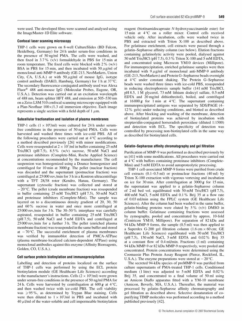
THP-1 cells were cultivated in the absence and presence ofPMA. The culture supernatants and cell lysates were analysedfor gelatinase activity by zymography and Western blotting. Theconditioned medium contained 94 kDa proMMP-9, expression ofwhich was enhanced considerably upon PMA stimulation (Fig-ure 1). An additional band migrating at the top of the zymogramgels represented the homodimer of proMMP-9 as observedunder non-reducing conditions of electrophoresis [48]. Theproduction of 72-kDa proMMP-2 was negligible in these cells(Figure 1A).
Zymographic analysis of cell lysates from untreated cellsrevealed the presence of gelatinolytic activity at 92–94 kDaconsisting of two single merging bands that were significantlyaugmented upon PMA treatment, which formed a major zoneof lysis between approx. 88 and 94 kDa (Figure 1A). Underreducing conditions, Western blot analysis using anti-MMP-9antibodies detected two distinct bands in cell lysates: a minorprotein with a molecular mass of 94 kDa, which presumablyrepresents intracellular proMMP-9 prior to secretion, and a major82 kDa form. Both MMP-9 species were augmented by PMAtreatment (Figure 1B). To assess whether the 82-kDa MMP-9was generated from 94-kDa proMMP-9 by the Triton X-100extraction procedure, conditioned medium comprising only the94-kDa enzyme was treated with extraction buffer for 18 h at37 ◦C, but no effect on 94 kDa proMMP-9 was observed (resultsnot shown), demonstrating that the 82 kDa form was not createdby the extraction technique.
c© The Authors Journal compilation c© 2007 Biochemical Society
Cell surface-associated 82 kDa proMMP-9 551
Figure 1 Detection of secreted and cell-associated MMP-9
THP-1 leukaemic cells (1 × 10⁶ cells/ml) were incubated under serum-free conditions for 24 hin the presence (+) or absence (−) of 50 ng/ml PMA. (A) Aliquots (3 µl) of THP-1 conditionedmedium (CM) and 10 µl aliquots of cell extract (EX) were analysed by zymography. (B) Forimmunoblot detection, 15 µl aliquots of gelatinases isolated from conditioned medium and30 µl aliquots of gelatinases from cell extracts containing 5–10 ng of protein were separated bySDS/PAGE under reducing conditions, blotted and probed with polyclonal antibodies directedagainst MMP-9. Molecular mass standards (M) were used as a reference, and their sizes aregiven in kDa to the left-hand and right-hand sides of (A) and (B) respectively.
Cell surface localization of MMP-9
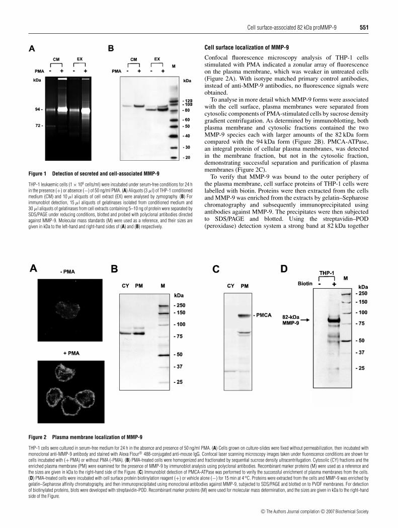
Confocal fluorescence microscopy analysis of THP-1 cellsstimulated with PMA indicated a zonular array of fluorescenceon the plasma membrane, which was weaker in untreated cells(Figure 2A). With isotype matched primary control antibodies,instead of anti-MMP-9 antibodies, no fluorescence signals wereobtained.
To analyse in more detail which MMP-9 forms were associatedwith the cell surface, plasma membranes were separated fromcytosolic components of PMA-stimulated cells by sucrose densitygradient centrifugation. As determined by immunoblotting, bothplasma membrane and cytosolic fractions contained the twoMMP-9 species each with larger amounts of the 82 kDa formcompared with the 94 kDa form (Figure 2B). PMCA-ATPase,an integral protein of cellular plasma membranes, was detectedin the membrane fraction, but not in the cytosolic fraction,demonstrating successful separation and purification of plasmamembranes (Figure 2C).
To verify that MMP-9 was bound to the outer periphery ofthe plasma membrane, cell surface proteins of THP-1 cells werelabelled with biotin. Proteins were then extracted from the cellsand MMP-9 was enriched from the extracts by gelatin–Sepharosechromatography and subsequently immunoprecipitated usingantibodies against MMP-9. The precipitates were then subjectedto SDS/PAGE and blotted. Using the streptavidin–POD(peroxidase) detection system a strong band at 82 kDa together
Figure 2 Plasma membrane localization of MMP-9
THP-1 cells were cultured in serum-free medium for 24 h in the absence and presence of 50 ng/ml PMA. (A) Cells grown on culture-slides were fixed without permeabilization, then incubated withmonoclonal anti-MMP-9 antibody and stained with Alexa Flour® 488-conjugated anti-mouse IgG. Confocal laser scanning microscopy images taken under fluorescence conditions are shown forcells incubated with (+PMA) or without PMA (-PMA). (B) PMA-treated cells were homogenized and fractionated by sequential sucrose density ultracentrifugation. Cytosolic (CY) fractions and theenriched plasma membrane (PM) were examined for the presence of MMP-9 by immunoblot analysis using polyclonal antibodies. Recombinant marker proteins (M) were used as a reference andthe sizes are given in kDa to the right-hand side of the Figure. (C) Immunoblot detection of PMCA-ATPase was performed to verify the successful enrichment of plasma membranes from the cells.(D) PMA-treated cells were incubated with cell surface protein biotinylation reagent (+) or vehicle alone (−) for 15 min at 4◦C. Proteins were extracted from the cells and MMP-9 was enriched bygelatin–Sepharose affinity chromatography, and then immunoprecipitated using monoclonal antibodies against MMP-9, subjected to SDS/PAGE and blotted on to PVDF membranes. For detectionof biotinylated proteins, blots were developed with streptavidin-POD. Recombinant marker proteins (M) were used for molecular mass determination, and the sizes are given in kDa to the right-handside of the Figure.
c© The Authors Journal compilation c© 2007 Biochemical Society

552 C. Ries and others
Figure 3 Purification of cell-associated and secreted MMP-9
Cell extracts and conditioned medium from PMA stimulated THP-1 cells were subjectedto gelatin–Sepharose affinity chromatography, followed by gel filtration chromatography.(A) Zymogram showing a representative analysis of fractions obtained by gel filtration separationof gelatin–Sepharose enriched material from cell extracts (G-Seph Pool). Fractions 9–13 con-taining the 94 kDa gelatinase (94k) and fractions 17–21 containing the 82 kDa gelatinase (82k)were pooled and concentrated. (B) and (C) Purified 82-kDa MMP-9 from cell extracts (82k) and94-kDa MMP-9 from conditioned medium (94k) were analysed by zymography (200 pg per lane)(B) and SDS/PAGE with subsequent silver staining (120 ng per lane) (C). HT1080 conditionedmedium containing MMP-9 (94 kDa), MMP-2 (72 kDa) and activated MMP-2 (66 kDa) wasused as a reference (M) in zymograms. Recombinant marker proteins (M) were applied formolecular mass determination in SDS/PAGE. Sizes are given in kDa to the left-hand side of (A)and the right-hand side of (B) and (C).
with a faint band of 94 kDa and additional weak proteinbands of low molecular masses were identified (Figure 2D).These results confirmed that the major MMP-9 detected on thesurface of THP-1 cells was the 82 kDa form.
Purification of cell-associated and secreted forms of MMP-9
To study the biochemical properties of the cell-associated 82-kDavariant in comparison with the secreted species of MMP-9, bothenzyme forms were purified from cell extracts and culture mediumof THP-1 cells treated with PMA. Gelatinases were first enrichedfrom crude cell extracts or the conditioned medium by gelatin–Sepharose affinity chromatography and were further purified bygel filtration chromatography, which separated the 82 kDa speciesfrom the 94 kDa form (Figure 3A). Analysis of the pooled andconcentrated fractions showed that the purified 82 kDa formmigrated with an apparent mass of approx. 90 kDa in zymograms(Figure 3B). This may be due to the absence of reducing agents inzymography, since under reducing conditions of SDS/PAGE withsubsequent silver staining of proteins it showed a single bandwith a molecular mass of 82 kDa (Figure 3C).
Microsequencing and MALDI–TOF-MS analysis of purifiedMMP-9 forms
As deduced from the molecular mass, we speculated that thecell-associated 82-kDa species could represent the activated formof mature MMP-9 lacking the N-terminal proenzyme domain.To address this question, the MMP-9 forms purified from cellextracts and conditioned media of PMA-stimulated THP-1cells were analysed by microsequencing and MS. Assessmentof the N-terminal sequence of the cell-associated 82 kDa variantrevealed a nine amino acid truncation for this enzyme comparedwith the regular proMMP-9 (Table 1). In contrast, secreted94 kDa proMMP-9 contained the complete N-terminus of thezymogen form (Table 1). Microsequencing of intracellular94 kDa proMMP-9 failed, which was possibly due to N-terminalblockage. Further analysis by MALDI–TOF-MS confirmed eachN-terminal sequence (Table 1). The tryptic peptides of boththe secreted and the cell-associated 94 kDa form of MMP-9showed the N-terminal peptide mass of 1345.7 Da, which agreedwith the predicted mass of that of the full-length proMMP-9.Mass mapping of the 82 kDa variant revealed a smaller fragmentof 916.5 Da, which matched the calculated molecular masslacking the nine N-terminal amino acids of proMMP-9 (Table 1).These results were in agreement with our findings obtained bymicrosequence analysis.
Studies on proMMP-9 glycosylation
Since proMMP-9 is known to be highly glycosylated [21], wehypothesized that alterations in the pattern of glycosylation might
Table 1 Sequencing and MALDI–TOF-MS analysis of purified MMP-9 forms
THP-1 cells (4.6 × 10⁹) were stimulated with 50 ng/ml of PMA for 24 h. Cell-associated 82- and 94-kDa gelatinases were isolated from cell extracts (EX) and secreted 94 kDa gelatinase fromconditioned medium (CM) by gelatin–Sepharose affinity chromatography. The samples were then subjected to SDS/PAGE under reducing conditions. For N-terminal microsequence analysis, proteinswere blotted and processed using a gas-phase sequencer. The N-terminal amino acid sequences obtained were compared with that of proMMP-9 [21]. For MS analysis, proteins were excised fromthe SDS-gel, digested with trypsin and analysed using a MALDI–TOF mass spectrometer. The molecular masses and the corresponding amino acid sequences of the N-terminal peptide fragmentgenerated by trypsin digestion of purified MMP-9 forms are shown, and compared with that of proMMP-9 [21]. n.d., not detected.
N-terminal peptide fragment after trypsin digestion
Molecular mass (Da)
Gelatinase N-terminal amino acid sequence Determined Calculated Amino acid sequence
1 10 20 6 17ProMMP-9 A P R Q R Q S T L V L F P G D L R T N L - 1345.7 Q S T L V L F P G D L R82 kDa (EX) V L F P G D - 916.5 916.5 V L F P G D L R94 kDa (EX) n.d. 1345.7 1345.7 Q S T L V L F P G D L R94 kDa (CM) A P R Q R Q S T L V L - 1345.7 1345.7 Q S T L V L F P G D L R
c© The Authors Journal compilation c© 2007 Biochemical Society

Cell surface-associated 82 kDa proMMP-9 553
Figure 4 Glycosylation studies
Aliquots of the purified secreted 94 kDa proMMP-9 (0.25 µg/ml) and the cell-associated82 kDa proMMP-9 (1 µg/ml) were reduced, denatured and then incubated for 3 h at 37◦Cwith either N-glycosidase F (N-GLYC) to remove N-linked oligosaccharides, with glycosidases(O-GLYC) that digest O-linked carbohydrates or with a combination of these enzymes. Shiftsin electrophoretical mobility were monitored by SDS/PAGE (4–12 % gels) under reducingconditions and subsequent immunoblotting using polyclonal antibodies against MMP-9. Resultsfrom a representative experiment of three independent determinations are shown. Recombinantmarker proteins (M) were used for molecular mass assessment and the sizes are shown in kDato the left-hand side of the Figure.
also account for the difference in molecular mass for the 82 and94 kDa species. Therefore the purified 82 and 94 kDa proMMP-9 forms were subjected to digestion with N-glycosidase F.Western blot analysis demonstrated that both forms of proMMP-9 were reduced in size by 6 kDa (Figure 4), indicating a similarextent of N-linked glycosylation for the two proenzymes. Whenthe proMMP-9 forms were incubated with glycosidases whichremove O-linked carbohydrates, the molecular mass of secreted94 kDa proMMP-9 was decreased by 9 kDa, whereas that ofthe 82 kDa variant remained unchanged (Figure 4). Removalof all sugar residues from the 82 and 94 kDa proMMP-9 usingboth N-linked and O-linked specific glycosidases yielded 76 and80 kDa proteins respectively (Figure 4). The molecular mass of the76 kDa protein is in accordance to that calculated from the aminoacid sequence of proMMP-9 [21], confirming that the 82 kDaproMMP-9 retains N-linked carbohydrates, but lacks O-linkedsugars, whereas the 94 kDa proMMP-9 contains both types ofglycan molecules.
Proteolytic activation of the 82 kDa proMMP-9
To investigate whether N-terminal truncation and lack of O-glycosylation influence activation of the 82-kDa proMMP-9, thepurified zymogen was incubated with stromelysin 1 (MMP-3),a potent physiological activator of proMMP-9, and the mode ofactivation was compared with that of the 94 kDa proMMP-9.As determined by zymography and Western blot analysis,treatment of the 94 kDa proMMP-9 with MMP-3 resulted incomplete conversion into a single 82 kDa MMP-9 within 1 h,with an intermediate form of approx. 86 kDa (Figure 5A), asdescribed by Ogata et al. [49]. In contrast, incubation of the 82 kDaproMMP-9 with MMP-3 initiated rapid conversion into a majorgelatinase with an apparent molecular mass of approx. 35 kDa,which migrated as a single protein of 35 kDa when analysed byWestern blotting under reducing conditions (Figure 5B). Owingto the high sensitivity of zymography additional bands withlower intensity ranging between approx. 25 and 40 kDa werealso detected, which may represent minor activation products(Figure 5B). Similar to the purified enzyme, 82 kDa proMMP-9present in plasma membrane fractions from THP-1 cells treatedwith MMP-3 led to the formation of 35 kDa gelatinase that was
stable for at least 6 h (Figure 5C). These findings indicate that the82 kDa proMMP-9, located in the plasma membrane, is differentfrom the regular secreted 94 kDa proMMP-9 in its susceptibilityto MMP-3 activation.
To examine the activity status of the different MMP-9 species,we utilized the property of the inhibitor α2-macroglobulin thatbinds only to active endopeptidases [50]. The 94 kDa proMMP-9pre-incubated with α2-macroglobulin and subsequently subjectedto zymography showed no significant decrease in gelatinolyticactivity, whereas for the activated 82 kDa species bound to α2-macroglobulin the majority of the activity had shifted to the topof the gel (Figure 5D). The 82 kDa proMMP-9 did not react withα2-macroglobulin (Figure 5D), suggesting that it is proteolyticallyinactive, like the 94 kDa proMMP-9. The activated 35 kDa speciesbound to α2-macroglobulin (Figure 5D), indicating that it is anactive enzyme.
Autocatalytic activation of 82 kDa proMMP-9
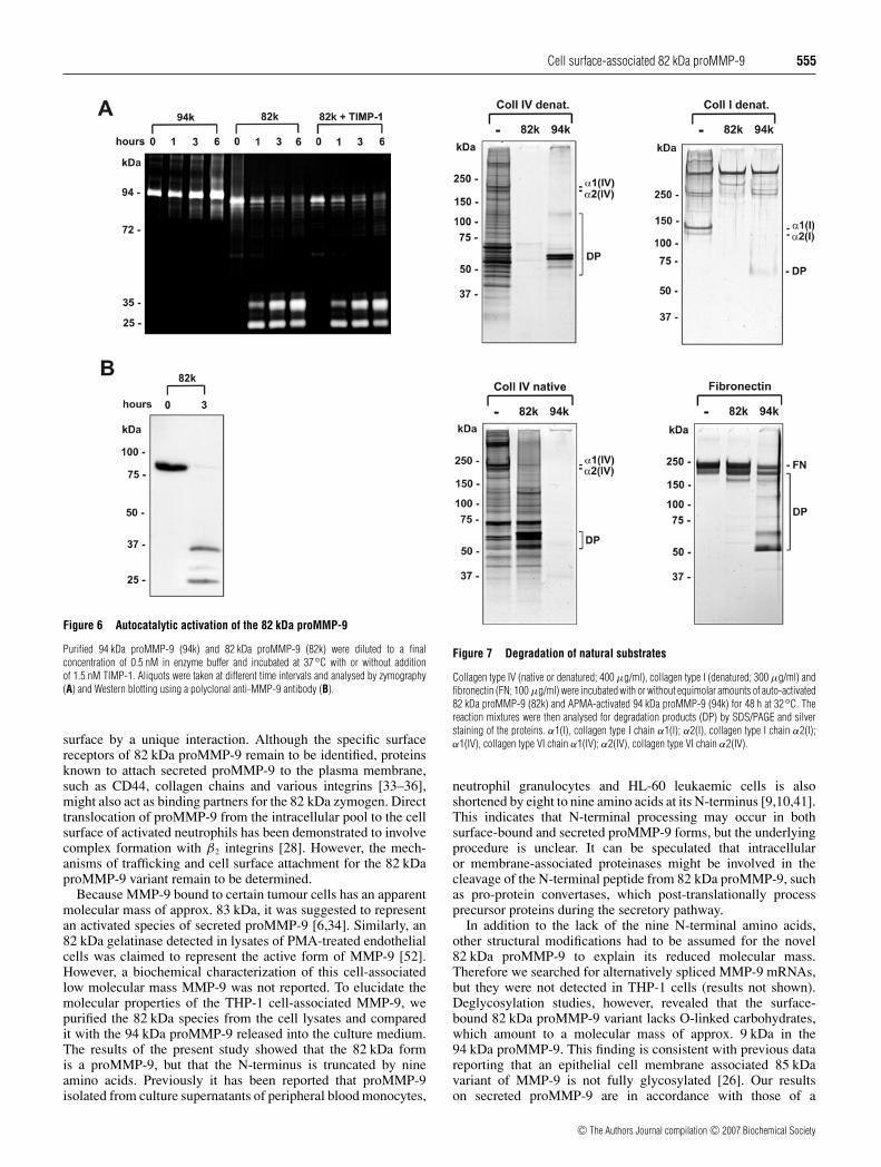
Incubation of the purified 82 kDa proMMP-9 in enzyme bufferin the absence of activating agents for 1–6 h at 37 ◦C graduallyconverted the enzyme into a major 35 kDa and a minor 25 kDagelatinase as determined by zymography (Figure 6A) andWestern blot analysis (Figure 6B). The conversion was almostcomplete after 6 h of incubation (Figure 6A). In contrast, the94 kDa proMMP-9 remained stable and showed no change inmolecular mass under these conditions (Figure 6A). Autocatalyticprocessing of the 82 kDa proMMP-9 was not affected by theaddition of TIMP-1 even when the inhibitor was present in a 3-fold molar excess (Figure 6A). These findings indicate that the82 kDa proMMP-9 is readily auto-activated compared withthe inert secreted 94 kDa proMMP-9.
Substrate specificities
Like the 94 kDa proMMP-9, the 82 kDa proMMP-9, whenactivated, digested denatured collagen type IV and type I(gelatins), native collagen type IV and fibronectin (Figure 7).The two enzymes, however, showed different extent in degradingthese substrates. Although activated 94 kDa proMMP-9 prefersto cleave native type IV collagen to the denatured gelatins, theactivated 82 kDa proMMP-9 digested denatured type I and type IVcollagens more readily than the 94 kDa form (Figure 7).
Inhibition of the 82 kDa and 94 kDa proMMP-9
To examine their susceptibility to inhibition, the activated 82 kDaand 94 kDa proenzymes were incubated with increasing concen-trations of TIMP-1, as well as a synthetic MMP inhibitor, andinhibition of their gelatinolytic activity was monitored. TIMP-1efficiently blocked the activated 94 kDa proMMP-9 with an IC50
of 1 nM (Figure 8A). In contrast, inhibition of the activated 82 kDaproMMP-9 by TIMP-1 was significantly weaker with an IC50
value of 82 nM (Figure 8A). Comparable results were obtainedby TIMP-2 (results not shown). The synthetic MMP inhibitor,GM6001, blocked both forms of MMP-9 with a similar efficiency,indicated by the IC50 value of 0.5 nM for the activated 94 kDaform and of 0.8 nM for the activated 82 kDa proMMP-9 (Fig-ure 8B).
Expression of the 82 kDa proMMP-9 in other leukaemic cells
The molecular mass of the 82 kDa proMMP-9 is identical to thatof the activated form (82 kDa) of 94 kDa proMMP-9. Todistinguish these two enzyme species, we used Western blotanalysis with antibodies that specifically recognize the propeptide,but not the activated form of MMP-9. As expected, both the82 kDa proMMP-9 and the 94 kDa proMMP-9, but not their
c© The Authors Journal compilation c© 2007 Biochemical Society

554 C. Ries and others
Figure 5 Proteolytic activation of proMMP-9 forms and binding to α2-macroglobulin
Purified 94 kDa proMMP-9 from culture supernatants (A) and 82 kDa proMMP-9 from cell extracts (B) were diluted to a final concentration of 50 ng/ml in enzyme buffer. The samples were thenincubated at 37◦C in the presence or absence of equimolar amounts of active MMP-3. Aliquots were taken at different time intervals and analysed by zymography and immunoblotting usinganti-MMP-9 polyclonal antibodies. (C) Enriched THP-1 plasma membranes containing the 94 kDa proMMP-9 (pro-94k) and the 82 kDa proMMP-9 (pro-82k) were incubated with MMP-3. Aliquotswere taken at different time intervals and analysed by zymography. (D) For examination of proteolytic activity the purified 94 kDa proMMP-9 and the 82 kDa proMMP-9 were treated with or withoutMMP-3 for 30 min at 37◦C, then incubated with an excess of α2-macroglobulin (500 µg/ml) for 30 min at 37◦C, and subsequently subjected to zymography. The dark protein band migrating ontop of the gel represents α2-macroglobulin (α2M), which binds and inhibits only active endopeptidases.
activated species, were detected by anti-(proMMP-9-prodomain)antibodies (Figure 9A).
Western blot analysis using the MMP-9 propeptide-directedantibodies was then applied to investigate whether the 82 kDaproMMP-9 was also present in other cell types. We first examinedHL-60 and NB4 cells, which originate from patients withacute myeloid leukaemia. The immunoblotting procedure clearlyshowed prominent amounts of the 82 kDa proMMP-9 and a weakband of 94 kDa proMMP-9 in extracts from PMA-stimulated HL-60 and NB4 cells, consistent with the observations in THP-1cells (Figure 9B). MMP-3 treatment of HL-60 and NB4 celllysates also generated the 35 kDa activated MMP-9 species(results not shown), proving the presence of 82 kDa proMMP-9in these cells. Western blot analysis of extracts from PMA-treatedex vivo leukaemic cells isolated from the peripheral blood of threepatients with acute myeloid leukemia clearly displayed productionof the 82 kDa proMMP-9 in these cells (Figure 9B). In contrast,proMMP-9 forms were absent from the lysates obtained fromPMA-stimulated mononuclear cells and neutrophil granulocytesfrom peripheral blood of healthy donors (Figure 9B).
DISCUSSION
Cell surface-associated proteinases concentrate proteolytic eventsat the sites of cell–matrix contact and mediate responses very
rapidly in contrast to secreted enzymes, which diffuse into thepericellular space after their release [51]. Previous studies havedemonstrated localization of MMP-9 on the plasma membraneof various tumour cells [6,24,26,31,33–36]. So far, it has beenassumed that surface-bound MMP-9 represents secreted enzymemolecules that re-associate with the plasma membrane. In thepresent study, we provide evidence for the existence of anunique variant of MMP-9 zymogen with a molecular mass of82 kDa that is not detectable in the culture medium, but ispresent as the prominent proMMP-9 on the surface of leukaemiccells. Moreover, this membrane associated proMMP-9 variantdiffers from the secreted zymogen form in several structural andfunctional aspects: it is truncated by nine amino acids at its N-terminus, it lacks O-linked glycosylation and it exhibits a distinctactivation process which produces an unusually small activeMMP-9 species with less susceptibility to TIMP-1 inhibition.
Previous studies in immortalized MCF10A breast epithelialcells demonstrated that secreted proMMP-9 was localized at theplasma membrane together with an 85 kDa species of MMP-9,which was not released from the cells even upon its stimulationwith PMA [26]. Thus, the latter form of MMP-9 appears to havesimilar trafficking properties to the 82 kDa proMMP-9 describedin our THP-1 cell system. This 82 kDa species was absent inmedium, but readily detected in cell lysates and the plasmamembrane, suggesting that this enzyme is retained at the cell
c© The Authors Journal compilation c© 2007 Biochemical Society
Cell surface-associated 82 kDa proMMP-9 555
Figure 6 Autocatalytic activation of the 82 kDa proMMP-9
Purified 94 kDa proMMP-9 (94k) and 82 kDa proMMP-9 (82k) were diluted to a finalconcentration of 0.5 nM in enzyme buffer and incubated at 37◦C with or without additionof 1.5 nM TIMP-1. Aliquots were taken at different time intervals and analysed by zymography(A) and Western blotting using a polyclonal anti-MMP-9 antibody (B).
surface by a unique interaction. Although the specific surfacereceptors of 82 kDa proMMP-9 remain to be identified, proteinsknown to attach secreted proMMP-9 to the plasma membrane,such as CD44, collagen chains and various integrins [33–36],might also act as binding partners for the 82 kDa zymogen. Directtranslocation of proMMP-9 from the intracellular pool to the cellsurface of activated neutrophils has been demonstrated to involvecomplex formation with β2 integrins [28]. However, the mech-anisms of trafficking and cell surface attachment for the 82 kDaproMMP-9 variant remain to be determined.
Because MMP-9 bound to certain tumour cells has an apparentmolecular mass of approx. 83 kDa, it was suggested to representan activated species of secreted proMMP-9 [6,34]. Similarly, an82 kDa gelatinase detected in lysates of PMA-treated endothelialcells was claimed to represent the active form of MMP-9 [52].However, a biochemical characterization of this cell-associatedlow molecular mass MMP-9 was not reported. To elucidate themolecular properties of the THP-1 cell-associated MMP-9, wepurified the 82 kDa species from the cell lysates and comparedit with the 94 kDa proMMP-9 released into the culture medium.The results of the present study showed that the 82 kDa formis a proMMP-9, but that the N-terminus is truncated by nineamino acids. Previously it has been reported that proMMP-9isolated from culture supernatants of peripheral blood monocytes,
Figure 7 Degradation of natural substrates
Collagen type IV (native or denatured; 400 µg/ml), collagen type I (denatured; 300 µg/ml) andfibronectin (FN; 100 µg/ml) were incubated with or without equimolar amounts of auto-activated82 kDa proMMP-9 (82k) and APMA-activated 94 kDa proMMP-9 (94k) for 48 h at 32◦C. Thereaction mixtures were then analysed for degradation products (DP) by SDS/PAGE and silverstaining of the proteins. α1(I), collagen type I chain α1(I); α2(I), collagen type I chain α2(I);α1(IV), collagen type VI chain α1(IV); α2(IV), collagen type VI chain α2(IV).
neutrophil granulocytes and HL-60 leukaemic cells is alsoshortened by eight to nine amino acids at its N-terminus [9,10,41].This indicates that N-terminal processing may occur in bothsurface-bound and secreted proMMP-9 forms, but the underlyingprocedure is unclear. It can be speculated that intracellularor membrane-associated proteinases might be involved in thecleavage of the N-terminal peptide from 82 kDa proMMP-9, suchas pro-protein convertases, which post-translationally processprecursor proteins during the secretory pathway.
In addition to the lack of the nine N-terminal amino acids,other structural modifications had to be assumed for the novel82 kDa proMMP-9 to explain its reduced molecular mass.Therefore we searched for alternatively spliced MMP-9 mRNAs,but they were not detected in THP-1 cells (results not shown).Deglycosylation studies, however, revealed that the surface-bound 82 kDa proMMP-9 variant lacks O-linked carbohydrates,which amount to a molecular mass of approx. 9 kDa in the94 kDa proMMP-9. This finding is consistent with previous datareporting that an epithelial cell membrane associated 85 kDavariant of MMP-9 is not fully glycosylated [26]. Our resultson secreted proMMP-9 are in accordance with those of a
c© The Authors Journal compilation c© 2007 Biochemical Society

556 C. Ries and others
Figure 8 Inhibition studies
Activated 82 kDa proMMP-9 and 94 kDa proMMP-9 (1 nM) were assayed with increasingconcentrations of the physiological MMP-9 inhibitor TIMP-1 (A) and the synthetic MMP-inhibitor GM6001 (B). Residual enzyme activity was quantified using fluorescence-labelledgelatin as a substrate and expressed in percentage of activity without inhibitor. Results are givenas means of duplicate experiments representative for three independent determinations.
detailed glycosylation analysis performed on proMMP-9 releasedfrom neutrophil granulocytes, demonstrating that N-linkedcarbohydrates of approx. 5 kDa and O-linked carbohydratesof approx. 10 kDa are attached to the 76 kDa zymogen coreprotein [53,54]. The biological significance of oligosaccharidesfor proMMP-9 function is far from clear, but they are considered toinfluence the backbone conformation of the enzyme, its stabilityand its interaction with other molecules. Recent studies usingrecombinant variants of proMMP-9 indicate that the abundantO-linked glycans present in this enzyme are important for itsinteraction with TIMP-1, but dispensable for catalytic activity[55]. Likewise, O-glycans present in membrane type 1 MMPare essential for binding to TIMP-2, but are not requiredfor its collagenolytic activity [56]. Additionally, aberrant O-glycosylation found in tumour cell-derived proMMP-9 comparedwith that from neutrophil granulocytes was shown to contributeto impaired interaction with galectin-3 [57]. We hypothesize thatN-terminal processing of the 82 kDa proMMP-9 alters its protein
Figure 9 Specific detection of 82 kDa proMMP-9 in leukaemic and non-leukaemic cells
(A) The zymogen forms (Pro) and activated species (Act.) of the 94 kDa proMMP-9 (94k) and the82 kDa proMMP-9 (82k) were analysed by zymography. Aliquots were subjected to SDS/PAGEunder reducing condition, blotted and incubated with monoclonal antibodies directed againstthe propeptide of proMMP-9 to allow specific detection of the 82 kDa proMMP-9 in distinctionto 82 kDa active MMP-9. (B) Cells were incubated in serum-free medium (1 × 10⁶ cells/ml)for 24 h in the presence of 50 ng/ml PMA. Proteins were extracted and 10 ng were subjected toWestern blotting analysis using an anti-(proMMP-9 prodomain) antibody. Results are shown forthe leukaemic cell lines THP-1, HL-60 and NB4, for mononuclear cells (MNC) and neutrophilgranulocytes (PMNC) from peripheral blood of healthy persons, and for leukaemic blast cellsfrom peripheral blood of patients with acute myeloid leukemia (Pat.#1-3).
structure in the endoplasmic reticulum and Golgi, which resultsin the failure of O-glycosylation. Consequently, the lack of O-glycans may favour surface binding and cause changes in itsinteraction with substrates, inhibition by TIMPs and accessibilityto (auto-)proteolytic cleavage.
MMP-3 is an effective proteolytic activator for secretedproMMP-9 [23,25,48,49] generating the active 82 kDa species.In the present study, we could demonstrate that it converts thesurface-associated 82 kDa proMMP-9 into an unusually small andso far undescribed 35 kDa active form. This species was found incell lysates and purified plasma membranes, but was absent in theculture supernatants. This failure may be due to a strong bindingto the cell surface or caused by dilution and limited stability in themedium. The 35 kDa MMP-9 has a substrate spectrum similarto the well-known 82 kDa MMP-9, but preferentially cleavesdenatured collagens, suggesting a biological role differing fromthat of secreted MMP-9. Moreover, autocatalytic conversion intoits active species occurred in the surface-bound 82 kDa proMMP-9, but not in the secreted 94 kDa zymogen. This auto-activationmay be promoted by the lack of a nonapeptide at the N-terminus
c© The Authors Journal compilation c© 2007 Biochemical Society

Cell surface-associated 82 kDa proMMP-9 557
of the 82 kDa proMMP-9, which may disrupt the cysteine–Zn2+ interaction (‘cysteine switch’) more readily. Although theautoproteolytic activation of the 82 kDa proMMP-9 occurred ata slower rate compared with the MMP-3 catalysed reaction, bothconditions led to the appearance of a major 35 kDa active speciesand a minor 25 kDa form detectable in zymograms, indicatingthat similar processing mechanisms may apply for autocatalyticand MMP-3-mediated activation. TIMP-1 is known to build atight complex with the secreted 94 kDa proMMP-9 by bindingwith its N-terminus to the catalytic site and with its C-terminusto the haemopexin domain of proMMP-9, thereby controllingits activation and activity [1,4,21,23,41,48]. Unexpectedly, co-incubation of the 82 kDa proMMP-9 with an excess of TIMP-1could not prevent or slow down the auto-activation, suggestinglimited reaction between the proenzyme variant and TIMP-1. Thismay be explained by the lack of O-glycans in 82 kDa proMMP-9,which were shown to be essential for optimal binding ofproMMP-9 to TIMP-1 [55].
To gain further insight into the structure/function relationshipof the 82 kDa proMMP-9, we performed comparative inhibitionstudies. The activated 94 kDa proMMP-9 was efficientlyblocked by equimolar concentrations of TIMP-1, consistentwith the results of previous studies on secreted proMMP-9[58]. In contrast, the 82 kDa proMMP-9 in its activated formrequired approx. 80-fold higher amounts of TIMP-1 to abrogategelatinolytic activity. It seems obvious that this dramaticallyreduced inhibition is related to the extensive truncationscharacterizing active 35-kDa MMP-9. In fact, antibodies directedagainst a C-terminal peptide of MMP-9 recognized 82 kDaproMMP-9, but not its activated 35 kDa form (results not shown),indicating C-terminal truncation in the latter. The C-terminus ofMMP-9 contains the haemopexin-like domain, which representsthe high-affinity binding site for TIMP-1 [55,58]. Therefore itis reasonable that extensive C-terminal processing of the 82 kDaproMMP-9 during activation reduces its susceptibility to TIMP-1inhibition and thereby increases its bioavailability. Presumably,the remaining 35 kDa mini MMP-9 essentially consists of thecatalytic domain of MMP-9. This area contains the fibronectin-type II repeats, which are important for gelatin binding, andincludes the zinc-binding sequence that is required for proteolyticactivity [1,4]. GM6001, a small synthetic metalloproteinaseinhibitor that binds directly to the enzyme’s active site [59],blocked the active 35 kDa and 82 kDa MMP-9 species with asimilar efficiency. This indicates the suitability of low molecularmass inhibitors in targeting both surface-associated and secretedMMP-9 activity, e.g. in the treatment of cancer.
The results of the present study indicate that THP-1 leukaemiccells endogenously produce a unique proMMP-9 variant exposedon the cell surface with reduced susceptibility to inhibitionby TIMP-1. Adopting an immunological method specificallydetecting this novel 82 kDa proMMP-9, we found the enzymeto be expressed in different leukaemic cell lines, but not in normalwhite blood cells. Its detection in patient-derived leukaemic blastcells indicates that the 82 kDa proMMP-9 also occurs in vivo,supporting the importance of this enzyme species. Physiologicalstimuli such as cytokines, chemokines or extracellular matrixcomponents may influence its synthesis and exposition on theplasma membrane. Therefore overexpression of the TIMP-1-insensitive MMP-9 variant on the surface of malignantly trans-formed cells may increase pericellular proteolysis and therebypromote cancer progression in vivo.
This work was supported by grants from the Deutsche Forschungsgemeinschaft(SFB 469), from the Wilhelm Sander-Stiftung (2002.122.1) and from the Wellcome Trust(No. 057508). We thank Professor Torsten Haferlach (Ludwig Maximilians University,
Munich, Germany) for his support in the collection of patient material and clinical data. Wealso thank Dr Irmgard Assfalg-Machleidt and Professor Werner Machleidt for assistancewith the performance of the inhibition kinetics, and Professor Christian Sommerhoff (all atLudwig-Maximilians-University of Munich, Munich, Germany) for critical reading of themanuscript.
REFERENCES
1 Nagase, H. and Woessner, J. F. (1999) Matrix metalloproteinases. J. Biol. Chem. 274,21491–21494
2 Stetler-Stevenson, W. G., Aznavoorian, S. and Liotta, L. A. (1993) Tumor cell interactionswith the extracellular matrix during invasion and metastasis. Annu. Rev. Cell Biol. 9,541–573
3 Egeblad, M. and Werb, Z. (2002) New functions for the matrix metalloproteinases incancer progression. Nat. Rev. Cancer 2, 161–174
4 Woessner, J. F. and Nagase, H. (2000) Matrix metalloproteinases and TIMPs,Oxford University Press, Oxford
5 Ries, C. and Petrides, P. E. (1995) Cytokine regulation of matrix metalloproteinase activityand its regulatory dysfunction in disease. Biol. Chem. 376, 345–355
6 Yu, Q. and Stamenkovic, I. (2000) Cell surface-localized matrix metalloproteinase-9proteolytically activates TGF-β and promotes tumor invasion and angiogenesis.Genes Dev. 14, 163–176
7 Van den Steen, P. E., Proost, P., Wuyts, A., van Damme, J. and Opdenakker, G. (2000)Neutrophil gelatinase B potentiates interleukin-8 tenfold by N-terminal processing,whereas it degrades CTAP-III, PF-4, and GRO-α and leaves RANTES and MCP-2 intact.Blood 96, 2673–2681
8 Schonbeck, U., Mach, F. and Libby, P. (1998) Generation of biologically active IL-1β bymatrix metalloproteinases: a novel caspase-1-independent pathway of IL-1β processing.J. Immunol. 161, 3340–3346
9 Masure, S., Proost, P., van Damme, J. and Opdenakker, G. (1991) Purification andidentification of 91-kDa neutrophil gelatinase: release by the activating peptideinterleukin-8. Eur. J. Biochem. 198, 391–398
10 Opdenakker, G., Masure, S., Proost, P., Billiau, A. and van Damme, J. (1991) Naturalhuman monocyte gelatinase and its inhibitor. FEBS Lett. 284, 73–78
11 Welgus, H. G., Campbell, E. J., Cury, J. D., Eisen, A. Z., Senior, R. M., Wilhelm, S. M. andGoldberg, G. I. (1990) Neutral metalloproteinases produced by human mononuclearphagocytes: enzyme profile, regulation, and expression during cellular development.J. Clin. Invest. 86, 1496–1502
12 Coussens, L. M., Tinkle, C. L., Hanahan, D. and Werb, Z. (2000) MMP-9 supplied bybone marrow-derived cells contributes to skin carcinogenesis. Cell 103, 481–490
13 Van Ranst, M., Norga, K., Masure, S., Proost, P., Vandekerckhove, F., Auwerx, J.,van Damme, J. and Opdenakker, G. (1991) The cytokine-protease connection:identification of a 96-kD THP-1 gelatinase and regulation by interleukin-1 and cytokineinducers. Cytokine 3, 231–239
14 Ries, C., Kolb, H. and Petrides, P. E. (1994) Regulation of 92-kD gelatinase release inHL-60 leukemia cells: tumor necrosis factor-α as an autocrine stimulus for basal- andphorbol ester-induced secretion. Blood 83, 3638–3646
15 Ismair, M. G., Ries, C., Lottspeich, F., Zang, C., Kolb, H. J. and Petrides, P. E. (1998)Autocrine regulation of matrix metalloproteinase-9 gene expression and secretion bytumor necrosis factor-α (TNF-α) in NB4 leukemic cells: specific involvement of TNFreceptor type 1. Leukemia 12, 1136–1143
16 Ries, C., Loher, F., Zang, C., Ismair, M. G. and Petrides, P. E. (1999) Matrixmetalloproteinase production by bone marrow mononuclear cells from normal individualsand patients with acute and chronic myeloid leukemia or myelodysplastic syndromes.Clin. Cancer Res. 5, 1115–1124
17 Janowska-Wieczorek, A., Marquez, L. A., Matsuzaki, A., Hashmi, H. R., Larratt, L. M.,Boshkov, L. M., Turner, A. R., Zhang, M. C., Edwards, D. R. and Kossakowska, A. E.(1999) Expression of matrix metalloproteinases (MMP-2 and -9) and tissue inhibitors ofmetalloproteinases (TIMP-1 and -2) in acute myelogenous leukaemia blasts: comparisonwith normal bone marrow cells. Br. J. Haematol. 105, 402–411
18 Klein, G., Vellenga, E., Fraaije, M. W., Kamps, W. A. and de Bont, E. S. (2004) Thepossible role of matrix metalloproteinase (MMP)-2 and MMP-9 in cancer, e.g. acuteleukemia. Crit Rev. Oncol. Hematol. 50, 87–100
19 Bernhard, E. J., Gruber, S. B. and Muschel, R. J. (1994) Direct evidence linkingexpression of matrix metalloproteinase 9 (92-kDa gelatinase/collagenase) to themetastatic phenotype in transformed rat embryo cells. Proc. Natl. Acad. Sci. U.S.A. 91,4293–4297
c© The Authors Journal compilation c© 2007 Biochemical Society

558 C. Ries and others
20 Hua, J. and Muschel, R. J. (1996) Inhibition of matrix metalloproteinase 9 expression by aribozyme blocks metastasis in a rat sarcoma model system. Cancer Res. 56,5279–5284
21 Wilhelm, S. M., Collier, I. E., Marmer, B. L., Eisen, A. Z., Grant, G. A. and Goldberg, G. I.(1989) SV40-transformed human lung fibroblasts secrete a 92-kDa type IV collagenase,which is identical to that secreted by normal human macrophages. J. Biol. Chem. 264,17213–17221
22 Moll, U. M., Youngleib, G. L., Rosinski, K. B. and Quigley, J. P. (1990) Tumorpromoter-stimulated Mr 92,000 gelatinase secreted by normal and malignant humancells: isolation and characterization of the enzyme from HT1080 tumor cells. Cancer Res.50, 6162–6170
23 Nagase, H. (1997) Activation mechanisms of matrix metalloproteinases. Biol. Chem. 378,151–160
24 Mazzieri, R., Masiero, L., Zanetta, L., Monea, S., Onisto, M., Garbisa, S. and Mignatti, P.(1997) Control of type IV collagenase activity by components of the urokinase–plasmin system: a regulatory mechanism with cell-bound reactants. EMBO J. 16,2319–2332
25 Ramos-DeSimone, N., Hahn-Dantona, E., Sipley, J., Nagase, H., French, D. L. andQuigley, J. P. (1999) Activation of matrix metalloproteinase-9 (MMP-9) via a convergingplasmin/stromelysin-1 cascade enhances tumor cell invasion. J. Biol. Chem. 274,13066–13076
26 Toth, M., Gervasi, D. C. and Fridman, R. (1997) Phorbol ester-induced cell surfaceassociation of matrix metalloproteinase-9 in human MCF10A breast epithelial cells.Cancer Res. 57, 3159–3167
27 Partridge, C. A., Phillips, P. G., Niedbala, M. J. and Jeffrey, J. J. (1997) Localization andactivation of type IV collagenase/gelatinase at endothelial focal contacts.Am. J. Physiol. 272, L813–L822
28 Stefanidakis, M., Ruohtula, T., Borregaard, N., Gahmberg, C. G. and Koivunen, E. (2004)Intracellular and cell surface localization of a complex between αMβ2 integrin andpromatrix metalloproteinase-9 progelatinase in neutrophils. J. Immunol. 172,7060–7068
29 Owen, C. A., Hu, Z., Barrick, B. and Shapiro, S. D. (2003) Inducible expression of tissueinhibitor of metalloproteinases-resistant matrix metalloproteinase-9 on the cell surface ofneutrophils. Am. J. Respir. Cell Mol. Biol. 29, 283–294
30 Rahat, M. A., Marom, B., Bitterman, H., Weiss-Cerem, L., Kinarty, A. and Lahat, N. (2006)Hypoxia reduces the output of matrix metalloproteinase-9 (MMP-9) in monocytes byinhibiting its secretion and elevating membranal association. J. Leukoc. Biol. 79,706–718
31 Bourguignon, L. Y., Gunja-Smith, Z., Iida, N., Zhu, H. B., Young, L. J., Muller, W. J. andCardiff, R. D. (1998) CD44v(3,8–10) is involved in cytoskeleton-mediated tumor cellmigration and matrix metalloproteinase (MMP-9) association in metastatic breast cancercells. J. Cell Physiol. 176, 206–215
32 Ellerbroek, S. M., Halbleib, J. M., Benavidez, M., Warmka, J. K., Wattenberg, E. V., Stack,M. S. and Hudson, L. G. (2001) Phosphatidylinositol 3-kinase activity in epidermalgrowth factor-stimulated matrix metalloproteinase-9 production and cell surfaceassociation. Cancer Res. 61, 1855–1861
33 Olson, M. W., Toth, M., Gervasi, D. C., Sado, Y., Ninomiya, Y. and Fridman, R. (1998)High affinity binding of latent matrix metalloproteinase-9 to the α2(IV) chain ofcollagen IV. J. Biol. Chem. 273, 10672–10681
34 Yu, Q. and Stamenkovic, I. (1999) Localization of matrix metalloproteinase 9 to the cellsurface provides a mechanism for CD44-mediated tumor invasion. Genes Dev. 13,35–48
35 Fridman, R., Toth, M., Chvyrkova, I., Meroueh, S. O. and Mobashery, S. (2003) Cellsurface association of matrix metalloproteinase-9 (gelatinase B). Cancer Metastasis Rev.22, 153–166
36 Stefanidakis, M. and Koivunen, E. (2006) Cell-surface association between matrixmetalloproteinases and integrins: role of the complexes in leukocyte migration and cancerprogression. Blood 108, 1441–1450
37 Bergers, G., Brekken, R., McMahon, G., Vu, T. H., Itoh, T., Tamaki, K., Tanzawa, K.,Thorpe, P., Itohara, S., Werb, Z. and Hanahan, D. (2000) Matrix metalloproteinase-9triggers the angiogenic switch during carcinogenesis. Nat. Cell Biol. 2, 737–744
38 Seiki, M. (2002) The cell surface: the stage for matrix metalloproteinase regulation ofmigration. Curr. Opin. Cell Biol. 14, 624–632
39 Kleiner, D. E. and Stetler-Stevenson, W. G. (1994) Quantitative zymography: detection ofpicogram quantities of gelatinases. Anal. Biochem. 218, 325–329
40 Heukeshoven, J. and Dernick, R. (1985) Simplified method for silver staining ofproteins in polyacrylamide gels and the mechanism of the staining. Electrophoresis 6,103
41 Ries, C., Lottspeich, F., Dittmann, K. H. and Petrides, P. E. (1996) HL-60 leukemia cellsproduce an autocatalytically truncated form of matrix metalloproteinase-9 with impairedsensitivity to inhibition by tissue inhibitors of metalloproteinases. Leukemia 10,1520–1526
42 Dean, D. D., Martel Pelletier, J., Pelletier, J. P., Howell, D. S. and Woessner, J. F. J. (1989)Evidence for metalloproteinase and metalloproteinase inhibitor imbalance in humanosteoarthritic cartilage. J. Clin. Invest. 84, 678–685
43 Eckerskorn, C., Mewes, W., Goretzki, H. and Lottspeich, F. (1988) A new siliconized-glassfiber as support for protein-chemical analysis of electroblotted proteins. Eur. J. Biochem.176, 509–519
44 Eckerskorn, C. and Lottspeich, F. (2001) Internal amino acid sequence analysis ofproteins separated by gel electrophoresis after tryptic digestion in polyacrylamide matrix.Chromatographia 28, 92–94
45 Meister, G., Buhler, D., Laggerbauer, B., Zobawa, M., Lottspeich, F. and Fischer, U. (2000)Characterization of a nuclear 20S complex containing the survival of motor neurons(SMN) protein and a specific subset of spliceosomal Sm proteins. Hum. Mol. Genet. 9,1977–1986
46 Suzuki, K., Kan, C. C., Hung, W., Gehring, M. R., Brew, K. and Nagase, H. (1998)Expression of human pro-matrix metalloproteinase 3 that lacks the N-terminal 34residues in Escherichia coli: autoactivation and interaction with tissue inhibitor ofmetalloproteinase 1 (TIMP-1). Biol. Chem. 379, 185–191
47 Huang, W., Meng, Q., Suzuki, K., Nagase, H. and Brew, K. (1997) Mutational studyof the N-terminal domain of human tissue inhibitor of metalloproteinases 1 (TIMP-1)locates an inhibitory region for matrix metalloproteinases. J. Biol. Chem. 272,22086–22091
48 Olson, M. W., Bernardo, M. M., Pietila, M., Gervasi, D. C., Toth, M., Kotra, L. P.,Massova, I., Mobashery, S. and Fridman, R. (2000) Characterization of the monomericand dimeric forms of latent and active matrix metalloproteinase-9: differential rates foractivation by stromelysin 1. J. Biol. Chem. 275, 2661–2668
49 Ogata, Y., Enghild, J. J. and Nagase, H. (1992) Matrix metalloproteinase 3 (stromelysin)activates the precursor for the human matrix metalloproteinase 9. J. Biol. Chem. 267,3581–3584
50 Barrett, A. J. and Starkey, P. M. (1973) The interaction of α2-macroglobulin withproteinases: characteristics and specificity of the reaction, and a hypothesis concerningits molecular mechanism. Biochem. J. 133, 709–724
51 Basbaum, C. B. and Werb, Z. (1996) Focalized proteolysis: spatial and temporalregulation of extracellular matrix degradation at the cell surface. Curr. Opin. Cell Biol. 8,731–738
52 Nguyen, M., Arkell, J. and Jackson, C. J. (1998) Active and tissue inhibitor of matrixmetalloproteinase-free gelatinase B accumulates within human microvascular endothelialvesicles. J. Biol. Chem. 273, 5400–5404
53 Mattu, T. S., Royle, L., Langridge, J., Wormald, M. R., van den Steen, P. E.,van Damme, J., Opdenakker, G., Harvey, D. J., Dwek, R. A. and Rudd, P. M. (2000)O-glycan analysis of natural human neutrophil gelatinase B using a combination ofnormal phase-HPLC and online tandem mass spectrometry: implications for the domainorganization of the enzyme. Biochemistry 39, 15695–15704
54 Rudd, P. M., Mattu, T. S., Masure, S., Bratt, T., van den Steen, P. E., Wormald, M. R.,Kuster, B., Harvey, D. J., Borregaard, N., van Damme, J. et al. (1999) Glycosylation ofnatural human neutrophil gelatinase B and neutrophil gelatinase B-associated lipocalin.Biochemistry 38, 13937–13950
55 van den Steen, P. E., van Aelst, I., Hvidberg, V., Piccard, H., Fiten, P., Jacobsen, C.,Moestrup, S. K., Fry, S., Royle, L., Wormald, M. R. et al. (2006) The hemopexin andO-glycosylated domains tune gelatinase B/MMP-9 bioavailability via inhibitionand binding to cargo receptors. J. Biol. Chem. 281, 18626–18637
56 Wu, Y. I., Munshi, H. G., Sen, R., Snipas, S. J., Salvesen, G. S., Fridman, R. and Stack,M. S. (2004) Glycosylation broadens the substrate profile of membrane type 1 matrixmetalloproteinase. J. Biol. Chem. 279, 8278–8289
57 Fry, S. A., Steen, P. E., Royle, L., Wormald, M. R., Leathem, A. J., Opdenakker, G.,McDonnell, J. M., Dwek, R. A. and Rudd, P. M. (2006) Cancer-associated glycoforms ofgelatinase B exhibit a decreased level of binding to galectin-3. Biochemistry 45,15249–15258
58 Olson, M. W., Gervasi, D. C., Mobashery, S. and Fridman, R. (1997) Kinetic analysis ofthe binding of human matrix metalloproteinase-2 and -9 to tissue inhibitor ofmetalloproteinase (TIMP)-1 and TIMP-2. J. Biol. Chem. 272, 29975–29983
59 Galardy, R. E., Grobelny, D., Foellmer, H. G. and Fernandez, L. A. (1994) Inhibition ofangiogenesis by the matrix metalloprotease inhibitor N-[2R-2-(hydroxamidocarbonymethyl)-4-methylpentanoyl]-L-tryptophan methylamide.Cancer Res. 54, 4715–4718
Received 5 February 2007/12 April 2007; accepted 10 May 2007Published as BJ Immediate Publication 10 May 2007, doi:10.1042/BJ20070191
c© The Authors Journal compilation c© 2007 Biochemical Society