ideas and innovations the buccal fat pad flap for ... · the buccal fat pad flap for periorbital...
TRANSCRIPT
Ideas and Innovations
The Buccal Fat Pad Flap for PeriorbitalReconstruction: A Cadaver Dissection andReport of Two CasesLuther H. Holton, III, M.D., Eduardo D. Rodriguez, D.D.S., M.D., Ronald P. Silverman, M.D.,Navin Singh, M.D., Anthony P. Tufaro, D.D.S., M.D., and Michael P. Grant, M.D., Ph.D.Baltimore, Md.
The buccal fat pad is a trilobed collection ofadipose tissue found in the midface. It is lo-cated behind the zygomatic arch, medial to themasseter muscle and lateral to the buccinatormuscle from which it derives its name.1–4 Itconsists of a body and four processes: the buc-cal, pterygoid, superficial, and deep tempo-ral.1–3 The body lies behind the zygomatic archand the processes extend from the body totheir respective surrounding tissue spaces.4
The buccal fat pad has recently been describedas having three lobes: an anterior lobe, anintermediate lobe, and a posterior lobe.4 Thepad receives an abundant vascular supply fromthree well-defined sources, namely, the facialand internal maxillary arteries and the deeptransverse facial vessels.2,4 The major vascularsupply enters the posterior lobe, requiring thatthis lobe remain attached when the buccal fatpad is used as a pedicled flap. Notably, the sizeof the pad remains relatively constant fromchildhood throughout adulthood, and it ap-pears to be resistant to wasting even in the mostcachectic patients.
The buccal fat pad has been proposed tohave a number of native functions. It fills themasticatory space and has traditionally beensuggested to have a role in mastication andsuckling, especially in the newborn. The padfunctions as a gliding pad during the contrac-tion of the masticatory and mimetic muscles.4In addition, it protects the deep facial neuro-vascular bundles from outside forces as well asfrom extrusion of muscle contraction. It may
serve as a venous network involved in cranialblood flow through the pterygoid plexus.5
Despite these various proposed functions,the buccal fat pad can be safely sacrificed in theadult patient.
Clinically, the buccal fat pad has been used foraesthetic and reconstructive applications.1–3,6,7,8–11
In cosmetic surgery, buccal fat pad lipectomy hasbeen used to improve midface width in full-facedindividuals.6–8 Similarly, buccal fat pad removalhas been used alone and in conjunction with facelift surgery to modify facial contours and en-hance the malar prominence.2,6,8,9 In addition, ithas been described in rhytidectomy for improve-ment of the nasolabial fold.4 As early as 1977, thereconstructive use of a pedicled buccal fat padflap was described by Egyedi10 for the closure oforoantral and oronasal communications follow-ing oncologic resection. In 1983, Neder11 eluci-dated its use as a free graft for the oral cavity. Anumber of other studies have further describedthe use of buccal fat pad flaps and grafts for thereconstruction of intraoral defects. The versatilityof the buccal fat pad is evident from the variety ofother novel reconstructive uses that have beendescribed, including coverage of irregularitiesfollowing bony trauma, closure of nasal defects,and use as vascularized filler material.3,10,11 Thereis, however, no literature describing its use inorbital reconstruction.
Using a cadaver study, we demonstrate thatthe buccal fat pad can be reliably mobilized toprovide sufficient coverage for orbital recon-structions. In addition, we present two case
From the University of Maryland Medical System and Johns Hopkins Hospital. Received for publication October 29, 2003; revised February5, 2004.
DOI: 10.1097/01.PRS.0000138257.44949.BB
1529

studies using this technique. We conclude bydiscussing the advantages and potential draw-backs of this novel approach for orbitalreconstruction.
MATERIALS AND METHODS
In the cadaver study, eight bilateral buccalfat pad dissections were performed on fourcadavers via a Weber-Ferguson approach. Acombination of blunt and sharp dissection wasused to expose and mobilize the anterior andintermediate lobes of the pad while the poste-rior lobe remained attached to the vascularsupply. Of note, in those patients who haveundergone maxillectomy and require orbitalfloor reconstruction, less length would beneeded to reach the orbit. After the pads weremobilized to allow the maximum cephaladreach, measurements were taken to ascertainthe length of the flaps. Length of flap wasdefined by a measurement taken from the al-veolar ridge at the level of the second molar tothe cephalad aspect of the mobilized flap.
RESULTS
The mean length of the buccal fat pad flapwas 9.34 cm (SD, �0.76 cm), with an averageapproximate volume of 24 cm3. In all eightdissections, the length of the flap and the vol-ume of mobilized tissue were easily sufficient tocover the inferior orbital rim, despite the fact
that none of the cadavers had undergone re-section of the maxilla. Figure 1 depicts therelevant regional anatomy and the arc ofrotation.
CASE REPORTS
Case 1A 74-year-old man presented with metastatic melanoma
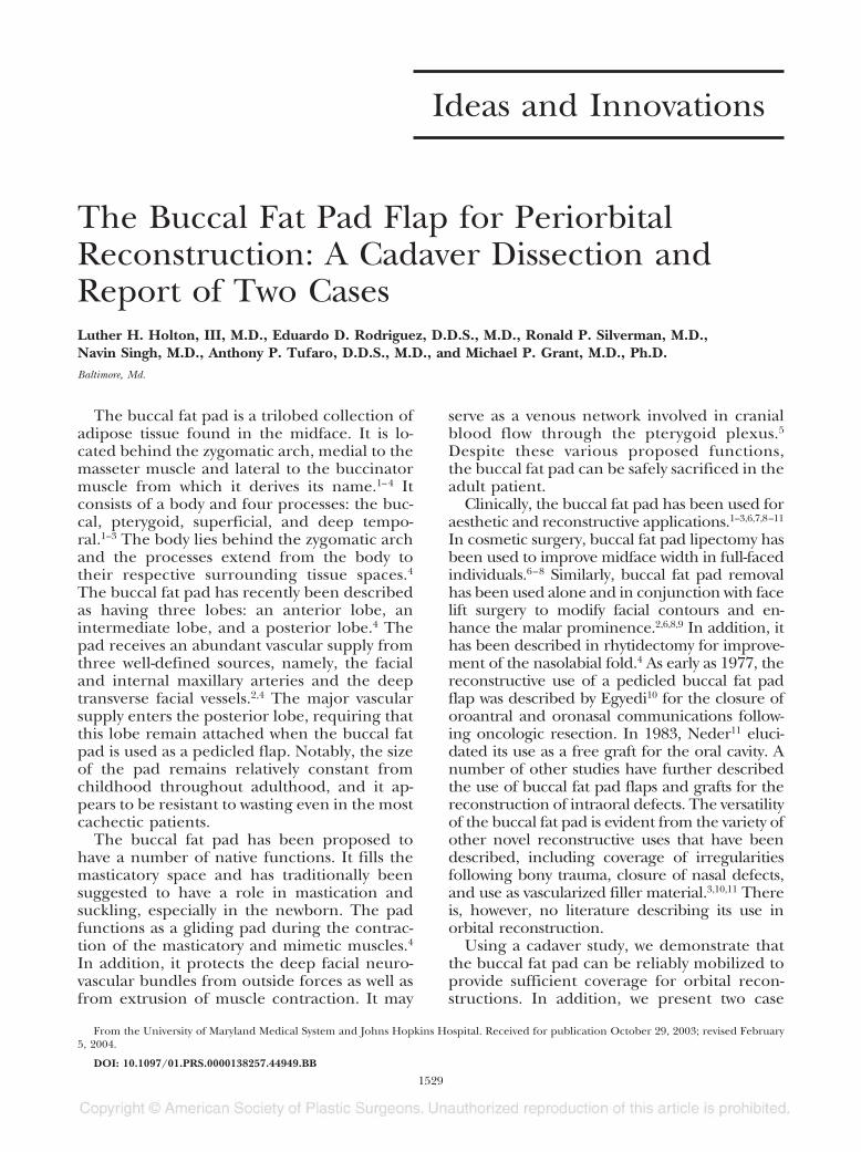
involving the left paranasal sinuses. The tumor involved thecheek, eyelid, nasal sidewall, anterior wall of the maxilla,orbital rim, and orbital floor. The tumor was extirpated fol-lowing a modified Weber-Ferguson approach (Fig. 2).
After resection of the soft and hard tissues, including apartial maxillectomy and 50 percent of the orbital floor,the orbit was reconstructed with a titanium plate along therim and porous polyethylene along the orbital floor. Dueto fears of exposure and infection, recruitment of vascular-ized tissue was indicated. The buccal fat pad was dissected andutilized to provide vascular lining for the synthetic implant(Fig. 3).
The buccal fat pad was harvested by mobilizing the ante-rior attachments while preserving the posterior lobe and itsvascular supply. The facial nerve was not disturbed during thedissection. The flap was advanced medially, protecting itsposterior vascular pedicle, and inset along the orbital floor,separating the implant from the maxillary sinus. The skindefect was closed using a cervicofacial rotation flap.
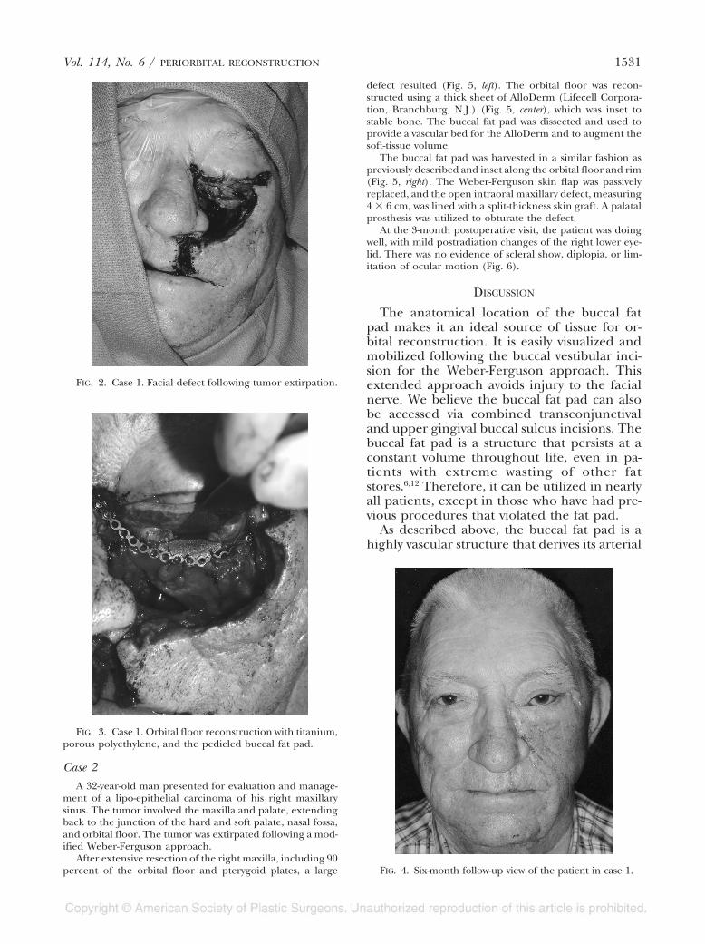
At his 6-month postoperative visit, the patient was doingwell and had minimal scleral show, no diplopia, and no dif-ficulties with his orbital floor implant (Fig. 4). By 7 months,his tumor was noted to have recurred and he went on to havea radical craniofacial resection of the previously recon-structed left orbital area.
FIG. 1. (Left) Illustration of the buccal space demonstrating its relationship to the buccinator and masseter muscles as wellas the blood supply. (Right) Illustration demonstrating the arc of rotation of the buccal fat pad.
1530 PLASTIC AND RECONSTRUCTIVE SURGERY, November 2004
Case 2A 32-year-old man presented for evaluation and manage-
ment of a lipo-epithelial carcinoma of his right maxillarysinus. The tumor involved the maxilla and palate, extendingback to the junction of the hard and soft palate, nasal fossa,and orbital floor. The tumor was extirpated following a mod-ified Weber-Ferguson approach.
After extensive resection of the right maxilla, including 90percent of the orbital floor and pterygoid plates, a large
defect resulted (Fig. 5, left). The orbital floor was recon-structed using a thick sheet of AlloDerm (Lifecell Corpora-tion, Branchburg, N.J.) (Fig. 5, center), which was inset tostable bone. The buccal fat pad was dissected and used toprovide a vascular bed for the AlloDerm and to augment thesoft-tissue volume.
The buccal fat pad was harvested in a similar fashion aspreviously described and inset along the orbital floor and rim(Fig. 5, right). The Weber-Ferguson skin flap was passivelyreplaced, and the open intraoral maxillary defect, measuring4 � 6 cm, was lined with a split-thickness skin graft. A palatalprosthesis was utilized to obturate the defect.
At the 3-month postoperative visit, the patient was doingwell, with mild postradiation changes of the right lower eye-lid. There was no evidence of scleral show, diplopia, or lim-itation of ocular motion (Fig. 6).
DISCUSSION
The anatomical location of the buccal fatpad makes it an ideal source of tissue for or-bital reconstruction. It is easily visualized andmobilized following the buccal vestibular inci-sion for the Weber-Ferguson approach. Thisextended approach avoids injury to the facialnerve. We believe the buccal fat pad can alsobe accessed via combined transconjunctivaland upper gingival buccal sulcus incisions. Thebuccal fat pad is a structure that persists at aconstant volume throughout life, even in pa-tients with extreme wasting of other fatstores.6,12 Therefore, it can be utilized in nearlyall patients, except in those who have had pre-vious procedures that violated the fat pad.
As described above, the buccal fat pad is ahighly vascular structure that derives its arterial
FIG. 4. Six-month follow-up view of the patient in case 1.
FIG. 2. Case 1. Facial defect following tumor extirpation.
FIG. 3. Case 1. Orbital floor reconstruction with titanium,porous polyethylene, and the pedicled buccal fat pad.
Vol. 114, No. 6 / PERIORBITAL RECONSTRUCTION 1531

blood supply from a rich network. Further-more, its venous plexus is robust, and the padhas been suggested to play a role in drainagefrom the pterygoid plexus. This robust vascu-larity obviously supports the viability of thisstructure in its reconstructive applications. Ac-cordingly, the buccal fat pad can be expectedto serve as a valuable source of vascular tissuewhen alloplastic or biologic implants are beingused. This is germane to orbital reconstruc-tion, as favored methods utilize titanium, po-
rous polyethylene implants, and various allo-graft materials, including AlloDerm.13,14
Although the flap is robust and accessible,potential drawbacks exist. Mobilization of thefat pad may create diminished midface volumeor asymmetry that can be aesthetically displeas-ing. In addition, this flap is limited by a rela-tively small volume of available tissue. This vol-ume is generally adequate for coverage ofperiorbital prosthetics, but for massive soft-tissue defects, other options, such as local tis-sue flaps (temporalis muscle flap) or free-tissuetransfer, should be considered.
Since the buccal fat pad is a highly vascularand fatty tissue, we believe this may improvethe tolerance to radiation therapy. However, todate, this has not been documented and maybe worthy of further investigation, since it isproposed for patients who have recently under-gone oncologic resection and may proceed toradiation therapy.
SUMMARY
We have shown that the buccal fat pad is arobust and reliable flap that may improve thesuccess rate of orbital reconstruction by provid-ing well-vascularized tissue of suitable volume.The buccal fat pad has been utilized for nu-merous aesthetic and reconstructive facial ap-plications and provides excellent vascularizedtissue support when alloplastic or allograft ma-terials are being used. We propose that usingthe pedicled buccal fat pad in conjunction with
FIG. 5. Case 2. (Left) Facial defect following tumor extirpation. (Center) Orbital floor reconstruction with AlloDerm. (Right)Orbital floor reconstruction with pedicled buccal fat pad.
FIG. 6. Three-month follow-up view of patient in case 2.
1532 PLASTIC AND RECONSTRUCTIVE SURGERY, November 2004

titanium, porous polyethylene mesh, poly-amide sheets, or various other allograft mate-rials can improve the success rates of recon-struction. This approach has not previouslybeen described in the literature. This tech-nique is an excellent compliment to periorbitalreconstruction given the ease of dissection,quality of vascularized tissue, and consistentvolume between patients.
Ronald P. Silverman, M.D.Division of Plastic and Reconstructive SurgeryUniversity of Maryland Medical Center22 South Greene Street, S8D12Baltimore, Md. [email protected]
REFERENCES
1. Dean, A., Alamillos, F., Garcia-Lopez, A., Sanchez, J., andPenalba, M. The buccal fat pad flap in oral recon-struction. Head Neck 23: 383, 2001.
2. Stuzin, J. M., Wagstrom, L., Kawamoto, H. K., Baker, T. J.,and Wolfe, S. A. The anatomy and clinical applica-tions of the buccal fat pad. Plast. Reconstr. Surg. 85: 29,1990.
3. Tideman, H., Bosanquet, A., and Scott, J. Use of thebuccal fat pad as a pedicled graft. J. Oral Maxillofac.Surg. 44: 435, 1986.
4. Zhang, H.-M., Yan, Y.-P., Qi, K.-M., Wang, J.-Q., and Liu,Z.-F. Anatomical structure of the buccal fat pad and
its clinical adaptations. Plast. Reconstr. Surg. 109: 2509,2002.
5. Racz, I., Maros, T. N., and Seres-Sturm, L. Structuralcharacteristics and functional significance of the buc-cal fat pad (corpus adiposum buccae). Morphol. Em-bryol. 35: 73, 1989.
6. Matarasso, A. Anatomy of the buccal fat pad and itsclinical significance. Plast. Reconstr. Surg. 103: 2061,1999.
7. Matarasso, A. Buccal fat pad excision: Aesthetic im-provement of the midface. Ann. Plast. Surg. 26: 413,1991.
8. Jackson, I. T. Anatomy of the buccal fat pad and itsclinical significance. Plast. Reconstr. Surg. 103: 2059,1999.
9. Ramirez, O. M. Buccal fat pad pedicle flap for midfaceaugmentation. Ann. Plast. Surg. 43: 109, 1999.
10. Egyedi, P. Utilization of the buccal fat pad for closureof oral-antral and/or oral-nasal communications.J. Maxillofac. Surg. 5: 241, 1977.
11. Neder, A. Use of the buccal fat pad for grafts. Oral Surg.Oral Med. Oral Pathol. 55: 349, 1983.
12. Xiao, H., Bayramicli, M., and Jackson, I. T. Volumetricanalysis of the buccal fat pad. Eur. J. Plast. Surg. 22: 177,1999.
13. Schubert, W., Gear, A. J., Lee, C., et al. Incorporationof titanium mesh in orbital and midface reconstruc-tion. Plast. Reconstr. Surg. 110: 1022, 2002.
14. Villarreal, P. M., Monje, F., Morillo, A. J., et al. Porouspolyethylene implants in orbital floor reconstruction.Plast. Reconstr. Surg. 109: 877, 2002.
Vol. 114, No. 6 / PERIORBITAL RECONSTRUCTION 1533