icu management of ecmo pt
TRANSCRIPT

ICU Management of ECMO Patient
Muhammad Asim RanaMBBS, MRCP(UK), EDIC, SFCCM, FCCP, FRCPE
Critical Care Services
Bahria International Hospital, Lahore

ICU Management of ECMO Patient
• Preparation
• Cannulation
• Initiation
• Maintenance
• Care of circuit
• Trouble shooting
• Complications
• Weaning

Preparation
• ICU staff dedicated for ECMO is responsible for preparation of cannulation trolley.
• Properly trained staff for cannulation.
• Cannulation is performed inside operation theaters usually for VA ECMO.

Cannulation

Selecting Cannulae

Example
• Drainage Cannula
– Femoral Vein
– Long to 60 cm
– Diameter 22 – 30 Fr
• Return Cannula
– Femoral Artery
– Shorter 20 – 25 cm
– Diameter 15 – 23 Fr

Securing Access and Returning Lines
• Once cannulae position have been confirmed femoral lines should be secured using a two stage prep to the patient’s leg and covering with fabric tape or Hypafix provided on the cannulation trolleys a mesentery dressing.
• The internal jugular line is directed over the patient’s head. The loop around the head is immobilized by strapping around the patient’s forehead. The cannulae are sutured to the dressing.

Securing Access and Returning Lines
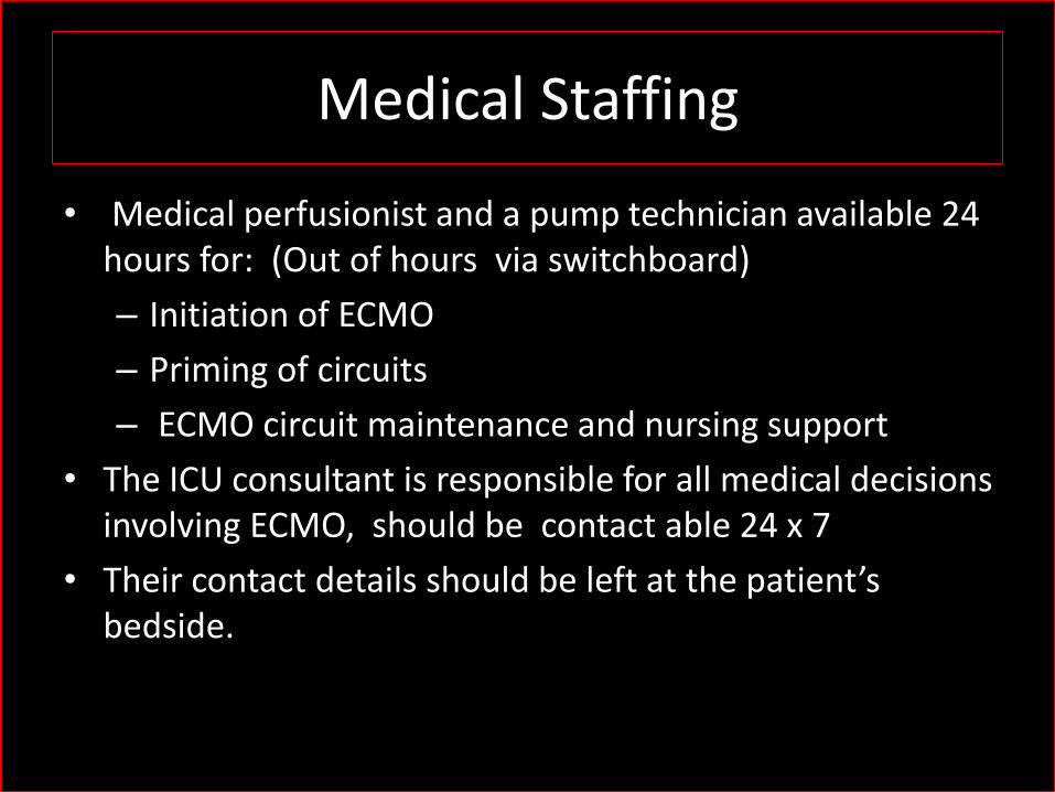
Medical Staffing
• Medical perfusionist and a pump technician available 24 hours for: (Out of hours via switchboard)
– Initiation of ECMO
– Priming of circuits
– ECMO circuit maintenance and nursing support
• The ICU consultant is responsible for all medical decisions involving ECMO, should be contact able 24 x 7
• Their contact details should be left at the patient’s bedside.

Commencement of ECMO
• Check ACT and ensure>200 seconds.
• Ensure O2 line is connected to oxygenator.
• Gas flow should be commenced at a rate equal to or greater than the anticipated circuit blood flow (usually 2-4L/min) with 100 % O2.
• Clean loop is opened and handed to the cannulating physician.

Commencement of ECMO
• The circuit is cut between two clamps allowing sufficient length on the access line and return line to prevent any tension on the circuit.
• Note the pump trolley is best kept at the “foot” end of the patient’s bed.
• Circuit is connected to cannulae ensuring no air is introduced.
• Clamps removed as circuit flows are gradually increased.

Commencement of ECLS
• Target flow rates:– For V-V ECMO target flow
must provide adequate arterial oxygenation
– For V-A ECMO target flows must provide adequate O2delivery.
• Check patient and circuit arterial blood gases.
• Reduce ventilator settings.• Establish baseline
anticoagulation sampling times.

Understanding Connections

Care of Equipment
• Ensure AC power alarm is turned on when using wall power
• Keep pump head in a position to minimize risk of accidental contact with other equipment e.g. X-ray machine
• Ensure heater cooler hoses & O2 flow tubing is not obstructed by feet, bed etc
• Don’t allow any part of the circuit to come in contact with alcohol or organic solvents

Respiratory Management
• Once adequate ECMO flows have been established
• Typical ventilatory goals would be:
– FiO2 <0.7,
– PIP < 35cmH2O,
– PEEP < 15cmH2O
– respiratory rate < 10bpm (V-A)

Pump Flow rates
V-V
– 2/3 of pts cardiac output, minimum 50% of patient’s cardiac output
– O2 flow rate twice ECMO flow rate
– Avoid increasing fluids to maintain pump flow as this may decrease respiratory function
– Lowest SaO2 85-90% or PaO2 of 55-60
V-A
– Flow rate 2.1- 2.4l/min/m2
– ECMO flows less than 2litres for long periods should be avoided to prevent clots in circuit
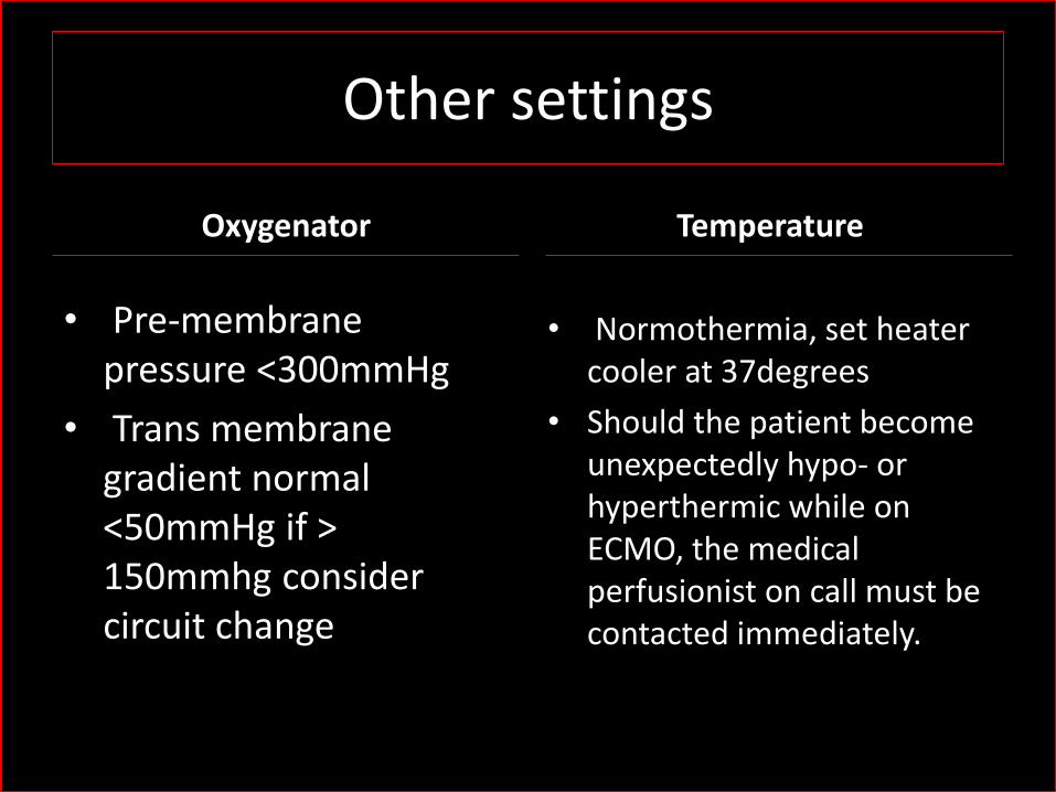
Other settings
Oxygenator
• Pre-membrane pressure <300mmHg
• Trans membrane gradient normal <50mmHg if > 150mmhg consider circuit change
Temperature
• Normothermia, set heater cooler at 37degrees
• Should the patient become unexpectedly hypo- or hyperthermic while on ECMO, the medical perfusionist on call must be contacted immediately.

Anticoagulation
• Although the ECMO circuit has an anticoagulant lining, low-dose heparin is usually administered to prevent clot formation
• First 24 hours ACT 6 hourly.
• A usual target for ACT in the non-bleeding patient with platelet count > 80,000 is 140–160.
• Beyond 24 hours, the APTT 4 times per day.
• A usual target for APTT in the non -bleeding patient with platelet count > 80,000 is 45-55sec.

Investigations for patients on ECMO
• Daily CXR • Daily bloods: CBC, RFTs, Electrolytes, LFT s• Clotting: ACT first 24 hours then APTT 6 hourly• Plasma free Hb is performed when clinically
indicated. The safe range for this is < 0.1g/L. • Blood cultures 3 times per week or as indicated.
samples should be taken from the circuit or through existing lines. Do NOT perform venipuncture.
• Other cultures as indicated

Sedation
• Initial deep sedation to inhibit respiratory efforts.
• Muscle relaxants may be necessary
• Sedation vacation
• Light sedation (Tube tolerant)

General management
• Doppler examination of the blood flow in the back-flow cannula is indicated if deteriorating leg perfusion is observed in the cannulated leg.
• Antibiotics (IV vancomycin) to prevent line sepsis are commenced at the start of ECMO. Other antibiotics are prescribed as indicated.
• Stress ulcer prophylaxis is standard.

Nursing Management
• Patient positioning
• Pressure area care
• Patients with “open sternum”
• Daily chest x-rays and other patient moves require a Jordan Frame and the presence of medical staff and/or perfusionist to ensure no change in circuit flow result as a consequence of movement.

Nursing Management
• Patients nursed at 1:1 nursing ratio
• The ECMO patient is dependent on the circuit for maintenance of oxygenation +/- cardiac output
• Any disruption to ECMO flow will result in rapid deterioration, which if not rapidly rectified will result in death
• The ECMO patient must NOT be left unattended at any time

Hourly observations
Related to PatientTemperatureCVP/MAPHaematuriaCirculation Limb temperature Limb color Pedal pulses Capillary refill Neuro-obsPain assesmentCPOT/RASS
Related to CircuitOozingKinkingLeakageJerks or shakesAnchors/DressingsPre & post membrane pressure Oxygen flow to oxygenator Flow on oxygen outlet
Paraphernalia•Ventilator•CVVHD•PA Cath•PiCCO•IABP
SVO2
Lactate
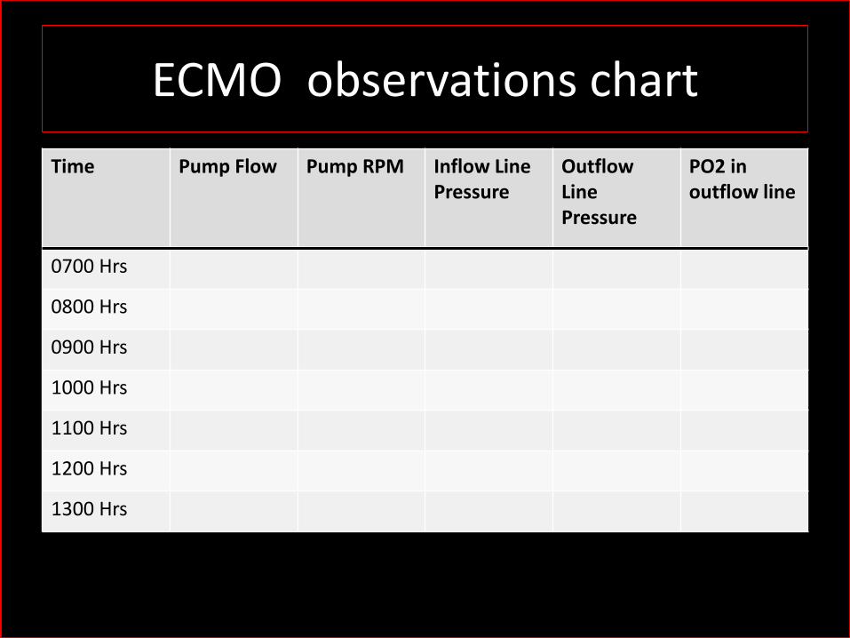
ECMO observations chart
Time Pump Flow Pump RPM Inflow Line Pressure
OutflowLine Pressure
PO2 in outflow line
0700 Hrs
0800 Hrs
0900 Hrs
1000 Hrs
1100 Hrs
1200 Hrs
1300 Hrs

Decreased ECMO Flow
• Fall in preload
• Hypovolemia
• Bleeding
• Mechanical Obstruction– Temponade
– Tension pneumothorax
– Abdominal Compartmental Syndrome
• Increased afterload
– Thrombus
– Increased SVR
– Kinking of tubes
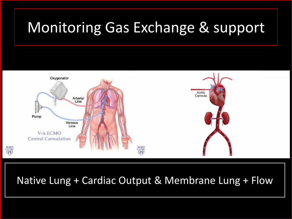
Monitoring Gas Exchange & support
Native Lung + Cardiac Output & Membrane Lung + Flow

Critical Limb Ischemia
• Due to the large bore cannulae distal arterial perfusion may be compromised in A-V ECMO, while the venous cannulae may lead to DVT formation

Critical Limb Ischemia-Prevention

TROUBLESHOOTING


Functional MechanicsCombating Gas Exchange
Modifiable VariablesPreload
AfterloadRPM of impeller
Static VariablesCannula length
Cannula diameter
Pumps preload dependent afterload sensitivevolFlow
Obstruction Flow In RPM Flow In RPM Flow
Gas exchange Diffusion gradient
Surface area
OxygenationBlood flow rate
FDO2
FDO2
CO2 elimination Sweep gas flow rate
Pre and post oxygenator samples reveal
in PaO2 & in PCO2

Hypoxia VENO- VENOUS ECMO
• Check • Pump flow is > 2/3 pt cardiac output
(e.g. CO 6l = 4l pump flow) • 100% O2 to oxygenator • Check Oxygenator: outflow
PO2>150mmHg
• Management • Increase pump flow • Increase ventilation • Cool pt to 35°C • Muscle relaxants • Correct anemia • Second access line to reduce shunt

HypercarbiaVeno-Venous ECMO
• Check
• Pump flow is > 2/3 cardiac output • Oxygen flow to oxygenator twice
pump flow rate( e.g. if pump flow 4l, O2 flow 8l)
• Management • Increase pump flow rate
otherwise consider that recirculation may be occurring
• Increase ventilation • Cool patient to 35°C • Administer muscle relaxants
Too close access & return cannulae may cause recirculation of blood, so Pump flow may not improve oxygenation

Differential Hypoxemia

VENO- ARTERIAL ECMO
• Differential hypoxaemia• Lower PO2 in upper body compared to lower body • Can occur when there is severe respiratory failure with a high cardiac output • Heart supplies upper body with de-oxygenated blood & ECMO supplies lower body
with oxygenated blood
• Check • Patient ABG sample from right radial arterial line (close to heart) • O2 saturation measured on right hand
• Management • Ensure proper functioning of oxygenator return line PO2 >150mmhg • Pump flow as high as possible • Increase ventilation/ PEEP / Fi02 • Resite return line from femoral to right subclavian artery

• Hypercarbia• Check • Adequate pump flow > 2/3 CO • O2 to oxygenator is twice pump flow rate
• Management • Increase pump flow rate • Increase ventilation • Cool pt • Paralysis pt

Bleeding
• Prevention is primary objective • ACT’s day 0; APTT 6hrly • Daily FBC, d-Dimer, fibrinogen • Anticoagulation management with heparin APTT 50-75
sec • If patient bleeding
– Cease heparin. Heparin coated circuits can run for couple days without heparin
– Investigate cause – Platelets, Cryoprecipitate, FFP, packed cells

Hemolysis
Causes Clot in the circuit or near cannulae orifice Access & return insufficiency or obstruction “over spinning” of pump speed
Signs Red or dark brown urine High K+ Renal failure Jaundice (late sign) Access line shaking due to changes in pressure
Management Plasma free haemoglobinIncrease volume Review pump flow settings TOE to ensure cannulae not obstructed Consider changing circuit

ECMO Flow decreased
Check Pre-membrane Pressure
Pre-membrane Pressure ↑ Pre-membrane Pressure ↔
Return line obstruction
Exclude Kinking in lines
Transmembrane gradient > 150Consider Changing
Exclude Kinking in line
IVC Collapse ↓Preload(Line jerks)
IV FluidsRepositioning of access line

“SIG” ALARM ON PUMP CONSOLE
• Contact perfusionist • Re- establish full ventilation • Stop pump slowly & clamp
inflow & outflow lines to pump head
• Remove pump head • Apply silicone cream • Reinsert head & close clip • Unclamp lines & slowly
restart flow • Wrap sensor in cling wrap
to prevent drying out
Flow rate indicator on pump says “SIG” while the pump is still functioning normally. RPM is unchanged Occurs as cream under the flow sensor has dried out

EMERGENCY COMPLICATIONS
Dramatic and life threatening that require immediate action
• General rules
– Call for help, Intensivist, CT surgeon, Perfusionist
– Clamp
– Ventilate, haemodynamic support
Pump Failure
• No flow due to electrical failure or pump head disengagement
• If circuit stopped for any period clotting is possible
• Prevention: – Always maintain the pump head in a position to minimize the risk of
contact especially with devices such as portable x-ray, hemofilter and TOE
– Minimize time on battery
– Ensure AC “Power Off” alarm is turned on when using wall power
– Console not in use, needs to be plugged into AC power and “on Switch” turned on in order to recharge battery

HAND CRANKING
• Take the disposable PUMP out of the CARDIOHELP:
• Fix the disposable to the CARDIOHELP Emergency Drive hand crank
• Open the lower locking device. Swing the disposable right up to the pump drive and release the lower locking device so that it fixes the disposable
• Unfold the hand crank handle. • Open the clamp on the venous
side and turn the hand crank clockwise
• When RPM is > 1500, unclamp lines and increase RPM

Accidental Decannulation
• V-V• Hypoxaemia• Cardiac arrest depending on
cardiac & respiratory reserve • Massive bleeding
• V-A
• Cardiac arrest • Catastrophic if central cannula
torn off aorta • Venous pulled out of atrium • Peripheral blood loss from site
controllable
• Prevention: • Anchoring the cannulae to the
patient • Use of a spotter to ensure that lines
remain free during patient manoeuvres
• Management • Call for help • Clamp circuit • Turn off pump • CPR • Establish ventilation & inotropic
support • Volume • Peripheral: apply pressure • Central: prepare chest opening

AIR EMBOLISM
• If embolus entered patient arterial system (VA) – Clamp arterial return
line
– Stop pump
– Patient head down
– Increase ventilation & inotropes
– Volume
– hypothermia
• If embolus entered venous system (VV) – Aspiration of right heart
using existing lines
– Clamp circuit
– Turn off pump
– Ensure pump head outlet is at 12 o’clock position
– Examine site for air & seal if possible

Cardiac Arrest
• V-V– No patient circulation
– ECMO flow decreases
– Patient in cardiac arrest with no output
• Management – Call for help
– CPR
– Reversible causes
• V-A
• Little hemodynamic effect if flow > 4l/min
• Management– Establish adequate flow
– Call for help
– Reversible causes
– CPR may not be needed unless pump compromised

Circuit Management

• Circuit change is indicated if : – There is a trend towards increasing transmembrane
pressures and / or worsening oxygenator function (oxygenator outflow PaO2 < 150mmHg).
– A normal transmembrane pressure gradient is <50mmHg.
– The decision to change the oxygenator will be based on the trend of transmembrane pressures and oxygenator performance and should also be considered if the ECMO circuit is thought to be a source of sepsis.
Circuit Management

Weaning ECMO
• V-V
• Maintain ECMO flow rate
• Re-establish pt full ventilation
• Turn off O2 to oxygenator
• 6hr stability then de-cannulation

• V-A
• Heparin so ACT >400 to decrease risk clotting
• Decrease pump flow 1litre while ventricular function assess by TOE
• Period of low flow ECMO before decannulation
• Respiratory function is a concern.
– If adequate oxygenation and CO2 removal can be maintained in the presence of this shunt it is likely that respiratory failure can be managed without ECMO.
• If O2 good & C02 managed by ventilation consider decannulation
Weaning ECMO

Physiotherapy during ECMO

Questions?
Thank You