i. prigogine stuart a. rice · peter schuster, institut fur theoretische chemie und strahlenchemie...
TRANSCRIPT

Advances in CHEMICAL PHYSICS
EDITED BY
I. PRIGOGINE
University of Brussels Brussels, Belgium
and University of Texas
Austin. Texas
AND
S T U A R T A. RICE
Department of Chemistry and
The James Franck Institute The University of Chicago
Chicago, Illinois
VOLUME LXXV
AN INTERSCIENCE@ PUBLICATION JOHN WILEY & SONS
NEW YORK CHICHESTER - BRISBANE T O R O N T O SINGAPORE


ADVANCES IN CHEMICAL PHYSICS
V O L U M E LXXV
EDITORIAL BOARD
C. J. BALLHAUSEN, Kobenhaven Universitets Fysisk-Kemiske Institut, Kemisk Labo-
BRUCE BERNE, Columbia University, Department of Chemistry, New York, New
RICHARD B. BERNSTEIN, University of California, Department of Chemistry, Los
G. CARERI, Instituto di Fisica “Guglielmo Marconi,” Universita delli Studi, Piazzle
MORREL COHEN, Exxon Research and Engineering Company, Clinton Township,
KARL F. FREED, The James Franck Institute, The University of Chicago, Chicago,
ROBERT GOMER, The James Franck Institute, The University of Chicago, Chicago,
RAYMOND E. KAPRAL, University of Toronto, Toronto, Ontario, Canada WILLIAM KLEMPERER, Department of Chemistry, Harvard University, Cambridge,
YV L. KLIMONTOVITCH, Moscow State University, Moscow, USSR V. KRINSKI, Institute of Biological Physics, USSR Academy of Science, Puschino,
M. MANDEL, Chemie-Complex der Rijks-Universiteit, Wassenaarseweg, Leiden,
RUDY MARCUS, Department of Chemistry, California Institute of Technology, Pasa-
PETER MAZUR, Institute Lorentz voor Theoretische Natuurkunde, Nieuwsteeg,
GREGOIRE NICOLIS, Pool de Physique, Faculte de Sciences, Universite Libre de
A. PACAULT, Centre de Recherches Paul Pascal, Domaine Universitaire, Talance,
YVES POMEAU, Commissariat a L’Energie Atomique, Centre d’Etudes Nucleares de
A. RAHMAN, Argonne National Laboratory, Argonne, Illinois, U.S.A. P. SCHUSTER, Institut fur Theoretische Chemie und Strahlenchemie, Universita Wien,
I. SHAVITT, Department of Chemistry, Ohio State University, Columbus, Ohio,
KAZUHISA TOMITA, Department of Physics, Faculty of Science, Kyoto University,
ratorium IV, Kobenhaven, Denmark
York, U.S.A.
Angeles, California, U.S.A.
delle Scienze, Rome, Italy
Annandale, New Jersey, U.S.A.
Illinois, U.S.A.
Illinois, U.S.A.
Massachusetts, U.S.A.
Moscow Region, USSR
Netherlands
dena, California, U.S:A.
Leiden, Netherlands
Bruxelles, Bruxelles, Belgium
France
Saclay, Division de la Physique, Gif-sur-Yvette, France
Wein, Austria
U.S.A.
Kyoto, Japan

Advances in CHEMICAL PHYSICS
EDITED BY
I. PRIGOGINE
University of Brussels Brussels, Belgium
and University of Texas
Austin. Texas
AND
S T U A R T A. RICE
Department of Chemistry and
The James Franck Institute The University of Chicago
Chicago, Illinois
VOLUME LXXV
AN INTERSCIENCE@ PUBLICATION JOHN WILEY & SONS
NEW YORK CHICHESTER - BRISBANE T O R O N T O SINGAPORE

An Interscience@ Publication
Copyright@ 1989 by John Wiley & Sons, Inc.
All rights reserved. Published simultaneously in Canada.
Reproduction or translation of any part of this work beyond that permitted by Section 107 or 108 of the 1976 United States Copyright Act without the permission of the copyright owner is unlawful. Requests for permission or further information should be addressed to the Permissions Department, John Wiley & Sons, Inc.
Library of Congress Catalog Number: 58-9935
ISBN 0-471-62219-2
Printed in the United States of America
10 9 8 7 6 5 4 3 2

CONTRIBUTORS TO VOLUME LXXV
GIUSEPPE ALLEGRA, Dipartimento di Chimica, Politecnico di Milano, Milan, Italy
V. A. BENDERSKII, Academy of Sciences of the USSR, Institute of Chemical Physics, Moscow, USSR
*ALEKSANDRA BORYSOW, Physics Department, University of Texas at Austin, Austin, Texas
CLIFFORD E. DYKSTRA, Department of Chemistry, University of Illinois, Urbana, Illinois
MANFRED EIGEN, Max Planck Institut fur Biophysikalische Chemie, Gottin- gen, Federal Republic of Germany
LOTHAR FROMMHOLD, Physics Department, University of Texas, Austin, Texas
FABIO GANAZZOLI, Dipartimento di Chimica, Politecnico di Milano, Milan, Italy
0. I. GERASIMOV, Katholieke Universitet Leuven, Laboratorium Voor Malek- uulfysika, Leuven, Belgium; Odessa State University, Department of Theoretical Physics, Odessa, USSR
V. I. GOL‘DANSKII, Academy of Sciences of the USSR, Institute of Chemical Physics, Moscow, USSR
ERICH, P. IPPEN, Department of Electrical Engineering and Computer Science, Massachusetts Institute of Technology, Cambridge, Massachusetts
PAOLO LAZZERETTI, Dipartimento di Chimica dell’Universit8 degli Studi di Modena, Modena, Italy
*Present address: Joint Institute for Laboratory Astrophysics, University of Colorado, Boulder. Colorado.

vi CONTRIBUTORS TO VOLUME LXXV
*SHI-YI LIU, Department of Chemistry, University of Illinois, Urbana, Illinois
JOHN M C C ~ ~ ~ ~ ~ ~ , Max Planck Institut fur Biophysikalische Chemie, Gottin- gen, Federal Republic of Germany
DAVID J. MALIK, Department of Chemistry, Purdue University at Indiana- polis, Indianapolis, Indiana
KEITH A. NELSON, Department of Chemistry, Massachusetts Institute of Technology, Cambridge, Massachusetts
PETER SCHUSTER, Institut fur Theoretische Chemie und Strahlenchemie der Universitat Wien, Wien, Austria
L. I. TRAKHTENBERG, Academy of Sciences of the USSR, Institute of Chemical Physics, Moscow, USSR
*Present address: Allied Signal Corporation, Des Plaines, Illinois.

INTRODUCTION
Few of us can any longer keep up with the flood of scientific literature, even in specialized subfields. Any attempt to do more and be broadly educated with respect to a large domain of science has the appearance of tilting at windmills. Yet the synthesis of ideas drawn from different subjects into new, powerful, general concepts is as valuable as ever, and the desire to remain educated persists in all scientists. This series, Advances in Chemical Physics, is devoted to helping the reader obtain general information about a wide variety of topics in chemical physics, which field we interpret very broadly. Our intent is to have experts present comprehensive analyses of subjects of interest and to encourage the expression of individual points of view. We hope that this approach to the presentation of an overview of a subject will both stimulate new research and serve as a personalized learning text for beginners in a field.
ILYA PRIGOGINE STUART A. RICE
vii


CONTENTS
FEMTOSECOND COHERENT SPECTROSCOPY By Keith A. Nelson and Erich P. Ippen
AB INITIO DETERMINATION OF MOLECULAR ELECTRICAL PROPERTIES By Clifford E. Dykstra, Shi-yi Liu, and David J. Malik
31
LINESHAPE IN EELS OF SIMPLE DISORDERED SYSTEMS By 0. I . Gerasimov
113
THE MOLECULAR QUASI-SPECIES 149 By Manfred Eigen, John McCaskill, and Peter Schuster
CHAIN CONFIGURATIONS A N D DYNAMICS I N THE GAUSSIAN APPROXIMATION By Giuseppe Allegra and Fabio Ganazzoli
265
QUANTUM CRYOCHEMICAL REACTIVITY OF SOLIDS 349 By V. I. Gol’danskii, V. A. Benderskii, and L. I . Trakhtenberg
COLLISION-INDUCED LIGHT SCATTERING: A BIBLIOGRAPHY By Aleksandra Borysow and Lothar Frommhold
439
GENERAL CONNECTIONS AMONG NUCLEAR ELECTROMAGNETIC SHIELDINS A N D POLARIZABILITIES 507 By Paolo Lozzeretti
AUTHOR INDEX SUBJECT INDEX
1x
55 1 561


ADVANCES IN CHEMICAL PHYSICS VOLUME LXXV


FEMTOSECOND COHERENT SPECTROSCOPY
KEITH A. NELSON*
Department of Chemistry Massachusetts Institute of Technology
Cambridge, Massachusetts 02139
ERICH P. IPPEN
Department of Electrical Engineering and Computer Science Massachusetts Institute of Technology
Cambridge, Massachusetts 02139
CONTENTS
I. Introduction 11. Electronic Phase Coherence
A. Measurement of Electronic Energy
B. Polarization Dephasing
A. Phase-Coherent Excitation Mechanisms 1. Impulsive Stimulated Scattering 2. Inverse Electro-optic Effect 3. Optical Absorption 4.
Relaxation and Dephasing
111. Nuclear Phase Coherence
Other Excitation Schemes: Phase-Coherent Chemistry B. Liquid State Molecular Dynamics C. Chemical Reaction Dynamics References
I. INTRODUCTION
The development of laser sources that can produce pulses of duration under 100 femtoseconds (fs) began in 1981 [l] and has progressed rapidly since [2]. Clearly this has great significance for many areas of physical chemistry. It is
*Alfred P. Sloan Fellow and Presidential Young Investigator Awardee.
1

2 KEITH A. NELSON AND ERICH P. IPPEN
now possible, in principle at least, to time-resolve almost any nuclear motion including collective vibrations in condensed media, molecular orientational motion and intermolecular collisions in gases and liquids, and even many intramolecular vibrations. Since these elementary motions of atoms and molecules are involved in most events of chemical interest (e.g., chemical bond formation and breakage in any phase of matter and local and collective structural rearrangement in condensed phases), the prospect exists for direct time-resolved observation of such events. It is also possible to time-resolve electronic excited-state relaxation and dephasing in many previously inac- cessible cases, permitting mechanistic questions concerned with electron- electron and electron-nuclear interactions to be addressed in detail.
Although speculation about femtosecond time-resolved physical chemis- try experiments has far outpaced their execution, it is fair to say that the outlook of experimental physical chemists has been altered significantly by the possibilities. In molecular spectroscopy, time-resolved observation of nonequilibrium molecular species including transition states is now discussed routinely. Molecular dynamics in liquids and other condensed phases are discussed in the context of experimental observation on the time scale of intermolecular collisions and comparison to corresponding computer simu- lations. The focus of study of electronic excited-state dynamics in semi- conductors and metals, molecular crystals, light-sensitive biological species, and other condensed materials is shifting increasingly toward the femto- second regime.
Several recent reviews have presented broad overviews of ultrafast time- resolved spectroscopy [3-61. We shall concentrate instead on a selected, rather small subset of femtosecond time-resolved experiments carried out (and to a very limited extent, proposed) to date. In particular, we shall review experiments in which phase-coherent electronic or, more often, nuclear motion is induced and monitored with time resolution of less than 100 fs. The main reason for selectivity on this basis is the rather ubiquitous appearance of phase-coherent effects (especially vibrational phase coherence) in femto- second spectroscopy. As will be discussed, nearly any spectroscopy exper- iment on molecular or condensed-phase systems is likely to involve phase- coherent vibrational motion if the time scale becomes short enough. Since the coherent spectral bandwidth of a femtosecond pulse often exceeds collective or molecular vibrational frequencies, such a pulse may perturb and be perturbed by a medium in a qualitatively different manner than a longer pulse of comparable peak power. The resulting spectroscopic possibilities are of special interest to these reviewers.
Measurements of electronic phase coherence and its decay are discussed in the next section. Pump-probe experiments with two incident laser pulses and

FEMTOSECOND COHERENT SPECTROSCOPY 3
polarization grating experiments with three incident pulses are described. Determination of homogeneous and inhomogeneous dephasing dynamics in organic dye molecules is reviewed.
The last section begins by showing that coherent vibrational (in general, nuclear) motion on ground- or excited-state potential surfaces can be in- itiated by a femtosecond pulse through either “impulsive” stimulated Raman scattering or optical absorption. Subsequent ultrashort pulses can be used to probe the coherently vibrating species at various stages of vibrationaldistor- tion, that is, to time-resolve individual cycles of vibrational oscillation. Experiments of this type on phonon and polariton dynamics in crystalline solids, molecular librational motion in liquids, and intramolecular vibrations are reviewed. Femtosecond time-resolved photochemistry experiments in which bond breakage or formation is monitored are also discussed. Finally, anticipated progress in optical control of molecular motion on ground- and excited-state potential surfaces is summarized, with attention focused on the prospects for “phase-coherent chemistry” and time-resolved observation of transition states.
11. ELECTRONIC PHASE COHERENCE
A. Measurement of Electronic Energy Relaxation and Dephasing
In the first reported measurements made with picosecond pulses, an optical beam splitter was used to pick off a portion of the pulse train and a variable optical delay path was introduced between the two beams [7]. The main beam was used to excite (pump) a dye sample, and the weak (probe) beam was used to monitor the recovery of dye transmission as a function of delay. Over the past two decades, this pump-probe method has been extended to a variety of measurement geometries and used to measure electronic polar- ization dephasing times as well as population lifetimes.
Interactions of pump and probe pulses with a material absorption are usually described by density matrix equations for a distribution of two-level or three-level systems. The formulation of these equations can be found in textbooks and other reviews [8,9]. We try here simply to describe in physical terms the ways in which the different parameters of the equations manifest themselves experimentally. The simplest theoretical case is the two-level system, in which T2 is the dephasing time of the coherently induced electronic polarization and TI is the energy relaxation time. We begin our discussion by simplifying even further and assuming that T2 is very short compared to the optical pulse durations. Then, the coherent polarization follows the optical

4 KEITH A. NELSON AND ERICH P. IPPEN
field, and the induced change in absorption (due to excitation of population) has a temporal behavior given by
where El ( t ) is the electric field of the incident pump pulse. The detected signal in a pump-probe measurement is the change in the energy of the transmitted probz pulse. It has the form
where E,(t) is the electric field of the probe pulse and t is the temporal delay between the pump and probe pulses. For a more complicated interaction in which the two fields may have different polarizations and the energy relaxa- tion is nonexponential, Eq. (2) is easily extended to
m
R , , , , ( t - t ’ ) E , ( t ‘ ) E , ( t ’ ) d t ‘ d t - m
(3)
which is the convolution of the energy (in general, susceptibility) relaxation function R,, , , with the (separately measureable) intensity correlation func- tion of the optical pulses. This description, however, ignores the optical interferences of the two pulses when they overlap in time. That interference gives rise to an additional detected signal of the form
This signal, often referred to as the “coherent artifact”, has been discussed by many authors [10-14]. It may be thought of as a scattering of the pump beam into the probe beam (and vice versa) by the periodic interference they produce in the medium. For the case E , ( t ) = E 2 ( t ) , y(0) = B(0). If the pump and probe have different polarizations, these magnitudes may not be equal. Reorien- tation (or loss of polarization memory within the medium) during the pulse can reduce /?(T). If pump and probe have different frequencies, B(T) depends upon the ability of the material response to follow the beat. Thus, this

FEMTOSECOND COHERENT SPECTROSCOPY 5
coherent coupling term may also be used to obtain information about both the medium and the optical pulses. Recent experiments have shown that it can have dramatic spectral as well as temporal manifestations [l5, 161.
Pumpprobe experiments of the type described in the preceding can be used to study energy relaxation in both excited and ground states. Even with pump and probe pulses of the same wavelength, vibrational relaxation in the electronic excited state, solvent reorganization, and other processes may be distinguished from T, recovery to ground state by their different temporal signatures [ 171. With a broadband femtosecond continuum probe, excited- state spectral dynamics can be studied in detail [18]. In these experiments, the coupling to vibrational modes may appear as transient sidebands of a spectral hole burned into the absorption spectrum by the pump pulse.
B. Polarization Dephasing
Different pumpprobe coupling phenomena arise if T2 is not negligibly small compared to the pulse duration. In this domain, studies of coherent transients [19] [the photon echo [20, 211 is a notable example] make it possible to distinguish between different, homogeneous and inhomogeneous, polariza- tion dephasing mechanisms. Consider first the effects of extended polariza- tion coherence on our two-pulse pumpprobe experiment. One direct conse- quence is a distortion of the pumpprobe signal described previously in the region of pulse overlap [14]. This distortion does not, however, depend strongly upon either T, or the dephasing mechanism. A more clear signature is given by light scattered (“self-diffracted”) into a different direction. See Fig. 1. When the noncollinear pump and probe pulses overlap in the absorber, they produce a periodic (gratinglike) excitation that couples them together. It also diffracts the probe beam into the direction defined by 2k, - k , , where k, and k, are the propagation vectors of the probe and pump beams, re- spectively. Even when the probe pulse arrives delayed with respect to the pump, it can still produce a grating and scatter into 2k, - k, if the coherent polarization created by the pump pulse has not completely died out (i.e., been dephased). In the limit of T2 much greater than the pulse durations, the scattered energy depends upon delay o between pump and probe as
if the system is homogeneously broadened. If the system is very inhomoge- neous, this dependence changes to
and the signal is an “echo” that appears after a delay z following the probe

6 KEITH A. NELSON AND ERICH P. IPPEN
pulse. When the approximation of long T, is not valid, the situation is more complicated, but the results are easily calculated from third-order density matrix perturbation theory. The symmetry of the situation implies that when the pump is delayed with respect to the probe, a similar scattering signature should be observed in the direction 2k1 - k,. Thus, when T, effects are present, there will be a temporal asymmetry between scattering in the two directions [22, 231. Conversely, such an asymmetry might be taken as an indication of polarization coherence. Unfortunately, this latter assumption is not always true. Asymmetry in the scattering depends not only on T, but also on Tl and the pulse shape [24]. Figure 2 illustrates this asymmetry for two different pulseshapes in the limit T, << Tp, the pulsewidth FWHM (full width at half maximum), and Tl b Tp. The experimental points were taken using a methanol solution of the dye malachite green as the sample. These data, along with intensity autocorrelation measurements of the scattered pulse durations, have been interpreted to mean that T, < 40 fs in this dye [24].
With a three-pulse geometry it is possible to remove the ambiguity caused by TI and pulseshape [25, 261. Such a geometry is given in Figure 3. A third pulse is used to monitor the grating established by the first two pulses. We emphasize, however, that in contrast to most transient grating experiments, including those described in the next section, the important parameter here is the delay between the first two pulses. The third pulse scatters (“diffracts”) into the background-free directions k, = k, + (k, - k,) and k, = k, - (k, - k,). For pulses much shorter that the inverse absorption spectral width of the sample, the scattered energies exhibit unique signatures of dephasing. If the absorbing system is homogeneously broadened, scattering into the two directions is always symmetric with regard to the delay between pulses 1 and 2:
Figure 1. Interaction geometry for two-pulse studies of polarization coherence.

FEMTOSECOND COHERENT SPECTROSCOPY 7
ENERGY (ARBITRARY UNITS)
INCIDENT PULSE SEPARATION A T (FWWMI
Figure 2. Total energy (integrated intensity) of scattered signal versus incident pulse separation ATin units of incident pulse FWHM. The experimental data were plotted assuming Gaussian incident pulses.
On the other hand, if the system is inhomogeneously broadened, scattering is no longer symmetric. In the limit of predominantly inhomogeneous broad- ening,
for z > 0, and
S4(t) N e4[IT2, S,( t ) = 0 (9)
for z < 0. Here positive t indicates that pulse 1 precedes pulse 2. In this case, the diffracted pulse may be described as a stimulated echo [27] delayed by z relative to pulse 3.
This asymmetry provides a clear and simple criterion for differentiating between the two types of line broadening. It can be explained in the following way. Following excitation by the first pulse, the coherent polarizations of the subsystems (of an inhomogeneous system) assume their natural frequencies

8 KEITH A. NELSON AND ERICH P. IPPEN
DETECTOR
FT-d L T I b
/
5
PULSE # 3 1 m PULSE # 2
k 2
4
BEAM
STOP ’’ \ \ \ E, \
‘ k u DETECTOR
Figure 3. Experimental geometry for dcphasing measurements by three-pulse scattering technique. Pump pulses (1 and 2) produce a grating response that diffracts probe pulse (3) into two orders. In grating experiments described in Section 111 (nuclear phase coherence), T = 0 and probe pulse is incident at the phase-matching angle for Bragg diffraction into only one order.
and develop relative phase shifts with time. The second pulse then interacts with these polarizations to produce a set of subsystem population gratings shifted spatially with respect to each other. Arrival of pulse 3 then generates for each grating a third-order polarization whose initial phase is determined by its spatial shift. At time T+ T, all of the polarization components interfere constructively to form a phased array for radiation in direction k, for T < 0 and k5 for T > 0. Thus, for a fixed T, scattering occurs preferentially in a single direction.
For pulses much longer than T2, the expression for the scattered energy is simply the envelope of the electric field autocorrelation squared for both the homogeneous and inhomogeneous cases:
E , ( t - T ) E : ( 2 ) d t . (10) I rm r SJT) = S , ( T ) =
This property has been used to measure the coherence properties of laser pulses [28]. Because it is readily obtainable by Fourier transform from the

FEMTOSECOND COHERENT SPECTROSCOPY 9
pulse spectrum, the T2 = 0 limit can be determined experimentally [25]. Therefore, fast dephasing times can be resolved by looking for small differ- ences between the scattering data and the transform-determined instanta- neous response. The results of two experimental comparisons are shown in Figure 4. Here, T2 is limited by the inverse dye absorption width, so dashed curves are calculated using the Fourier transform of the pulse spectrum multiplied by the dye absorption spectrum. Since the absorption curves of the two dyes overlap the pulse spectrum differently, different scattering curves are expected. The solid lines are thermal grating scattering data obtained to confirm this prediction. Information about system inhomogeneity was then obtained by comparing these curves to data taken using orthogonal polariza- tions for pulses 1 and 2 to eliminate the thermal effect. Data for a variety of dyes in solution at room temperature indicated homogeneous dephasing on a time scale of less than 20 fs [26].
True evidence of inhomogeneous broadening has been obtained from three-pulse studies of dye molecules in thin films of polymethyl methacrylate (PMMA) [29]. Figure 5 shows the experimental results. The curves of Figure 5a demonstrate that the system is homogeneous at room temperature. Scattering in both directions is symmetric about T = 0 and is determined by pulse coherence. The data of Figure 5b clearly show asymmetry indicating that inhomogeneous broadening is present. It is interesting that most of the
- DATA
'"-400 - 2 0 0 0 200 4 0 0
r (fsec)
Figure 4. Three-pulse scattering data for Rhodamine 640 and Nile Blue in methanol using parallel polarizations. Solid lines, integrated scattered intensity; dashed lines, obtained by Fourier transform from pulse spectrum modified by dye absorption.

10
t 0
W 2 W
0 W
W c c 4 V v)
a
a
t u w z w
a
n w 0: W c I- 4 U VI
KEITH A. NELSON AND ERICH P. IPPEN
60 f8eC
Figure 5. Measured scattering energy for cresyl violet in PMMA as function of delay between pulses 1 and 2. The delay of the third pulse was set to 1.3 ps. Temperatures: (a) 290 K; (b) 15 K.
asymmetry is in the peak shift; there is no corresponding exp( - 4 I z I / T2) behavior in the wings of the curves. The explanation apparently lies in the fact that the molecular absorption must be described by a multilevel system rather than two-level structures. Unlike inhomogeneous dephasing, beating

FEMTOSECOND COHERENT SPECTROSCOPY 11
between several lines of a single molecule leads to irreversible dephasing of the coherent polarization associated with it. The asymmetry of the data in Figure 5b indicates that the complicated spectra of individual molecules are indeed resolvable at low temperatures but that the irregularity of their level spacings suppresses the tail of the time domain data. As the temperature increases, the individual lines of a molecule become unresolvable. Each multilevel mdecule behaves as a homogeneously broadened two-level system with a linewidth equal to the total absorption width. In this context, it should be pointed out that it is possible to burn holes in the absorption spectra of multilevel molecules even though they are homogeneously broadened from a dephasing part of view. The difference between the multilevel case and that of an inhomogeneous distribution of two-level systems is that in the former each molecule has the same absorption spectrum.
The three-pulse scattering geometry offers the possibility of extracting still more information about the sample under investigation by variation of T, the arrival time of the third pulse. As time progresses after interference of pulses 1 and 2, the spatially chirped population gratings of an inhomogeneous system can lose their inhomogeneous character through spectral cross-relaxation. If this spectral cross-relaxation proceeds more rapidly than energy relaxation through TI, the asymmetry of the scattering will also change as a function of T. Observation of this change would provide information about configura- tional or environmental fluctuation in the medium. With dye molecules in the polymer, no spectral cross-relaxation could be detected [24], implying that for the temperature at which the medium became inhomogeneous very little environmental change occurs within T I . There should, however, be other molecular systems for which this probe of spectral cross-relaxation would be especially useful.
Finally, it should be noted that dephasing dynamics may also be inves- tigated with temporally incoherent pulses [3&32]. To the extent that the two- and three-pulse T2 measurements described in the preceding depend upon the electric field correlation and not upon the intensity autocorrela- tions, femtosecond temporal resolution may be obtained with any broadband source. Spectral cross-relaxation and a variety of TI effects, however, can reduce the effectiveness of this technique.
111. NUCLEAR PHASE COHERENCE
When a femtosecond laser pulse passes through nearly any medium, coherent vibrational excitation (in general, initiation of coherent wavepacket propa- gation) is likely [33, 341. One- or two-photon absorption of a visible Oi ultraviolet pulse into an electronic excited state can result in phase-coherent motion in the excited-state potential [35]. Impulsive stimulated Raman scattering can initiate phase-coherent vibrational motion in the electronic

12 KEITH A. NELSON AND ERICH P. IPPEN
ground state [33, 341. In noncentrosymmetric materials, phonon-polariton modes can be excited coherently through the inverse electro-optic effect [36]. The main criterion for any of these coherent excitation mechanisms is that the laser pulse duration be short compared to a single oscillation cycle of the vibrational mode that is to be excited. Equivalently, the transform-limited bandwidth of the pulse must exceed the vibrational frequency.
The coherent motion initiated by an excitation pulse can be monitored by variably delayed, ultrashort probe pulses. Since these pulses may also be shorter in duration than the vibrational period, individual cycles of vibra- tional oscillation can be time resolved and spectroscopy of vibrationally distorted species (and other unstable species) can be carried out. In the first part of this section, the mechanisms through which femtosecond pulses may initiate and probe coherent lattice and molecular vibrational motion are discussed and illustrated with selected experimental results. Next, experi- ments in the areas of liquid state molecular dynamics and chemical reaction dynamics are reviewed. These important areas can be addressed incisively by coherent spectroscopy on the time scale of individual molecular collisions or half-collisions,
A. Phase-Coherent Excitation Mechanisms
1. Impulsive Stimulated Scattering
Most stimulated scattering experiments are carried out by overlapping spatially and temporally two laser outputs of appropriately tuned frequencies and wave vectors (w,, k,) and (w2, k,) to excite coherent phonons (or other Raman-active modes) of the difference frequency and wave vector (42 = w1 - w,, q, = k, - k,) [37]. The excitation process can be described by the stimulated scattering equation of motion [9] for a damped, nondispersive vibrational mode,
where Q is the normal vibrational coordinate, p is the reduced mass density or moment of inertia, 0, is the natural undamped frequency, and y is the dephasing rate of the mode. The right-hand side of Eq. (1 1) is the driving term in which E is the net electric field and ai j = (aeij/aQ),, where E is the dielectric tensor, is the light-scattering coupling constant for incident and scattered light of polarizations i and j . For long pulses or continuous-wave (cw) lasers, the two excitation fields take the form e i (o l t -k l . r ) + C.C. (complex conjugate) and ei(w2t- k 2 * r ) + c.c., and the product contains difference-frequency terms
FEMTOSECOND COHERENT SPECTROSCOPY 13
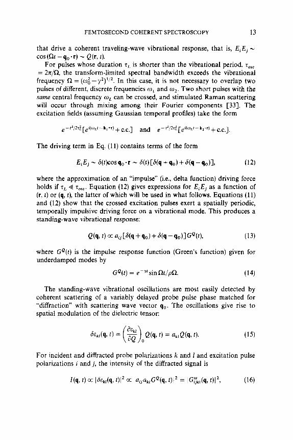
that drive a coherent traveling-wave vibrational response, that is, EiEj - cos (Rt - qo * r) - Q (r, t ) .
For pulses whose duration T~ is shorter than the vibrational period, T,,, = 2n/R, the transform-limited spectral bandwidth exceeds the vibrational frequency R = (m i - y 2 ) ’ I 2 . In this case, it is not necessary to overlap two pulses of different, discrete frequencies w1 and m2. Two short pulses with the same central frequency wL can be crossed, and stimulated Raman scattering will occur through mixing among their Fourier components [33]. The excitation fields (assuming Gaussian temporal profiles) take the form
The driving term in Eq. ( 1 1) contains terms of the form
where the approximation of an “impulse” (i.e., delta function) driving force holds if zL 4 T,,,. Equation (12) gives expressions for EiEj as a function of (r, t ) or (9, t), the latter of which will be used in what follows. Equations (1 1) and (12) show that the crossed excitation pulses exert a spatially periodic, temporally impulsive driving force on a vibrational mode. This produces a standing-wave vibrational response:
where GQ(t) is the impulse response function (Green’s function) given for underdamped modes by
(14) GQ(t) = e - ?‘ sin Rt/pn.
The standing-wave vibrational oscillations are most easily detected by coherent scattering of a variably delayed probe pulse phase matched for “diffraction” with scattering wave vector qo. The oscillations give rise to spatial modulation of the dielectric tensor:
For incident and diffracted probe polarizations k and 1 and excitation pulse polarizations i and j , the intensity of the diffracted signal is

14 KEITH A. NELSON AND ERICH P. IPPEN
where G" is the dielectric tensor impulse response function (often called the nonlinear optical susceptibility and labeled 2). For an underdamped vibra- tional mode, Eqs. (14) and (16) show that
Impulsive stimulated Raman scattering (ISRS) signal shows oscillations at twice the vibrational frequency and decay at twice the vibrational dephasing rate.
In general, several Raman-active modes may be excited simultaneously by the ISRS excitation pulses and may contribute to coherent scattering of the probe pulse. The dielectric response tensor G"(q, t ) may be written as a sum of terms representing different material modes. In addition, depending on the polarizations used, several tensor components of GEE", t ) may be sampled in a given experiment. For simplicity, we write
where the scalar GEe is taken to mean the projection of GEE that is sampled by the polarizations chosen [33, 381.
Using picosecond laser pulses, acoustic phonons in liquids and solids have been characterized through impulsive stimulated Brillouin scattering [3943]. With femtosecond pulses, higher frequency excitations including optic phonons C44-461 and molecular vibrations [34,4749] can be studied through ISRS. Figure 6 shows temperature-dependent ISRS data from optic phonons in the organic molecular crystal a-perylene, whose excimer forma- tion reaction will be discussed further in what follows. The data show a "beating" pattern because two phonon modes of energies 80 and 104 cm-' are excited. In terms of Eq. (16), the signal takes the form I ( t ) ci (a:e-Y1fsin R,t + aie-Y2'sinR2t)2, where the subscripts label the mo- des. Sum and difference frequency terms contribute to the data. From this type of data, temperature-dependent phonon dephasing rates were deter- mined. Similar data have been recorded from the isomorphous excimer- forming crystal pyrene [45] and other crystals [33].
Figure 7 shows ISRS data from dibromomethane liquid. The oscillations in the data correspond to oscillations in the 173-cm-' bromine "bending" mode. The data also contain a nonoscillatory contribution due to molecular orientational motion. Both vibrational and orientational dynamics are ac- counted for in the fit [34, 471.
The impulsive stimulated scattering (ISS) experiment is a stimulated, time domain analog of spontaneous, frequency domain light-scattering (LS) spec-

FEMTOSECOND COHERENT SPECTROSCOPY 15
PERYLENE ISRS DATA ( a axis)
TIME (ps)
Figure 6. ISRS data from a-perylene crystal at two temperatures, recorded with transient grating experimental arrangement. Oscillations in each sweep due to 80- and 104-cm-' optic phonons. Data contain sum and difference frequencies that produce "beating" pattern.
troscopy. The connection between the two is clear from the classical limit expressions for the LS spectrum [SO, 511,
where the frequency-dependent response function G"(co) is the Fourier transform of G"(t). The second part of Eq. (19) relates the LS spectrum to the dielectric tensor time correlation function C"(t), whose elements are c$';,(q, t ) = (E$(q, o)&k,(q, t ) ) . The connection between ISS data and C" is made through the relation
A detailed comparison of ISS and LS techniques, including simulated data

16
h c,
Cn C cu c, C H
.d
KEITH A. NELSON AND ERICH P. IPPEN
r
CHa13rz V-V - /FIT------/ I
Y
J . A A A A
Figure 7. Time-resolved observation of molecular vibrations in CH,Br, liquid recorded with a transient grating experimental arrangement using V-polarized pulses. The 5.2-THz oscillations correspond to oscillations of 173-cm- ' bromine “bending” mode which was excited coherently through ISRS.
and a discussion of their relative advantages in different cases, has been presented [33, 521.
The data shown in the preceding were collected using spatially and temporally overlapped femtosecond excitation pulses as shown in Figure 1 (with ‘t = 0). However, it was predicted theoretically [53] and later shown experimentally [47] that a single ultrashort laser pulse will also excite a coherent vibrational response through ISRS. For an i-polarized, z-propagat- ing excitation pulse the driving force in Eq. (1 1) contains terms of the form E,? - J(t’ ) , where the finite speed c/n of the pulse in the sample (neglected earlier) is accounted for by defining a “local time” variable t’ = t - znjc. The single pulse exerts a temporally impulsive, spatially uniform force on a Raman-active mode. The vibrational response is therefore also spatially uniform:
Q(z, t ) a aiiGQ(t’) a e-Y(r-zn’c)sin(Rt-qz), (21)
where q = Rn/c is the vibrational wave vector that is collinear with that of the pulse (i.e., along the z axis). Physically, the excitation pulse first drives a vibrational response in the front of the sample, then the middle, and then the bdck, so the vibrational phase varies linearly with z. The vibrational wave- length is equal to the distance light travels in the sample during one vibrational period; that is, the optical and vibrational phase velocities are