i. differentiation of the lutein cell from the granulosa
TRANSCRIPT

O V A R I A N S T E R O I D C E L L S
I. Differentiation of the Lutein Cell
from the Granulosa Follicle Cell during the
Preovulatory Stage and under the Influence
of Exogenous Gonadotrophins
E. J O A N B L A N C H E T T E
From the Departments of Anatomy and Obstetrics and Gynecology, Columbia University, College of Physicians and Surgeons, New York
A B S T R A C T
The granulosa follicle cell of the Graafian follicle of the rabbit ovary differentiates into a lutein cell involved in steroid synthesis. Cytological events which occur within the granulosa cell of the normally stimulated follicle prior to ovulation have been duplicated by the intra- follicular injection of exogenous gonadotrophin. The luteinization of the granulosa cells involves the accumulation of 250- to 300-A, electron-opaque, spherical granules, dispersed within the cytoplasmic matrix, which have been identified as glycogen with the PAS- staining procedure. Further development of the granulosa cell following ovulation involves an increase in cell size, a decrease in the number of RNP particles, and an accumulation of an abundant system of intracellular membranes (agranular endoplasmic reticulum). Glycogen granules first appear in the granulosa cells as the separate, monoparticulate form. After follicle rupture and the formation of agranular endoplasmic reticulum, glycogen particles are present in a rosette arrangement within membrane-bounded vacuoles. The rosette arrangement of glycogen particles is also found dispersed within the cytoplasmic matrix of the lutein cell during the later stages of the cell life-span. Injection of luteinizing hormone or human chorionic gonadotrophin into a mature follicle also produces a marked accumulation of monoparticulate glycogen in the majority of granulosa ceils, within 30 rain. Cytoplasmic extensions which contain the glycogen masses are noticeably free of RNP particles.
I N T R O D U C T I O N
The ovary is a heterogeneous tissue containing endocrinologically active structural subunits in- cluding the Graafian follicle and corpus luteum which provide an ideal system to study the differ- entiation, maturation, and degeneration of cells involved in steroid synthesis and secretion. A timed series of stages during cell differentiation
prior to follicle rupture may be obtained from the rabbit, since it is a reflex ovulator. Recent investi- gations, both in vivo and in vitro, have provided information concerning the role of the vesicular follicle in steroid metabolism (12, 16). Although preovulatory progestin release after mating (10) or gonadotrophic stimulation (16, 17) has been de-
501
Dow
nloaded from http://rupress.org/jcb/article-pdf/31/3/501/1068157/501.pdf by guest on 12 February 2022

termined in the rabbit, the cellular site of origin of
this secretion is not known. Both the theca follicle cells and granulosa cells have been implicated in
the formation of steroid metabolites present in the follicular fluid (9, 12). The synthetic activity of the granulosa cells has been established in the
formation of mucopolysaccharides using S 3~ as a tracer (24) and in relation to protein turnover in
the follicular fluid (23). Variat ion in function is to be expected between
the parietal granulosa cells and the cells surround- ing the oocyte that comprise the corona radiata, which is released with the secondary oocyte at follicle rupture. The parietal granulosa cells re- main, hypertrophy, and reorganize into a com- pact lutein body (6, 32). I t is these cells that have been examined in the present study. Although gonadotrophins such as luteinizing hormone (LH) and human chorionic gonadotrophin (HCG)
have been shown to stimulate steroid synthesis in vitro and in vivo (11, 16) and also follicle rupture in vivo (14), their effect on cellular differentiation
has not previously been elucidated. I t is the purpose of this study to report the fine
structural changes which occur in the granulosa cell compar tment of the pre- and postovulatory follicles in the rabbit : ( 1 ) during the differentiation of the granulosa cell into a lutein cell, and (2) after a local injection of gonadotrophins into the follicle. Subsequent reports will deal with struc- tural changes in the cells of other ovarian com- par tments during various physiological conditions
(2).
M A T E R I A L 8 A N D M E T H O D S
Female Dutch rabbits varying in weight from 2.0 to 3.0 kg were used for this study. They were received at 5 to 6 months of age and maintained for l month before mating. It was determined that 12 hr after mating most of the mature follicles had rupture points on their surfaces. Mature Graaflan follicles are prominent in the ovary and are characterized by a light pink color and translucent appearance. Specimens of mature, intact preovulatory follicles and of ruptured follicles were sectioned completely, to further establish the presence or absence of the oocyte. One to several Graafian follicles were col- lected from each ovary in a total of twenty animals.
Various fixatives and buffer solutions were em- ployed. In some cases tissue specimens were subjected to more than one procedure, while in other instances only one fixative per ovary was used. In the latter case, cold fixative at 4°C buffered at pH 7.4 was flushed onto the ovarian surface in situ for 1 rain.
Then, specimens were removed and put into fresh cold fixative. The variation in preservation between the Graafian follicle fixed in toto and the follicle diced during fixation was minimal.
The majority of follicles were fixed in 2% OsO4 in phosphate buffer at pH 7.4. Other preparations were fixed with glutaraldehyde (31). After fixation in the cold OsO4 for 2 hr, the tissue was carried through graded acetones and then transferred to Epon. Polymerization of the Epon was carried out in gelatin capsules at 45°C for 10 hr, and 60°C for 48 hr. Sections were cut on a Porter-Blum microtome and stained with Karnovsky's mixture A lead stain (19) for 5 min or a solution of 1% sodium borate saturated with uranyl acetate for 30 rain (35). Sec- tions were examined with RCA EMU-3C and 3F microscopes.
The gonadotrophins employed were luteinizing hormone (NIH-ovine-LH-S7) and human chorionic gonadotrophin (APL-Ayerst). Consistent results were obtained with intrafollicular injections containing 8 /zg of LH in 1 /zl of saline solution and 1.0 IU of HCG in 1 #1 of sterile diluent. Injections of 1 #1 of isotonic saline containing 1 #g of bovine albumin (Nutritional Biochemical Corp., Cleveland, Ohio) as the control protein were also carried out.
The injection was accomplished with a sterile 10-#1 syringe with a 1-in., 30-gauge needle or glass capillaries drawn out to a fine tip much smaller than a 30-gauge needle. Although the steel needle penetrated the follicle more easily than the glass needle, it had the disadvantage of clogging after 2 or 3 injections. The solutions were injected directly into the mature Graafian follicle. Volumes of 1 to 5 ~1 were tested. A 1-/zl injection was tolerated without noticeable distention of the follicle under the dissecting microscope. A larger volume produced an enlargement of the follicle, with escape of material at the penetration point.
Granulosa cell size and shape did not vary in rela- tion to the method of injection, although disruption of the follicle occurred in every case. Injection of solution into the follicle can be accomplished on the first penetration of the needle, which is then slowly withdrawn 1 min later, allowing no obvious flow of fluid from the follicle. In some cases, material was allowed to escape from the follicle by withdrawing the needle once before the injection or by producing a second rupture point in the follicular wall.
It was possible to inject three Graafian follicles per ovary by careful notation of the position of the follicles on the ovarian surface. The injected follicle was removed 15 and 30 min later, along with a normal noninjected follicle, from each ovary.
Both injected and noninjected follicles were pre- pared for electron microscopy a n d also for light microscopy, allowing the identification of glycogen. Tissue fixed in Rossman's fluid and embedded in
502 T ~ . JOURNAL OF CELL BIOLOGY " VOLUME 31, 1966
Dow
nloaded from http://rupress.org/jcb/article-pdf/31/3/501/1068157/501.pdf by guest on 12 February 2022

FmVRE 1 Light micrograph of a mature Graafian follicle of rabbit. The oval follicle is 1 mm across its longest diameter. The germinal epithelium (GE) covers the surface of the ovary facing the peritoneal cavity (PC). The granulosa follicle cells (GF), comprise the follicular wall beneath the ovarian surface. Strands of granulosa cells extend into the follicular antrum (A) toward the cells of the corona radiate (CR) surround- ing the oocyte. The basement membrane separates the cells of the theca interna (TI) from the follicle cells. Epon section stained with toluidine blue. X 160.
paraplast was utilized for the PAS procedure. The test employed for glycogen was the lack of staining of sections subjected to a solution of malt diastase before the PAS staining.
R E S U L T S
Mature Graafian follicles were studied in both nonstimulated and stimulated animals at varying intervals during the pre- and postovulatory pe- riods.
Follicle during Estrus
The organization of the mature Graafian follicle, which protrudes from the surface of the adult rab- bit ovary, is illustrated in the light micrograph of an Epon-embedded section stained with toluidine blue (Fig. 1). The oval follicle is about 1 mm across its largest axis. Granulosa follicle cells (GF) approximately one to four layers deep corn-
prise the major portion of the follicular wall im- mediately beneath the ovarian surface. At the basal end of the follicle, many more cell layers are present. Strands of granulosa follicle cells (the retinaculum) extend from the surface toward the cells of the corona radiata (CR) which surround the oocyte. The theca interna (TI) is a narrow layer separated from the follicle cells by a base- ment membrane consisting of a light, homogeneous component and a thin, denser one. The basement membrane is immediately adjacent to the basal surface of the granulosa cell (Fig. 2).
The granulosa cells in the adult rabbit Graafian follicle vary in shape, being columnar and 12 to 14 /z in height (Fig. 2) in the periphery, and cuboidal and extremely irregular within the fol- licle. The peripheral cells exhibit polarity, with the nucleus situated toward the basal region and their irregular cytoplasmic prolongations extend-
E. JOAN BLANCHETTE Ovarian Steroid Cells. I 503
Dow
nloaded from http://rupress.org/jcb/article-pdf/31/3/501/1068157/501.pdf by guest on 12 February 2022
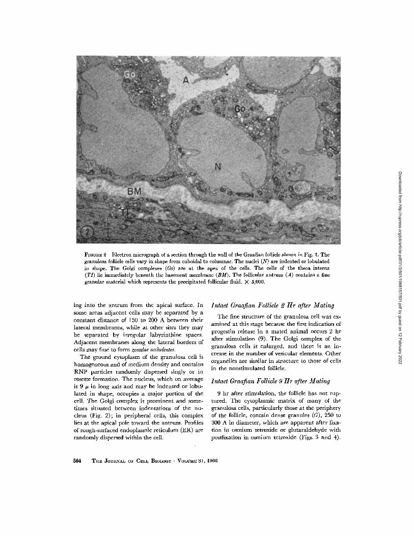
Fmtra~, ~ Electron micrograph of a section through the waU of the Graafian follicle shown in Fig. 1. The granulosa follicle cells vary in shape from cuboidal to columnar. The nuclei (N) are indented or lobulated in shape. The Golgi complexes (Go) are at the apex of the cells. The cells of the theca interna (TI) lie immediately beneath the basement membrane (BM). The follicular antrum (A) contains a fine granular material which represents the precipitated follicular fluid. X 5,000.
ing into the antrum from the apical surface. In some areas adjacent cells may be separated by a constant distance of 150 to 200 A between their lateral membranes, while at other sites they may be separated by irregular labyrinthine spaces. Adjacent membranes along the lateral borders of ceils may fuse to form zonulae occludentes.
The ground cytoplasm of the granulosa cell is homogeneous and of medium density and contains R N P particles randomly dispersed singly or in rosette formation. The nucleus, which on average is 9/z in long axis and may be indented or lobu- lated in shape, occupies a major portion of the cell. The Golgi complex is prominent and some- times situated between indentations of the nu- cleus (Fig. 2); in peripheral cells, this complex lies at the apical pole toward the antrum. Profiles of rough-surfaced endoplasmic reticulum (ER) are randomly dispersed within the cell.
Intact Graafian Follicle 2 Hr after Mating
The fine structure of the granulosa cell was ex- amined at this stage because the first indication of progestin release in a mated animal occurs 2 hr after stimulation (9). The Golgi complex of the granulosa cells is enlarged, and there is an in- crease in the number of vesicular elements. Other organelles are similar in structure to those of cells in the nonstimulated follicle.
Intact Graafian Follicle 9 Hr after Mating
9 hr after stimulation, the follicle has not rup- tured. The cytoplasmic matrix of many of the granulosa cells, particularly those at the periphery of the follicle, contain dense granules (G), 250 to 300 A in diameter, which are apparent after fixa- tion in osmium tetroxide or glutaraldehyde with postfixation in osmium tetroxide (Figs. 3 and 4).
504 'l~v. J'OVaNAL OV CEI,L BIoLoGY • VOLtrME 31, 1966
Dow
nloaded from http://rupress.org/jcb/article-pdf/31/3/501/1068157/501.pdf by guest on 12 February 2022

FIGURE 3 Granulosa follicle cells 9 hr after mating. Small, electron-opaque granules (G) ~50 to 300 A in diameter are present in the cytoplasm. They are numerous in the upper cell. RNP particles (P), nucleus (N) of the cell, and the irregular plasma membrane (CM) air labeled. X 18,000.
These granules are regular, homogeneous spheres without discernible substructure and are randomly distributed throughout the cell. They are dis- persed among the free RNP (P) particles which occur singly or as rosettes. Rough-surfaced endo- plasmic reticulum is present, and in some cells the smooth-surfaced vesicles and tubules are increased in amount (Figs. 3 and 4).
Ruptured Follicles 12 Hr after Mating
In a newly ruptured follicle, the granulosa ceils are disarranged and occur in compact groups. Some of the cells are hypertrophied to a diameter of about 15 #. The spaces between epithelial cells are increased markedly, particularly at the pe- riphery of the follicle. The epithelial cells have rounded contours and few cytoplasmic extensions. The agranular endoplasmic reticulum (AER) is now abundan t in many cells (Fig. 5). Aggregates of irregular tubules of the reticulum leave the
plane of section and thus appear vesicular. In the whorled patterns of smooth endoplasmic reticu- lure, linear densities are obvious (Fig. 5) which are produced, in part, by the apposition of the membranous cisternal and tubular walls. This finding is not an artifact of sectioning, since these profiles appear in various planes, not necessarily parallel to the plane of the section. Small dense granules (G), 250 to 300 A in diameter, are present along the periphery of the cell and also dispersed among the interconnected tubules of the agranular endoplasmic reticulum (Fig. 6). In many sites, 250- to 300-A granules appear, in a single section, to be present within smooth membrane-bounded vesicles (Fig. 6, at G). However, it may be that these vesicles are enlarged cisternal fenestrations. Both free RNP particles and profiles of granular endoplasmic reticulum are decreased in amount in the epithelial cells of the ruptured follicle.
E. JOAN BLANCHETTE Ovarian 8retold Cells. I 505
Dow
nloaded from http://rupress.org/jcb/article-pdf/31/3/501/1068157/501.pdf by guest on 12 February 2022

FIGURE 4 A section similar to that of Fig. 3, showing two adjacent granulosa cells within a follicle 9 hr after mating. Granules (G) 250 to 300 A in diameter stain intensely with lead hydroxide and are larger than the RNP particles (P) associated with the membranes of the endoplasmic reticulum. The nucleus (N) and mitochondria (M) are shown. )< ~0,000.
Mature Graafian Follicle 30 Min after Local
Injection of L H or HCG
The granulosa cell is approximately 10 to 11 /z in diameter. Many cells have a rounded or oval contour. The plasma membrane on the surface not in contact with adjacent cells is thrown into many outpocketings which protrude into the fol- licular antrum (Fig. 12). This elaboration of the cell surface is in the form of elongated as well as circular extensions (Fig. 14) and is found in fol- licles injected with either LH or HCG. The granu- lar endoplasmic reticulum is in the form of mem- brane-bounded cisternae, often dilated, lying parallel to the circular contours of the cell border (Fig. 12).
Dense 250- to 300-A granules (G) are present within the cells (Figs. 11 to 13). They are regular in contour and extremely osmiophilic. They occur singly or as strings of a few to several granules (Figs. 12 and 14) associated with a fine filamentous
cytoplasmic material. The granules are present throughout the cytoplasm (Fig. 11) and are also aggregated in the outpocketings and extensions (CP) of the cell surface (Fig. 12). In the latter instances, the cytoplasmic matrix surrounding the granules is very sparse. Few organelles are present in the cytoplasmic extensions.
Sections of the Graafian follicle stained with the periodic acid-Schiff reaction and controlled for the identification of glycogen exhibit a strong posi- tive reaction after the intrafollicular injection of L H (Figs. 9 and 10) or HCG. Electron micro- graphs of the same follicle as shown in those figures indicate an accumulation of 250 to 300 A spherical granules in the cells (Fig. 11). The PAS-positive reaction identifies the granules as glycogen. In preparations for electron microscopy, the granules exhibit an affinity for lead, which is demonstrated by the highly stained glycogen in Fig. 11. R N P granules which are distributed in the cytoplasm are often not numerous at sites in which glycogen
506 THE JOURNAL OF CELL BIOLOGY • VOLUME 31, 1966
Dow
nloaded from http://rupress.org/jcb/article-pdf/31/3/501/1068157/501.pdf by guest on 12 February 2022
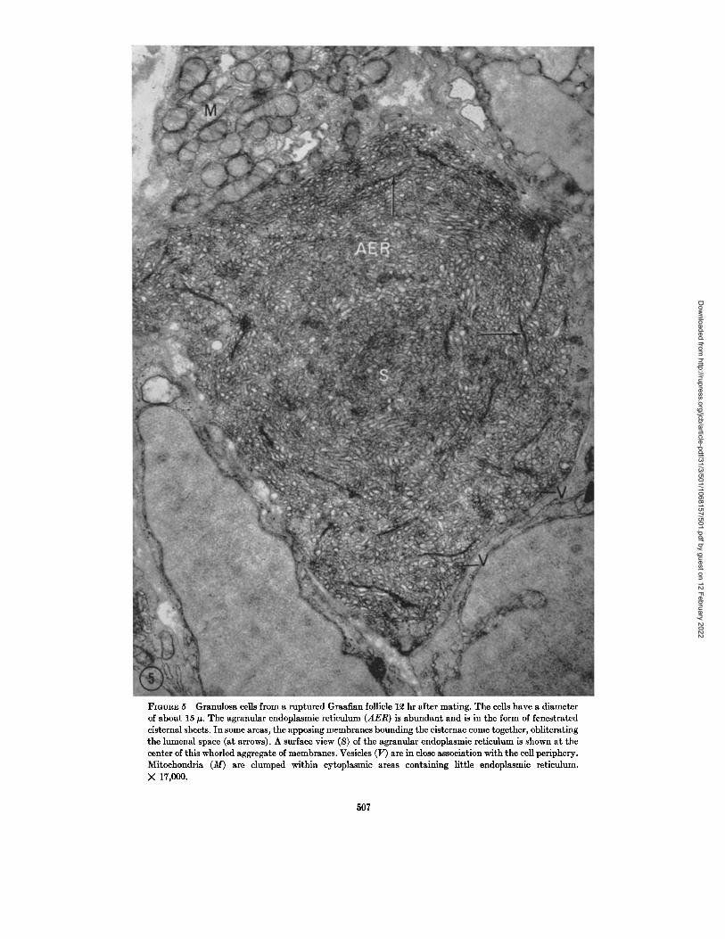
FIOURE 5 Granulosa cells from a ruptured Graafian follicle 12 hr after mating. The cells have a diameter of about 15 ~. The agranular endoplasmie reticulum (AER) is abundant and is in the form of fenestrated cisternal sheets. In some areas, the apposing membranes bounding the cisternae come together, obliterating the lumenal space (at arrows). A surface view (S) of the agranular endoplasmic reticulum is shown at the center of this whorled aggregate of membranes. Vesicles (V) are in close association with the cell periphery. Mitoehondria (M) are clumped within cytoplasmic areas containing little endoplasmic reticulum. X 17,000.
507
Dow
nloaded from http://rupress.org/jcb/article-pdf/31/3/501/1068157/501.pdf by guest on 12 February 2022
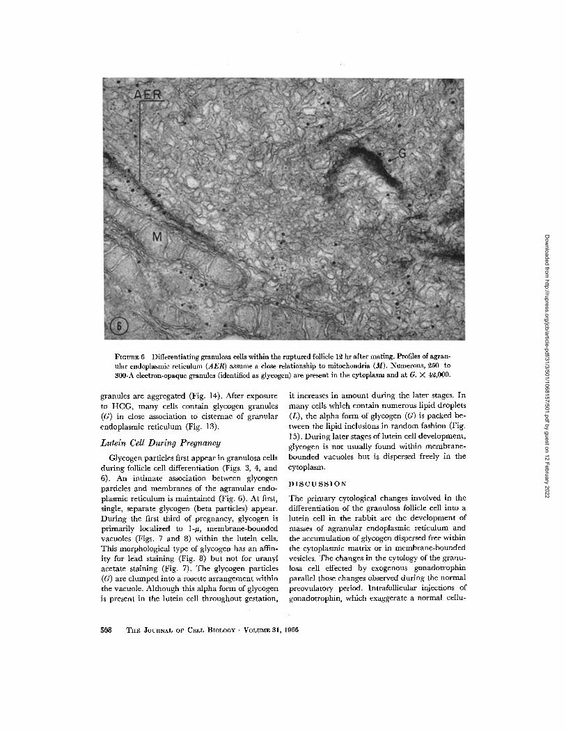
Fioun~: 6 Differentiating granulosa cells within the ruptured follicle 1~ hr after mating. Profiles of agran- ular endoplasmic reticulum (AER) assume a close relationship to mitochondria (M). Numerous, ~50- to $00-A electron-opaque granules (identified as glycogen) are present in the cytoplasm and at G. )< 4~,000.
granules are aggregated (Fig. 14). After exposure to HCG, many cells contain glycogen granules (G) in close association to cisternae of granular endoplasmic reticulum (Fig. 13).
Lutein Cell During Pregnancy
Glycogen particles first appear in granulosa cells during follicle ccll differentiation (Figs. 3, 4, and 6). An intimate association between glycogen particles and membranes of the agranular endo- plasmic reticulum is maintained (Fig. 6). At first, single, separate glycogen (beta particles) appear. During the first third of pregnancy, glycogen is primarily localized to 1-#, membrane-bounded vacuoles (Figs. 7 and 8) within the lutein cells. This morphological type of glycogen has an affin- ity for lead staining (Fig. 8) but not for uranyl acetate staining (Fig. 7). The glycogen particles (G) are clumped into a rosette arrangement within the vacuole. Although this alpha form of glycogen is present in the lutein cell throughout gestation,
it increases in amount during the later stages. In many cells which contain numerous lipid droplets (L), the alpha form of glycogen (G) is packed be- tween the lipid inclusions in random fashion (Fig. 15). During later stages of lutein cell development, glycogen is not usually found within membrane- bounded vacuoles but is dispersed freely in the cytoplasm.
D I S C U S S I O N
The primary cytological changes involved in the differentiation of the granulosa follicle cell into a lutein cell in the rabbit are the development of masses of agranular endoplasmic reticulum and the accumulation of glycogen dispersed free within the cytoplasmic matrix or in membrane-bounded vesicles. The changes in the cytology of the granu- losa cell effected by exogenous gonadotrophin parallel those changes observed during the normal preovulatory period. Intrafollicular injections of gonadotrophin, which exaggerate a normal cellu-
508 THE ffOURNAL OF CELL BIOLOGY • VOLUME 81, 1966
Dow
nloaded from http://rupress.org/jcb/article-pdf/31/3/501/1068157/501.pdf by guest on 12 February 2022

FIGURE 7 A l-it membrane-bounded vacuole containing glycogen (G) within a lutein cell during the first third of pregnancy. Stained with uranyl acetate. )< 54,000.
FIGVaE 8 A membrane-bounded vacuole containing glycogen (G) in a lutein cell, similar to that of Fig. 7. Stained with lead hydroxide. X 54,000.
lar response, become a useful device for the identi- fication of glycogen and the study of its accumula- tion within the cytoplasmic matrix. Previous reports on the fine structure of the granulosa cell of the rabbit have been concerned with maturing follicles (15, 22) and not the cells of the Graafian follicle during the preovulatory stage. One report has shown that the granulosa cells in the primary, vesicular, and postovulatory follicles of the rat have an abundance of free RNP particles and a limited amount of granular endoplasmie reticulum (5). Luteinization in the rat, which occurs before ovu- lation, is accompanied by mitochondrial changes and the accumulation of agranular endoplasmic reticulum (5).
The differentiation of the lutein cell (6, 13) represents the formation of a cell primarily in- volved with steroid metabolism (30). However, the metabolic activity of the granulosa cells in- volves the secretion of mucopolysaccharides into the follicular antrum as a component of the pri- mary liquor (24, 37) and perhaps the formation of
an enzyme which depolymerizes the acid muco- polysaccharides during the preovulatory phase, producing a less viscous secondary liquor, follicular swelling, and rupture (18, 38). The presence of many free RNP particles in the cytoplasm of the granulosa cell (Fig. 2) is characteristic of "retain- ing cells" or rapidly enlarging and undifferenti- ated cells, and not of those particularly active in secretion (4). There is no clear cytological evidence in the rabbit that the preovulatory steroid secre- tory response of the ovary resides in the luteinizing (a descriptive term that is not defined chemically) granulosa ceils. 2 hr after mating, when progestin activity is present in the blood (10), the ceils show a slight increase in the number of Golgi vesicles. A few lipid droplets are present in cells of the estrous follicle and those of later stages, increasing in number after follicle rupture. The osmiophilic properties of the lipid droplets may be correlated with their sterol content (2). An increase in the amount of smooth-surfaced reticulum is apparent as early as 12 hr after mating (Fig. 5). However,
E. JOAN BLANCHETTE Ovarian Steroid Cells. I 509
Dow
nloaded from http://rupress.org/jcb/article-pdf/31/3/501/1068157/501.pdf by guest on 12 February 2022

an accumulation of glycogen precedes the forma- tion of large quantities of agranular endoplasmic reticulum. There is a variation in the amount of glycogen present in the parietal granulosa cells. Uniform, 250- to 300-A granules are scattered in some cells and not in others (Figs. 3 and 4). Al- though within the size range of glycogen particles, the granules have a regularity of contour and density that is not the same as that noted for monoparticulate glycogen (3, 28). A PAS reaction when applied to these follicles is not positive. How- ever, 30 min after injection of LH or H C G into a mature follicle, sections of the follicle exhibit a PAS-positive reaction which is removed by diastase digestion (Figs. 9 and 10). The large accumula- tions, within the cell, of electron-opaque, spherical granules morphologically identical to those within the cells of the preovulatory follicle (compare Figs. 4 and 11) are believed to be responsible for the positive PAS reaction, and on this evidence the particles arc considered to be glycogen. The glycogen granules observed within the granulosa follicle cell show a uniformity of contour and density.
Two types of glycogen particles (alpha and beta) have been distinguished in pellets and tissue sections for electron microscopy (3, 8, 29). I t has been established that glycogen fractions have the same morphology and staining characteristics as intracellular glycogen (7, 29). The beta particles, or monoparticulate glycogen, 150 to 400 A in diameter, are roughly spherical in shape, with irregular contours, and have a subunit structure as determined on negatively stained glycogen pellets (7) or on unfixed, shadowed preparations (28). I t has been suggested that the punctate appearance
of monoparticulate glycogen may represent an alignment of structure within the glycogen granule. Since glycogen is a branched polysaccharide, there may be a relationship between the morpho- logical unit structure and the molecular arrange- ment within glycogen particles (28). The absence of punctate appearance of the glycogen of the lutein cell may indicate that the molecule is slightly branched.
In the present study, the spherical glycogen granules are randomly distributed in the granulosa cells 9 hr after mating. The cytoplasm at this stage contains profiles of granular endoplasmic reticu- lure and free RNP particles. Glycogenesis within the granulosa cell occurs before the accumulation of agranular endoplasmic reticulum and is not associated with these membranous elements of the cytoplasm. The association of glycogen with structural elements of the cell cytoplasm has fostered a divergence of opinion concerning the functional significance of these relationships in the process of glycogenesis and glycogenolysis. Par- ticularly, the presence of agranular endoplasmic reticulum in glycogen-rich areas of the cytoplasm has led to speculation concerning their enzymatic activity in these regions. An association between glycogen and smooth endoplasmic reticulum in liver cells has been noted in studies on the effect of azo dyes and of starvation in rats (25). Glycogen granules have also been observed within Golgi vesicles as well as free in the cytoplasmic matrix in developing chick liver cells (20). Clarification of glycogen metabolism has been provided by biochemical studies showing that the UDPG l-
1 UDPG = uridine diphosphate glucose.
FmuuE 9 Light micrograph of a Graafian follicle 30 min after injection of LH into the follicle. Stained for PAS-positive material. The surface of the follicle (S) and the follicular fluid (FF) are labeled. The PAS-positive material of the granulosa ceils, identified as glycogen, is illustrated at arrows. X 200.
FmVRE 10 A high magnification of the area outlined within the box in Fig. 9. The PAS-positive cytoplasm (at arrows) contrasts with the nuclei (N) which were counter- stained with fast green. X 450.
FmvnE 11 Granulosa follicle cell from the same follicle shown in Figs. 9 and 10, a portion of which was fixed for electron microscope observations. Accumulations of 250- to 800-A glycogen granules (G) are present throughout the cytoplasm. The granules exhibit an affinity for lead and appear extremely electron opaque after staining. The cell nucleus (N) is labeled. X 15,000.
510 THE JOURNAL OF CELL BIOLOGY • VOLUME 31, 1966
Dow
nloaded from http://rupress.org/jcb/article-pdf/31/3/501/1068157/501.pdf by guest on 12 February 2022

E. JOAN BLANCHETTE Ovarian Steroid Cells. I 511
Dow
nloaded from http://rupress.org/jcb/article-pdf/31/3/501/1068157/501.pdf by guest on 12 February 2022

F m ~ E 12 Granulosa follicle cell 30 rain after intrafollieular injection of LH. Two bulbous cytoplasmic projections (CP) are obvious at the cell periphery. Within one of these cytoplasmic areas, numerous dense granules (G) are present but other cell organdies are absent. The lumen of the endoplasmie retieulum (Lu) is extremely dilated. Note that the granules (G) are present in the expanded cytoplasmic area and not with- in the endoptasmic reticulum. Adjacent cells in the disrupted follicle are separated by large follicular spaces (F). The cisternae of the granular endoplasmic reticulum (GER) are aligned parallel to the granulosa cell surface. X 14,000.
glycogen transglucosylase necessary for glyco- genesis is present in the glycogen fraction (21) and that the phosphorylase (21) and phosphorylase- activating enzyme (26) necessary for glycogenol- ysis are localized in the postmicrosomal superna- tant and not bound to cell organelles (21).
In the granulosa follicle cells exposed to LH and HCG, areas in which glycogen granules are most abundant are sparse of RNP particles. This, of course, could be a fixation artifact, but it may also indicate a synthesis of glycogen at the expense of RNP particles. A similar accumulation of glyco- gen granules in association with RNP particles has been observed in the cytotrophoblast of the human placenta (36), and it was proposed that the uridine nucleotides contributing to the uridine pathway
for glycogen synthesis are liberated through a decomposition of the RNP particles.
The aggregation of glycogen dur ing differentia- tion may he for later use as a substrate when, it is conceivable, lipogenesis is occurring in relationship to glycogenolysis. Large accumulations of mem- branes of the smooth endoplasmic reticulum (Fig. 5) and lipid inclusions are the cytological evidence that lipid synthesis is an important function of lutein cells which are undergoing hypertrophy. Other studies which suggest a similar relationship between lipogenesis and glycogen metabolism, resulting in an increase in size of fat droplets and a decrease in the amount of glycogen have been performed on the inguinal fat body of fetal mice after birth (34), and during fatty metamorphosis
512 THE JOURNAL OF CELL BIOLOGY • VOLUME 31, 1966
Dow
nloaded from http://rupress.org/jcb/article-pdf/31/3/501/1068157/501.pdf by guest on 12 February 2022

FIOURE 13 Granulosa follicle cell 30 min after intrafollicular injection of HCG. The basal portion of the cell contains dilated granular endoplasmic reticulum, with associated RNP particles (P). Note the align- ing of glycogen granules (G) in close association with the endoplasmic reticulum. Mitoehondria (M) are labeled. X ~0,000.
FmtTRE 14 ~50- to 300-A granules (G) within a cytoplasmic extension of a granulosa follicle cell, from an LH-injected follicle 30 min after injection. The granules occur singly or in strings of a few to several (at arrows), associated with a fine filamentous material. The follicular antrum (FA) is labeled. X 40,000.
in the rat liver (1). I t is reasonable to suggest that the granulosa follicle cell synthesizes glycogen when glucose is available, perhaps from the follicu- lar fluid, or when a stimulation is present, perhaps gonadotrophic, for future use in a lipogenic ca- pacity. I t appears from this study that the accumu- lation of glycogen is an early morphological indi- cator of lutein cell differentiation in a normal, nonatretic follicle. An increase in glycogen content of the developing corpus luteum from 24 to 120 hr after mat ing has been reported (27). I t is during this period of glycogen content increase that num- erous beta glycogen particles have been identified dispersed in the granulosa cells. The subsequent plateau in glycogen accumulation (27) correlates with the presence of membrane-bounded glycogen aggregates. This second form of glycogen, the
alpha particle, is a complex unit which consists of clumps of beta particles, described as a rosette for- mation of glycogen (8). The size of the aggregate varies greatly. Membrane bounded glycogen ag- gregates are a conspicuous feature of the lutein cell during the first half of pregnancy. The character- istics of the alpha glycogen are such that the parti- cles are not stained by uranyl acetate (Fig. 7) but exhibit a deep staining quality with lead saIts (Fig. 8). This differential staining has been utilized as a morphological criterion of alpha glycogen granules (28) present during the later stages while a similar intense lead staining of the monoparticulate, beta glycogen has been corre- lated with the more reliable criterion of PAS stain- ing. Glycogen accumulations which increase dur- ing the later stages of the lutein cell life-span occur
:E. ~OAN BLANCHETTE Ovarian Steroid Cells. 1 513
Dow
nloaded from http://rupress.org/jcb/article-pdf/31/3/501/1068157/501.pdf by guest on 12 February 2022

lq'IOURE 15 Luteln cell 25 days after mating. Glycogen accumulations (G) are packed between lipid drop- lets (L) of little electron opacity. This alpha glycogen consists of clumps of individual (beta) particles. Mitochondria (M) are also labeled. )< 27,000.
as glycogen aggregates freely dispersed in the cytoplasmic matrix (Fig. 15). At this stage, the cells have also accumulated numerous, light-ap- pearing lipid droplets which are present duri~ag presumed "storage" phases of cellular activity (2). It is believed that glycogen masses in the lutein cell during later stages of pregnancy are another indication of reduced synthetic activity. Glycogen has been reported to occur in the rabbit lutein cell at maximum histochemical levels during the first third of pregnancy and to subsequently decrease in amount (33). The present morphological ob- servations also indicate that glycogen is most prevalent immediately after follicle rupture. Lu- tein cells containing dispersed alpha glycogen (Fig. 15) are not numerous at later stages. The significance of the early sequestering of alpha glycogen within membrane-bounded vacuoles and the subsequent free dispersion of a morphologically similar particle is not known. It may represent transitory structural fluctuations concomitant with the metabolic utilization of this molecule.
Membrane-bounded glycogen aggregates, be-
lieved to be related to the Golgi area, as well as glycogen granules free in the cytoplasmic matrix have been described in the developing chick em- bryo (20).
The observations made in this study indicate that the pattern of events include the formation of glycogen in a monoparticulate form, the pre- sumed utilization of this glycogen in a lipogenic capacity, and the subsequent sequestering of the glycogen in an aggregated fashion as the life-span of the lutein cell reaches its culmination.
This study was supported in part by United States Public Health Service Grants, Training Grant 571-GM-256, Division of General Medical Sciences, NB-03448-05, and by a grant from the Ford Founda- tion.
The work was submitted in partial fulfillment of the degree of Doctor of Philosophy, Anatomy De- partment, Columbia University.
The author wishes to express her appreciation to Dr. George D. Pappas for his guidance and advice during the course of this work. Received for publication 16 May 1966.
514 THE JOURNAL OF CELL BIOLOGY • VOLUME 31, 1966
Dow
nloaded from http://rupress.org/jcb/article-pdf/31/3/501/1068157/501.pdf by guest on 12 February 2022
R E F E R E N C E S
1. AMICK, C. J., and STENGER, R. J. , Ultrastruc- rural alterations in acute hepatic fatty meta- morphosis, Lab. Inv., 1964, 13, 128.
2. BLANCI-mTaX, E. J., Ovarian steroid cells. II. The lutein cell, J. Cell Biol., 1966, 31, 517.
3. BIAVA, C., Identification and structural forms of human particulate glycogen, Lab. Inv., 1963, 12, 1179.
4. BIRBECK, M. S. C., and MERCER, E. H., Cytol- ogy of ceils which synthesize protein, Nature (London), 1961, 189, 558.
5. BJSRKMAN, N. A., A study of the ultrastructure of the granulosa cells of the rat ovary, Acta Anat., 1962, 51, 125.
6. DEANSLFV, R., The development and vascu- larization of the corpus luteum in the mouse and rabbit, Proc. Roy. Soc. London, Series B, 1930, 106, 578.
7. DROCHMANS, P., Mise en 6vidence du glycog~ne dans la cellule h6patique par microscopic 61ectronique, J. Biophysic, and Biochem. Cytol., 1960, 8, 553.
8. DROCHMANS, P., Morphologie du glycog&ne, J. Ultrastruct. Research, 1962, 6, 141.
9. FALCK, B., Site of production of oestrogen in rat ovary as studied in microtransplants, Acta Physiol. Scan&, 1959, 47, suppl., 163, 1.
10. FORBES, T. R., Pre-ovulatory progesterone in the peripheral blood of the rabbit, Endocrinol., 1953, 53, 79.
11. GOSPODAROWICZ, D., The action of follicle stimu- lating and of human chorionic gonadotrophin upon steroid synthesis by rabbit ovarian tissue in vitro, Acta Endocrinol., 1964, 47, 293.
12. GOSPODAROWICZ, D., The in vitro production of androgens by follicular tissue of rabbits, Acta Endocrinol., 1964, 47, 306.
13. HAMMOND, J., and MARSHALL, F. H. A., Repro- duction in the Rabbit, London, Oliver and Boyd, Ltd., 1925.
14. HARPER, M. J., The time of ovulation in the rabbit following the injection of luteinizing hormone, or. Endocrinol., 1961, 22, 147.
15. HASHIMOTO, M., TSUTOMU, K., MITSUO, K., YORISATO, M., KMORI, A., SHIMOYAMA, T., and KATSUHIDE, A., Electron microscopic studies on the fine structure of the rabbit ovarian follicles, reports I and II, J. ,lap. Obstet. Gynec., 1960, 7, 228 and 267.
16. HILLIARD, J., ARCHIBALD, D., and SAWYER, C. H., Gonadotrophic activation of pre- ovulatory synthesis and release of progestin in the rabbit, Endoerinol., 1963, 72, 59.
17. HILLIARD, J., ENDROCZI, E., and SAWYER, C. H., Stimulation of progestin release from rabbit
ovary in vivo, Soc. Exp. Biol. and Med. Proc., 1961, 108, 154.
18. JENSSN, C. E., and ZACHARIAE, F., Studies on the mechanism of ovulation (Isolation and analysis of acid mucopolysaccharides in bovine follicular fluid), Acta Endocrinol., 1958, 27, 356.
19. KARNOVSKY, M. J., Simple methods for "staining with lead" at high pH in electron microscopy, J. Biophysic. and Biochem. Cytol., 1961, 11, 729.
20. KARRER, H. E., Electron-microscopic study of glycogen in chick embryo liver, J. Ultrastruct. Research, 1960, 4, 191.
21. LucK, D. J. L., Glycogen synthesis from uridine diphosphate glucose. The distribution of the enzyme in liver cell fractions, J. Biophysic. and Biochem. Cytol., 1961, 10, 195.
22. MERK~R, H. J., Elektronenmikroskopisehe Un- tersuchungen fiber die Bildung der Zona Pellucida in den Follikeln des Kaninchenovars, Z. Zellforsch., 1961, 54, 677.
23. MI~ILLER, H. G., and LINNERTZ-MIKLAS, A., Autoradiographische Untersuchungen fiber die GrSsse der Eiweiss-Synthese der weiblichen Genitalorgane im Metoestrus bei Ratte und Maus, Arch. Gynak., 1960, 194, 48.
24. ODEBLAD, E., and BOSTROM, H., A time-picture relation study with autoradiography on the uptake of labelled sulphate in the Graafian follicles of the rabbit, Acta Radiol., Stockholm, 1953, 39, 137.
25. PORTER, K. R., and BRUNI, G., An electron microscope study of the effects of 3-Me-DAB on rat liver cells, Cancer Research, 1959, 1% 997.
26. RALL, T. W., SUTHERLAND, E. W., and BERTHET, J. , The relationship of epinephrine and gluca- gon to liver phosphorylase. IV. Effect of epi- nephrine and glucagon on the reactivation of phosphorylase in liver homogenates, J. Biol. Chem., 1957, 224, 463.
27. RENNIE, P., DAVENPORT, G. R., and WELBORN, W., Cellular changes in protein and glycogen content in developing rabbit corpora lutea, Anat. Rec., 1965, 133, 289.
28. REVEL, J. P., Electron microscopy of glycogen, J. Histochem. and Cytochem., 1964, 12, 104.
29. REVEL, J. P., NAPOLITANO, L., and FAWCETT, D. W., Identification of glycogen in electron micrographs of thin tissue sections, J. Biophysic. and Biochem. Cytol., 1960, 8, 575.
30. RYAN, K. J., and SMITH, O. W., Biogenesis of estrogens by the human ovary. III. Conversion of cholesterol-4-C 14 to estrone, J. Biol. Chem., 1961, 236, 2204.
E. JOAN BLANCHETTE Ovarian Steroid Cells. I 515
Dow
nloaded from http://rupress.org/jcb/article-pdf/31/3/501/1068157/501.pdf by guest on 12 February 2022

31. SABATINI, D. D., B~NSCH, K. G. and BARRNETT, R. J. Cytochemistry and electron microscopy. The preservation of cellular ultrastructure and enzymatic activity by aldehyde fixation, J. Cell Biol., t963, 17, 19.
32. TO~ARI, C., On the corpus luteum of the rabbit, Folia Anat. Jap., 1926, 4, 347.
33. TOGARI, C., On the appearance of glycogen in the female reproductive glands of rodents, with special reference to their histology, Folia Anat. Jap., 1927, 5, 429.
34. WASSERM~, F., and McDONALD, T. F., Electron microscopic study of adipose tissue (fat organs) with special reference to the transport of lipid between blood and fat cells, Z. Zellforsch., 1963, 59, 326.
35. WATSOS, M. L., Staining of tissue sections for
electron microscopy with heavy mesals, J . Biophysic. and Biochem. Cytol., 1958, 4, 475 and 727.
36. YOSHIDA, Y., Ultrastructure and secretory func- tion of the syncytial trophoblast of human placenta in early pregnancy, Exp. Cell Research, 1964, 34, 305.
37. ZACHARXAE, F., Studies on the mechanism of ovulation. Autoradiographic investigations on the uptake of radioactive sulphate (35S) into the ovarian follicular mucopolysaccharides, Acta Endocrinol., 1957, 26, 215.
38. ZACHARIAE, F., and JENSEN, C. E., Studies on the mechanism of ovulation (Histochemical and physico-chemical investigations on genuine follicular fluids), Acta Endocrinol., 1958, 27, 343.
516 ThE JOURNAL OF CELL BIOLOGY " VOLUME 31, 1966
Dow
nloaded from http://rupress.org/jcb/article-pdf/31/3/501/1068157/501.pdf by guest on 12 February 2022