hypothesis open access epistemology of the origin of
TRANSCRIPT

Brücher and Jamall BMC Cancer 2014, 14:331http://www.biomedcentral.com/1471-2407/14/331
HYPOTHESIS Open Access
Epistemology of the origin of cancer: a newparadigmBjörn LDM Brücher1,2,3,4,5,6,7* and Ijaz S Jamall1,2,3,4,5,6,8*
Abstract
Background: Carcinogenesis is widely thought to originate from somatic mutations and an inhibition of growthsuppressors, followed by cell proliferation, tissue invasion, and risk of metastasis. Fewer than 10% of all cancers arehereditary; the ratio in gastric (1%), colorectal (3-5%) and breast (8%) cancers is even less. Cancers caused by infection arethought to constitute some 15% of the non-hereditary cancers. Those remaining, 70 to 80%, are called “sporadic,” becausethey are essentially of unknown etiology. We propose a new paradigm for the origin of the majority of cancers.
Presentation of hypothesis: Our paradigm postulates that cancer originates following a sequence of events that include(1) a pathogenic stimulus (biological or chemical) followed by (2) chronic inflammation, from which develops (3) fibrosiswith associated changes in the cellular microenvironment. From these changes a (4) pre-cancerous niche develops,which triggers the deployment of (5) a chronic stress escape strategy, and when this fails to resolve, (6) a transitionof a normal cell to a cancer cell occurs. If we are correct, this paradigm would suggest that the majority of the findingsin cancer genetics so far reported are either late events or are epiphenomena that occur after the appearance of thepre-cancerous niche.
Testing the hypothesis: If, based on experimental and clinical findings presented here, this hypothesis is plausible, thenthe majority of findings in the genetics of cancer so far reported in the literature are late events or epiphenomena thatcould have occurred after the development of a PCN. Our model would make clear the need to establish preventivemeasures long before a cancer becomes clinically apparent. Future research should focus on the intermediate steps ofour proposed sequence of events, which will enhance our understanding of the nature of carcinogenesis. Findings oninflammation and fibrosis would be given their warranted importance, with research in anticancer therapies focusing onsuppressing the PCN state with very early intervention to detect and quantify any subclinical inflammatory change andto treat all levels of chronic inflammation and prevent fibrotic changes, and so avoid the transition from a normal cell toa cancer cell.
Implication of the hypothesis: The paradigm proposed here, if proven, spells out a sequence of steps, one or more ofwhich could be interdicted or modulated early in carcinogenesis to prevent or, at a minimum, slow down theprogression of many cancers.
Keywords: Cancer, Paradigm, Inflammation, Fibrosis, Carcinogenesis, Tumor, Neoplasm
BackgroundCancer is a complex and heterogeneous set of diseaseswith no simple definition [1]. A century ago, tumor growthalone was considered the fundamental derangement, andtumors were classified and described in terms of theirgrowth rates: (1) slow, (2) moderately rapid, and (3) rapid[2]. Today, carcinogenesis is thought to be triggered by
* Correspondence: [email protected]; [email protected]®, Munich, Germany2Theodor-Billroth-Academy®, Richmond, VA, USAFull list of author information is available at the end of the article
© 2014 Brücher and Jamall; licensee BioMed CCreative Commons Attribution License (http:/distribution, and reproduction in any mediumDomain Dedication waiver (http://creativecomarticle, unless otherwise stated.
mutations [3] and an inhibition of growth suppressors,which, in turn, gives rise to the cell proliferation, tissueinvasion, and risk of metastasis [4].
Mutation and polymorphismOver the past several decades, the theory that somaticmutations are the primary trigger for carcinogenesis hasbecome the predominant paradigm to explain the originof most cancers. In fact, the German surgeon and cancerresearcher, Karl-Heinrich Bauer (1928), on observing
entral Ltd. This is an Open Access article distributed under the terms of the/creativecommons.org/licenses/by/4.0), which permits unrestricted use,, provided the original work is properly credited. The Creative Commons Publicmons.org/publicdomain/zero/1.0/) applies to the data made available in this

Brücher and Jamall BMC Cancer 2014, 14:331 Page 2 of 15http://www.biomedcentral.com/1471-2407/14/331
mutations in plants and animals, offered the then plausiblebiological explanation that cancers were likely caused bymutations [5]. Some rare cancers have indeed been shownto involve mutations, most notably the deoxyribonucleicacid (DNA) damage that ensues from exposure to non-lethal doses of ionizing radiation [6]. The Watson andCrick discovery, aided by Rosalyn Franklin’s X-ray dif-fraction study of DNA [7], achieved in large measure by“theoretical conversation…little experimental activity” [8],served to elucidate the three-dimensional structure ofDNA [9] and gave credence to the concept that damage toDNA molecules can lead to cancer. Although some50 years ago, Ashley stated that cancer may be the resultof just 3 to 7 mutations [10], and since then, others haveproposed different possible numbers of critical mutations[11,12], the number necessary to cause a normal cell tochange to a cancer cell is not yet known. The clinicaland laboratory evidence suggests that carcinogenesisrequires more than mutations since, in order for a can-cer to develop, the DNA repair mechanism would have tobe absent, defective, or inefficient, as seen, for example,in children with Xeroderma pigmentosum [13]. Somaticmutations are increasingly questioned as drivers of car-cinogenesis [14,15], and some cancers are not associatedwith any mutation [16,17]. Furthermore, the inactivationof tumor suppressor genes is also involved in the celltransformation process [18]. In this context, one group ofresearchers has suggested illuminating the process bycomparing genomes among different species for example,those of a mouse or rat to those of the naked mole rat,which is resistant to cancer [19]. In recent years, thecontribution of chronic inflammation to cell transformationhas been revisited, although the mechanism of inflamma-tion and its importance have yet to be elucidated [20]. Longthought to play a role in the development of cancer, inflam-mation is again under scrutiny, in light of recent data.Until recently, the source of cancers was thought to be
(1) hereditary, (2) infectious or (3) sporadic. Hereditarycancers occur in 5 to 10% of all cancers and in some 8%of breast and ovarian cancers, which are associated withgenetic changes as BRCA1 or BRCA2 [21]; the equivalentfigure for colorectal cancer is between 3 and 5%. Some15% are thought to be caused by infection [22,23], a ratioperhaps misleading, as it is about 60% of gastric cancersand as high as 80% of hepatic cancer [24]. The remainingcancers (70-80%) are considered sporadic, a euphemismfor “unknown cause”. Only 15% of sporadic cancers aretraced to somatic mutations [25], but a carrier is notautomatically afflicted, although his risk for the associatedcancer may be greater than 50%. Intra-patient hetero-geneity and variability have always hampered the searchfor uniform and effective therapies, and heterogeneityremains a huge impediment to assigning one origin tomany different types of cancer.
Fully 99.9% of all mutations that occur within the codingregions of the genome are not understood, nor have theybeen investigated. Additionally, the number of mutatedgenes and mutations per cancer are, a small percentageof mutations in a coding region varies greatly [26]: 97%of mutations are single-base substitutions and about 3%are insertions or deletions. Furthermore, of the reportedsingle-base mutations, 90.7% are missense changes, 7.6%are nonsense, and 1.7% involve splice sites located innon-translated regions that immediately follow a startor stop codon. The number of mutated genes varies, witha smaller number of somatic mutations observed in thepopulation of younger patients with a cancer than thatof older patients with the same cancer. The number ofobserved mutations varies among tissues of the sourcecancer: tissue of cancers with high rates of cell division, suchas the colon, exhibit more mutations per cell than that ofcancers in slowly dividing tissues, such as the brain [26,27].The enormous variability of mutations, combined with
the fact that more than half of these occur even beforethe cancer phenotype is established, leads to an elevated“noise to signal” ratio in the exon sequencing data [26,27].Mutations are assumed to occur over long periods oftime - even as long as several decades. Because of thelong time frame, it is reasonable to assume that thedata from sequencing vary greatly according to the timeof sample collection. Investigation to understand mutationsis of significant importance to understanding even moreprofound underlying biological processes.Genetic polymorphism is also important for understand-
ing the processes, as two or more different phenotypes mayexist in the same individual. Biologists usually investigatecertain point mutations in the genotype, such as single-nucleotide polymorphisms (SNPs) or variations in homolo-gous DNA by restriction fragment length polymorphisms(RFLPs), with chromatography, chromosome cytology, orby exploiting genetic data. Neither the mechanisms nor thedistribution of different polymorphisms among individualgenes are well understood, although the latter is considereda major reason for the evolutionary disparity that survivesnatural selection [28]. Polymorphisms are necessary tounderstanding biology - including tumor biology - butare not be the key to solving cancer genomics.The reasons why polymorphisms are not a viable route
for unraveling cancer genomics are multiple: (1) We do notunderstand how polymorphisms reflect a disease or re-spond to a treatment, or even if they react in coordinationwith other polymorphisms in other genes. (2) On 23 July2013, the number of SNPs published in the Single Nucleo-tide Polymorphism Database (dbSNP) was 62,676,337[29]. (3) Human beings have 23 paired chromosomes(46 in each cell) and, according to data from the HumanGenome Project, humans probably have 21,000 haploidcoding genes with approximately 3.3 × 10⁹ base pairs [30].

Brücher and Jamall BMC Cancer 2014, 14:331 Page 3 of 15http://www.biomedcentral.com/1471-2407/14/331
(4) Chromosome 1 of the 46, with its 249,250,621 basepairs, has 4,401,091 variations [31]. (5) The mutationrate is estimated to be 10−6 to 10−10 in eukaryotes [32],a piece of data that could permit a calculation of thepossible combinations. (6) However, the number ofpseudogenes - about 13,000 [30] - and (7) the wide vari-ation of transposable (mobile) genetic DNA sequencescomplicate such a calculation [33,34]. For example, Aluhas about 50,000 active copies/genome, while another,LINE-1 (=long interspersed element 1), has 100. (8) Tothe best of our knowledge, mobile genetic elements -classified under CLASS I DNA transposons as LTRs(long terminal transposanable retroposons) and non-LTRs,such as long interspersed elements (=LINEs) and shortinterspersed elements (=SINEs), and CLASS II DNAtransposons - account for more than 40% of the totalgenetic elements [35].In addition to these eight reasons, we note that neither
the genetic information nor the different cells aloneinfluence biological processes [36]; the extracellular matrix(ECM) is essential for cellular differentiation and thusinfluences that differentiation directly, as well as providingstabilizing ligament fibroblasts [37]. Moreover, only 50%of patients with disseminated tumor cells and circulatingtumor cells (CTCs) develop clinically evident metastaticcancer, and only 0.01% of disseminated cells and CTCsdevelop metastasis [38,39]. Even the fact that cancerouscells have been observed in vitro without inflammation orfibrosis does not account for the vast majority of cancersfor which mutations cannot explain their development.Normal cellular processes that damage DNA include the
generation of reactive oxygen species (ROS), alkylation,depurination, and cytidine deamination [40]. The magni-tude of DNA damage affected by normal cellular processesis enormous, estimated at approximately ten thousanddepurinated sites generated per cell per day; an evengreater number of alterations results from ROS [41,42].This DNA damage is continuously monitored andrepaired; over 130 DNA repair products have beenidentified [43]. In normal cells, DNA replication andchromosomal segregation are exceptionally accurateprocesses. Measurements of the mutagenesis of cellsgrown in culture yield values of approximately 2×10⁻¹⁰
single base substitutions per nucleotide in DNA per celldivision, or 1×10⁻⁷ mutations/gene/cell division. Aneven lower number has been demonstrated in culturedstem cells [40,44]. Taking into account this very lowfrequency of mutation, the spontaneous mutation rateof normal cells seems insufficient to generate the largenumber of genetic alterations observed in human cancercells. If a cancer arises in a single stem cell, then the spon-taneous mutation rate would account for less than onemutation per tumor. That discrepancy led to a hypothesis,as yet unproven, of a “mutator phenotype,” which - by
envoking genomic instability - might account for thegreater number of somatic mutations observed [45].These sobering considerations reflect the complexity
of biological processes. We think it unlikely, logically andcomputationally, to find the needle - the origin of cancers -in this huge haystack. After depending on the somaticmutation paradigm for some 85 years, these consider-ations justify contemplating a paradigm shift. Biologicalprocesses as well as cell-cell communication and signal-ing are themselves a multidimensional musical opera indifferent acts, which are played differently by differentsymphony orchestras rather than by a soloist. Even thecomposition of the music, which is needed before it canbe played, is not well understood.We propose an alternate hypothesis for the origin of
the majority of cancers. Our paradigm postulates thatcancer originates following a sequence of events thatinclude (1) a pathogenic stimulus (biological or chemical),followed by (2) subclinical chronic inflammation, fromwhich develops (3) fibrosis with associated changes inthe cellular microenvironment. From these changes, (4)a pre-cancerous niche (PCN) develops, which triggers(5) deployment of a chronic stress escape strategy (CSES)with (6) a normal cell-cancer cell transition (NCCCT)(Figure 1). In this paper, we justify our hypothesis byshowing why it deserves consideration as the explanationfor the genesis of most cancers.
Presentation of the hypothesis
(1) Pathogenic Stimulus
The earliest information a cell receives is a pathogenic(biological or chemical) stimulus. The first receiverseems to play a major role in processing the stimulus.Chemical carcinogenesis is thought to be a two-stepprocess: in the first step, called “initiation,” thecarcinogen damages or binds to nuclear DNA; in thesecond step, referred to as “promotion,” some otherchemical or physiologic event facilitates the aberrantgrowth that ultimately results in cancer. The classicexample was reported by Yamigawa and Ichikawa in1915, when they applied coal tar derivatives to rabbitears and observed skin cancer [46]. Subsequent workshowed that dermal application of several differentpolyaromatic hydrocarbons (PAHs), such as benzo[a]pyrene and benzo[a]anthracene, followed by aphorbol ester (a promoter), generated skin cancersin a dose-dependent manner. Over time, alkylatingagents, such as sulfur mustard, ethylene dibromide,and many nitrosoamines, were included in the listof chemicals that could give rise to cancer, both inexperimental animals and in humans. The list grew toinclude arsenic, hexavalent chromium, mycotoxins -notably aflatoxins - ionizing and ultraviolet radiation,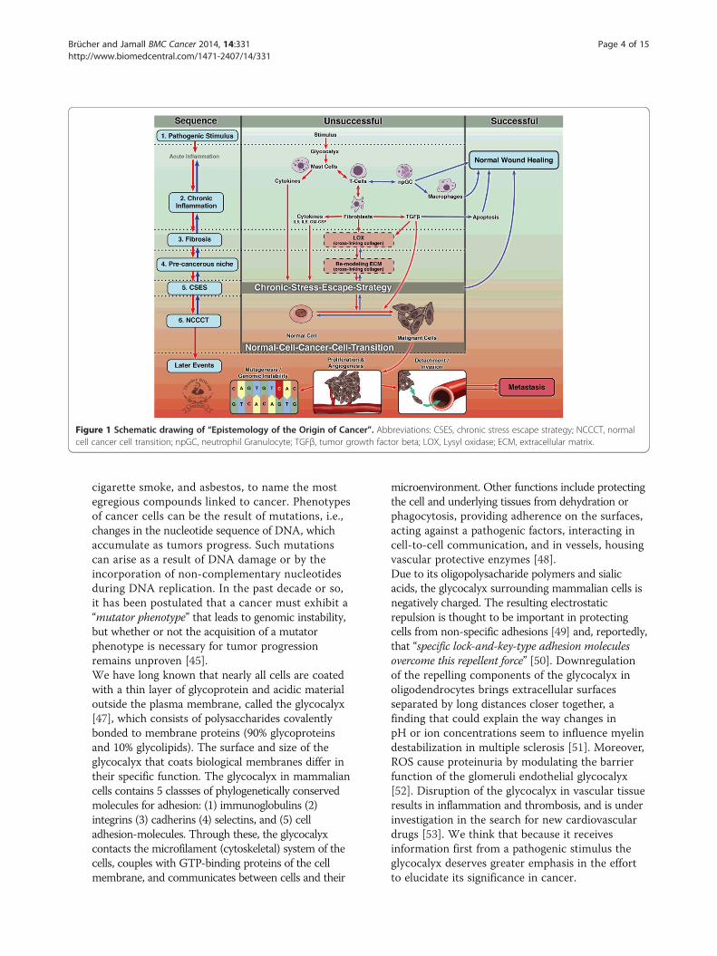
Figure 1 Schematic drawing of “Epistemology of the Origin of Cancer”. Abbreviations: CSES, chronic stress escape strategy; NCCCT, normalcell cancer cell transition; npGC, neutrophil Granulocyte; TGFβ, tumor growth factor beta; LOX, Lysyl oxidase; ECM, extracellular matrix.
Brücher and Jamall BMC Cancer 2014, 14:331 Page 4 of 15http://www.biomedcentral.com/1471-2407/14/331
cigarette smoke, and asbestos, to name the mostegregious compounds linked to cancer. Phenotypesof cancer cells can be the result of mutations, i.e.,changes in the nucleotide sequence of DNA, whichaccumulate as tumors progress. Such mutationscan arise as a result of DNA damage or by theincorporation of non-complementary nucleotidesduring DNA replication. In the past decade or so,it has been postulated that a cancer must exhibit a“mutator phenotype” that leads to genomic instability,but whether or not the acquisition of a mutatorphenotype is necessary for tumor progressionremains unproven [45].We have long known that nearly all cells are coatedwith a thin layer of glycoprotein and acidic materialoutside the plasma membrane, called the glycocalyx[47], which consists of polysaccharides covalentlybonded to membrane proteins (90% glycoproteinsand 10% glycolipids). The surface and size of theglycocalyx that coats biological membranes differ intheir specific function. The glycocalyx in mammaliancells contains 5 classses of phylogenetically conservedmolecules for adhesion: (1) immunoglobulins (2)integrins (3) cadherins (4) selectins, and (5) celladhesion-molecules. Through these, the glycocalyxcontacts the microfilament (cytoskeletal) system of thecells, couples with GTP-binding proteins of the cellmembrane, and communicates between cells and their
microenvironment. Other functions include protectingthe cell and underlying tissues from dehydration orphagocytosis, providing adherence on the surfaces,acting against a pathogenic factors, interacting incell-to-cell communication, and in vessels, housingvascular protective enzymes [48].Due to its oligopolysacharide polymers and sialicacids, the glycocalyx surrounding mammalian cells isnegatively charged. The resulting electrostaticrepulsion is thought to be important in protectingcells from non-specific adhesions [49] and, reportedly,that “specific lock-and-key-type adhesion moleculesovercome this repellent force” [50]. Downregulationof the repelling components of the glycocalyx inoligodendrocytes brings extracellular surfacesseparated by long distances closer together, afinding that could explain the way changes inpH or ion concentrations seem to influence myelindestabilization in multiple sclerosis [51]. Moreover,ROS cause proteinuria by modulating the barrierfunction of the glomeruli endothelial glycocalyx[52]. Disruption of the glycocalyx in vascular tissueresults in inflammation and thrombosis, and is underinvestigation in the search for new cardiovasculardrugs [53]. We think that because it receivesinformation first from a pathogenic stimulus theglycocalyx deserves greater emphasis in the effortto elucidate its significance in cancer.

Brücher and Jamall BMC Cancer 2014, 14:331 Page 5 of 15http://www.biomedcentral.com/1471-2407/14/331
(2)Chronic inflammationSome 230 years ago, the British physician, SirPercival Pott, reported a high incidence of scrotalcancers in chimney sweeps, suggesting that irritationby soot led to a chronic inflammation of thescrotum and that, in turn, resulted in the scrotalcancers in this cohort [54]. Later, in 1863, Virchowobserved leukocytes in neoplastic tissue [55],indicative of inflammation, but he could notdetermine whether the inflammation was a cause oran effect of the accompanying neoplasia. JohnChalmers da Costa reported two cases of squamouscell carcinoma within chronic ulcers and noted, “[itis] believed, that cancer may arise … in an area ofchronic inflammation” [56]. As mentioned above, inthe early 20th century, Yamagiwa and Ishikawarepeatedly applied coal tar to rabbit ears andobserved the resultant tumor growth, which waspreceded by chronic inflammation [46]. William Gyeused acriflavine, other antiseptics, and heattreatment to inactivate filtrates from the Roussarcoma, which were free of tumor cells, anddemonstrated that these filtrates gave rise to chronicinflammation before the onset of the cancer [57].All organisms attempt to resolve the disruption ofcells and tissues caused by inflammation, a complexand multifactorial process that usually results inwound healing. Persistent acute inflammation due tonon-degradable pathogenic stimuli such as a viral orbacterial infection, a persistent foreign body, or anautoimmune reaction results in unresolved woundhealing with consequent chronic inflammation.Between acute and chronic inflammation lye awide range of overlapping processes; the kind ofinflammation found at the midway point of thatrange is often referred to as sub-acute inflammation[1]. In addition to the differences between acute andchronic inflammation, a difference between local andsystemic wound responses, in terms of inter-tissue andorgan communications, also exists [58]. Modulation ofcell interacting junctions is maintained for epithelialintegrity and, in particular, desmosomes, connexins,and adhesion complexes are downregulated at thewound edge [59,60]. The major cells involved aremononuclear: monocytes, lymphocytes, plasmacells, fibroblasts, and, especially, mast cells (MCs).Paul Ehrlich, in 1878, first described MCs in detail[61]; more recently, they have been reported as acomponent of the tumor microenvironment reviewedin [62]. MCs are thus a significant communication linkbetween a pathogenic stimulus, the glycocalyx, and thecell stroma directly and/or via fibroblasts. MCs can beactivated directly by a pathogen or indirectly by bindingto such receptors as the high-affinity immunoglobulin
E (IgE) receptor FcεRI, as well as through patternrecognition receptors (PRRs), e.g., toll-like receptors(TLRs) [63,64] and G-protein-coupled receptors(GPCRs) [63]. MCs present native protein antigens toCD4+ T-cells and act as antigen-presenting cells(APC); both cell types influence each other in anantigen-dependent manner [65]. CD4+ T-cellpopulations, with their regulatory interactions, playa role in the host response to pathogenic stimuli [66].Contact-mediated activation of endothelial cells byT-cells involving a ligand such as CD40 may serve asone mechanism for the continuous progression ofinflammatory diseases in atherosclerosis andrheumatoid arthritis [67]. Immune cells and theircytokines have been reported to be associated withcarcinogenesis and T-cell-infiltrating tumors such asovarian, breast, prostate, renal, esophageal, colorectalcarcinomas, and melanomas, all of which have beencorrelated with patient outcome [68-74]. Stromalcell-related cytokines of inflammation such as tumornecrosis factor alpha (TNF-α) activate the nuclear factorkappa-light-chain-enhancer of activated B cells (NF-κB),which plays an important role - not completelyunderstood - in carcinogenesis [75,76]. Inflammation“associated” cells, as well as the tumor microenviron-ment, interacts with all different types of immune cells[20,77], and MCs effectively communicate amongvascular, nerve, and immune system cells [78].To date, some 15% of all human cancers arereported to originate from infectious disease [22,23].However, the majority of cancers arises spontaneouslyand is attributed to an unknown etiology. Althoughformally designated as “unknown etiology,” under theexisting paradigm an accumulation of a number ofsomatic mutations greater than some threshold notyet defined is considered to be the principal triggeringfactor. Chronic inflammation is known to lead toderangement in signaling processes and to a localmicroenvironment described as lying somewherebetween pre-cancerous stromal cells and cancercells [79], even as the details of the steps in thetransformation to a cancer cell are incompletelyunderstood [80]. Earlier findings [81], recentlyrevisited [82], demonstrated that wound healingleads to a microenvironment similar to thehospital-observed stroma of tumors. The tumorswere compared to wounds that do not heal [83]. Acomplex biological and immunological process [84]leads to all of the five signs of cancer first noted byCelsus and Galen [85]: dolor (pain), calor (heat),rubor (redness), tumor (swelling) and function laesa(loss of function).It has been stated that “the direct link betweenpathogen-specific gene products and a stereotypical

Brücher and Jamall BMC Cancer 2014, 14:331 Page 6 of 15http://www.biomedcentral.com/1471-2407/14/331
altered host response key to disease development ismissing” [86]. Observations in epidemiology andlaboratory research have generated sufficient evidencethat chronic inflammation evokes an increasedsusceptibility to cancer [87]. The association ofchronic inflammation and cancer makes the fact thata low-dose aspirin regimen, known to suppressprostaglandin-H2-synthase (COX-1, COX-2), couldhave an anticancer effect in colorectal cancer [88]. Wehave no data on the prevalence of “silent” inflammation,as it is often low-level and sub-clinical, but we doknow that a weakened immune system may facilitatethe initiation of tumor growth [89]. Eliminating thetriggering event for infection or inflammation typicallyresults in healing and tissue repair. If the infection orconsequent inflammation is not completely resolved,it simmers as a chronic inflammatory condition [90],setting up one of the pre-conditions for transformingnormal cell to cancerous cells.The primary mediators of cells involved ininflammation are IFN-γ (equivalent to macrophage-activating factor), other cytokines, growth factors,ROS (released by macrophages), and hydrolyticenzymes. ROS are toxic for the organism and thetissue, and both are usually protected against ROS byalpha-1-microglobulin, superoxide dismutases (SOD),catalases, lactoperoxidases, glutathione peroxidases,and peroxiredoxins [91]. Exogenous ROS can comefrom pollutants, tobacco, smoke, xenobiotics, orradiation; endogenous ROS are produced intracellu-larlily through multiple mechanisms. Depending onthe cell and tissue, the major ROS sources are thededicated producers: NADPH oxidase, (NOX)complexes (7 distinct isoforms) in cell membranes,mitochondria, peroxisomes, and the endoplasmicreticulum [92]. The resulting oxidative stress affectsnot only cells but also the ECM, which is thought toenjoy less antioxidant capacity than do cells: Madsenand Sahai stated that the “cytoskeleton of a typicalepithelial cell and many cancer cells is not adapted towithstand stresses” and that the microenvironment ofacute inflammation differs significantly from that ofchronic inflammation [93]. Additionally, the proteinsof connexins, Cx43 and Cx32, are synthesized andintegrated into the cell membranes of MCs [94],monocytes [95], leukocytes [96], and Kupffer cells [97].They have also been found in cells associated withbrain tumors [98], reviewed in [99]. Thus, cell typessuch as those of the brain and immune system cancommunicate with their microenvironment viaexpressed connexins.Cancer has been linked to various pathogens,including the Epstein-Barr virus (EBV) in Burkitt’slymphoma and nasopharyngeal carcinomas [100]
and human papilloma virus (HPV) in cervicalcancer [101]. In 2005, the Nobel Prize honored thediscovery that infection by Helicobacter pylori (H.pylori) leads to inflammation, gastritis, and pepticulcer [102]. The fact that H. pylori increases the riskof gastric cancer is widely accepted [103]. When itinfects, H. pylori attaches to cell-cell interfaces andthe bacterium changes it shape, adhering to the celland secreting outer membrane vesicles [104]. It hasbeen shown that the extent of “loss or dysfunction ofE-cadherin was proportional to the migratory behaviorof tumor cells and its metastatic potential” [104-106].Loss of E-cadherin is associated with loss of cell-celladherens and increased epithelial permeability.Within 48 hours after H. pylori infection, asignificant proportion of E-cadherin was found insmall vesicles within the cell [107]; furthermore,vacuolating cytotoxin VacA from H. pylori enhancedthe association of intracellular H. pylori vesiclescontaining lipopolysaccharide [108]. We assumethese are the effects of the chronic inflammatoryprocesses because, according to the Kuehn andKesty review [109], so-called membrane vesicles ofbacteria contain not just lipopolysacharides, but alsochromosomal, plasmid, and phage DNA [110-112].Why do all chronic inflammations not result incancer? If chronic inflammation, per se, were asentinel event in the transformation of a normal cellto a cancer cell, one would expect a high incidenceof cancer in patients with chronic arthritis, but thatis not evident. The nature of the inflammation thatcan facilitate the development of cancer, and ofthat that does not, is as yet unexplained. Patientswith rheumatoid arthritis have a greater risk thannon-arthritic patients for lymphoma, melanoma, andlung cancer, but not of colon cancer or breast cancer[113]. We do know, however, that severe pneumonitisassociated with either bacterial pneumonia ortuberculosis resolves completely with treatment,whereas inflammation associated with H. pylori canresult in gastric cancer in about 60% of cases, and withhepatitis B or C, in liver cancer in as many as 80% ofchronic infections [24]. Perhaps the distinctive featurein the inflammation that promotes the conversion ofa normal cell to a cancerous one is its ability totrigger the onset of fibrosis. For example, pulmonarymesothelioma, known to be caused by exposure toasbestos, generally presents decades after exposure. Itsappearance is always preceded by inflammation andby severe fibrosis [114]. No increase in the numbersomatic mutations has been associated with asbestoscarcinogenesis. In a mouse model of experimentalhepatocellular carcinoma (HCC), injection of a singledose of an initiator such as diethylnitrosamine

Brücher and Jamall BMC Cancer 2014, 14:331 Page 7 of 15http://www.biomedcentral.com/1471-2407/14/331
(DEN), followed by repeated sub-toxic doses ofcarbon tetrachloride (promotor), resulted in bothinflammation and fibrosis, as well as a 100% incidenceof HCC that mimicked the human disease [115].Furthermore, only recently, ultraviolet radiation-induced inflammation has been demonstrated topromote angiotropism and metastasis in melanoma;blocking the inflammation alone markedly reducedthe incidence of metastasis [116,117]. Patients withchronic inflammatory diseases can develop cancer aftervariable latency periods. For example, a long-termfollow-up of patients with oral pre-cancerous lesionsdemonstrated an increased risk for oral cancers after 5and 10 years of about 5% and 10%, respectively [118].
(3)Fibrosis and changes in the microenvironmentSince chronic scars were first linked to the onset ofcancer, well over 100 years ago, chronicinflammation has been associated with fibrosis [119];Hepatitis B and C infections are related tohepatocellular carcinoma (HCC) in patients who firstdevelop liver fibrosis [120]. A recent review of cell-cellcommunication between MCs and fibroblasts states,“The remodeling phase of inflammation may explainchronic fibrosis”; preventing the accumulation of MCsand their interference of fibroblast activation viaconnexins may offer a new approach to prevent excessscarring [121]. The process of fibrogenesis, an integralpart of wound healing as the organism tries to resolvechronic inflammation, is governed by three factors:continuous stimulus, an imbalance of collagen synthesisversus degradation, and a decrease in the activity of thedegradative enzymes involved in removingcollagen [122]. One key enzyme for the permanentcross-linking of single triple-helix collagen molecules(multiple tropocollagen molecules) is the copper(Cu)-dependent amine oxidase, lysyl oxidase (LOX),discovered by Pinnell and Martin in 1968 [123].LOX is an extracellular amine oxidase that catalyzesthe covalent crosslinking of ECM fibers. Collagen I,a component of both desmoplastic tumor stromaand organ fibrosis is a major substrate for LOX andhas been shown to be a key component of bothprimary and metastatic tumor microenvironments[124,125]. Elevated levels of procollagen I, a collagenI precursor, have been found in the serum ofpatients with recurrent breast cancer [126]. Theyalso have been shown to drive the activation ofdormant D2.OR cells seeded to the lung [127].LOX activity was reported to be greater in humanbreast cancer than in normal tissues [128], a findingthat suggests that LOX plays a key role in creatingthe cellular microenvironment necessary for apre-cancerous niche (PCN), one of the prerequisitesfor the induction of cancer. LOX overexpression is
found in myofibroblasts and myoepithelial cellsaround in situ tumors and at the invasion front ofinfiltrating breast cancers [129]. It was shown to beessential for hypoxia-induced metastasis [130] and,more recently, it has been rather elegantly demonstratedthat targeting LOX prevents both fibrosis and metastaticcolonization [131]. Furthermore, LOX modulatesthe ECM and also cell migration and growth [132].Studies in the blind mole rat, Spalax, revealed thatthe fibroblasts in this species suppress the growth ofhuman cancer cells in vitro [133] and decrease theactivity of hyaluronan synthase 2 [134]. This specieswas also resistant to chemical carcinogenesis. Thesedata constitute evidence that fibrosis is necessaryfor establishing the PCN stage, an intermediatestage on the path from a normal cell to a cancercell. Additionally, it has been shown that necroticwounds induced in Spalax by chemical carcinogensheal with no sign of malignancy [133], a findingthat supports our hypothesis that the PCN stageis key to the transformation of a normal cell to acancer cell.Some of the LOX findings are paradoxical [135]; weassume the paradoxes are due to the fact that earlyinvestigators did not differentiate among thedifferent LOX isoforms. That LOX was expressed in79% of human breast cancers revealed theattenuated metastasis of human breast cancer cellsby a downregulation of adhesion kinase and thepaxillin-signaling pathway [128,136]. SNPs in theLOX-like protein 4 were reported in patients withendometriosis, a semi-malignant tumor [137]. LOXoverexpression can be found in myofibroblasts andmyoepithelial cells around in situ tumors and at theinvasion front of infiltrating breast cancers [129].Further, LOX is downregulated in squamous cellskin carcinomas [138], head and neck cancers [139],upper gastrointestinal carcinomas [140-142], andrenal carcinomas [143]. LOX expression was shownto be upregulated only in the presence of fibroblasts,suggesting that stromal fibroblasts directly influenceLOX regulation [144]. This finding is concordantwith one previously described, that targeting LOXprevents fibrosis and metastatic colonization [131].The ECM itself provides biochemical and physicalsignaling to modulate and sustain surrounding tissueand cells (tumor microenvironment). LOX inductionis mediated by both tumor growth factor beta(TGFβ-) and Smad and non-Smad JNK/AP-1signaling pathways; it has been shown in vitro thatLOX expression is blocked by “TGFβ inhibitors as wellas by inhibitors of the canonical Smad2, −3, and −4signaling and non-Smad JNK/AP-1 signaling pathways”.[145] This regulation of LOX is mediated in endothelial

Brücher and Jamall BMC Cancer 2014, 14:331 Page 8 of 15http://www.biomedcentral.com/1471-2407/14/331
cells by such adhesion molecules as P-selectin, vascularcell adhesion molecule (VCAM-1), intracellularadhesion molecule (ICAM-1), and monocytechemotactic protein (MCP-1) [146]. Furthermore,Cx43 expression is paralleled closely by that of adhesionmarkers such as VCAM-1, ICAM-1, and MCP-1 [147].A number of reasons could explain the discrepanciesreported of the down- and upregulation in LOX.Among these are the following: (1) Biomarkers, such astissue inhibitors exhibit different levels of expression intumor tissue compared to the tumor invasion zone ornormal tissue. For example, Kopitz et al. investigatedtissue inhibitor of metalloproteinase 1 (TIMP-1) inliver metastasis with reported significantly differentexpression levels in (a) tumor tissue, (b) invasion zonetissue, and (c) normal tissue [148]. (2) RemodeledECM (pre-cancerous niche - PCN) as well as normal-cell-to-cancer-cell transitions were in different stagesof completion. The LOX concentrations that differedaccording to the type of tumor may alsoreflect that both re-modeled ECM (pre-cancerousniche - PCN) and normal-cell-to-cancer-cell transitionswere encountered in different stages of completion, andthus the resulting expression levels were different. (3)The finding of LOX upregulation in the invasion zoneof breast cancer tissue has been reported [129]. (4)Researchers on LOX usually do not differentiateamong the known isoforms of the enzyme (LOX,LOX1, LOX2, LOX3 and LOX4), although - eventhough they catalyze the same biochemical reaction -they differ in their amino acid sequence [149,150].The LOX isoforms are encoded by different genes (onchromosomes 5, 15, 8, 2, and 10, respectively), havedifferent molecular weights, differ in their percentageof similarities to the LOX domain (100, 85, 58, 65, and62, respectively), and exhibit different protein sizes aswell as different tissues, depending on their mRNAexpression rates [151]. Moreover, LOX isoenzymesare expressed differently in different tissues [152]. (5)Different methodological approaches and protocolsfor measuring LOX could account for some of thereported differences. These five factors might explainsome of the paradoxical findings reported for LOX.The assumption that fibrosis is a necessary andthus a key step in the sequence of events precedingthe transformation of normal cells to cancer cells issupported by the following evidence: (1) The presenceof fibrosis is reported to increase the risk of acquiringcancer [153]. (2) Fibrosis with chronic inflammation isreported with a number of pre-cancerous lesions,e.g., actinic keratosis, Crohn’s disease, and Barrett’smetaplasia [154-156]. (3) Ongoing fibrosis, with fibroticfoci, has been observed in postmortem pancreaticcancer specimens [157]. (4) In cancer-resistant
species such as the blind mole rat, Spalax, fibroblastssuppress the growth of cancers as well as the activityof hyaluronic synthase [133,134]. (5) In mice, chroniclow-grade systemic inflammation leads to architecturalchanges that permit a mild level of alveolarmacrophage infiltration [158]. (6) One of the featuresof oral submucosal fibrosis (OSF), a pre-cancerouscondition, is chronic inflammation of the buccalmucosa accompanied by a progressive sub-epithelialfibrotic disorder [159].
(4) Pre-cancerous niche and (5) Chronic-Stress-Escape-Strategy (CSES)The microenvironment of an acute inflammatorycondition differs significantly from that of chronicinflammation, in which the host cannot eliminatethe offending agent (a microorganism, a disease, or atoxin) because the “cytoskeleton of a typicalepithelial cell and many cancer cells is not adaptedto withstand stresses” [93]. Pathogenic stimuli inducechronic inflammation that, in turn, remodels themicroenvironment, which itself develops fibrosis.This leads to a modulation of the ECM that, followingexposure to chronic stress, may promote theformation of a pre-cancerous niche (PCN). Findingsin the Tasmanian Devil, with its contagious cancer,led to an allograft theory [160]. Other authors havesuggested that the near 100% mortality in this specieswas caused by the transmitted clonal tumor throughdownregulation of major histocompatibility complex(MHC) molecules [161], and they proposed animmunological escape strategy [162,163]. In anorganism, the pathogenic stimulus, the chronicinflammation, and the fibrosis, which lead to apre-cancerous niche, become a “vicious circle” thoughtto be resolved through a chronic-stress escape strategy(CSES). Histopathological investigations of 549 gastriculcer patients revealed that about 70% of the lesionspresented intestinal metaplasia within the regenerativeepithelium, where chronic inflammation was consid-ered the precursor of a pre-cancerous lesion [164].We propose that chronic inflammation, with chronicTGFβ induction, serves to sustain a persistent stressin the cells of the host tissue. Furthermore, thedistinction between the inflammation that promotesthe development of a normal cell and that for acancerous one lies in the ability of the inflammationto cause the onset of fibrosis. Asbestos leads topulmonary mesothelioma decades after the exposurereveals fibrosis and, although no increase in somaticmutations has been reported in asbestos causedcarcinogenesis, chronic inflammation has beenobserved in every instance of asbestos-inducedmesothelioma [114]. These differences, in light ofthe proposed paradigm, are the duration of

Brücher and Jamall BMC Cancer 2014, 14:331 Page 9 of 15http://www.biomedcentral.com/1471-2407/14/331
exposure to the pathogenic stimulus which reflectsthe importance of chronic inflammation and fibrosisin carcinogenesis.The continuous release of TGFβ that is triggered bychronic inflammation has many effects: (1) TGFβrepresses E-cadherin and occludin, increasing theadherens junction disassembly [165]. InhibitingTGFβ receptor type-I has been shown to decreaseits invasiveness [166]. (2) TGFβ induces miR21, akey regulator of mesenchymal phenotype transition[167], but increased levels also have been observedin early chronic fibrosis in COPD patients [168]. (3)TGFβ activates protein kinase B (AKT or PKB)through phosphoinositide-3 kinase (PI3K) [169],activating the mechanistic targets of rapamycincomplex 1 (mTORC1) and mTORC2 [170].Furthermore, TORC activates the translation ofproteins important for cell growth and development,and the PI3K/TmTORC1 pathway has recently beenshown both essential for cancer-associated inflammation[171]. (4) LOX and matrix metalloproteinase (MMPs)are induced by TGFβ [172], and (5) LOX activates PI3K[173]. (6) The phosphorylation of glycogen synthasekinase-3beta (GSK3beta) by AKT stabilizes SNAIL[174], which leads to an increase of TGFβ-inducedSNAIL [175]. (7) SNAIL stability and activity,furthermore, are activated by LOX [176]. (8) TGFβeffects the dissociation of the long isoform of p120from the membrane and its accumulation in thecytoplasm [177] and Figure two B in [178].The chronic release of TGFβ and the continuousLOX activation trigger an accumulation of p120 inthe cytoplasm, inducing remodeling of the ECM,which forms the pre-cancerous niche. This processmay be seen as the starting point for the chronic-stressescape strategy. The p120 accumulation stimulatesCdc42 - a cell-division control protein and a memberof the family of Rho small guanosine triphosphatases(GTPases) - and activates Ras-related C3 botulinumtoxin substrate 1 (Rac1), decreasing thereby E-cadherin[179,180], microtubule polymerization [181], andintegrin clustering [182]. Thus, the contact to thebasal membrane is destabilized [183], promotingcell migration. In addition, p120 suppresses Rhoactivity by binding to exchange factor Vav2 and, inso doing, activates Rac1 [177]. As adherens junctionsare regulated by Rho GTPases, suppressing Rhodestabilizes the adherens junctions, increasing thedysregulation in the formation of cell-cell complexes.When microM antisense oligonucleotide waschallenged by p120, after 4 h a decrease of 50% in theratio of in vitro LOX cells in mitosis was observedand, after 8 to 72 h, as much as 70% [184]. Thesefindings, together with the increase in both p120 and
LOX activity, may indicate a p120 effect with anadditional increase of LOX. SNAIL itself results ina decrease of E-cadherin [185,186], occludins [187],claudins [186,187], desmoplakin, and plakoglobin[188], and an increase in MMPs [189], fibronectinand vimentin [189], twist-related protein 1(TWIST), zinc finger E-box-binding homeobox 1(ZEB1), and ZEB2 [190]. With these cell interactionsand communication mechanisms, all necessaryconditions for cell transition have been accounted for:the formation of cell-cell complexes are deregulated,the stability of adherens junctions decreased, andthe apical-basal polarity and re-organization of thecytoskeletal architecture lost.
(6) Normal Cell-Cancer Cell Transition (NCCCT)The transition from one cell function to another,as well as the transition of one cell type to anotherseems to be a routine event rather than a rare one.Embryological studies have shown that the complex-building pancreatic homeodomain protein (PDX1)with pre-B-cell leukemia transcription factor 1(PBX1) and the PBX-related homeobox gene MRG1(MEIS2) results in building a multimeric complexwhich then switches the nature of its transcriptionalactivity in exocrine versus endocrine cells [191,192].Additionally, it has been shown that an epithelialmesenchymal transition (EMT) in embryogenesis/morphogenesis acts in a direction opposite to that ofa mesenchymal-epithelial transition (MET) [193].Furthermore, EMT can induce non-cancer stem cellsto become cancer stem cells [194,195].Armin Braun recognized some 60 years ago that agram negative bacterium Agrobacterium tumefaciens(A. tumefaciens) could initiate the in vitrotransformation of normal plant cells into tumorcells; he showed that transformation occurs in ashort time period, resulting in tumor cells withslower growth and less progression [196-198]. IvoZaenen et al. revealed, and Mary-Ann Chilton’s groupsubsequently proved, that a small DNA plasmidwithin A. tumefaciens was responsible for thetransformation [199]: tumor inducing DNA (Ti-DNA),after infection, was integrated into the plant genomein tobacco plants [200]. Chilton also showed thatBraun’s findings were based on the same principle:although the T-DNA from the A. tumefaciensTi-plasmids was not at first detected [201], it waslater proven to be in the nuclear DNA fraction ofcrown-gall tumors [202]. More evidence comes fromresearch on mesothelial cells. In 1966, Eskeland, basedon silver-staining electron microscopy studies, firstsuggested that injured or destroyed mesothelial cellsare replaced in location and function by free-floating“peritoneal macrophages,” which are transformed

Brücher and Jamall BMC Cancer 2014, 14:331 Page 10 of 15http://www.biomedcentral.com/1471-2407/14/331
from their original role to that of mesothelial cells[203,204]. This hypothesis was supported by furthermicroscopy and electron microscopy studies from thesame group [205,206] and by the later findings of Ryanand Watters [207,208]. As a consequence of apathogenic stimulus such as inflammation orwound healing, EMTcan change MCs into cells withmesenchymal or epithelial characteristics [122]. Xinreported supportive findings in prostate cancer that“the cells of origin of cancer are the cells within tissuesthat serve as the target for transformation” [209].Similarly, studies in which Cx43 was knocked out toinhibit cell transition in corneal cells in vivo haveshown that multifactorial regulated cell transition isinfluenced by cell-cell communication [210]. There isfurther evidence that a decrease in cell-cell adhesionis crucial for cell transition [211].Because, under special circumstances, one type ofhuman cell can transition to another, proposing thata normal cell transition to a cancer cell as oneimportant sequence in carcinogenesis is justified.Additionally, evidence has been presented that apathogenic stimulus gives rise to a molecular link ofhost immune response and genotoxic events,followed by inflammation also associated withcarcinogenesis [212]. We propose that theobservations in both the plant and animal kingdomsdescribed above, taken together with the discoveryof H. pylori, the finding that EBV can transformlymphocytes into cancer [213], and the identificationof HPV 16 DNA [214] and HPV 18 [215] in cervicalcancers (HPV infection is a precondition for about75% of human cervical cancers) further support ourhypothesis. EMT and MET were described asnecessary for tissue repair and for migration,invasion, and metastasis [193]. We assume, incontrast, that - after a latency period in the CSES - aPCN results from chronic inflammation and fibrosis,and those conditions lead to a NCCCT.To the extent that the above discussion provesthe principle that chronic inflammation, includingsub-clinical inflammation, can - after a latencyperiod in the PCN stage - induce the a transformationof a normal cell to a cancer cell, finding biomarkersto define this sequence of events is important. Thechronic inflammation and the fibrotic changes,including perhaps LOX activity, could explain theconsiderable aggression of many cancer cells, oncetransformed.
Testing the hypothesisWe have described a new paradigm for the origin of themajority of cancers, based on observations and experi-mental findings in plants, animals, and humans. The
paradigm postulates that most cancers originate from astimulus and are followed by chronic inflammation,fibrosis, and a change in the tissue microenvironmentthat leads to a pre-cancerous niche (PCN). The organismresponds with a chronic stress escape strategy (CSES),which, if not completely resolved, can induce a normalcell-cancer cell transition (NCCCT) (Figure 1).If, based on experimental and clinical findings presented
here, this hypothesis is plausible, then the majority offindings in the genetics of cancer so far reported in theliterature are late events or epiphenomena that couldhave occurred after the development of a PCN. Ourmodel would make clear the need to establish prevent-ive measures long before a cancer becomes clinicallyapparent. Future research should focus on the intermedi-ate steps of our proposed sequence of events, which willenhance our understanding of the nature of carcinogen-esis. Findings on inflammation and fibrosis would be giventheir warranted importance, with research in anticancertherapies focusing on suppressing the PCN state with veryearly intervention to detect and quantify any subclinicalinflammatory change and to treat all levels of chronicinflammation and prevent fibrotic changes, and so avoidthe transition from a normal cell to a cancer cell.
Implication of the hypothesisWe suggest that the majority of findings reported on thegenetics of cancer are either late events or epiphenomenaand that the different observations from basic and clinicalresearch, combined with those from the plant, animal, andhuman world, justify our hypothesis. The development ofcancer traces the following pathway: 1) pathogenic stimu-lus, 2) chronic inflammation, 3) fibrosis, 4) changes in thecellular microenvironment that result in a pre-cancerousniche, 5) deployment of a chronic-stress escape strategy,and 6) a transition from normal cell to cancer cell. Theparadigm proposed here, if proven, spells out a sequenceof steps, one or more of which could be interdicted ormodulated early in carcinogenesis to prevent or, at a mini-mum, slow down the progression of many cancers.
AbbreviationsAkt: Protein kinase B; APC: Antigen presenting cell; BRCA1: Breast cancer 1,early onset; BRCA2: Breast cancer 2, early onset; COX-1: Cyclooxygenase-1(=Prostaglandin G/H synthetase 1); COX-2: Cyclooxygenase-2 (=ProstaglandinG/H synthetase 2); CSES: Chronic stress escape strategy; CTC: Circulating tumorcells; Cx43: Connexin 43; Cx32: Connexin 32; dbSNP: Single nucleotidepolymorphism database; DEN: Diethylnitrosamine; DNA: Deoxyribonucleic acid;EBV: Epstein-Barr virus; ECM: Extracellular matrix; EMT: Epithelial- mesenchymaltransition; GPCR: G protein-coupled receptors; GSK3beta: Glycogen synthasekinase-3beta; GTPase: Small guanosine triphosphateses; HCC: Hepatocellularcarcinoma; HPV: Human papilloma virus; ICAM-1: Intracellular adhesionmolecule 1; IFN-γ: Macrophage-activating factor; IgE: Immunoglobulin E;LINE-1: Long interspersed element 1; LTR: Long terminal transposanableretroposon; LOX: Lysyl oxidase; MC: Mast cell; MCP-1: Monocyte chemotacticprotein; MEIS2: PBX-related homeobox gene MRG1; MET: Mesenchymal-epithelial transition; MHC: Major histocompatibility complex; MMP: Matrixmetalloproteinase; NCCCT: Normal cell-cancer cell transition; NF-κB: Nuclear

Brücher and Jamall BMC Cancer 2014, 14:331 Page 11 of 15http://www.biomedcentral.com/1471-2407/14/331
factor kappa-light-chain-enhancer of activated B cells; NOX: NADPH oxidase;PBX1: Pre-B-cell leukemia transcription factor 1; PCN: Pre-cancerous niche;PDX1: Pancreatic homeodomain protein; PI3K: Phosphoinositide-3 kinase;PRR: Pattern recognition receptor; Rac1: Ras-related C3 botulinum toxinsubstrate 1; RFLP: Restriction fragment length polymorphism; Rho: Rashomolog gene; ROS: Reactive oxygen species; SINE: Short interspersedelement; SNP: Single-nucleotide polymorphism; SOD: Superoxide dismutase;TNFα: Tumor necrosis factor alpha; TGFβ: Tumor growth factor beta;TIMP-1: Tissue inhibitor of metalloproteinase 1; TLR: Toll-like receptor;TORC1: Target of rapamycin complex 1; TORC2: Target of rapamycin complex 2;TWIST: Twist-related protein 1; VCAM-1: Vascular cell adhesion molecule;ZEB1: Zinc finger E-box-binding homeobox 1.
Competing interestsNeither author has a competing interest to disclose.
Authors’ contributionsThis manuscript contains original material that has not been previouslypublished. Both authors equally contributed in thinking, discussing, and writingfor the manuscript. Both author read and approved the final manuscript.
Authors’ informationBB www.linkedin.com/in/bruecher.IS www.linkedin.com/pub/ijaz-jamall-ph-d-dabt/1b/69/b92.
AcknowledgementWe gratefully acknowledge Professor Dr. Karl Daumer, Professor emeritus inBiology, Munich, and Dr.rer.nat.Dipl.Phys.Martin Daumer, whose kindness indiscussing the immunology of plants with us in 2013 was of greatimportance to the development of our thinking.
Author details1Theodor-Billroth-Academy®, Munich, Germany. 2Theodor-Billroth-Academy®,Richmond, VA, USA. 3Theodor-Billroth-Academy®, Sacramento, CA, USA.4INCORE, International Consortium of Research Excellence of the Theodor-Billroth-Academy®, Munich, Germany. 5INCORE, International Consortium ofResearch Excellence of the Theodor- Billroth-Academy®, Richmond, Virginia,USA. 6INCORE, International Consortium of Research Excellence of theTheodor- Billroth-Academy®, Sacramento, CA, USA. 7Bon Secours CancerInstitute, Richmond, VA, USA. 8Risk-Based Decisions, Inc., Sacramento, CA, USA.
Received: 14 March 2014 Accepted: 6 May 2014Published: 10 May 2014
References1. Anderson WAD: Pathology, Volume One. 6th edition. St. Louis: The CV Mosby
Company; 1971.2. Howard WT, Schultz OS: Studies in the Biology of Tumor Cells. New York:
The Rockefeller Institute of Medical Research; 1911.3. Vogelstein B, Kinzler KW: Cancer genes and the pathways they control.
Nat Med 2004, 10(8):789–799.4. Hanahan D, Weinberg RA: Hallmarks of cancer: the next generation.
Cell 2011, 144(5):646–674.5. Bauer KH: Mutationstheorie der Geschwulst-Entstehung. Berlin: Julius Springer
Verlag; 1928.6. Knudson A: Mutation and cancer: statistical study in Retinoblastoma.
Proc Natl Acad Sci U S A 1971, 68(4):820–823.7. Watson JD, Crick FH: Molecular structure of nucleic acids; a structure for
deoxyribose nucleic acid. Nature 1953, 171(4356):737–738.8. Friedman M, Friedland GW: Medicine’s 10 Greatest Discoveries. Yale University
Press; 1998.9. Cobb M: 1953: when genes become “information”. Cell 2013,
153(3):503–506.10. Ashley DJB: The two “hit” and multiple “hit” theories of carcinogenesis.
Br J Cancer 1969, 23(2):313–328.11. Fearon ER, Vogelstein B: A genetic model for colorectal tumorigenesis.
Cell 1990, 61(5):759–767.12. Hanahan D, Weinberg RA: The hallmarks of cancer. Cell 2000, 100(1):57–70.13. Cleaver JE: Photosensitivity brings light to a new transcription-coupled
DNA repair cofactor. Nat Genet 2012, 44(5):447–478.
14. Rosenfeld S: Are the somatic mutation and tissue organization fieldtheories of carcinogenesis incompatible? Cancer Inform 2013, 12:221–229.
15. Versteeg R: Cancer: tumours outside the mutation box. Nature 2014,506(7489):438–439.
16. Mack SC, Witt H, Piro RM, Gu L, Zuyderduyn S, Stütz AM, Wang X, Gallo M,Garzia L, Zayne K, Zhang X, Ramaswamy V, Jäger N, Jones DT, Sill M, PughTJ, Ryzhova M, Wani KM, Shih DJ, Head R, Remke M, Bailey SD, Zichner T,Faria CC, Barszczyk M, Stark S, Seker-Cin H, Hutter S, Johann P, Bender S,et al: Epigenomic alterations define lethal CIMP-positive ependymomasof infancy. Nature 2014, 506(7489):445–450.
17. Parker M, Mohankumar KM, Punchihewa C, Weinlich R, Dalton JD, Li Y,Lee R, Tatevossian RG, Phoenix TN, Thiruvenkatam R, White E, Tang B,Orisme W, Gupta K, Rusch M, Chen X, Li Y, Nagahawhatte P, Hedlund E,Finkelstein D, Wu G, Shurtleff S, Easton J, Boggs K, Yergeau D, Vadodaria B,Mulder HL, Becksford J, Gupta P, Huether R, et al: C11orf95-RELA fusionsdrive oncogenic NF-κB signalling in ependymoma. Nature 2014,506(7489):451–455.
18. Roche B, Sprouffske K, Hbid H, Missé D, Thomas F: Peto’s paradox revisited:theoretical evolutionary dynamics of cancer in wild populations.Evol Appl 2013, 6(1):109–116.
19. Kim EB, Fang X, Fushan AA, Huang Z, Lobanov AV, Han L, Marino SM, Sun X,Turanov AA, Yang P, Yim SH, Zhao X, Kasaikina MV, Stoletzki N, Peng C,Polak P, Xiong Z, Kiezun A, Zhu Y, Chen Y, Kryukov GV, Zhang Q, Peshkin L,Yang L, Bronson RT, Buffenstein R, Wang B, Han C, Li Q, Chen L, et al:Genome sequencing reveals insights into physiology and longevity ofthe naked mole rat. Nature 2011, 479(7372):223–227.
20. Grivennikov SI, Greten FR, Karin M: Immunity, Inflammation, and Cancer.Cell 2010, 140(6):883–899.
21. Tomlinson IP, Novelli MR, Bodmer WF: The mutation rate and cancer.Proc Natl Acad Sci U S A 1996, 93(25):14800–14803.
22. Blattner WA: Human retroviruses: their role in cancer. Proc Assoc AmPhysicians 1999, 111(6):563–572.
23. Parkin DM: The global health burden of infection-associated cancers inthe year 2002. Int J Cancer 2006, 118(12):3030–3044.
24. Pisani P, Parkin DM, Muñoz N, Ferlay J: Cancer and infection: estimates ofthe attributable fraction in 1990. Cancer Epidemiol Biomarkers Prev 1997,6(6):387–400.
25. Liu B, Nicolaides NC, Markowitz S, Willson JK, Parsons RE, Jen J,Papadopolous N, Peltomaki P, de la Chapelle A, Hamilton SR, Kinzler KW,Vogelstein B: Mismatch repair gene defects in sporadic colorectal cancerswith microsatellite instability. Nat Genet 1995, 9(1):48–55.
26. Vogelstein B, Papadopoulos N, Velculescu VE, Zhou S, Diaz LA Jr, Kinzler KW:Cancer genome landscapes. Science 2013, 339(6127):1546–1558.
27. Tomasetti C, Vogelstein B, Parmigiani G: Half or more of the somaticmutations in cancers of self-renewing tissues originate prior to tumorinitiation. Proc Natl Acad Sci U S A 2013, 110(6):1999–2004.
28. Da Cunha AB: Genetic analysis of the polymorphism of color pattern inDrosophila polymorphia. Evolution 1949, 3(3):239–251.
29. National Center for Biotechnology Information, United States NationalLibrary of Medicine: NCBI dbSNP build 138 for human. 2013, http://www.ncbi.nlm.nih.gov/mailman/pipermail/dbsnp-announce/2013q3/000133.html.
30. Human Genome Project 2013: The science behind the human genomeproject: understanding the basics. http://web.ornl.gov/sci/techresources/Human_Genome/project/info.shtml.
31. European Bioinformatics Institute (EBI) and Wellcome Trust Sanger:Ensemble database 2013. http://useast.ensembl.org/Homo_sapiens/Location/Chromosome?r=1.
32. Watson JD, Baker TA, Bell SP, Gann A, Levine M, Losick R: Molecular Biologyof the Gene. In 5th edition. Pearson: CSHL Press; 2004:732. BenjaminCummings Publishers, San Francisco, CA; ISBN: 0-8053-4635-X.
33. Brouha B: Hot L1s account for the bulk of retrotransposition in thehuman population. Proc Natl Acad Sci U S A 2003, 100(9):5280–5285.
34. Bennett EA, Keller H, Mills RE, Schmidt S, Moran JV, Weichenrieder O, DevineSE: Active Alu retrotransposons in the human genome. Genome Res 2008,18(12):1875–1883.
35. Wicker T, Sabot F, Hua-Van A, Bennetzen JL, Capy P, Chalhoub B, Flavell A,Leroy P, Morgante M, Panaud O, Paux E, SanMiguel P, Schulman AH:A unified classification system for eukaryotic transposable elements.Nat Rev Genet 2007, 8(12):973–982.
36. Slavkin HC, Greulich RC: Extracellular Matrix Influences on Gene Expression.New York: Academic Press Inc; 1975:833pp.

Brücher and Jamall BMC Cancer 2014, 14:331 Page 12 of 15http://www.biomedcentral.com/1471-2407/14/331
37. Mecham RP, Madaras JG, Senior RM: Extracellular matrix-specific inductionof elastogenic differentiation and maintenance of phenotypic stability inbovine ligament fibroblasts. J Cell Biol 1984, 98(5):1804–1812.
38. Zhe X, Cher ML, Bonfil RD: Circulating tumor cells: finding the needle inthe haystack. Am J Cancer Res 2011, 1(6):740–751.
39. Fidler JJ: Metastasis: guantitative analysis of distribution and fate oftumor embolilabeled with 125 I-5-iodo-2′-deoxyuridine. J Natl Cancer Inst1970, 45(4):773–782.
40. Loeb LA: Endogenous carcinogenesis: molecular oncology into the twenty-first century–presidential address. Cancer Res 1989, 49(20):5489–5496.
41. Lindahl T: Instability and decay of the primary structure of DNA. Nature1993, 362(6422):709–715.
42. Ames BN, Gold LS, Willett WC: The causes and prevention of cancer. Proc NatlAcad Sci U S A 1995, 92(12):5258–5265.
43. Wood RD, Mitchell M, Sgouros J, Lindahl T: Human DNA repair genes.Science 2001, 291(5507):1284–1289.
44. Cervantes RB, Stringer JR, Shao C, Tischfield JA, Stambrook PJ: Embryonicstem cells and somatic cells differ in mutation frequency and type.Proc Natl Acad Sci U S A 2002, 99(6):3586–3590.
45. Wogan GN, Hecht SS, Felton JS, Conney AH, Loeb LA: Environmental andchemical carcinogenesis. Semin Cancer Biol 2004, 14(6):473–486.
46. Yamagiwa K, Ichikawa K: Experimentelle Studie über die Pathogenese derEpithelialgeschwülste [Experimental study of the pathogenesis ofepithelial tumours]. Mitt Med Fak Tokyo 1915, 15:295–344.
47. Rambourg A, Leblond CP: Electron microscope observations on thecarbohydrate-rich cell coat present at the surface of cells in the rat.J Cell Biol 1967, 32(1):27–53.
48. Choi Y, Chung H, Jung H, Couchman JR, Oh ES: Syndecans as cell surfacereceptors: unique structure equates with functional diversity. Matrix Biol2011, 30(2):93–99.
49. Curry FE, Adamson RH: Endothelial glycocalyx: permeability barrier andmechanosensor. Ann Biomed Eng 2012, 40(4):828–839.
50. Sackmann E, Groennenwein: Cell adhesion as dynamic interplay of lock-and-key, generic and elastic forces. Prog Theor Phys Suppl 2006, 165:78–99.
51. Bakhti M, Snaidero N, Schneider D, Aggarwal S, Möbius W, Janshoff A,Eckhardt M, Nave KA, Simons M: Loss of electrostatic cell-surface repulsionmediates myelin membrane adhesion and compaction in the centralnervous system. Proc Natl Acad Sci U S A 2013, 110(8):3143–3148.
52. Singh A, Ramnath RD, Foster RR, Wylie EC, Fridén V, Dasgupta I, Haraldsson B,Welsh GI, Mathieson PW, Satchell SC: Reactive oxygen species modulate thebarrier function of the human glomerular endothelial glycocalyx. PLoS One2013, 8(2):e55852.
53. Drake-Holland AJ, Noble MI: The important new drug target incardiovascular medicine–the vascular glycocalyx. Cardiovasc HematolDisord Drug Targets 2009, 9(2):118–123.
54. Pott P: Chirurgical observations Volume 3. London: L Hawes, W Clark, and RCollins; 1775:177–183.
55. Virchow R: Ueber bewegliche thierische Zellen. Arch Path Anat Physiol1863, 28:237–240.
56. Da Costa JC, III: Carcinomatous changes in an area of chronic ulceration,or Marjolin’s ulcer. Ann Surg 1903, 37(4):496–502.
57. Gye WE: The cancer problem. Br Med J 1926, 2(3436):865–870.58. Lee WJ, Miura M: Mechanisms of systemic wound response in Drosophila.
Curr Top Dev Biol 2014, 108:153–183.59. Beaudry VG, Ihrie RA, Jacobs SB, Nguyen B, Pathak N, Park E, Attardi LD:
Loss of the desmosomal component perp impairs wound healingin vivo. Dermatol Res Pract 2010, 2010:759731.
60. Gingalewski C, Wang K, Clemens MG, De Maio A: Posttranscriptionalregulation of connexin 32 expression in liver during acute inflammation.J Cell Physiol 1996, 166(2):461–467.
61. Ehrlich P: Beiträge zur Theorie und Praxis der histologischen Färbung.Dissertation at Leipzig University; 1878.
62. Dyduch G, Kaczmarczyk K, Okoń K: Mast cells and cancer: enemies orallies? Pol J Pathol 2012, 63(1):1–7.
63. Gilfillan AM, Tkaczyk C: Integrated signalling pathways for mast-cellactivation. Nat Rev Immunol 2006, 6(3):218–230.
64. Trivedi NH, Guentzel MN, Rodriguez AR, Yu JJ, Forsthuber TG, Arulanandam BP:Mast cells: multitalented facilitators of protection against bacterialpathogens. Expert Rev Clin Immunol 2013, 9(2):129–138.
65. Suurmond J, van Heemst J, van Heiningen J, Dorjée AL, Schilham MW,van der Beek FB, Huizinga TW, Schuerwegh AJ, Toes RE: Communication
between human mast cells and CD4(+) T cells through antigen-dependent interactions. Eur J Immunol 2013, 43(7):1758–1768.
66. Powrie F, Correa-Oliveira R, Mauze S, Coffman RL: Regulatory interactionsbetween CD45RBhigh and CD45RBlow CD4+ T cells are important for thebalance between protective and pathogenic cell-mediated immunity.J Exp Med 1994, 179(2):589–600.
67. Monaco C, Andreakos E, Young S, Feldmann M, Paleolog E: T cell-mediatedsignaling to vascular endothelium: induction of cytokines, chemokines,and tissue factor. J Leukoc Biol 2002, 71(4):659–668.
68. Zhang L, Conejo-Garcia JR, Katsaros D, Gimotty PA, Massobrio M, Regnani G,Makrigiannakis A, Gray H, Schlienger K, Liebman MN, Rubin SC, Coukos G:Intratumoral T cells, recurrence, and survival in epithelial ovarian cancer.N Engl J Med 2003, 348(3):203–213.
69. Marrogi AJ, Munshi A, Merogi AJ, Ohadike Y, El-Habashi A, Marrogi OL,Freeman SM: Study of tumor infiltrating lymphocytes and transforminggrowth factor-beta as prognostic factors in breast carcinoma. Int J Cancer1997, 74(5):492–501.
70. Vesalainen S, Lipponen P, Talja M, Syrjanen K: Histological grade,perineural infiltration, tumour-infiltrating lymphocytes and apoptosisas determinants of long-term prognosis in prostatic adenocarcinoma.Eur J Cancer 1994, 30A(12):1797–1803.
71. Nakano O, Sato M, Naito Y, Suzuki K, Orikasa S, Aizawa M, Suzuki Y, Shintaku I,Nagura H, Ohtani H: Proliferative activity of intratumoral CD8(+) T-lymphocytesas a prognostic factor in human renal cell carcinoma: clinicopathologicdemonstration of antitumor immunity. Cancer Res 2001, 61(13):5132–5136.
72. Schumacher K, Haensch W, Roefzaad C, Schlag PM: Prognostic significanceof activated CD8(+) T cell infiltrations within esophageal carcinomas.Cancer Res 2001, 61(10):3932–3936.
73. Naito Y, Saito K, Shiiba K, Ohuchi A, Saigenji K, Nagura H, Ohtani H: CD8+ Tcells infiltrated within cancer cell nests as a prognostic factor in humancolorectal cancer. Cancer Res 1998, 58(16):3491–3494.
74. Halpern AC, Schuchter LM: Prognostic models in melanoma. Semin Oncol1997, 24(1 Suppl 4):S2–S7.
75. Greten FR, Eckmann L, Greten TF, Park JM, Li ZW, Egan LJ, Kagnoff MF, Karin M:IKKβ links inflammation and tumorigenesis in a mouse model ofcolitis-associated cancer. Cell 2004, 118(3):285–296.
76. Pikarsky E, Porat RM, Stein I, Abramovitch R, Amit S, Kasem S, Gutkovich-PyestE, Urieli-Shoval S, Galun E, Ben-Neriah Y: NF-ƙB functions as a tumour pro-moter in inflammation-associated cancer. Nature 2004, 431(7007):461–466.
77. Grivennikov SI, Karin M: Immunity and oncogenesis: a vicious connection.Curr Opin Genet Dev 2010, 20(1):65–71.
78. Silver R, Curley JP: Mast cells on the mind: new insights andopportunities. Trends Neurosci 2013, 36(9):513–521.
79. Yang J, Weinberg RA: Epithelial-mesenchymal transition: at the crossroadsof development and tumor metastasis. Dev Cell 2008, 14(6):818–829.
80. Nathan C, Ding A: Nonresolving inflammation. Cell 2010, 140(6):871–882.81. Dvorak HF: Tumors: wounds that do not heal. Similarities bewtween tumor
stroma generation and wound healing. N Engl J Med 1986, 315(26):1650–1659.82. Chaffer CL, Weinberg RA: A perspective on cancer cell metastasis. Science
2011, 331(6024):1559–1564.83. Hirshberg A, Leibovich P, Horowitz I, Buchner A: Metastatic tumors to
postextraction sites. J Oral Maxillofac Surg 1993, 51(12):1334–1337.84. Scott A, Khan KM, Cook JL, Duronio V: What is inflammation? Are we
ready to move beyond Celsus? Inflammation 2004, 38(3):248–249.85. Porth C: Essentials of pathophysiology: concepts of altered health states.
Hagerstown, MD: Lippincott Williams & Wilkins; 2007:270.86. Karin M, Lawrence T, Nizet V: Innate immunity gone awry: linking microbial
infections to chronic inflammation and cancer. Cell 2006, 124(4):823–835.87. Mantovani A: Molecular Pathways liking inflammation and cancer.
Curr Mol Med 2010, 10(4):369–373.88. Kozak W, Kluger MJ, Tesfaigzi J, Kozak A, Mayfield KP, Wachulec M,
Dokladny K: Molecular Mechanisms of fever and endogenous antipyresis.Ann N Y Acad Sci 2000, 917:121–134.
89. Prehn RT, Lappe MA: An immuno stimulation theory of tumordevelopment. Transplant Rev 1971, 7:26–54.
90. Medzhitov R: Inflammation 2010: new adventures of an old flame.Cell 2010, 140(6):771–776.
91. Olsson MG, Nilsson EJ, Rutardóttir S, Paczesny J, Pallon J, Akerström B:Bystander cell death and stress response is inhibited by the radicalscavenger α(1)-microglobulin in irradiated cell cultures. Radiat Res 2010,174(5):590–600.
Brücher and Jamall BMC Cancer 2014, 14:331 Page 13 of 15http://www.biomedcentral.com/1471-2407/14/331
92. Szasz T, Thakali K, Fink GD, Watts SW: A comparison of arteries and veinsin oxidative stress: producers, destroyers, function, and disease. Exp BiolMed 2007, 232(1):27–37.
93. Madsen CD, Sahai E: Cancer dissemination – lessens from Leukocytes.Cell 2010, 19(1):13–26.
94. Vliagoftis H, Hutson AM, Mahmudi-Azer S, Kim H, Rumsaeng V, Oh CK,Moqbel R, Metcalfe DD: Mast cells express connexins on their cytoplasmicmembrane. J Allergy Clin Immunol 1999, 103(4):656–662.
95. Eugenin EA, Branes MC, Berman JW, Saez JC: TNF-alpha plus IFN-gammainduce connexin43 expression and formation of gap junctions betweenhuman monocytes/macrophages that enhance physiological responses.J Immunol 2003, 170(3):1320–1328.
96. Jara PI, Boric MP, Saez JC: Leukocytes express connexin 43 after activationwith lipopolysaccharide and appear to form gap junctions withendothelial cells after ischemia-reperfusion. Proc Natl Acad Sci U S A 1995,92(15):7011–7015.
97. Gonzalez HE, Eugenin EA, Garces G, Solis N, Pizarro M, Accatino L, Saez JC:Regulation of hepatic connexins in cholestasis: possible involvement ofKupffer cells and inflammatory mediators. Am J Physiol Gastrointest LiverPhysiol 2002, 282(6):G991–G1001.
98. Aronica E, Gorter J, Jansen G, Leenstra S, Yankaya B, Troost D: Expression ofconnexin 43 and connexin 32 gap-junction proteins in epilepsy-associatedbrain tumors and in the perilesional epileptic cortex. Acta Neuropathol2001, 101(5):449–459.
99. Eugenin EA: Role of Connexin/Pannexin containing channels in infectiousdiseases. FEBS Lett 2014, 588(8):1389–1395.
100. Henle W, Henle G: Epidemiologic aspects of Epstein-Barr-Virus (EBV)-associated diseases. Ann N Y Acad Sci 1980, 354:326–331.
101. Waldboomers JMM, Jacobs MV, Manos MM, Bosch FX, Kummer JA, Shah KV,Snijders PJ, Peto J, Meijer CJ, Muñoz N: Human Papillomavirus is a necessarycause of invasive cervical cancer worldwide. J Pathol 1999, 189(1):12–19.
102. Marshall BJ: The pathogenesis of non-ulcer dyspepsia. Med J Aust 1985,143(7):319.
103. Blaser MJ, Perez-Perez GI, Kleanthous H, Cover TL, Peek RM, Chyou PH,Stemmermann GN, Nomura A: Infection with Helicobacter pylori strainspossessing cagA is associated with an increased risk of developingadenocarcinoma of the stomach. Cancer Res 1995, 55(10):2111–2115.
104. Heczko U, Smith VC, Meloche RM, Buchan AM, Finlay BB: Characteristics ofHelicobacter pylori attachment to human primary antral epithelial cells.Microbes Infect 2000, 2(14):1669–1676.
105. Ramesh S, Nash J, McCulloch PG: Reduction in membranous expression ofbeta-catenin and increased cytoplasmic E-cadherin expression predictpoor survival in gastric cancer. Br J Cancer 1999, 81(8):1392–1397.
106. Jawhari AU, Noda M, Farthing MJ, Pignatelli M: Abnormal expression andfunction of the E-cadherin-catenin complex in gastric carcinoma celllines. Br J Cancer 2000, 80(3–4):322–330.
107. Conlin VS, Curtis SB, Zhao Y, Moore ED, Smith VC, Meloche RM, Finlay BB,Buchan AM: Helicobacter pylori infection targets adherens junctionregulatory proteins and results in increased rates of migration in humangastric epithelial cells. Infect Immun 2004, 72(9):5181–5192.
108. Parker H, Chitcholtan K, Hampton MB, Heenan JI: Uptake of Helicobacterpylori outer membrane vesicles by gastric epithelial cells. Infect Immun2010, 78(12):5054–5061.
109. Kuehn MJ, Kesty NC: Bacterial outer membrane vesicles and thehost-pathogen interaction. Genes Dev 2005, 19(22):2645–2655.
110. Dorward DW, Garon CF: DNA-binding proteins in cells and membraneblebs of Neisseria gonorrhoeae. J Bacteriol 1989, 171(8):4196–4201.
111. Kolling GL, Matthews KR: Export of virulence genes and Shiga toxin bymembrane vesicles of Escherichia coli O157:H7. Appl Environ Microbiol1999, 65(5):1843–1848.
112. Yaron S, Kolling GL, Simon L, Matthews KR: Vesicle-mediated transfer ofvirulence genes from Escherichia coli O157:H7 to other enteric bacteria.Appl Environ Microbiol 2000, 66(10):4414–4420.
113. Mellemkkjaer L, Linet MS, Gridley G, Frisch M, Møller H, Olsen JH: Rheumatoidarthritis and cancer risk. Eur J Cancer 1996, 32A(10):1753–1757.
114. Lotti M, Bergamo L, Murer B: Occupational toxicology of asbestos-relatedmalignancies. Clin Toxicol (Phila) 2010, 48(6):485–496.
115. Uehara T, Ainslie GR, Kutanzi K, Pogribny IP, Muskhelishvili L, Izawa T,Yamate J, Kosyk O, Shymonyak S, Bradford BU, Boorman GA, Bataller R,Rusyn I: Molecular mechanisms of fibrosis-associated promotion of livercarcinogenesis. Toxicol Sci 2013, 132(1):53–63.
116. Bald T, Quast T 2, Landsberg J, Rogava M, Glodde N, Lopez-Ramos D, KohlmeyerJ, Riesenberg S, van den Boorn-Konijnenberg D, Hömig-Hölzel C, ReutenR, Schadow B, Weighardt H, Wenzel D, Helfrich I, Schadendorf D, Bloch W,Bianchi ME, Lugassy C, Barnhill RL, Koch M, Fleischmann BK, Förster I,Kastenmüller W, Kolanus W, Hölzel M, Gaffal E, Tüting T: Ultraviolet-radiation-induced inflammation promotes angiotropism and metastasis inmelanoma. Nature 2014, 507(7490):109–113.
117. Coffelt SB, de Visser KE: Cancer: Inflammation lights the way tometastasis. Nature 2014, 507(7490):48–49.
118. Lian IB, Tseng YT, Su CC, Tsai KY: Progression of precancerous lesions tooral cancer: results based on the Taiwan National health InsuranceDatabase. Oral Oncol 2013, 49(5):427–430.
119. Smith RW: Observation upon the “Warty ulcer of Marjolin”. Dublin Q JMed Sci 1850, 9:257–274.
120. Perz JF, Armstrong GL, Farrington LA, Hutin YJ, Bell BP: The contributionsof hepatitis B virus and hepatitis C virus infections to cirrhosis andprimary liver cancer worldwide. Hepatol 2006, 45(4):529–538.
121. Ehrlich HP: A snapshot of direct cell-cell communications in wound healingand scarring. Adv Wound Care (New Rochelle) 2013, 2(4):113–121.
122. Mutsaers SE, Bishop JE, McGrouther G, Laurent GJ: Mechanisms of tissue repair:from wound healing to fibrosis. Int J Biochem Cell Biol 1997, 29(1):5–17.
123. Pinnell SR, Martin GR: The cross-linking of collagen and elastin: enzymaticconversion of lysin in peptide linkage to alpha-aminoadipic-delta-semial-dehyde (allysine) by an extract from bone. Proc Natl Acad Sci U S A 1968,61(2):708–716.
124. Paszek M, Zahir N, Johnson KR, Lakins JN, Rozenberg GI, Gefen A, Reinhart-KingCA, Margulies SS, Dembo M, Boettiger D, Hammer DA, Weaver VM: Tensionalhomeostasis and the malignant phenotype. Cancer Cell 2005, 8(3):241–254.
125. Egeblad M, Rasch MG, Weaver VM: Dynamic interplasy between thecollagen scaffold and tumor evolution. Curr Opin Cell Biol 2010,22(5):697–706.
126. Jensen BV, Johansen JS, Skovsgaard T, Brandt J, Teisner B: Extracellularmatrix building marked by the N-terminal propeptide of procollagentype I reflect aggressiveness of recurrent breast cancer. Int J Cancer 2002,98(4):582–589.
127. Barkan D, El Touny LH, Michalowski AM, Smith JA, Chu I, Davis AS, WebsterJD, Hoover S, Simpson RM, Gauldie J, Green JE: Metastatic growth fromdormant cells induced by a col-1-enriched fibrotic environment. CancerRes 2010, 70(14):5706–5716.
128. Chen LC, Tu SH, Huang CS, Chen CS, Ho CT, Lin HW, Lee CH, Chang HW,Chang CH, Wu CH, Lee WS, Ho YS: Human breast cancer cell metastasis isattenuated by lysyl oxidase inhibitors through down-regulation of focaladhesion kinase and the paxillin-signaling pathway. Breast Cancer ResTreat 2012, 134(3):989–1004.
129. Peyrol S, Raccurt M, Gerard F, Gleyzal C, Grimaud JA, Sommer P: Lysyloxidase gene expression in the stromal reaction to in situ and invasiveductal breast carcinoma. Am J Pathol 1997, 150(2):497–507.
130. Erler JT, Bennewith KL, Nicolau M, Dornhöfer N, Kong C, Le QT, Chi JT,Jeffrey SS, Giaccia AJ: Lysyl oxidase is essential for hypoxia-inducedmetastasis. Nature 2006, 440(7088):1222–1226.
131. Cox TT, Bird D, Baker AM, Barker HE, Ho MW, Lang G, Erler JT: LOX-mediated collagen crosslinking is responsible for fibrosis-enhancedmetastasis. Cancer Res 2013, 73(6):1721–1732.
132. Mammoto T, Jiang E, Jiang A, Mammoto A: ECM structure and tissuestiffness control postnatal lung development through the LRP5-Tie2signaling system. Am J Respir Mol Biol 2013, 49(6):1009–1018.
133. Manov I, Hirsh M, Iancu TC, Malik A, Sotnichenko N, Band M, Avivi A, ShamsI: Pronounced cancer resistance in a subterranean rodent, the blindmole-rat, Spalax: in vivo and in vitro evidence. BMC Biol 2013, 11:91.
134. Tian X, Azpurua J, Hine C, Vaidya A, Myakishev-Rempel M, Ablaeva J, Mao Z,Nevo E, Gorbunova V, Seluanov A: High-molecular-mass hyaluronanmediates the cancer resistance of the naked mole rat. Nature 2013,499(7458):346–349.
135. Nishioka T, Eustace A, West C: Lysyl oxidase: from basic science to futurecancer treatment. Cell Struct Funct 2012, 37(1):75–80.
136. Payne SL, Fogelgren B, Hess AR, Seftor EA, Wiley EL, Fong SF, Csiszar K,Hendrix MJ, Kirschmann DA: Lysyl oxidase regulates breast cancer cellmigration and adhesion through a hydrogen peroxide-mediatedmechanism. Cancer Res 2005, 65(24):11429–11436.
137. Ruiz LA, Dutil J, Ruiz A, Fourquet J, Abac S, Laboy J, Flores I: Single-nucleotidepolymorphisms in the lysyl oxidase-like protein 4 and complement

Brücher and Jamall BMC Cancer 2014, 14:331 Page 14 of 15http://www.biomedcentral.com/1471-2407/14/331
component 3 genes are associated with increased risk for endometriosisand endometriosis-associated infertility. Fertil Steril 2011, 96(2):512–515.
138. Bouez C, Reynaud C, Noblesse E, Thépot A, Gleyzal C, Kanitakis J, Perrier E,Damour O, Sommer P: The lysyl oxidase LOX is absent in basal andsquamous cell carcinomas and its knockdown induces an invadingphenotype in a skin equivalent model. Clin Cancer Res 2006, 12(5):1463–1469.
139. Rost T, Pyritz V, Rathcke IO, Görögh T, Dünne AA, Werner JA: Reduction ofLOX- and LOXL2-mRNA expression in head and neck squamous cellcarcinomas. Anticancer Res 2003, 23(2B):1565–1573.
140. Kaneda A, Kaminishi M, Yanagihara K, Sugimura T, Ushijima T: Identificationof silencing of nine genes in human gastric cancers. Cancer Res 2002,62(22):6645–6650.
141. He J, Tang HJ, Wang YY, Xiong MH, Zhou F, Shao K, Li TP: Expression oflysyl oxidase gene in upper digestive tract carcinomas and its clinicalsignificance. Ai Zheng 2002, 21(6):671–674.
142. Kaneda A, Wakazono K, Tsukamoto T, Watanabe N, Yagi Y, Tatematsu M,Kaminishi M, Sugimura T, Ushijima T: Lysyl oxidase is a tumor suppressorgene inactivated by methylation and loss of heterozygosity in humangastric cancers. Cancer Res 2004, 64(18):6410–6415.
143. Tsuchiya MI, Okuda H, Takaki Y, Baba M, Hirai S, Ohno S, Shuin T: Renal cellcarcinoma-and pheochromocytoma-specific altered gene expressionprofiles in VHL mutant clones. Oncol Rep 2005, 13(6):1033–1041.
144. Kirschmann DA, Seftor EA, Fong SF, Nieva DR, Sullivan CM, Edwards EM,Sommer P, Csiszar K, Hendrix MJ: A molecular role for lysyl oxidase inbreast cancer invasion. Cancer Res 2002, 62(15):4478–4483.
145. Sethi A, Mao W, Wordinger RJ, Clark AF: Transforming growth factor-betainduces extracellular matrix protein cross-linking lysyl oxidase (LOX)genes in human trabecular meshwork cells. Invest Ophthalmol Vis Sci2011, 52(8):5240–5250.
146. Chen H, Li D, Saldeen T, Mehta JL: Transforming growth factor-beta(1)modulates oxidatively modified LDL-induced expression of adhesionmolecules: role of LOX-1. Circ Res 2001, 89(12):1155–1160.
147. Toubas J, Beck S, Pageaud AL, Huby AC, Mael-Ainin M, Dussaule JC, ChatziantoniouC, Chadjichristos CE: Alteration of connexin expression is an early signal forchronic kidney disease. Am J Physiol Renal Physiol 2011, 301(1):F24–F32.
148. Kopitz C, Gerg M, Ister D, Pennington CJ, Hauser S, Krell HW, Brew K, NagaseH, Stangl M, von Weyhern CWH, Brücher BLDM, Coussens LM, Edwards DR,Krüger A: Elevated host TIMP-1 establishes an invasion-promoting geneexpression signature in experimental and clinical liver metastasis. CancerRes 2007, 67(18):8615–8623.
149. Hunter RL, Markert CL: Histochemical demonstration of enzymes separatedby zone electrophoresis in starch gels. Science 1957, 125(3261):1294–1295.
150. Kim Y, Boyd CD, Csiszar K: A new gene with sequence and structuralsimilarity to the gene encoding human lysyl oxidase. J Biol Chem 1995,270(13):7176–7182.
151. Hornstra IK, Birge S, Starcher B, Bailey AJ, Mecham RP, Shapiro SD: Lysyloxidase is required for vascular and diaphragmatic development inmice. J Biol Chem 2003, 278(16):14387–14393.
152. Szabó Z, Light E, Boyd CD, Csiszár K: The human lysyl oxidase-like genemaps between STS markers D15S215 and GHLC.GCT7C09 on chromo-some 15. Hum Genet 1997, 101(2):198–200.
153. Maisonneuve P, Marshall BC, Knapp EA, Lowenfels AB: Cancer risk in cysticfibrosis: a 20-year nationwide study from the United States. J Natl CancerInst 2013, 105(2):122–129.
154. Abraham SC, Krasinskas AM, Correa AM, Hofstetter WL, Ajani JA, Swisher SG,Wu TT: Duplication of the muscularis mucosae in Barrett esophagus:underrecognized feature and it implication for staging ofadenocarcinoma. Am J Surg Pathol 2007, 31(11):1719–1725.
155. Bailey JR, Bland PW, Tarlton JF, Peters I, Moorghen M, Sylvester PA, ProbertCS, Whiting CV: IL-13 promotes collagen accumulation in Crohn’s diseasefibrosis by down-regulation of fibroblast MMP synthesis: a role for innatelymphoid cells? PLoS One 2012, 7(12):e52332.
156. Takahara M, Chen S, Kido M, Takeuchi S, Uchi H, Tu Y, Moroi Y, Furue M:Stromal CD10 expression, as well as increased dermal macrophages anddecreased Langerhans cells, are associated with malignanttransformation of keratinocytes. J Cutan Pathol 2009, 36(6):668–674.
157. Detlefsen S, Sipos B, Feyerabend B, Loppel G: Pancreatic fibrosis with ageand ductal papillary hyperplasia. Virchows Arch 2005, 447(5):800–805.
158. Arimura K, Aoshiba K, Tsuji T, Tamaoki J: Chronic low-grade systemicinflammation causes DNA damage in the lungs of mice. Lung 2012,190(6):613–620.
159. Bag S, Conjeti S, Das RK, Pal M, Anura A, Paul RR, Ray AK, Sengupta S,Chatterjee J: Computational analysis of p63(+) nuclei distribution patternby graph theoretic approach in an oral pre-cancer (sub-mucous fibrosis).J Pathol Inform 2013, 4:35.
160. Pearse AM, Swift K: Allograft theory: transmission of devil facial-tumourdisease. Nature 2006, 439(7076):549.
161. Siddle HV, Kreiss A, Eldridge MD, Noonan E, Clarke CJ, Pyecroft S, WoodsGM, Belov K: Transmission of a fatal clonal tumor by biting occurs due todepleted MHC diversity in a threatened carnivorous marsupial. Proc NatlAcad Sci U S A 2007, 104(41):16221–16226.
162. Siddle HV, Kreiss A, Tovar C, Yuen CK, Cheng Y, Belov K, Swift K, Pearse AM,Hamede R, Jones ME, Skjødt K, Woods GM, Kaufman J: Reversibleepigenetic down-regulation of MHC molecules by devil facial tumourdisease illustrates immune escape by a contagious cancer. Proc Natl AcadSci U S A 2013, 110(13):5103–5108.
163. Siddle HV, Kaufman J: A tale of two tumours: comparison of the immuneescape strategies of contagious cancers. Mol Immunol 2013, 55(2):190–193.
164. Oohara T, Tohma H, Aono G, Ukawa S, Kondo Y: Intestinal metaplasia ofthe regenerative epithelia in 549 gastric ulcers. Hum Pathol 1983,14(12):1066–1071.
165. Hurst V IV, Goldberg PL, Minnear FL, Heimark RL, Vincent PA:Rearrangement of adherens junctions by transforming growth factor-beta1: role of contraction. Am J Physiol 1999, 276(4Pt1):L582–L595.
166. Shinto O, Yashiro M, Kawajiri H, Shimizu K, Shimizu T, Miwa A, Hirakawa K:Inhibitory effect of a TGFbeta receptor type-I inhibitor, Ki26894, oninvasiveness of scirrhous gastric cancer cells. Br J Cancer 2010, 102(5):844–851.
167. Wang T, Zhang L, Shi C, Sun H, Wang J, Li R, Zou Z, Ran X, Su Y: TGF-β-induced miR-21 negatively regulates the antiproliferative activity but hasno effect on EMT of TGF-β in HaCaT cells. Int J Biochem Cell Biol 2012,44(2):366–376.
168. Xie L, Wu M, Lin H, Liu C, Yang H, Zhan J, Sun S: An increased ratio ofserum miR-21 to miR-181a levels is associated with the early pathogenicprocess of chronic obstructive pulmonary disease in asymptomaticheavy smokers. Mol Biosyst 2014, Epub ahead of print.
169. Viñals F, Pouysségur J: Transforming growth factor beta1 (TGF-beta1)promotes endothelial cell survival during in vitro angiogenesis via anautocrine mechanism implicating TGF-alpha signaling. Mol Cell Biol 2001,21(21):7218–7230.
170. Zeng Z, dos Sarbassov D, Samudio IJ, Yee KW, Munsell MF, Ellen Jackson C,Giles FJ, Sabatini DM, Andreeff M, Konopleva M: Rapamycin derivativesreduce mTORC2 signaling and inhibit AKT activation in AML. Blood 2007,109(8):3509–3512.
171. Thiem S, Pierce TP, Palmieri M, Putoczki TL, Buchert M, Preaudet A, Farid RO,Love C, Catimel B, Lei Z, Rozen S, Gopalakrishnan V, Schaper F, Hallek M,Boussioutas A, Tan P, Jarnicki A, Ernst M: mTORC1 inhibition restrictsinflammation-associated gastrointestinal tumorigenesis in mice. J ClinInvest 2013, 123(2):767–781.
172. Xie J, Wang C, Huang DY, Zhang Y, Xu J, Kolesnikov SS, Sung KL, Zhao H:TGF-beta1 induces the different expressions of lysyl oxidases and matrixmetalloproteinases in anterior cruciate ligament and medial collateralligament fibroblasts after mechanical injury. J Biomech 2013, 46(5):890–898.
173. Pez F, Dayan F, Durivault J, Kaniewski B, Aimond G, Le Provost GS, Deux B,Clézardin P, Sommer P, Pouysségur J, Reynaud C: The HIF-1-inducible lysyloxidase activates HIF-1 via the Akt pathway in a positive regulation loopand synergizes with HIF-1 in promoting tumor cell growth. Cancer Res2011, 71(5):1647–1657.
174. Schlessinger K, Hall A: GSK-3beta sets Snail’s pace. Nat Cell Biol 2004,6(10):913–915.
175. Peinado H, Quintanilla M, Cano A: Transforming growth factor beta-1induces snail transcription factor in epithelial cell lines: mechanisms forepithelial mesenchymal transitions. J Biol Chem 2003, 278(23):21113–21123.
176. Peinado H, Olmeda D, Cano A: Snail, Zeb and bHLH factors in tumourprogression: an alliance against the epithelial phenotype? Nat Rev Cancer2007, 7(6):415–428.
177. Noren NK, Liu BP, Burridge K, Kreft B: p120 catenin regulates the actincytoskeleton via Rho family GTPases. J Cell Biol 2000, 150(3):567–580.
178. Yilmaz M, Christofori G: Mechanisms of motility in metastasizing cells.Mol Cancer Res 2010, 8(5):629–642.
179. Hsu CL, Muerdter CP, Knickerbocker AD, Walsh RM, Zepeda-Rivera MA, DepnerKH, Sangesland M, Cisneros TB, Kim JY, Sanchez-Vazquez P, Cherezova L, ReganRD, Bahrami NM, Gray EA, Chan AY, Chen T, Rao MY, Hille MB: Cdc42 GTPase

Brücher and Jamall BMC Cancer 2014, 14:331 Page 15 of 15http://www.biomedcentral.com/1471-2407/14/331
and Rac1 GTPase act downstream of p120 catenin and require GTP exchangeduring gastrulation of zebrafish mesoderm. Dev Dyn 2012, 241(10):1545–1561.
180. Yanagisawa M, Anastasiadis PZ: p120 catenin is essential for mesenchymalcadherin-mediated regulation of cell motility and invasiveness. J Cell Biol2006, 174(7):1087–1096.
181. Semina EV, Rubina KA, Rutkevich PN, Voyno-Yasenetskaya TA, Parfyonova YV,Tkachuk VA: T-cadherin activates Rac1 and Cdc42 and changes endothelialpermeability. Biochemistry (Mosc) 2009, 74(4):362–370.
182. Bialkowska K, Kulkarni S, Du X, Goll DE, Saido TC, Fox JE: Evidence thatbeta3 integrin-induced Rac activation involves the calpain-dependentformation of integrin clusters that are distinct from the focal complexesand focal adhesions that form as Rac and RhoA become active. J Cell Biol2000, 151(3):685–696.
183. Migeotte I, Omelchenko T, Hall A, Anderson KV: Rac1-dependent collectivecell migration is required for specification of the anterior-posterior bodyaxis of the mouse. PLoS Biol 2010, 8(8):e1000442.
184. Perlaky L, Smetana K, Busch RK, Saijo Y, Busch H: Nucleolar and nuclearaberrations in human lox tumor cells following treatment with p120antisense oligonucleotide ISIS-3466. Cancer Lett 1993, 74(1–2):125–135.
185. Roura S, Domínguez D: Inducible expression of p120Cas1B isoformcorroborates the role for p120-catenin as a positive regulator ofE-cadherin function in intestinal cancer cells. Biochem Biophys ResCommun 2004, 320(2):435–441.
186. Bezdekova M, Brychtova S, Sedlakova E, Langova K, Brychta T, Belej K:Analysis of snail-1, e-cadherin and claudin-1 expression in colorectaladenomas and carcinomas. Int J Mol Sci 2012, 13(2):1632–1643.
187. Ohkubo T, Ozawa M: The transcription factor Snail downregulates thetight junction components independently of E-cadherin downregulation.J Cell Sci 2004, 117(Pt 9):1675–1685.
188. Cano A, Pérez-Moreno MA, Rodrigo I, Locascio A, Blanco MJ, del Barrio MG,Portillo F, Nieto MA: The transcription factor snail controls epithelial-mesenchymal transitions by repressing E-cadherin expression. Nat CellBiol 2000, 2(2):76–83.
189. Miyoshi A, Kitajima Y, Sumi K, Sato K, Hagiwara A, Koga Y, Miyazaki K: Snailand SIP1 increase cancer invasion by upregulating MMP family inhepatocellular carcinoma cells. Br J Cancer 2004, 90(6):1265–1273.
190. Guaita S, Puig I, Franci C, Garrido M, Dominguez D, Batlle E, Sancho E,Dedhar S, De Herreros AG, Baulida J: Snail induction of epithelial tomesenchymal transition in tumor cells is accompanied by MUC1repression and ZEB1 expression. J Biol Chem 2002, 277(42):39209–39216.
191. Wells JM, Melton DA: Vertebrate endoderm development. Annu Rev CellDev Biol 1999, 15:393–410.
192. Swift GH, Liu Y, Rose SD, Bischof LJ, Steelman S, Buchberg AM, Wrigth CVE,MacDonald RJ: An endocrine-exocrine switch in the activity of the pancre-atic homeodomain protein PDX1 through formation of a trimeric complexwith PBX1b and MRG1 (MEIS2). Mol Cell Biol 1998, 18(9):5109–5120.
193. Thiery JP, Acloque H, Huang RY, Nieto MA: Epithelial-mesenchymaltransitions in development and disease. Cell 2009, 139(5):871–890.
194. Mani SA, Guo W, Liao MJ, Eaton EN, Ayyanan A, Zhou AY, Brooks M,Reinhard F, Zhang CC, Shipitsin M, Campbell LL, Polyak K, Brisken C, Yang J,Weinberg RA: The epithelial-mesenchymal transition generates cells withproperties of stem cells. Cell 2008, 133(4):704–715.
195. Morel AP: Generation of breast cancer stem cells through epithelial-mesenchymal transition. PLoS One 2008, 3(8):e2888.
196. Braun AC: Thermal studies on the factors responsible for tumor initiationin crown gall. Am J Biol 1947, 34(4):234–240.
197. Braun AC: Cellular Autonomy in crown gall. Phytopathology 1951, 41:963–966.198. Braun AC: A physiological basis for autonomous growth of the
crown-gall tumor cell. Proc Natl Acad Sci U S A 1958, 44(4):344–349.199. Zaenen I, Van Larebeke N, Teuchy H, Van Montagu M, Schell J: Supercoiled
circular DNA in crown-gall inducing Agrobacterium strains. J Mol Biol 1974,86(1):109–127.
200. Chilton MD, Drummond MH, Merio DJ, Sciaky D, Montoya AL, Gordon MP,Nester EW: Stable incorporation of plasmid DNA into higher plant cells:the molecular basis of crown gall tumorigenesis. Cell 1977, 11(2):263–271.
201. Chilton MD, Currier TC, Farrand SK, Bendich AJ, Gordon MP, Nester EW:Agrobacterium tumefaciens DNA and PS8 bacteriophage DNA not detectedin crown gall tumors. Proc Natl Acad Sci U S A 1974, 71(9):3672–3676.
202. Chilton MD, Saiki RK, Yadav N, Gordon MP, Quetier F: T-DNA fromAgrobacterium Ti plasmid is in the nuclear DNA fraction of crown galltumor cells. Proc Natl Acad Sci U S A 1980, 77(7):4060–4064.
203. Eskeland G, Kjaerheim A: Regeneration of parietal peritoneum in rats. 2.An electron microscopical study. Acta Pathol Microbiol Scand 1966, 68(3):379–395.
204. Eskeland G: Regeneration of parietal peritoneum in rats. 1. A lightmicroscopical study. Acta Pathol Microbiol Scand 1966, 68(3):353–378.
205. Eskeland G: Growth of autologous peritoneal fluid cells in intraperitonealdiffusion chambers in rats. 1. A light microscopical study. Acta PatholMicrobiol Scand 1966, 68(4):481–500.
206. Eskeland G, Kjaerheim A: Growth of autologous peritoneal fluid cells inintraperitoneal diffusion chambers in rats. 2. An electron microscopicalstudy. Acta Pathol Microbiol Scand 1966, 68(4):501–516.
207. Ryan GB, Grobetry J, Main OG: Mesothelial injury and recovery. Am J Path1973, 71(1):93–112.
208. Watters WB, Buck RC: Scanning electron microscopy of mesothelialregeneration in the rat. Lab Invest 1972, 26(5):604–609.
209. Xin L: Cells of origin for cancer: an updated view from prostate cancer.Oncogene 2013, 32(32):3655–3663.
210. Nakano Y, Oyamada M, Dai P, Nakagami T, Kinoshita S, Takamatsu T:Connexin43 knockdown accelerates wound healing but inhibitsmesenchymal transition after corneal endothelial injury in vivo. InvestOphthalmol Vis Sci 2008, 49(1):93–104.
211. González-Mariscal L, Lechuga S, Garay E: Role of tight junctions in cellproliferation and cancer. Prog Histochem Cytochem 2007, 42(1):1–57.
212. Arthur JC, Perez-Chanona E, Mühlbauer M, Tomkovich S, Uronis JM, Fan TJ,Campbell BJ, Abujamel T, Dogan B, Rogers AB, Rhodes JM, Stintzi A, SimpsonKW, Hansen JJ, Keku TO, Fodor AA, Jobin C: Intestinal inflammation targetscancer-inducing activity of the microbiota. Science 2012, 338(6103):120–123.
213. Henle W, Diehl V, Kohn G, zur Hausen H, Henle G: Herpes-type virus andchromosome marker in normal leukocytes after growth irradiated Burkittcells. Science 1967, 157(3792):1064–1065.
214. Dürst M, Gissmann L, Ikenberg H, zur Hausen H: A papillomavirus DNA from acervical carcinoma and its prevalence in cancer biopsy samples fromdifferent geographic regions. Proc Natl Acad Sci U S A 1983, 80(12):3812–3815.
215. Boshart M, Gissmann L, Ikenberg H, Kleinheinz A, Scheurlen W, Zur Hausen H:A new type of papillomavirus DNA, its presence in genital cancer biopsiesand in cell lines derived from cervical cancer. EMBO J 1984, 3(5):1151–1157.
doi:10.1186/1471-2407-14-331Cite this article as: Brücher and Jamall: Epistemology of the origin ofcancer: a new paradigm. BMC Cancer 2014 14:331.
Submit your next manuscript to BioMed Centraland take full advantage of:
• Convenient online submission
• Thorough peer review
• No space constraints or color figure charges
• Immediate publication on acceptance
• Inclusion in PubMed, CAS, Scopus and Google Scholar
• Research which is freely available for redistribution
Submit your manuscript at www.biomedcentral.com/submit