hypoplasticanaemia in infancyand erythroid - … · steroids were administered in large doses (up...
TRANSCRIPT
HYPOPLASTIC ANAEMIA IN INFANCY AND CHILDHOOD:ERYTHROID HYPOPLASIA
BY
D. W. O'GORMAN HUGHESFrom the Royal Alexandra Hospital for Children, Sydney, Australia
(RECEIVED FOR PUBLICATION JULY 7, 1960)
In 1936, Josephs reported 'a hypoplastic or aplastictype of anaemia confined to a failure of erythro-poiesis' in two children. Diamond and Blackfan(1938) gave a more detailed description of the dis-order in four children, and emphasized the featuresof a chronic progressive anaemia beginning earlyin infancy, with a tendency to moderate depressionof the leucocyte and thrombocyte counts, andselective hypoplasia of the red cell precursors in thebone marrow. Several reports of this disease haveappeared since (Rinvik, 1940; Kohlbry, 1941;H0yer, 1942; Rubell, 1942; Robson and Sweeney,1948; Smith, 1949, 1953, 1959; Palmen and Vahl-quist, 1950; Cathie, 1950; Lelong, Joseph, Polonow-ski, Desmonts and Colin, 1951; Anderson, 1952;Donnelly, 1953; Aldridge and Kidd, 1953; Kass andSundal, 1953; Verger and Leger, 1953; Fisher andAllen, 1953; Arrowsmith, Burris and Segaloff, 1953;Burgert, Kennedy and Pease, 1954; Diamond,1954; Harper and Geikie, 1955; Calvert andRobson, 1956; Sundal, 1956; Pearson and Cone,1957; Gasser, 1957), and a total of about 70cases have been recorded. The reports haveconformed to the original descriptions, with theexception that marrow examinations in someinstances have revealed normal or increased numbersof normoblasts (Cathie, 1950). Various terms usedto designate this haematological disorder includecongenital hypoplastic anaemia, chronic hypoplasticanaemia (Josephs-Diamond-Blackfan type), erythro-genesis imperfecta, congenital pure red cell anaemia,chronic erythroblastopenia and pure red cellaplasia.The aetiology is unknown, but is considered by
many to rest in a congenital anomaly of the erythron,and the finding of associated congenital anomaliesprovides some support for this theory. Theidentification of anthranilic acid in the urine of somechildren with this disease suggested the presence ofan inborn error of tryptophane metabolism; theadministration of riboflavin to these patients resultedin a decrease in the excretion of anthranilic acid,
but did not alter the haematological status (Altmanand Miller, 1953). Smith (1949) reported thehistory of a child in whom anaemia developed inearly infancy; the maternal antibody titre of anti:Awas 1:128,000. It appears that isoimmunization mayproduce extreme and prolonged depression of thered cell precursors in infancy.
Erythroid hypoplasia, in more general terms, isof varied aetiology, and may occur at all ages.Acute hypoplasia may result from toxic, infectiveor allergic factors (Gasser, 1957), and suddencrises may develop in congenital spherocytosis(Owren, 1948), sickle cell anaemia (Chernoff andJosephson, 1951), and acquired haemolytic anaemia(Seip, 1955). Chronic forms of the disease havebeen attributed to chemical agents and autoimmuneprocesses (Eisemann and Dameshek, 1954; BonhamCarter, Cathie and Gasser, 1954; Bousser, Christol,Dausset, Rampon, Jallut and Mery, 1955), andhave been associated with the presence of thymictumours. The subject of chronic erythroid hypo-plasia in adults has been reviewed by Tsai and Levin(1957), but this report has included cases onlywhere erythroid proportions were reduced in marrowexaminations. Various forms of refractory anaemiawith hypercellular bone marrow patterns have beendescribed more recently by Dacie, Smith, White andMollin (1959), and Vilter, Jarrold, Will, Mueller,Friedman and Hawkins (1960). In many instancesthe aetiology is unknown, and occasionally therehave been reports, both in adults and children, ofa familial incidence of the disease (Burgert et al.,1954; Diamond, 1954; Loeb, Moore and Dubach,1953; Wallman, 1956).For some time effective therapy of erythroid
hypoplasia in infancy and childhood was restrictedto the frequent administration of blood transfusions,though spontaneous remissions were known tooccur. Three of 12 children cited by Diamondunderwent spontaneous remission. From 1953onwards, reports have been published concerningthe successful use of corticotrophin and cortico-
349
copyright. on 26 January 2019 by guest. P
rotected byhttp://adc.bm
j.com/
Arch D
is Child: first published as 10.1136/adc.36.188.349 on 1 A
ugust 1961. Dow
nloaded from

ARCHIVES OF DISEASE IN CHILDHOODTABLE
ERYTHROID HYPOPLASIA: CLINICAL
1 2 3a 3bCase N.G. S.McK. J.M. J.M.
Age when pallor first noted.. .. 4 months ? 2 months 18 monthsAge of first admission 11 months 4j months 18 months 9 years 10 months
Sex . . . Male Female Male Male
Blood counts:Initially:Haemoglobin (g.lOOml-) 3-1 3-0 3-3 8-1Red cell count (x l0f/c.mm.) 0-98 1-0 1-0 2-1Reticulocytes (x I03!c.mrn.) 0 10
% 0-0 <0-5
Smear .Normocytic, normochromic Normocytic, normochromic Normocytic, normochromic MacrocyticLeucocytes (c.mmn) 9,500 8.100 5,400Neutrophils (c.mm.) 5,400 2,000 2,500Thrombocytes ( x 103lc.mm.) Numerous Numerous 318 330
Subsequent range:Leucocytes (x 10/cmun.) 3-1-14 8-0-22 4-0-11 5-4-8-0Neutrophils (x 103/c.mm.) 2-1-9-2 3-8-11 1-2-4-7 2-5-4-4Thrombocytes (x lO3ic.mm.) 80-500 130-260 320-0 380
Maximum reticulocyte countsbefore steroid therapy
(X 1032c.mm.) <20 58 <50 <10
Other investigations:Blood urea (mg. 100 ml.) 27 28Direct Coombs' test NegativeSerum bilirubin (mg./100 ml.) <0-5Blood group: Patient -- .. A Rh positive A Rh positive A Rh negative
Mother . . A Rh positive 0 Rh positiveLeucocyte chromatin patternm Positive (F.) Negative (M.)
Physical features Dwarfism; bilateral urinary Dwarfism; absence of sexual Normal growth; invertedmalformation development; blue sclerae nipples
3a First admission to this hospital.
steroids in some patients, and these agents have nowprovided the mainstay of drug therapy in thisdisease. Arrowsmith et al. (1953) reported anunusual history of a child in whom megaloblasticerythropoiesis developed during steroid therapy, andnormal erythropoiesis was obtained with the use ofcortisone, folic acid and vitamin B]2. After splenec-tomy, cortisone and vitamin B52 therapy wererequired for some time to maintain a full haemato-logical remission.
Present Investiatn
The present series comprises eight patients witherythroid hypoplasia who developed anaemiawithin the first 18 months of life. All were admittedto the Royal Alexandra Hospital for Children,Sydney, between January 1953 and March 1960.The following criteria were required for inclusionin this report:
(I) A chronic aregenerative anaemia with rela-tively normal leucocyte and thrombocyte counts,appearing in the early months of life; (2) exclusionof cases if underlying systemic disease or postnatal
3b Relapse with megaloblastic features.
physico-chemical hazards were considered to be ofmajor aetiological signifiance. Bone marrow studieswere performed on all eight patients, usually byaspiration from the iliac crest or tibia, and occa-sionally by means of bone trephine. In all smears,no abnormality of the myeloid cells or mega-karyocytes was detected. The term 'erythroidhypoplasia' was used to signify a failure of matura-tion of adequate numbers of erythroid marrow cellsto reticulocytes and erythrocytes, 'erythroblastichypoplasia' being used more specifically to denotedepression of the normoblasts to an arbitrary levelof 10%/ or less of the total nucleated cells in themarrow. Defects in normoblastic maturation in-cluded multi-lobed and distorted nuclei, abnormalnuclear chromatin structure, scanty cytoplasm, anddefective haemoglobinization of the nucleated redcells. Over the past three years, section of theclotted marrow specimen has been used as a guideto cellularity. With regard to earlier specimens, ifit were considered that dilution with bloodinterfered with the correct interpretation, nocomment concerning marrow cellularity wasmade.
350
copyright. on 26 January 2019 by guest. P
rotected byhttp://adc.bm
j.com/
Arch D
is Child: first published as 10.1136/adc.36.188.349 on 1 A
ugust 1961. Dow
nloaded from

ER YTHROID HYPOPLASIA
FEATURES AND LABORATORY FINDINGS
5S.L.I9
3 months
Female
4-51*3
403
Moderate anisocytosis
13,0004,200
Numerous
7 0-192-2-14
300
53
36-52Negative<0 5
A Rh positive0 Rh positivePositive (F.)
Dwarfism; webbed neck;exomphalos; invertednipples widely spacedeyes
6K.W.
birth2 months
Female
3 90-9813<0 5
Moderate macrocytosis
7,0004,000240
7*0-101-7-6-1
Numerous
<20
22
<0 5A Rh positiveB Rh positivePositive (F.)
Small; epicanthic folds;wide fontanelle; widelyspaced eyes; short neck
7D.McC.
I~~~~~~~~~ ek 3 mnth2 weeks5 months
Female
2-50 71
<0 5
Moderate macrocytosis
11,0003,900210
8 0-222-8-13190-760
<10
22-44Negative<0 5
O Rh positiveO Rh positive
Dwarfism; wide fontanelle;blue sclerae
8J.A.
3 months5 months
Female
3-3
Normocytic, normochromic
8,0006,300
Numerous
5-0-8-01 2-6- 3130-360
30
22Negative<0 5
A Rh negativeA Rh positivePositive (F.)
Dwarfism; skeletal abnor-mality; short neck; bluesclerae; inverted nipples
The significant clinical laboratory features arelisted in Table 1. Bone marrow findings aresummarized in Table 2.
Case ReportsCase 1. N.G., a male infant, was born in June 1943.
Pallor was noted from the age of 4 months, and when8 months old he received his first blood transfusion.He was admitted to this hospital at the age of 11 months,when a severe aregenerative orthochromic anaemia wasdetected. The child received 120 blood transfusions,representing a total of 82 litres of blood, over the ensuing10 years, and at no stage did signs of red cell regenerationappear. Stunted growth was a prominent feature.Pyuria was detected from the age of 5 years onwards.Bone marrow examinations in 1945 and 1949 revealedextreme depression of the red cell precursors. Thechild died of renal failure in 1954, at the age of years.Autopsy revealed extensive haemosiderosis, grosshepatosplenomegaly, bilateral double ureters, congenitalbilateral hydronephrosis and pyelonephritis, and amoderately hypocellular bone marrow with markeddepression of the erythroid elements. No abnormalityof the myeloid or megakaryocyte elements was detected.Comment: The presence of erythroid hypoplasia in
infancy and the urinary malformations suggest a
common congenital origin. The duration of the anaemiashould exclude the possibilities of azotaemia and chronicinfection as major aetiological factors.
Case 2. S.McK., a female infant, was born on July 23,1947. Transient jaundice developed on the fourth dayof life. At the age of 4i months she was admitted tothis hospital with a severe normocytic normochromicanaemia, and between 1947 and 1958 she received 32blood transfusions at intervals varying between 33 and402 days. Her progress was consistent with that of an
aregenerative anaemia with periodic remissions; duringrelapse reticulocytopenia was constantly noted. In 1958steroids were administered in large doses (up to 48 mg.of methylprednisolone daily) for three weeks withouthaematological improvement. A blood transfusion wasadministered and methylprednisolone therapy wascontinued in a dose of 8 mg. daily for six months and4 mg. daily for eight months. In January 1960, thehaemoglobin value was 15 g. pet 100 ml., and the childhad not received a transfusion for 14 months. She wasmarkedly dwarfed, her height being only 4 ft. 4 in. atthe age of 121 years. Bone marrow studies were per-formed on several occasions: in June 1948, erythroblastichypoplasia was noted; in October 1958 (before adminis-tering steroid therapy), the erythroid proportions werenormal, and defective normoblastic maturation and
351
4G.J.
1 month3 months
Male
3 -71*3
13<0 5
Slight anisocytosis
17.0005,600
Numerous
5 *0-141 2-8 *7200
<15
34Negative<0 5
B Rh positiveAB Rh positive
thic folds; vced eyes; n,wth
Picanspagro
widelyiormal
I~1~
copyright. on 26 January 2019 by guest. P
rotected byhttp://adc.bm
j.com/
Arch D
is Child: first published as 10.1136/adc.36.188.349 on 1 A
ugust 1961. Dow
nloaded from

ARCHIVES OF DISEASE IN CHILDHOODTABLE 2
ERYTHROID HYPOPLASIA: BONE MARROW FINDINGS
Case Date Age Method Clinical Statusyrs mths
1. N.G. 20.6.45 2 Trephine Anaemia15.3.49 6 Aspiration Anaemia
Mveloid: ICellu- Eryth-larity roid
Ratio Total
Cellular69 1
4.11.54 11 6 Section Autopsy Slightlvhypo-cellular
2. S.McK. 24.6.48 11 Trephine Anaemia Cellular 12-4 57.10.58 11 3 Aspiration Anaemia Cellular 3-5 18
16.12.58 11 5 Aspiration Post transfusion; Cellularsteroids
16.1.60 12 6 Aspiration Remission; steroids Cellular 4
3. J.M. 15.4.52 2 I Aspiration Anaemia Cellular 4-6
4.5.53 3 2 Aspiration Anaemia Cellular21.2.58 7 11 Aspiration Anaemia Cellular8.1.60 9 10 Aspiration Relapse; no steroids Cellular
for four months22.1.60 9 10 Aspiration Steroids; Hb 9 3 g.' Cellular
100 ml.4.2.60 9 11 Aspiration Remission; steroids. Cellular
folic acid, vitaminB1i
4. G.J. 4.10.56 4 Aspiration Anaemia8.8.57 1 2 Aspiration Rembision; steroids
23.10.58 2 4 Aspiration Remission; steroids14.1.60 4 Aspiration Relapse Cellular
5. S.L. 1.6.56 5 Aspiration Anaemia
25.10.57 1 7 Aspiration Anaemia
7.1.5814.1.60
6. K.W. 14.10.58
219-8
18
16
14
0- 57
5
45
3 1
8 8-3 8- 32-7 22 22
4-6 13 133 21 21
2-5 17 17
Ccllular 2-4 29 29
2 Aspiration Remission; steroids Cellular 2-3 244 Aspiration Relapse; no steroids Cellular 1 -6 27
8 weeks Aspiration Anaemia 1-3 10
20.10.58 9 weeks Aspiration Post transfusion
5.12.58 4 Aspiration Anaemia; folic acid Cellular
2427
9
62- 1
4-6 6 5-5
N.R.C. *eONormo- Megalo-blasts blasts
Comments
Erythroblastic hypoplasiaErythroblastic hypoplasiaErythroblastic hypoplasia. haemosiderosis
5
18
Erythroblastic hypoplasiaMaturation defect; scant- cytoplasm of late
nucleated red cells; haemosiderin-ladenmacrophages engulfing late normoblasts(erythrophagocytosis)
Similar to previous aspirate
16 - Normal erythroid proportions; irregularcytoplasm in some late nucleated red cells
14 - Defective haemoglobinization and nucleardistortion of late nucleated red cells
0- 5 - Erythroblastic hypoplasia7 - Erythroblastic hypoplasia4 1 Erythroblastic hypoplasia. abnormal nuclear
structure, some megaloblasts25 20 Numerous megaloblasts, defective normo-
blastic maturation29 2 Improvement; occasional megaloblasts.
maturation of normoblasts less defective
Erythroblastic hypoplasia- Normal marrow
Normal marrowNormal marrow
- Defective maturation of nucleated red cells;numerous cells with minimal cytoplasmresembling early nucleated red cells notincluded in count
Defective cytoplasm and nuclear maturation;difficult to determine stages of develop-ment
ImprovementNormal marrow
I Erythroblastic hypoplasia. occasionalmegaloblasts
Erythroblastic hypoplasia. occasionalmegaloblasts, hypersegmentation of poly-morphs
0-5 Erythroblastic hypoplasia, occasionalmegaloblasts
2.3.59 7 'Aspiration Anaemia; folic acid 21-5 2 2 Erythroblastic hypoplasia
7. D.McC. 22.10.58 5 Aspiration Anaemia 56 1 1 - Erythroblastic hypoplasia21.1.60 1 8 Aspiration Remission; steroids Cellular 1-9 29 29 - Defective cytoplasm of some nucleated red
l~~~~~~~~~~~~~~~~~~~~~~~~~el8. J.A. 4.2.60 5 Aspiration Anaemia Cellular 16 5 5 - Erythroblastic hypoplasia
23.3.60 6 Aspiration Regression; steroids Cellular 33 8 25 Marked megaloblastic changes; defectivematuration of other nucleated red cells
1.4.60 64 Aspiration' Regression; steroids Cellular Similar piture; numerous megaloblasts12.4.60 7 Aspiration Remission; steroids. Cellular 30 25 5 Only few megaloblasts; defective maturation
folic acid of nucleus and cytoplasmn of manynucleated red cells
erythrophagocytosis were detected; in December 1958,the marrow picture was essentially similar to that of theprevious aspirate; and in January 1960, minor defectswere present in the cytoplasm of the late normoblasts.Comment: The long intervals between transfusions
indicated that spontaneous marrow regeneration some-times occurred. The present remission is the longestthe child has obtained, and may be related to the con-tinuation of steroid therapy; the recent haemoglobinlevel of 15 g. per 100 ml. is probably higher than wouldbe expected if the marrow were producing red cells
unaided. Erythrophagocytosis was detected in themarrow examination at one stage, suggesting the pos-sibility of an immune or haemolytic process. However,there was no collateral clinical evidence to suggest thepresence of haemolysis.
Case 3. (Fig. I) J.M., a male, was born in March1950. His progress was normal until the age of 18months when a severe normocytic normochromicanaemia with reticulocytopenia developed. In the first31 years of life only two blood transfusions were adminis-
352
copyright. on 26 January 2019 by guest. P
rotected byhttp://adc.bm
j.com/
Arch D
is Child: first published as 10.1136/adc.36.188.349 on 1 A
ugust 1961. Dow
nloaded from

ER YTHROID HYPOPLASIAtered, but blood transfusions were required subsequentlyfor relief of an aregenerative anaemia on 25 occasions,at intervals of approximately three to four months.Bone marrow aspirations were performed on threeoccasions before the institution of steroid therapy.At the age of 2 years defective normoblastic maturationwith normal erythroid proportions was noted, whilstat the age of 3 years erythroblastic hypoplasia alone waspresent, and at the age of 8 years erythroblastic hypo-plasia was associated with defective maturation. Spleno-megaly had been detected intermittently since 1954.In February 1958, prednisolone therapy was commencedin a dose of 40 mg. daily, and was accompanied byreticulocytosis and correction of the anaemia. Thedose was reduced gradually and the drug was withdrawn18 months later, at which stage mild anaemia was present.In January 1960, the haemoglobin level was 8 - 1 g. per100 ml., and reticulocytopenia was evident. Marrowexamination revealed erythroblastic hypoplasia (Fig. 1),defective normoblastic maturation, and a small propor-tion of megaloblasts. Prednisolone (20 mg. daily) wasadministered; marked reticulocytosis was apparentwithin a week, but no appreciable rise in the haemoglobinlevel occurred in the next fortnight, and marked macro--cytosis was detected in the peripheral blood. A furtherbone marrow study at that stage demonstrated erythro-blastic hyperplasia, the bizarre normoblasts and megalo-
Z 14-TRANSFUSIONS
13-012
I 9- /
[~~I~~T51~~~1 ~BONEE4E11180N
2 3 4 MARROW EXAMINATIONS7 5 45 31 %1 N.R.C
300-i
,E 200ak3U .1
<Juz
blasts being even more prominent than on the previousoccasion. Additional folic acid and vitamin B,. therapywere administered, and the dose of steroids was increasedfor two days because of vomiting. The haemoglobinlevel increased in three days and rose by 3- 7 g. per100 ml. over the ensuing fortnight; the reticulocytecount was usually raised, but was variable. Furthermarrow examinations two weeks after the commence-ment of combined therapy revealed improvement and aconsiderable reduction in the proportion of megaloblastsand abnormal erythroid elements. With reductions inthe dose of prednisolone to 10 mg. daily the haemoglobinlevel fell, to rise again in one week when the dose wasincreased to 20 mg. daily. Tests performed in March1960, revealed no evidence of achlorhydria or mal-absorption, and the Schilling test was normal. The childis now receiving 15 mg. prednisolone and 20 mg. folicacid daily. He is 10 years of age and is normal inphysical development.Comment: Anaemia was not apparent until the age of
18 months, but as long remissions obtained in the first31 years of life, it is possible that the defect of erythro-poiesis was relatively minor in infancy, and became moremarked in later childhood. He subsequently receivednumerous transfusions before steroid therapy was com-menced at the age of 8 years, when rapid haematologicalresponse ensued. Relapse occurred when steroid therapy
FEB. MAR. JAN fEB. MAR. APR1957 1958 1959 1960
FIG. 1.-Progress of J.M. (Case 3) from the time of commencement of steroid therapy at the age of 8 years. Serial bone marrow findings(1-4) are illustrated by circles within the blocks. Each blackened circle represents 10% nucleated red cells; larger ones depict megalo-
blasts and smaller ones represent normoblasts.
353
copyright. on 26 January 2019 by guest. P
rotected byhttp://adc.bm
j.com/
Arch D
is Child: first published as 10.1136/adc.36.188.349 on 1 A
ugust 1961. Dow
nloaded from

ARCHIVES OF DISEASE IN CHILDHOOD
- m
S N D'J F M A M J J A S O N D J F M A M J J A 5 O N D J F M A1956 1957 1958
FIG. 2.-Progress of G.J. (Case 4). For legend see Fig. 1.
was withdrawn, and was associated with partial megalo-blastic erythropoiesis, which became more obviousduring subsequent steroid therapy. Combined vitaminB15, folic acid, and steroid therapy was associated withcorrection of the anaemia and improvement in the marrowpicture, suggesting that folic acid and vitamin B52administration were partly responsible for the haemato-logical remission.
Case 4. (Fig. 2) G.J., a male infant, was born onJune 4, 1956. Increasing pallor was noted from theage of 1 month, and he was admitted to this hospitalat the age of 12 1 weeks with a severe orthochromicanaemia and reticulocytopenia. Blood transfusionswere administered on five occasions at intervals ofapproximately two months, without evidence of redcell regeneration. After the last transfusion pred-nisolone was administered in initial doses of 25 mg.daily, and reticulocytosis and rise in haemoglobin levelsappeared within a week. Steroid therapy was dis-continued five weeks later. Recurrence of the anaemiawith reticulocytopenia developed five times betweenAugust 1957 and January 1960, and on each occasionthe administration of steroids was associated with cor-rection of the anaemia. A satisfactory haemoglobinlevel was maintained without therapy for 12 monthsbetween September 1958 and September 1959. Theinitial marrow examination revealed erythroblastichypoplasia, but subsequent studies during remission andrelapse have shown a normal picture. The child is now3 years of age and is normal in mental and physicaldevelopment. He has widely set eyes and prominentepicanthic folds.
M J J A S O N D J F M A1959 1960
Comment: On five occasions haematological remissionoccurred during steroid therapy, and the intervalsbetween therapy are probably increasing. It is interest-ing to note that the last marrow picture during haemato-logical relapse was entirely normal.
Case 5. (Fig. 3) S.L., a female infant of Greekparentage, was born on January 23, 1956. Exomphaloswas present at birth, and required immediate surgery.At the age of 3 months the child was admitted to thishospital with a severe orthochromic anaemia; reticulo-cytosis was absent. Over the ensuing 21 months shereceived blood transfusions on 12 occasions for correc-tion of an aregenerative anaemia. Transient pyuria wasdetected at the age of 8 months, and chemotherapy wasadministered with success. No renal abnormality wasdemonstrable by pyelography. The foetal haemoglobinconcentration of the blood at the age of 5 months was3-8%. In November 1957, prednisolone therapy wasinstituted, but it was not until 200 mg. of cortisone andlater 60 mg. of prednisolone daily were administeredthat reticulocytosis and a rise in haemoglobin levelswere detected. The dose of prednisolone was graduallyreduced to 5 mg. daily over the succeeding six months,and therapy was withdrawn in October 1959. InJanuary 1960, anaemia became apparent and the reinsti-tution of steroid therapy was followed by haemato-logical remission. Bone marrow studies performedbefore the commencement of steroid therapy revealednormal or increased erythroid proportions and defectivenormoblastic maturation (Fig. 3). Subsequent exami-nations showed improvement, and in January 1960, themarrow appearances were normal when the child was in
z
-oV °0-
I
U EO U
uta, X
354
copyright. on 26 January 2019 by guest. P
rotected byhttp://adc.bm
j.com/
Arch D
is Child: first published as 10.1136/adc.36.188.349 on 1 A
ugust 1961. Dow
nloaded from
ERYTHROID HYPOPLASIA
1958 1959
FIG. 3.-Progress of S.L. (Case 5) from time of commencement of steroid therapy.
haematological relapse. She is now 4 years old and ismarkedly stunted in growth and underweight. She haswidely-spaced eyes, a flat nose, receding chin, webbedneck, and inverted nipples. The leucocyte chromatinpattern is positive.Comment: The presence of exomphalos, webbed neck
and dwarfism provides supportive evidence of a con-
genital origin of the anaemia. High doses of steroidswere required to induce haematological remission,
TRANSFUSIONS
I I I RBONE MARROW EXAMINATIONS
1 1 1 112 3 4
z
G o-
UJ8C
I
143
12,.
10-9-8-7-
6-5-4-
uJw
E2- 200.
uO
I. X
although smaller amounts were adequate for maintenancetherapy. The marrow picture was normal while the childwas in haematological relapse.
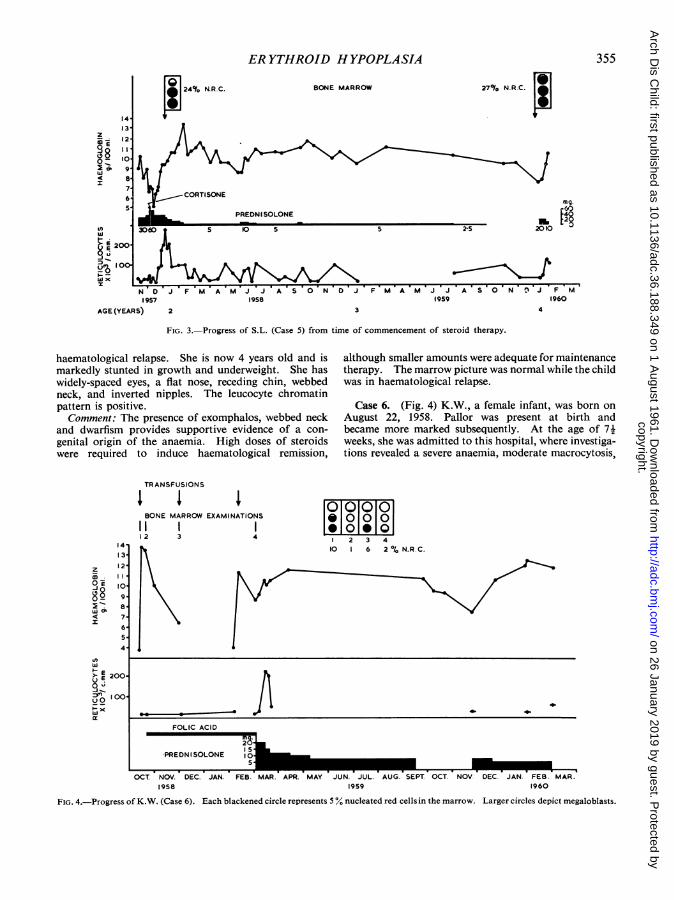
Case 6. (Fig. 4) K.W., a female infant, was born onAugust 22, 1958. Pallor was present at birth andbecame more marked subsequently. At the age of 71weeks, she was admitted to this hospital, where investiga-tions revealed a severe anaemia, moderate macrocytosis,
510M0
2 3 410 6 2% N.R.C.
-~~~~~~~~~~~~~~~~~~~~~~~~~~~~~~~~~~~~4-_ _ 3 4. -
FOLIC ACID
-PREDN ISOLONE 0ISOCT. NOV. DEC. JAN. FEB. MAR. APR. MAY JUN. JUL. AUG. SEPT. OCT. NOV DEC. JAN. FEB. MAR.
1958 1959 1960
FIG. 4.-Progress of K.W. (Case 6). Each blackened circle represents 5% nucleated red cellsin the marrow. Larger circles depict megaloblasts.
355
z
43V_
wO-
I
Ln
I i 2"X(
N D J F M A M1957
AGE (YEARS) 2
19604
copyright. on 26 January 2019 by guest. P
rotected byhttp://adc.bm
j.com/
Arch D
is Child: first published as 10.1136/adc.36.188.349 on 1 A
ugust 1961. Dow
nloaded from

ARCHIVES OF DISEASE IN CHILDHOOD
and reticulocytopenia. Marrow studies revealed erythro-blastic hypoplasia and the presence of occasional megalo-blasts. Blood transfusions were administered on threeoccasions at intervals of two months. Folic acid wasadministered for a period of six months without evidenceof haematological improvement; megaloblasts did notdisappear from the marrow smears for several months,but were not evident just before the commencement ofsteroid therapy. On March 2, 1959, folic acid therapywas withdrawn and steroid therapy was commenced.Reticulocytosis and a rise in haemoglobin level werenoted nine days later, and satisfactory haemoglobinvalues were maintained on small doses of prednisolone,which was withdrawn in September 1959. Within twomonths anaemia and reticulocytopenia became apparent;prednisolone therapy was recommenced in a dose of7 5 mg. daily and the haemoglobin value rose. At theage of 17 months, the child was of slightly less thanaverage weight and height. She had a snub nose,widely-spaced eyes, prominent epicanthic folds and amoderately widely patent fontanelle.Comment: Anaemia was probably present at birth
and progressed slowly. Marrow examinations revealederythroblastic hypoplasia and associated megaloblasticchanges which did not disappear for many monthsdespite a prolonged course of folic acid therapy, butwere not apparent at the stage when steroid therapy wascommenced. The anaemia was corrected followingsteroid administration and recurred when the drug waswithheld.
Case 7. (Fig. 5) D.McC., a female infant, was bornon May 18, 1958. Mild jaundice occurred in the firstfew days of life; pallor was noted at the age of 2 weeks,and became progressive. At the age of 5 months she
I I% N.R.C. BOI
TRANSFUSIONS
2z_ -z0-
0000 8
6,
5- |PREDNISOLONE4-
w
u 8 100
0 SO~-0cul
was admitted to this hospital, where investigationsrevealed a severe anaemia, moderate macrocytosis andreticulocytopenia. Pyuria was detected shortly after-wards, and chemotherapy was successfully prescribed.Bone marrow examinations revealed erythroblastichypoplasia. After the administration of blood trans-fusions, prednisolone was given in relatively smallquantities (7 5 mg. daily). Moderate anaemia withfluctuating reticulocyte counts was present over the nextfew months; in January 1959, the dose of prednisolonewas increased to 10 mg. daily, and the haemoglobinreached a satisfactory level after several months. Thedose of prednisolone was reduced to 2-5 mg. daily inMay 1959, but by October anaemia had become marked.An increase in the dose of steroids was followed bycorrection of the anaemia. At the age of 20 months thechild was well, but was less than the tenth centile inheight and weight for her age. Blue sclerae and amoderately widely patent fontanelle were noted. Thespleen was just palpable. Marrow studies at that stagerevealed normal proportions of red cell precursors,some of which showed imperfect haemoglobinization.Comment: When relatively small amounts of pred-
nisolone (7 5 to 5 mg. daily) were used initially, noimpressive rise in haemoglobin values occurred. Anincrease in dosage was followed by correction of theanaemia over a period of several months. Relapseoccurred when only 2 5 mg. of prednisolone were ad-ministered daily, and remission followed the institutionof larger doses. Evidence for small stature was presentbefore steroids were administered.
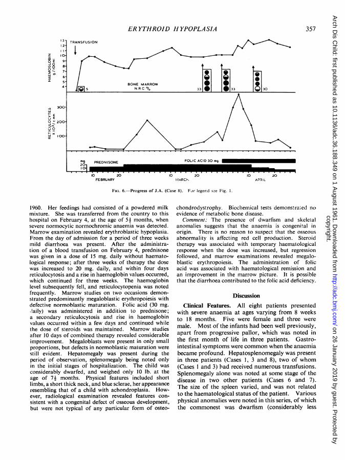
Case 8. (Fig. 6) J.A., a female infant, was born onAugust 29, 1959. Pallor was noted at the age of 3Amonths and she received a blood transfusion in January
ONE MARROW 29°/o N.R.C. I
FIG. 5.-Progress of D.McC. (Case 7).
1958 1959 1960
356
copyright. on 26 January 2019 by guest. P
rotected byhttp://adc.bm
j.com/
Arch D
is Child: first published as 10.1136/adc.36.188.349 on 1 A
ugust 1961. Dow
nloaded from
ER YTHROID HYPOPLASIA
0 0 19133_0_ 0_33 _8I30
FEBRUARY MARCh APRIL
FIG. 6.-Progress of J.A. (Case 8). For legend sze Fig. 1.
1960. Her feedings had consisted of a powdered milkmixture. She was transferred from the country to thishospital on February 4, at the age of 5! months, whensevere normocytic normochromic anaemia was detected.Marrow examination revealed erythroblastic hypoplasia.From the day of admission for a period of three weeksmild diarrhoea was present. After the administra-tion of a blood transfusion on February 4, prednisonewas given in a dose of 15 mg. daily without haemato-logical response; after three weeks of therapy the dosewas increased to 20 mg. daily, and within four daysreticulocytosis and a rise in haemoglobin values occurred,which continued for three weeks. The haemoglobinlevel subsequently fell, and reticulocytopenia was notedfrequently. Marrow studies on two occasions demon-strated predominantly megaloblastic erythropoiesis withdefective normoblastic maturation. Folic acid (30 mg..laily) was administered in addition to prednisone;a seconidary reticulocytosis and rise in haemoglobinvalues occurred within a few days and continued whilethe dose of steroids was maintained. Marrow studiesafter 10 days of combined therapy revealed considerableimprovement. Megaloblasts were present in only smallproportions, but defects in normoblastic maturation werestill evident. Hepatomegaly was present during theperiod of observation, splenomegaly being noted onlyin the initial stages of hospitalization. The child wasconsiderably dwarfed, and weighed only 10 lb. at theage of 7! months. Physical features included shortlimbs, a short thick neck, and blue sclerae, her appearanceresembling that of a child with achondroplasia. How-ever, radiological examination revealed features con-sistent with a congenital defect of osseous development,but were not typical of any particular form of osteo-
chondrodystrophy. Biochemical tests demonstra.ed noevidence of metabolic bone disease.
Comment: The presence of dwarfism and skeletalanomalies suggests that the anaemia is congenital inorigin. There is no reason to suspect that the osseousabnormality is affecting red cell production. Steroidtherapy was associated with temporary haematologicalresponse when the dose was increased, but regressionfollowed, and marrow examinations revealed megalo-blastic erythropoiesis. The administration of folicacid was associated with haematological remission andan improvement in the marrow picture. It is possiblethat the diarrhoea contributed to the folic acid deficiency.
Discussion
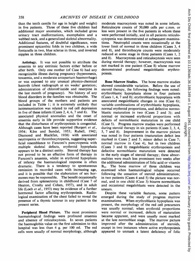
Clinical Features. All eight patients presentedwith severe anaemia at ages varying from 8 weeksto 18 months. Five were female and three weremale. Most of the infants had been well previously,apart from progressive pallor, which was noted inthe first month of life in three patients. Gastro-intestirial symptoms were common when the anaemiabecame profound. Hepatosplenomegaly was presentin three patients (Cases 1, 3 and 8), two of whom(Cases 1 and 3) had received numerous transfusions.Splenomegaly alone was noted at some stage of thedisease in two other patients (Cases 6 and 7).The size of the spleen varied, and was not relatedto the haematological status of the patient. Variousphysical anomalies were noted in this series, of whichthe commonest was dwarfism (considerably less
357
copyright. on 26 January 2019 by guest. P
rotected byhttp://adc.bm
j.com/
Arch D
is Child: first published as 10.1136/adc.36.188.349 on 1 A
ugust 1961. Dow
nloaded from
ARCHIVES OF DISEASE IN CHILDHOOD
than the tenth centile for age in height and weight)in five patients. Three of these five children hadadditional major anomalies, which included grossurinary tract malformations, exomphalos and awebbed neck, and a generalized skeletal abnormality.Additional features, of doubtful significance, wereprominent epicanthic folds in two children, a widefontanelle in two, blue sclerae in three, and invertednipples in three children.
Aetiology. It was not possible to attribute theanaemia to any extrinsic factors either before orafter birth. Only one mother (of Case 6) had anyrecognizable illness during pregnancy (hyperemesis,toxaemia, and a moderate antepartum haemorrhage)or was exposed to any unusual physico-chemicalhazards (chest radiograph at six weeks' gestation;administration of chlorotHiazide and reserpine inthe last month of pregnancy). No history of anyblood disorders in the families was obtainable. Theblood groups of the mothers and patients areincluded in Table 1; it is extremely unlikely thatisoimmunization was related to red cell depressionin five of these infants (Cases 2, 4, 6, 7 and 8). Theassociated physical anomalies and the onset ofanaemia early in life provide supportive evidencethat the disturbance of erythropoiesis is congenital.Although cases have been reported (Burgert et al.,1954; Koass and Sundal, 1953; Rubell, 1942;Diamond and Blackfan, 1938) with associatedneutropenia or thrombocytopenia, suggesting super-ficial resemblance to Fanconi's pancytopenia withmultiple skeletal defects, erythroid hypoplasiaappears to be a distinct entity. Steroid therapy hasnot proved to be an effective form of therapy inFanconi's anaemia, whilst in erythroid hypoplasiaof infancy the haematological response is oftendramatic. There is a tendency to spontaneousremission in recorded cases with increasing age,and it is possible that the elaboration of sex hor-mones may be responsible. The benefit occasionallyderived from splenectomy in childhood (Case 7 ofHeaton, Crosby and Cohen, 1957), and in adultlife (Loeb et al., 1953) may be evidence of a furtherhormonal factor affecting erythropoiesis. Radio-logical examination of the chest failed to reveal thepresence of a thymic tumour in any patient in thepresent series.
Peripheral Blood Picture. The most prominenthaematological findings were profound anaemiaand absence of reticulocytosis. In seven patientsthe haemoglobin value on the first admission to thishospital was less than 4 g. per 100 ml. The redcells were usually of normal morphology, although
moderate macrocytosis was noted in some infants.Reticulocyte counts of 50,000 cells per c.mm. orless were present in the five patients in whom thesewere performed initially, and in all patients reticulo-cytopenia was demonstrated at subsequent examina-tions. Neutrophil counts were occasionally at thelower limit of normal in three children (Cases 3, 4and 8), and thrombocyte counts were moderatelyreduced at some stage in three patients (Cases 1, 2and 8). Macrocytosis and reticulocytosis were seenduring steroid therapy; however, macrocytosis wasnot marked in one patient (Case 8) whose marrowdemonstrated profound megaloblastic erythro-poiesis.
Bone Marrow Studies. The bone marrow studiesshowed considerable variation. Before the use ofsteroid therapy, the following findings were noted:erythroblastic hypoplasia alone in four patients(Cases 1, 4, 7 and 8); erythroblastic hypoplasia withassociated megaloblastic changes in one (Case 6);variable combinations of erythroblastic hypoplasia,normal erythroid proportions, and defective normo-blastic maturation in two (Cases 2 and 3); andnormal or increased erythroid proportions withdefects of normoblastic maturation in one child(Case 5). Progress marrow studies were performedduring steroid therapy in six patients (Cases 2, 3, 4,5, 7 and 8). Improvement in the marrow picturewas noted in four patients (maturation defect lessmarked in Cases 2 and 5; minor defect in Case 7;normal marrow in Case 4), but in two children(Cases 3 and 8) megaloblastic erythropoiesis anddefective normoblastic maturation were detectedin the early stages of steroid therapy; these abnor-malities were much less prominent two weeks afterthe additional administration of folic acid or vitaminB12. The bone marrow of three children wasexamined when haematological relapse occurredfollowing the cessation of steroid administration;in two patients (Cases 4 and 5) the picture was nor-mal, and in one child (Case 3) bizarre normoblastsand occasional megaloblasts were detected in thesmears.
Despite these variable features, some patternemerged during the study of progress marrowexaminations. When erythroblastic hypoplasia waspresent, the morphology of the red cell precursorswas usually normal; when erythroid proportionswere normal or increased, defects of maturationbecame apparent, and were usually most markedat the late normoblast stage. The marrow pictureimproved after the administration of steroids,except in two instances where active erythropoiesisappeared to unmask a latent deficiency of folic
358
copyright. on 26 January 2019 by guest. P
rotected byhttp://adc.bm
j.com/
Arch D
is Child: first published as 10.1136/adc.36.188.349 on 1 A
ugust 1961. Dow
nloaded from

ER YTHROID HYPOPLASIA
acid or vitamin B2. The administration of theselatter agents with steroids was associated withhaematological improvement.
Other Laboratory Investigations. Blood urealevels showed transient elevation in two patients(Cases 5 and 7); the results are not available forCase 1. Pyuria was detected in three children(Cases 1, 5 and 7) and was transient in two (Cases5 and 7); the duration of the anaemia excludedthe possibilities of chronic azotaemia or infectionas major aetiological factors. The direct Coombs'test was negative and the serum bilirubin levelwas within normal limits in all patients where thesetests were performed (see Table 1). A thymictumour was not detected in the radiological exami-nation of the chest in any patients.
Course and Therapy. Before the advent of steroidtherapy, the only consistent benefit in these patientswas derived from repeated blood transfusions.Spontaneous remissions occurred in two children(Cases 2 and 3) for periods as long as 14 months.Three patients in this series received more than25 blood tranfusions, and the remainder at least two.The only child who died (Case 1) received 82 litresof blood, and was found to have extensive haemo-siderosis at autopsy.
Steroid therapy was administered to seven patients,and was associated with haematological improve-ment in all. In one (Case 2), intensive therapy forthree weeks failed to induce haematological im-provement, and a transfusion was administered.Steroid administration was continued subsequentlyand she has remained in haematological remissionfor over 14 months. As spontaneous remissionshad occurred previously for periods as long as14 months, it is possible that her present satisfactorystate may be unrelated to steroid therapy. Of theother six patients, none has required transfusionssince the time when steroids were first administered,representing intervals of 25, 37, 29, 13, 18 andtwo and a half months. There is no doubt thatsteroid therapy was effective, as haematologicalremission occurred on five occasions in one child(Case 4), and in other patients (Cases 3, 5, 6 and 7)haematological relapse developed when the drugwas withdrawn or reduced in dose, to be followedby remission with the reinstitution of adequatetherapy. In general, amounts equivalent to 20-30mg. of prednisolone daily were sufficient to initiateincreased red cell production, and such a responsewas usually evident within four to 11 days. In twopatients (Cases 5 and 8) reticulocytosis and a rise inhaemoglobin levels did not appear until the dose
was increased, when signs of red cell regenerationbecame apparent in four to five days. The natureof this response suggests that the adjustment ofsteroid dosage may be critical, although one cannotexclude the possibility that this form of therapymay be required for several weeks to induce effectiveerythropoiesis.Numerous megaloblasts were noted in marrow
examinations of two children (Cases 3 and 8) duringsteroid therapy, and reversion to normoblasticerythropoiesis followed the additional administra-tion of folic acid and vitamin B12. In one (Case 8),megaloblastic erythropoiesis was very marked, anda well-defined haematological response followedthe use of folic acid. In the other patient (Case 3),megaloblastic erythropoiesis was present to a lesserdegree, and haematological improvement after theadministration of vitamin B,2, folic acid and pred-nisolone was less dramatic; moreover, the dailydose of prednisolone administered during the criticalperiod was considerably smaller than that used toinduce the first remission two years earlier. Theprogress of J.A. (Case 8) is similar to that of thechild described by Arrowsmith et al. (1953). Whena suboptimal haematological response follows anadequate trial of steroid therapy, it seems advisableto perform further marrow examination to excludethe possibility of megaloblastic erythropoiesis; inCase 8 there was little evidence of macrocytosis inthe peripheral blood picture.
Maintenance steroid therapy was usually effectivewhen amounts equivalent to 5-10 mg. of predniso-lone .were administered daily, and was associatedwith no harmful side-effects. Relapse occurredwithin a few weeks or months in five patients(Cases 3, 4, 5, 6 and 7) when the drug was with-drawn or drastically reduced in dosage, so thatprolonged maintenance therapy or intermittenttherapy was required. It is likely that the intervalsbetween relapse will become longer with increasingage, as suggested by the progress of Case 4. Thelongest period of observation of a patient withsteroid administration is three years (Case 4).
It has been suggested (Gasser, 1957; Smith, 1959)that steroid therapy is of little avail when trans-fusion haemosiderosis has developed. Only oneof the present group of patients could be consideredunduly refractory to treatment (Case 2), althoughonly two patients (Cases 2 and 3) had received manyblood transfusions before steroid therapy was tried.A variety of therapeutic regimes was adopted for
patients in this series. It is suggested that bloodtransfusions be administered on the initial two orthree occasions when anaemia develops. In doingso, it should be possible to assess the nature of the
359
copyright. on 26 January 2019 by guest. P
rotected byhttp://adc.bm
j.com/
Arch D
is Child: first published as 10.1136/adc.36.188.349 on 1 A
ugust 1961. Dow
nloaded from
360 ARCHIVES OF DISEASE IN CHILDHOOD
disease and the intervals between transfusionswhich may be required. Moreover, haematologicalremissions may occur for long periods, as illustratedin Cases 2 and 3. Steroid therapy may be institutedsubsequently, and it is convenient to continuemaintenance treatment for a period of six to 12months, at the end of which time the patient'srequirements may be judged according to haemato-logical progress. High doses of steroids may berequired for several weeks to induce haematologicalremission, and the reason for therapeutic failures inother case reports may be in the administration ofinadequate initial doses of steroids. Suboptimalhaematological response during steroid therapyshould prompt investigation into the possibilityof megaloblastic erythropoiesis.
SummaryThe literature concerning erythroid hypoplasia
in infancy and childhood is reviewed briefly, andthe details of eight further patients are presented.
All children developed a severe aregenerativeanaemia at ages varying between 8 weeks and18 months. A high incidence of associated physicalanomalies was noted in this series, and includeddwarfism, urinary tract malformations, exomphalos,skeletal abnormalities and webbed neck. Theassociation of these anomalies with the onset ofanaemia early in life provides supportive evidencethat the aetiology of the anaemia rests in a con-genital defect of the erythron.Bone marrow studies revealed considerable
variation. Erythroid elements were present indecreased, normal or increased proportions, anddefects of normoblastic maturation were noted atsome stage in several patients. During steroidtherapy the marrow picture showed improvementin four patients, but megaloblastic erythropoiesisbecame prominent in two other children, andhaematological improvement followed the additionaladministration of folic acid or vitamin B12.One patient died; the remainder are still living.
Spontaneous remission for long periods occurred intwo patients before the advent of steroid therapy.Steroids were administered to seven children, ofwhom six received definite, and one possiblebenefit.High doses of steroids for several weeks may be
required to induce haematological remission; sub-optimal responses to adequate therapy should sug-gest the possibility of megaloblastic erythropoiesis.A method of management of these patients is
briefly outlined.I wish to express my gratitude to Dr. R. D. K. Reye
for his suggestions and interpretation of the bone
marrow slides; to Professor Lorimer Dods for his adviceand encouragement; to Drs. S. E. L. Stening, M. L.Edwards, S. E. J. Robertson and J. Alexander for theirencouragement and permission to publish the casehistories; to my colleagues and members of the Depart-ment of Pathology.
REFERENCES
Aldridge, A. G. V. and Kidd, P. (1953). Correspondence. Brit.med. J., 1, 729.
Altman, K. I. and Miller, G. (1953). A disturbance of tryptophanmetabolism in congenital hypoplastic anaemia. Nature (Lond.),172, 868.
Anderson, D. E. (1952). Chronic aregenerative anaemia of thenewborn. Med. J. Aust., 1, 573.
Arrowsmith, W. R., Burris, M. B. and Segaloff, A. (1953). Produc-tion of megaloblastic marrow by administration of cortisone inaplastic anemia, with subsequent response to vitamin B.12A relationship not previously described. J. Lab. clin. Med.,42, 778.
Bonham Carter, R. E., Cathie, I. A. B. and Gasser, C. (1954).Aplastische Anamie (chronische Erythroblastophthise) bedingtdurch Autoimmunisierung. Schweiz. med. Wschr., 84, 1114.
Bousser, J., Christol, D. Dausset, J., Rampon, S., Jallut, H. andMery, J. P. (1955). l:pisode erythroblastopenique prolong6ayant marque le d6but d'une anemie hemolytique chroniqueavec auto-anticorps. Sang, 26, 804.
Burgert, 0. Jr., Kennedy; R. L. J. and Pease, G. L. (1954). Con-genital hypoplastic anemia. Pediatrics, 13, 218.
Calvert, R. J. and Robson. T. (1956). Cortisone therapy in erythro-genesis imperfecta. Arch. Dis. Childh., 31, 177.
Cathie, I. A. B. (1950). Erythrogenesis imperfecta. Ibid., 25, 313.Chernoff, A. I. and Josephson, A. M. (1951). Acute erythroblasto-
penia in sickle-cell anemia and infectious mononucleosis.A.M.A. Amer. J. Dis. Child., 82, 310.
Dacie, J. V., Smith, M. D., White, J. C. and Mollin, D. L. (1959).Refractory normoblastic anaemia: a clinical and haematologicalstudy of seven cases. Brit. J. Haemat., 5, 56.
Diamond, L. K. (1954). Editorial comment, Year Book ofPediatrics,1954-1955 series, p. 308. Year Book Publishers, Chicago.and Blackfan, K. D. (1938). Hypoplastic anemia. Amer. J.Dis. Child., 56, 464.
Donnelly, M. (1953). A case of uimary red cell aplasia. Brit.med. J., 1, 438.
Eisemann, G. and Dameshek, W. (1954). Splenectomy for 'purered-cell' hypoplastic (aregenerative) anemia associated withautoimmune hemolytic disease. (Report of a case.) NewEngl. J. Med., 251, 1044.
Fisher, 0. D. and Allen, F. M. B. (1953). Erythrogenesis imperfectaor congenital hypoplastic anaemia (Diamond-Blackfan type).Arch. Dis. Childh., 28, 363.
Gasser, C. (1957). Aplasia oferythropoiesis. Pediat. Clin. N. Amer.,May, 1957, p. 445.
Harper, M. and Geikie, G. (1955). Congenital pure red cell anaemiaor erythrogenesis imperfecta. Med. J. Aust., 2, 245.
Heaton, L. D., Crosby, W. H. and Cohen, A. (1957). Splenectomyin the treatment of hypoplasia of the bone marrow, with areport of twelve cases. Ann. Surg., 146, 637.
Hoyer, K. (1942). Aregenerative, probably congenital, anaemiain an infant with deficiency of erythroblasts in the bone marrow.Nord. med., 14, 1097.
Josephs, H. W. (1936). Anaemia of infancy and early childhood.Medicine (Baltimore), 15, 307.
Kiss, A. and Sundal, A. (1953). Anaemia hypoplastica congenita(anaemia typus Josephs-Diamond-Blackfan). Report of a casetreated with adrenocorticotropin with effect. Acta paediat.(Uppsala), 42, 265.
Kohlbry, C. 0. (1941). Congenital hypoplastic anemia. Casereport. J. Pediat., 19, 662.
Lelong, M., Joseph, R., Polonowski, C., Desmonts, G. and Colin, J.(1951). L'anemie chronique avec arret de la maturation normo-blastique (type Blackfan-Diamond). Arch. franc. Pediat.,8, 473.
Loeb, V., Jr., Moore, C. V. and Dubach, R. (1953). The physiologicevaluation and management of chronic bone marrow failure.Amer. J. Med., 15, 499.
Owren, P. A. (1948). Congenital hemolytic jaundice. The patho-genesis of the 'hemolytic crisis'. Blood, 3, 231.
Palmen, K. and Vahlquist, B. (1950). Stationary hypoplasticanemia. Acta haemat. (Basel), 4, 273.
Pearson, H. A. and Cone, T. E., Jr. (1957). Congenital hypoplasticanemia. Pediatrics, 19, 192.
Rinvik, R. (1940). Two cases of idiopathic hypoplastic anemia ininfants. Acta paediat. (Uppsala), 28, 304.
Robson, T. and Sweeney, P. J. (1948). Chronic hypoplastic anaemiaarising in infancy. Arch. Dis. Childh., 23, 294.
Rubell, I. (1942). Hypoplastic congenital anemia. J. Pediat.,20, 756.
copyright. on 26 January 2019 by guest. P
rotected byhttp://adc.bm
j.com/
Arch D
is Child: first published as 10.1136/adc.36.188.349 on 1 A
ugust 1961. Dow
nloaded from
ERYTHROID HYPOPLASIA 361Seip, M. (1955). Aplastic crisis in a case of immuno-hemolytic
anemia. Acta med. scand., 153, 137.Smith, C. H. (1949). Chronic congenital aregenerative anemia
(pure red-cell anemia) associated with iso-immunization by theblood group factor 'A'. Blood, 4, 697.(1953). Hypoplastic and aplastic anemias of infancy and child-hood: with a consideration of the syndrome of nonhemolyticanemia of the newborn. J. Pediat., 43, 457.(1959). Pure red-cell anemia. Ibid., 54, 609.
Sundal, A. (1956). Anemia hypoplastica congenita treated withcortisone. Correction of the anemia with cortisone treatmentduring 4 years. Acta paediat. (Uppsala), 45, 456.
Tsai, S. Y. and Levin, W. C. (1957). Chronic ervthrocytic hypo-plasia in adults. Amer. J. Med., 22, 322.
Verger and Leger (1953). Anemie hypoplastique de la premiereenfance (anemie de Blackfan-Diamond). Arch. franf. Pediat,10, 328.
Vilter, R. W., Jarrold, T., Will, J. J., Mueller, J. F., Friedman, B. I.and Hawkins, V. R. (1960). Refractory anemia with hyper-plastic bone marrow. Blood, 15, 1.
Wallman. I. S. (1956). Hereditary red cell aplasia. Med. J. Aust.2, 488.
copyright. on 26 January 2019 by guest. P
rotected byhttp://adc.bm
j.com/
Arch D
is Child: first published as 10.1136/adc.36.188.349 on 1 A
ugust 1961. Dow
nloaded from