hypobaric-lschemic conditions produce glutamate-like cytopathology in infant rat brain
TRANSCRIPT
The Journal of Neuroscience, May 1969, g(5): 1693-1700
Hypobaric-lschemic Conditions Produce Glutamate-like Cytopathology in Infant Rat Brain
C. Ikonomidou, M. T. Price, J. L. Mosinger, G. Frierdich, J. Labruyere, K. Shahid Salles, and J. W. Olney
Department of Psychiatry, Washington University School of Medicine, St. Louis, Missouri 63110
We present a new animal model of perinatal hypoxic/isch- emit brain damage and compare this type of brain damage with the excitotoxic type of damage previously described in the brains of infant rats and monkeys treated systemically with glutamate (Glu). Ten-d-old rats with unilateral occlusion of the common carotid artery were subjected to hypobaric conditions for 75 min and sacrificed O-4 hr later for light and electron microscopic brain examination. The mortality rate was relatively low (12%) and brain damage was evident ipsilateral to the ligated carotid in 94% of surviving animals 4 hr after termination of the hypobaric event. Regions most frequently affected were the medial habenulum, dentate gy- rus, caudate nucleus, frontoparietal neocortices, olfactory tubercle, and several thalamic nuclei. The acute cytopatho- logical changes, primarily edematous degeneration of neu- ronal dendrites and cell bodies, evolved very rapidly, with some neurons manifesting end-stage necrosis at 0 hr (im- mediately after hypobaric exposure) and others developing such changes over a l-4-hr period. We conclude that the neurodegenerative reaction induced in infant rat brain by hypoxia/ischemia is indistinguishable from the excitotoxic type of damage exogenous Glu is known to cause. Moreover, in a companion study (Olney et al., 1989) we show that MK- 801, a powerful antagonist of the N-methyl-D-aspartate re- ceptor complex (subtype of Glu receptor), protects against neuronal degeneration in this hypobaric/ischemic model. Our results reinforce other recent evidence suggesting that hyp- oxiclischemic brain damage is mediated by endogenous Glu or related excitotoxins.
Glutamate (Glu) and aspartate (Asp) are major excitatory trans- mitters (Watkins, 1978) and neurotoxins (excitotoxins) that have the potential to destroy central neurons by an excitatory mech- anism (Olney, 1969a; Olney et al., 197 1). Subcutaneous or oral administration of Glu to infant rodents or monkeys induces acute neuronal necrosis in certain brain regions that lack blood brain barriers (Olney 1969a, 197 1; Olney and Ho, 1970; Olney et al., 1972). The neurodegenerative process is exceedingly acute; vulnerable neurons display swelling within 15-30 min after Glu administration and progress to end-stage necrosis over a 1-4- hr period (Olney 1971; Olney et al., 197 1, 1972). Recent evi- dence suggests that endogenous Glu and/or Asp may be re-
Received Aug. 19, 1988; revised Oct. 3, 1988; accepted Oct. 7, 1988.
Supported in part by NIMH Research Scientist Award MH 38894 (J.W.O.), HHS arants HD 24237 and ES 07066. and a erant from Pfizer Pharmaceutical.
Correspondence should be addressed to Johi W. Olney, M.D., Department of Psychiatry, Washington University School of Medicine, 4940 Audubon Ave., St. Louis, MO 63110. Copyright 0 1989 Society for Neuroscience 0270-6474/89/051693-08$02.00/O
sponsible for hypoxic/ischemic neuronal degeneration in con- ditions such as stroke, cardiac arrest, and perinatal asphyxia (Benveniste et al., 1984; Rothman, 1984; Simon et al., 1984; Weiss et al., 1986; Gill et al., 1987; Lawrence et al., 1987; McDonald et al., 1987; Olney et al., 1987, 1988; Prince and Freeser, 1988). If endogenous Glu is the responsible pathogenic agent in hypoxic/ischemic brain damage, it should be possible to show that hypoxia/ischemia causes the same type of cyto- pathological changes in brain that exogenous Glu typically caus- es, Here we describe a new model of hypoxic/ischemic brain damage in the infant rat and provide light and electron micro- scopic evidence that the acutely evolving cytopathology asso- ciated with hypoxia/ischemia is indistinguishable from that in- duced by exogenous Glu.
Materials and Methods Our method for producing hypoxic/ischemic brain damage in infant rats is similar to that described by Levine (1960) as modified by Rice et al. (1980) but it involves exposure of infant animals to a partial vacuum rather than an environment of reduced oxygen and increased nitrogen. Ten-d-old Sprague-Dawley rats were used in all experiments. A unilat- eral carotid ligation was performed under halothane anesthesia. During and immediately after surgery the pups were kept on a warming pad that maintained their body temperature at approximately 36°C as mea- sured by a skin surface microprobe. After recovery from anesthesia, a group of lo-12 pups (total body wt = 200 f 10 gm) was placed in a hypobaric chamber that measured 2370 ml in volume and was im- mersed in a water bath that maintained the temperature inside the chamber at 36 t 0.5”C. The air pressure in the chamber was gradually reduced from 760 to 225 mm Hg over a 1-min period and maintained at 225 mm for 75 min, then brought back to 760 mm over a 1-min period. Either immediately or l-4 hr after removal from the hypobaric chamber, the rats were anesthetized with chloral hydrate and perfused through the heart and ascending aorta with a solution of paraformal- dehyde (lo/o) and glutaraldehyde (1.5%) in phosphate buffer. In some cases the neck incision was opened prior to perfusion and the suture removed from the carotid artery (a piece of polyethylene tubing origi- nally placed between the suture and vessel wall facilitated removal of the suture without damage to the vessel); however, this was not found necessary in order to achieve adequate preservation of the brain. After perfusion for 15 min the brains were removed and sliced in l-mm transverse slabs that were additionally fixed in osmium tetroxide (l%), dehydrated in graded ethanols, cleared in toluene, and embedded in araldite. Sections 1 pm thick were cut with i/z” glass knives at 40-pm intervals on a Sorval MT-2B ultratome, stained with methylene blue/ azure II, and evaluated by light microscopy. Ultrathin sections were cut from areas of special interest from the same blocks, stained with lead citrate and uranyl acetate, and examined with a JEOL 100 B transmis- sion electron microscope. These histological methods are the same as were used in previous studies of Glu-induced brain damage (Olney, 1971; Olney et al., 1972).
Results
In pilot experiments, it was observed that unilateral carotid ligation alone did not induce brain damage, nor did exposure
1694 lkonomidou et al. l Hypobaric-lschemic Conditions Cause Glu-like Brain Damage
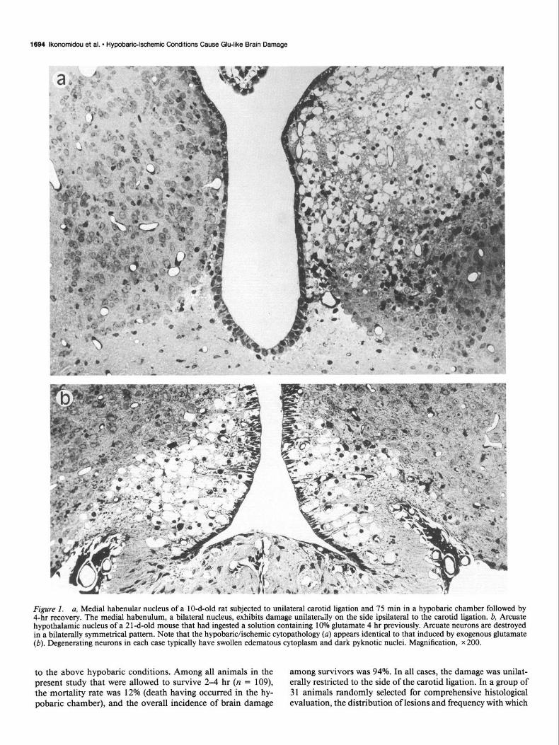
Figure 1. a, Medial habenular nucleus of a lo-d-old rat subjected to unilateral carotid ligation and 75 min in a hypobaric chamber followed by 4-hr recovery. The medial habenulum, a bilateral nucleus, exhibits damage unilaterally on the side ipsilateral to the carotid ligation. b, Arcuate hypothalamic nucleus of a 2 1 -d-old mouse that had ingested a solution containing 10% glutamate 4 hr previously. Arcuate neurons are destroyed in a bilaterally symmetrical pattern. Note that the hypobaric/ischemic cytopathology (a) appears identical to that induced by exogenous glutamate (b). Degenerating neurons in each case typically have swollen edematous cytoplasm and dark pyknotic nuclei. Magnification, x 200.
to the above hypobaric conditions. Among all animals in the among survivors was 94%. In all cases, the damage was unilat- present study that were allowed to survive 2-4 hr (n = 109), erally restricted to the side of the carotid ligation. In a group of the mortality rate was 12% (death having occurred in the hy- 3 1 animals randomly selected for comprehensive histological pobaric chamber), and the overall incidence of brain damage evaluation, the distribution of lesions and frequency with which

The Journal of Neuroscience, May 1989, 9(5) 1695
each brain region was involved is given in Table 1. A striking lesion was consistently found in the medial habenulum (Fig. 1). Also quite consistently, we found a cytopathological reaction affecting granule neurons in the ventral blade of the dentate gyrus immediately overlying the damaged medial habenulum. Other regions frequently damaged (Fig. 2) were the frontal and parietal neocortices, caudate nucleus, medial dorsal and lateral dorsal nuclei of the thalamus, olfactory tubercle, dorsal subicu- lum, and islands of Calleja.
The acute cytopathological changes appeared by light mi- croscopy to consist predominantly of edematous swelling of dendritic processes (confirmed ultrastructurally; see below) and acute edematous degeneration of neuronal cell bodies with pyk- notic changes occurring quite early in the nucleus. Comparison of the acute cytopathological reaction in the hypothalamus of a mouse treated orally with Glu and that in the medial habenulum of an infant rat subjected to hypobaric/ischemic conditions (Fig. 1) demonstrates that the 2 cytopathological processes are in- distinguishable at the light microscopic level. Some neurons, primarily in the hippocampus, cortex, thalamus, and caudate nucleus, underwent a more slowly developing process of vac- uolar condensation in which both the cytoplasm and the nu- cleoplasm became uniformly dense and dark-staining except for numerous spherical vacuoles in the cytoplasm. This type of degenerative reaction, sometimes referred to as “coagulative” degeneration, has been described previously as a feature of the cytopathology associated with cerebral ischemia (Van Reempts, 1984), status epilepticus (Olney et al., 1986a), and systemic administration of Glu to infant rodents or monkeys (Olney, 1969a, 1986).
To study the time course of the acutely evolving lesion, we killed infant rats at intervals from 0 to 4 hr post-hypoxia. Sur- prisingly, animals killed at 0 hr (immediately after removal from the hypobaric chamber) consistently displayed cytopathological changes, including end-stage neuronal necrosis. In fact, one fea- ture of the cytopathology, dendritic swelling, was more extensive in certain brain regions at 0 hr than at 4 hr. The other major feature, degeneration of cell bodies (both edematous degener- ation and vacuolar condensation), was more extensively devel- oped at 4 hr.
By electron microscopy, it was readily confirmed that the major tissue components initially affected were neuronal den- drites (Fig. 3) and cell bodies (Fig. 4); indeed, every feature of the acute cytopathology in either dendritic or somal neural ele- ments was identical to that previously described (Olney, 197 1; Olney et al., 1972) in infant rodent or monkey brain following systemic administration of exogenous Glu (Figs. 3, 4). In nerve cells destined to undergo acute edematous degeneration, mi- tochondria exhibited the first sign of pathological change; before cytoplasmic swelling occurred, mitochondria became transient- ly condensed, with their membranes assuming a thickened, dark, compacted appearance (Fig. 5). Subsequently, as signs of edem- atous degeneration began to pervade the entire cytoplasmic compartment, mitochondria became swollen (Fig. 5). Finally, in later stages of neuronal necrosis, mitochondria appeared less swollen and typically displayed a small and spherical (“rounded- up”) profile (Fig. 4). These several stages of mitochondrial trans- formation could be detected simultaneously in a given brain. For example, the lesion affecting dentate granule neurons tended to develop more slowly than the lesion affecting medial haben- ular neurons. Thus, at 0 to 1 hr it was possible to find swollen mitochondria in medial habenular neurons that were undergo-
Table 1. Frequency of damage in various brain regions
Brain region Freauencv (%)
Habenular nucleus, medial Dentate gyms (granule cells) Caudate nucleus Thalamus, mediodorsal nucleus Neocortex, parietal Olfactory tubercle Subiculum, dorsal Thalamus, lateral dorsal nucleus Islands of Calleja Neocortex, frontal Septum Hippocampus, CA 1 pyramids Thalamus, ventrobasal nucleus Habenular nucleus, lateral Hippocampus, CA3 pyramids Pyriform cortex Amygdala, cortical nucleus
94 81 71 71 65
63
61
58
56
52
38
35
32
32
16
10
ing edematous degeneration and condensed mitochondria in dentate granule neurons that had not yet begun manifesting edematous degeneration (Fig. 5). But at 4 hr, dentate granule neurons exhibited edematous degeneration and contained mi- tochondria that displayed swollen profiles (Fig. 5). By this time (4 hr), medial habenular neurons were in an advanced necrotic stage, and their mitochondria had become small and spherical (Fig. 4). Other features of the edematous degenerative reaction affecting the cytoplasmic compartment of neuronal cell bodies included dissolution of the endoplasmic reticular system with formation of spherical vacuoles bounded by rough membranes, disaggregation of polysomes, and dispersal of nondescript par- ticulate material in the cytoplasm (Fig. 4). Changes in the neu- ronal nucleus typically consisted of chromatin clumping with the clumps finally coalescing centrally to form a pyknotic nu- cleus (Fig. 4).
The evacuation of degenerating elements was very rapid in periventricular regions, especially the medial habenulum. Four hours after hypobaric exposure non-neuronal phagocytic cells were enveloping and incorporating neuronal cell bodies, and within 24 hr much of the evacuation process had transpired. In the thalamus and neocortex, biological debridement proceeded more slowly and was still evident as an ongoing process 4 d post-ischemia.
Discussion
We have shown that unilateral ligation of the common carotid artery and subsequent exposure of 1 O-d-old rat pups to a partial vacuum causes a reproducible pattern of neuronal degeneration in several brain regions ipsilateral to the ligation. We also have studied brains of infant rats subjected to the hypoxic/ischemic protocol described by Rice et al. (1980), which consists of uni- lateral common carotid artery ligation followed by 2-3 hr in a reduced oxygen environment, and demonstrated (Olney, 1988) that the cytopathological changes induced by that approach are identical to the hypobaric/ischemic changes described here. However, with this approach we were unable to find a set of conditions yielding a consistently high incidence of lesions with- out an unacceptably high mortality rate. We abandoned this

1696 lkonomidou et al. * Hypobaric-lschemic Conditions Cause Glu-like Brain Damage
Figure 2. Ten-d-old infant rat brain 4 hr after the animal was subjected to hypobaric/ischemic conditions. Neurons in the parietal cortex contralateral to the carotid ligation have a normal appearance (a). Ipsilateral to the carotid ligation, acutely degenerating neurons in the parietal cortex (b), caudate nucleus (c), and olfactory tubercle (d) display changes that are indistinguishable from those induced by exogenous Glu (Fig. 1 b). Magnification, x 260.

The Journal of Neuroscience, May 1989, 9(5) 1697
Figure 3. a, Swollen dendrite (0) undergoing acute edematous degeneration in the medial dorsal nucleus of the thalamus of a lo-d-old rat 4 hr after hypobaric/ischemic exposure. This degenerating dendrite is in contact with a presynaptic axon terminal (A), which retains a normal appearance. This cytopathology is typical of Glu-induced damage as shown in b, where a swollen dendrite (0) is undergoing acute edematous degeneration in the arcuate hypothalamic nucleus of an infant rhesus monkey 4 hr after oral administration of Glu (0.5 g/kg); note the presence of a presynaptic axon terminal (A) that retains a normal appearance. Magnification, x 27,000.
model after finding that the hypobaric/ischemic approach yield- ed more favorable results regarding lesion incidence and mor- tality rate while simultaneously being easier and less expensive and entailing less stress (shorter period of hypoxia) for the ex- perimental animal.
Brain damage that occurs during the perinatal period in the human is often referred to as hypoxic/ischemic because typically it appears to be caused by a combination of reduced oxygen content of blood and regional reductions in cerebral blood flow (Johnston and Silverstein, 1987). A pathological sequence is thought to occur in which anoxia triggers systemic hypotension and reduced cardiac output, which may be further complicated by failure of cerebral blood vessels to autoregulate under anoxic conditions (Lou et al., 1979). The brain damage model described here can well be characterized as hypoxic/ischemic, since low blood oxygen tension coupled with inadequate vascular perfu- sion of the affected side of the brain is the primary factor con- tributing to its occurrence. Perinatal brain damage in the human and hypobaric/ischemic brain damage in the infant rat have additional features in common. In human infants dying during or shortly after a hypoxic episode, histological changes charac- terized by swollen vacuous cytoplasm and clumping or karyor- rhexis of nuclear chromatin have been described (Banker and Larroche, 1962). Neurons that are sensitive to hypoxic/ischemic
damage in the infant human include those in the cerebral cortex, basal ganglia, various thalamic nuclei, and certain periventric- ular zones (Banker and Larroche, 1962; Brierley and Graham, 1984). These regions are also consistently affected in the hy- pobaric/ischemic infant rat brain. It has been proposed that vulnerability of periventricular regions in the immature human brain relates to a lag in development of ventriculopetal arteries that are destined to supply these regions (Banker and Larroche, 1962). Conceivably, this might account for the extreme vul- nerability of medial habenular neurons to hypobaric/ischemic injury observed in the present study. Selective degeneration of other neuronal groups might be explained in the rat model by the watershed phenomenon, a concept often invoked to explain human neuropathology, in which neurons that lie between distal tributaries of separate arterial trunks are hypervulnerable to degeneration under hypoxic/ischemic circumstances. Further cross-species comparisons may not be warranted, since the pat- tern of neuropathology resulting from hypoxic/ischemic con- ditions may vary as a function of developmental age at the time of insult and it is difficult to know exactly what point in human ontogenesis corresponds with the tenth postnatal day in the rat.
Several lines of recent evidence support the hypothesis that hypoxic/ischemic brain damage is mediated by endogenous Glu or a related excitotoxin. Ischemia of the adult rat brain is as-

1696 lkonomidou et al. l Hypobaric-lschemic Conditions Cause Glu-like Brain Damage
Figure 4. a, Neuron undergoing acute degeneration in the medial habenular nucleus of the lo-d-old rat 4 hr after hypobaric/ischemic exposure. b, neuron undergoing acute degeneration in the arcuate hypothalamic nucleus of an infant rhesus monkey 4 hr after oral administration of Glu (1 g/kg). Note the typical features of Glu-induced neuronal degeneration in both figures, including vacuolated endoplasmic reticulum (v), spherical mitochondria (m), and clumped chromatin (arrow) that is consolidating centrally to produce the typical appearance of a pyknotic nucleus. The hypobaric/ischemic changes exhibited by the neuron in a are indistinguishable from those induced by Glu (b). Magnification, x 20,000.
sociated with an accumulation of Glu and Asp in the extracel- lular compartment, where these agents can exert excitotoxic activity at postsynaptic dendrosomal receptors (Benveniste et al., 1984). The uptake mechanism for removal of Glu and Asp from the extracellular compartment is impaired during hypoxiaJ ischemia in the infant rat brain (Silverstein et al., 1986). Tran- section of the excitatory (probably glutamergic) afferents to the hippocampus protects hippocampal neurons from hypoxic/isch- emit damage (Wieloch et al., 1985). EAA antagonists reportedly protect against anoxic and/or ischemic neuronal degeneration in cell cultures of hippocampal neurons (Rothman, 1984) or cerebrocortical neurons (Weiss et al., 1986), in the ex vivo chick embryo retina (Olney et al., 1987), in the in vivo adult hippo- campus of the rat (Simon et al., 1984) and gerbil (Gill et al., 1987; Lawrence et al., 1987), and in several regions of the in vivo infant rat brain (McDonald et al., 1987; Prince and Freeser, 1988).
A side-by-side comparison of the hypobaric/ischemic neu- rodegenerative reaction in infant rat brain with that induced in immature rodent or monkey brain by systemic administration of exogenous Glu reveals many similarities. In both syndromes postsynaptic dendrosomal neural elements are preferentially af- fected, with conspicuous sparing of presynaptic axons and iden- tical changes occurring in cytoplasmic organelle systems and nuclear chromatin. Moreover, the time course of the neurode- generative reaction is often remarkably acute, with the most
sensitive neurons undergoing degenerative changes within min- utes and progressing to end-stage necrosis within a few hours after either Glu administration or exposure to hypobaric/isch- emit conditions. A less acute in vivo neurodegenerative process following Glu administration has also been described (Olney, 1969b, 1986) in which the neuronal cytoplasm becomes vacu- olated and the entire cell assumes a condensed “dark cell” ap- pearance. In the present study, a similar rather protracted “dark cell” degenerative reaction was seen in several regions of the infant rat brain following exposure to hypobaric/ischemic con- ditions. Recently, 2 types of excitotoxic reaction to Glu have been described in in vitro preparations: an exceedingly acute process (Rothman, 1985; Olney et al., 1986b) that is not de- pendent on CaZ+ influx, and a delayed process (Choi, 1987) that is Caz+ dependent. Although the present study does not address the issue of CY+ dependency, it documents that hypoxic/isch- emit neuronal degeneration, like Glu-induced neuronal de- generation, may have either an acute or a delayed time course. Thus, both on the basis of pathomorphological criteria and time course, the neurodegenerative reaction to hypobaric/ischemic conditions in the infant rat brain is indistinguishable from the well-known reaction of CNS neurons to exogenous Glu.
One of our motivations for developing the infant rat prepa- ration described here was to have an hypoxic/ischemic model in which neurons clearly manifest the remarkably acute type of dendrosomal pathology that traditionally has been associated

The Journal of Neuroscience, May 1989, 9(5) 1699
Figure 5. Dentate granule neurons in the lo-d-old infant rat brain at 0 hr (a) and 4 hr (b) after hypobaric/ischemic exposure. a, Very early stage of degeneration, in which the only detectable sign of pathological change is the dense, compacted appearance of the mitochondria (arrow). b, Later stage of degeneration, in which the cytoplasmic compartment is becoming edematous, nuclear chromatin is beginning to form coarse clumps, and mitochondria (arrows) are spherical, edematous, and swollen. Each of these types of mitochondrial change has been described as a typical feature of Glu-induced neuronal degeneration (Olney, 1986). Magnification, ~45,000.
with Glu toxicity. In addition, since much of the in vivo research pertaining to the role of excitotoxins in hypoxic/ischemic neu- ronal degeneration (Simon et al., 1984; Gill et al., 1987; Law- rence et al., 1987) has focused exclusively on a single type of neuron, the CA1 hippocampal pyramidal neuron, there is a need for new models pertaining to other neuronal populations. Fi- nally, in order to determine whether anti-excitotoxic drugs can prevent hypoxic/ischemic neuronal degeneration, it is essential to have a model in which the same degree and pattern of brain damage are consistently reproduced and in which the mortality rate is low. The model described here meets the several criteria sought; that this model may be of value for studying methods of preventing perinatal hypoxic/ischemic brain damage is cor- roborated in a companion article (Olney et al., 1989).
References Banker, B. Q., and J. C. Larroche (1962) Periventricular leukomalacia
in infancy: A form of neonatal anoxic encephalopathy. Arch. Neurol. 7: 386-410.
Benveniste, H., J. Drejer, A. Schousboe, and N. H. Diemer (1984) Elevation of extracellular concentrations of glutamate and aspartate in rat hippocampus during transient cerebral ischemia monitored by intracerebral microdialysis. J. Neurochem. 43: 1369-1374.
Brierley, J. B., and D. I. Graham (1984) Hypoxia and vascular dis- orders of the central nervous system. In GreenfieldS Neuropathology, J. H. Adams, J. A. N. Corsellis, and L. W. Duchen, eds., pp. 125- 207, Arnold, London.
Choi, D. W. (1987) Ionic dependence of glutamate neurotoxicity. J. Neurosci. 7: 369-379.
Gill, R., A. C. Foster, and G. N. Woodruff (1987) Systemic admin- istration of MK-801 protects against &hernia-induced hippocampal neurodegeneration in the gerbil. J. Neurosci. 7: 3343-3349.
Johnston, M. V., and F. S. Silverstein (1987) Perinatal anoxia. In Animal Models of Dementia, pp. 223-25 1, Liss, New York.
Lawrence, J J., T. A. Fuller, and J. W. Olney (1987) MK-801 and PCP protect against ischemic neuronal degeneration in the gerbil hippocampus. Sot. Neurosci. Abstr. 13: 1079.
Levine, S. (1960) Anoxic-ischemic encephalopathy in rats. Am. J. Pathol. 36: 1-17.
Lou, H. C., N. A. Lassen, and B. Friis-Hansen (1979) Impaired au- toregulation of cerebral blood flow in the distressed newborn. J. Pe- diatr. 94: 118-127.
McDonald, J. W., F. S. Silverstein, and M. V. Johnston (1987) MK- 80 1 protects the neonatal brain from hypoxic/ischemic damage. Eur. J. Pharmacol. 140: 359-361.
Olney, J. W. (1969a) Brain lesions, obesity and other disturbances in mice treated with monosodium glutamate. Science 164: 7 19-72 1.
Olney, J. W. (1969b) Glutamate-induced retinal degeneration in neo- natal mice. Electron microscopy of the acutely evolving lesion. J. Neuropathol. Exn. Neurol. 28: 455-474.
Olney, J.W. (197 lj Glutamate-induced neuronal necrosis in the infant
1700 lkonomidou et al. * Hypobaric-lschemic Conditions Cause Glu-like Brain Damage
mouse hypothalamus: An electron microscopic study. J. Neuropathol. Exp. Neurol. 30: 75-90.
Olney, J. W. (1986) Inciting excitotoxic cytocide among central neu- rons. In Excitatory Amino Acids and Epilepsy, R. Schwartz and Y. Ben-Ari, eds., pp. 631-645, Plenum, New York.
Olney, J. W. (1988) Endogenous excitotoxins and neuropathological disorders. In Excitatory Amino Acids in Health and Disease, D. Lodge, ed., pp. 337-35 1, Wiley, London.
Olney, J. W.,,and 0. L. Ho (1970) Brain damage in infant mice following oral intake of glutamate, aspartate or cysteine. Nature 227: 609-610.
Olney, J. W., 0. L. Ho, and V. Rhee (197 1) Cytotoxic effects of acidic and sulphur-containing amino acids on the infant mouse central ner- vous system. Exp. Brain Res. 14: 6 l-76.
Olney, J. W., L. G. Sharpe, and R. D. Feigin (1972) Glutamate-induced brain damage in infant primates. J. Neuropathol. Exp. Neurol. 31: 464-488.
Olney, J. W., R. C. Collins, and R. S. Sloviter (1986a) Excitotoxic mechanisms of eoileotic brain damage. Adv. Neurol. 44: 857-877.
Olney, J. W., M. T: P&e, L. Samson, and J. Labruyere (1986b) The role of specific ions in glutamate neurotoxicity. Neurosci. Lett. 65: 65-71.
Olney, J. W., M. T. Price, J. Labruyere, E. Silverman, and M. Mueller (1987) Comparative efficacy of various agents in preventing gluta- mate-induced or ischemic neuronal degeneration in chick retina. Sot. Neurosci. Abstr. 13: 1030.
Olney, J. W., C. Ikonomidou, J. L. Mosinger, and G. Frierdich (1989) MK-80 1 prevents hypobaric-ischemic neuronal degeneration in in- fant rat brain. J. Neurosci. 9: 1701-1704.
Prince, D. A., and H. R. Freeser (1988) Dextromethorphan protects against cerebral infarction in a rat model of hypoxia-ischemia. Neu- rosci. Lett. 85: 291-296.
Rice, J. E., R. C. Vannuci, and J. B. Brierley (1980) The influence of immaturity on hypoxic-ischemic brain damage in the rat. Ann. Neu- rol. 9: 131-141.
Rothman, S. M. (1984) Synaptic release of excitatory amino acid neurotransmitter mediates anoxic neuronal death. J. Neurosci. 4: 1884- 1891.
Rothman, S. M. (1985) The neurotoxicity of excitatory amino acids is produced by passive chloride influx. J. Neurosci. 5: 1483-1489.
Silverstein, F. S., K. Buchanan, and M. V. Johnston (1986) Perinatal hypoxia-ischemia disrupts striatal high affinity [3H]-glutamate uptake into synaptosomes. J. Neurochem. 47: 16 14-l 6 19.
Simon, R. P., J. H. Swan, T. Griffiths, and B. S. Meldrum (1984) Blockade of N-methyl-D-aspartate receptors may protect against isch- emit damage in the brain. Science 226: 850-853.
Van Reempts, J. (1984) The hypoxic brain: Histological and ultra- structural aspects. Behav. Brain Res. 14: 99-108.
Watkins, J. C. (1978) Excitatory amino acids. In Kainic Acid as a Tool in Neurobiology, E. McGeer, J. W. Olney, and P. McGeer, eds., pp. 37-69, Raven, New York.
Weiss, J., M. P. Goldberg, and D. W. Choi (1986) Ketamine protects cultured neocortical neurons from hypoxic injury. Brain Res. 380: 186-190.
Wieloch, T., 0. Lindrall, P. Blomqvist, and F. Gage (1985) Evidence for amelioration of ischemic neuronal damage in the hippocampal formation by lesions of the perforant path. Neurol. Res. 7: 24-26.