hypertrophic cardiomyopathy in two young men: a …
TRANSCRIPT

HYPERTROPHIC CARDIOMYOPATHY IN TWO YOUNG MEN: A CASE SERIES
Dodiyi-manuel S.T., Ezennaka R.C.
Department of Internal Medicine, University of Port Harcourt Teaching
Hospital.
CORRESPONDENCE :
Email: [email protected]
Phone: 08033419562
ABSTRACT
Background:
Hypertrophic cardiomyopathy (HCM) is a genetic disease of the myocardium
with an autosomal dominant inheritance. The hallmark of HCM is left
ventricular hypertrophy (LVH), which usually affects the interventricular
septum in an asymmetric fashion; however, almost any pattern of LVH is
possible including concentric LVH and hypertrophy localized to only one or two
myocardial segments. The diagnosis requires a high index of suspicion because
the disease is often asymptomatic and sudden death may be the initial
presentation especially in young people.
Objective:
To highlight the importance of using echocardiography for evaluation of
patients presenting with cardiovascular symptoms and the existence of
hypertrophic cardiomyopathy in our environment.
Methods:
The medical record of the patients and relevant literature were reviewed.
Case Reports:
A 29 year old businessman and a 40 year old civil servant both presented with
recurrent breathlessness on exertion, dull retrosternal chest pain, regular
unprovoked palpitations and light headedness. Transthoracic
echocardiography done on both patients revealed HCM.
Conclusion:
Hypertrophic cardiomyopathy is a cause of sudden cardiac death (SCD), though
it is relatively uncommon, it does occur in our environment and may mimic
other cardiac diseases.
Key words: Hypertrophic cardiomyopathy, left ventricular hypertrophy,
sudden cardiac death.
INTRODUCTION
Hypertrophic cardiomyopathy is a genetic disorder of the myocardium caused
by an autosomal dominant mutation in genes that encode sarcomere proteins
or sarcomere associated proteins.1 It is the most common genetic heart disease
and evidence has shown that eight genes are known to definitively cause HCM:
beta-myosin heavy chain, myosin-binding protein C, troponin I, troponin T,
alpha tropomyosin, actin, regulatory light chain and essential light chain.2,3 The
overall prevalence of HCM is low and has been estimated to occur in 0.05-0.2%
of the population,4 with a mortality rate of approximately 1%.5 HCM may be
asymptomatic until adulthood. It is the most common cause of SCD in young
people6 and is responsible for 36% of sudden death in competitive
athletes.7Most patients have obstruction of the Left ventricular outflow tract
(LVOT) which may be present at rest or after physiological provocation in most
patients with HCM.8
Electrocardiography (ECG) shows LVH in 75% to 95% of patients with HCM.9
The clinical diagnosis of HCM is conventionally made with cardiac imaging,
most commonly with 2-dimensional echocardiography (2D ECHO) and
increasingly with cardiac magnetic resonance.1 An unexplained maximal left
ventricular wall thickness on 2D ECHO greater than 15mm in any myocardial
segment is sufficient to make a diagnosis of HCM in adults.10
CASE SERIES
1.Mr OS, a 29-year old businessman who presented with a history of recurrent
breathlessness on exertion of 2 years with a non-radiating retrosternal chest
pain, regular unprovoked palpitations and light headedness. There was no
syncope, orthopnoea, paroxysmal nocturnal dyspnoea or bilateral leg swelling.
Past medical history revealed childhood hospital admissions for difficulty in

breathing and exemption from strenuous physical activities at school. He was
not a known hypertensive or diabetic patient. He had significant alcohol and
tobacco history. There was no family history of SCD or heart disease.
On examination, he was not pale, afebrile, not cyanosed and with no
dependent oedema. Cardiovascular examination revealed a pulse of 72
beats/min, full volume and regular. Blood pressure was 120/80mmHg. Jugular
venous pulsation was not visibly elevated. Apex beat was located at the 5th left
intercostal space, lateral to the mid-clavicular line and was heaving. There was
an S4 with a grade 3 systolic murmur at the left sternal border and the apex.
The respiratory rate was 18 cycles/min and the breath sounds were vesicular.
The abdominal examination revealed normal findings.
The investigations showed cardiomegaly on chest radiograph. ECG showed
sinus rhythm with significant ST elevation in the precordial leads from V1 to V3
with poor R wave progression, anterolateral and inferior walls myocardial
ischemia. Cardiac Troponin I was elevated. Fasting lipid profile showed
dyslipidaemia. Fasting plasma glucose was 4.2mmol/L. Uric acid, liver function
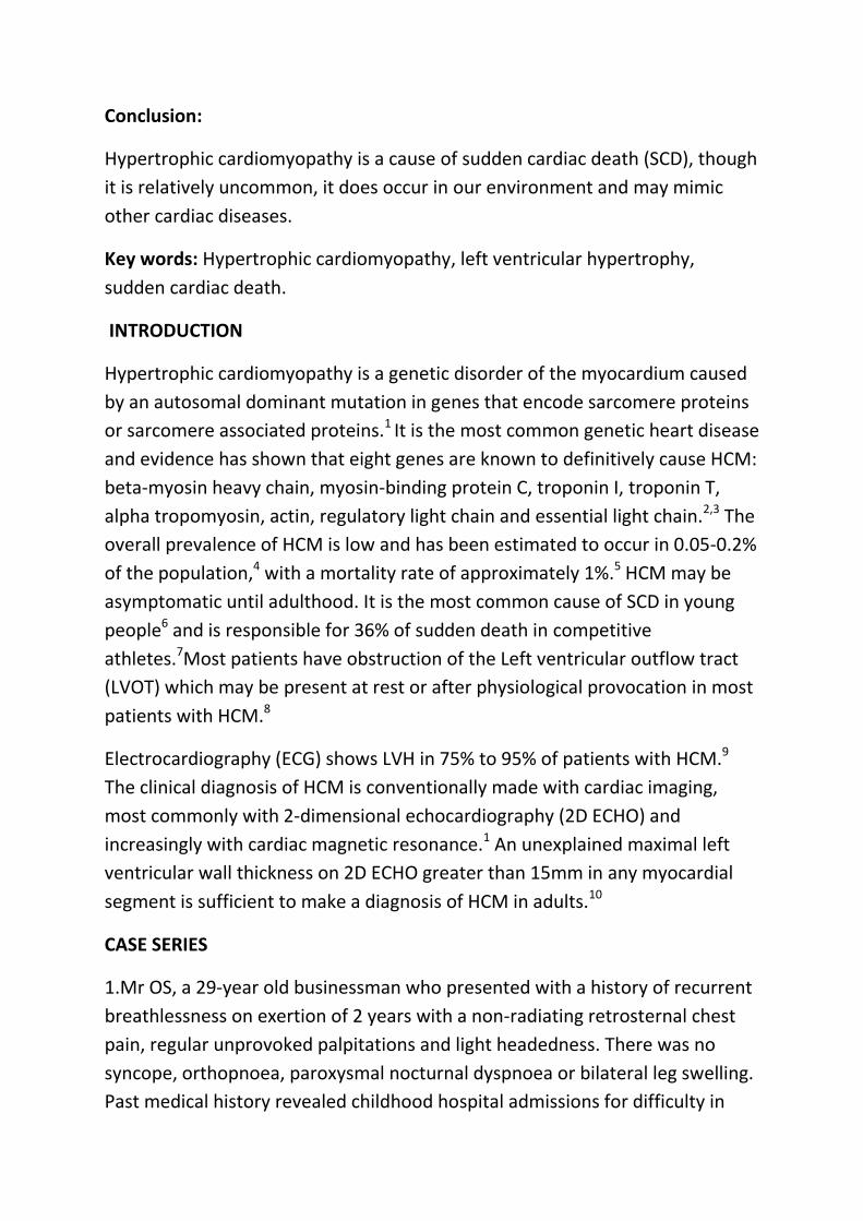
test, electrolyte, urea and creatinine were normal. Echocardiography revealed
grossly hypertrophied left ventricle with asymmetric septal hypertrophy (ASH).
The interventricular septal wall thickness (IVS) was 30mm and the left
ventricular posterior wall (LVPW) was 13.3mm with IVS/LVPW of 2.25 and a
small left ventricular cavity (Figure 1 & 2). There was Systolic Anterior Motion
(SAM) of the anterior mitral valve leaflet (Figure 3) and dilated left atrium.
There was gross hypokinesia of the interventricular septum (IVS). Ejection
fraction was 78.6% with restrictive left ventricular diastolic dysfunction. There
was LVOT obstruction with a peak systolic pressure gradient between the left
ventricle and the ascending aorta of 190 mmHg at rest. Medical treatment was
started with a β blocker (tablets metoprolol 25mg twice daily), tablets
rosuvastatin 10mg nocte and aspirin 75mg daily. Metoprolol was increased and
maintained at 50mg twice daily. Other medications were continued at the
same dose. He attended monthly follow up visits and made significant clinical
improvement as his symptoms of palpitation, retrosternal chest pain and light
headedness have resolved. He was then referred for implantable cardioverter
defibrillator (ICD) for primary prevention of SCD.

Figure 1: 2D guided M mode echocardiogram revealed grossly hypertrophied
left ventricle with asymmetric septal hypertrophy (ASH) and small LV cavity.
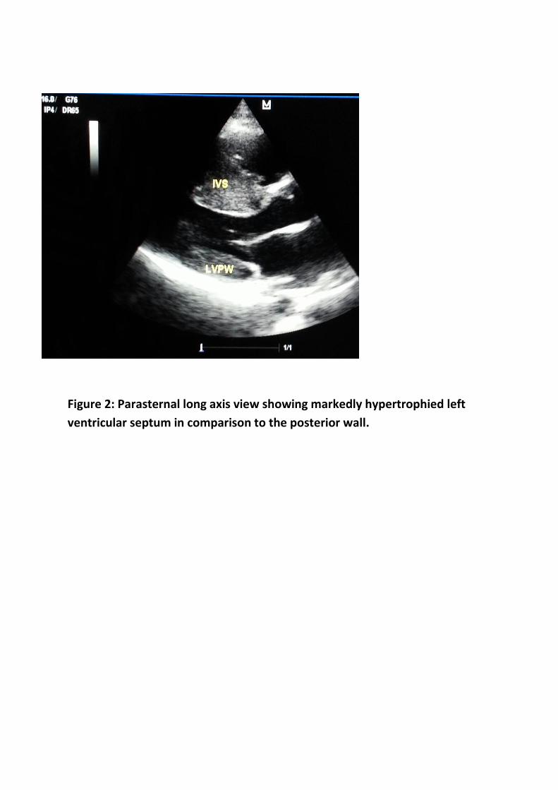
Figure 2: Parasternal long axis view showing markedly hypertrophied left
ventricular septum in comparison to the posterior wall.
Figure 3: 2D guided M mode echocardiogram showing systolic anterior
motion (SAM) of the mitral valve leaflet.
2. Mr AC a 40 year old civil servant who presented with a history of previous
syncopal attack, recurrent light headedness, unprovoked palpitations, easy
fatiguability and recurrent breathlessness. He was previously being evaluated
for rheumatic heart disease. There was no orthopnea, paroxysmal nocturnal
dyspnoea or bilateral leg swelling. He had a previous history of easy
fatiguability from childhood and avoided strenuous activities since then. He
was not a known hypertensive or diabetic. No significant alcohol or tobacco
history. No family history of sudden cardiac death or heart disease.
On examination he was not pale, afebrile, not cyanosed, not dyspnoeic at rest
with no dependent edema. His pulse was 92 beats per minute, full volume and
regular. Blood pressure was 124/70mmHg. Jugular venous pressure was not
elevated. The apex beat was located at the 5th left intercostal space lateral to
the midclavicular line and was heaving. The 4th, 1st and 2nd heart sounds
were heard. The breath sounds were vesicular with a respiratory rate of 16
cycles per minute. Electrocardiography showed ST depression and deep T wave

inversion in V4 – V6, AVL and I. It also showed left ventricular hypertrophy.
Echocardiography revealed thickened interventricular septum of 2.27cm and
left ventricular posterior wall of 2cm (Figure 4). There was systolic anterior
motion of the anterior mitral valve leaflet (Figure 5) and dilated left atrium.
The ejection fraction was 87.4%.The peak systolic pressure gradient between
the left ventricle and the ascending aorta was 192mmHg. Laboratory
investigations revealed dyslipidaemia but the fasting plasma glucose, uric acid
and kidney function tests were normal. He was commenced on tablet
rosuvastatin 10mg nocte, tablets aspirin 75mg daily and metoprolol 25mg
twice daily. He was also counselled on the need for implantable cardioverter
defibrillator but was lost to follow up.
Figure 4: 2D guided M mode echocardiogram of Case 2 revealed thickened left
ventricular walls.

Figure 5: 2D guided M mode echocardiogram of Case 2 also showing SAM.
DISCUSSION
Hypertrophic cardiomyopathy was thought to be a rare disease in black
Africans in the pre-echocardiography era11 but this fact has been disputed by
subsequent echocardiographic studies.12,13 There is paucity of data on the
prevalence of HCM in Nigeria, however Mbakwem et al14 reported a much
higher prevalence of 2% in Lagos. Both patients presented with dyspnoea on
moderate exertion (DOE) in New York Heart association(NYHA) class ll.
Dyspnoea on exertion, chest pain and palpitations are symptoms commonly
associated with HCM. This was similar to the symptoms found by Mbakwem et
al14 in which half of the patients had palpitation and chest pain, while 42.9%
had DOE and another 42.9% were asymptomatic. The symptoms found in HCM
are non-specific and could mimic ischemic heart disease and other cardiac
diseases.
The ECG and TTE in both patients were typical for HCM. The ECG for Mr OS
showed significant ST elevation in the precordial leads from V1 to V3 which

was in keeping with an acute myocardial infarction. This was in keeping with
the ECG found in a study in Germany by Daralammouri et al.15 Other diagnostic
modalities include a 48 hour ambulatory Holter ECG to detect non-sustained
ventricular tachycardia and stress test.
Medical treatment with a beta adrenergic blockers or non-dihydropyridine
calcium channel blocker which are known to decrease the obstructive gradient
in HCM by decreasing cathecholamine –mediated contractility has been found
useful in patients with mild to moderate symptoms16. Beta adrenergic receptor
blockers are useful in relieving symptoms of heart failure in both obstructive
and non-obstructive HCM by slowing the heart rate and reducing the force of
left ventricular contraction thus augmenting LV filling and decreasing
myocardial oxygen consumption.
Surgical septal myomectomy is done for symptomatic patients in New York
heart association class lll /lV who do not respond to medical therapy9. It is also
the indicated procedure in patients with obstruction to LV outflow under basal
conditions or following physiological exercise.(LV outflow gradient ≥
50mmHg)1,5.During resection of part of the septal muscle and ventricular wall,
complications with papillary muscle and left bundle branch block may occur17.
For management of severely symptomatic patients with apical hypertrophic
cardiomyopathy in this setting, Said and colleagues proposed a new surgical
technique that they described as “apical ventriculotomy” to remove a portion
of the thickened muscle to permit greater filling of the left ventricle in
diastole18. When left ventricular outflow tract obstruction was present, an
additional trans aortic resection was accomplished18.
Percutaneous alcohol septal ablation has also been shown to reduce or
eliminate the obstruction in 90% of cases.9
Dual chamber pacing has been promoted as an alternative to myomectomy
for patients with refractory heart failure symptoms5.
Implantable cardioverter defibrillator (ICD) is the mainstay of therapy for both
primary and secondary prevention of SCD.19 Sudden death is the most feared
complication of HCM and may be prevented by the use of ICD. Secondary
prevention is relevant in patients with a prior cardiac arrest and sustained
ventricular tachycardia (VT). Indications for Primary prevention, especially in
patients < 50yrs includes (a) A family history of 1 or more premature HCM
related deaths particularly if sudden (b) hypotensive /attenuated blood

pressure response to exercise (c) unexplained syncope especially if recent (d)
multiple or prolonged non sustained bursts of VT on serial or ambulatory ECG.
(e)Massive LV hypertrophy ≥ 30mm. (f) Late gadolinium enhancement on
cardiac magnetic resonance imaging.6,19,20
CONCLUSION
Hypertrophic cardiomyopathy does exist in our environment and may often be
missed because of unavailability and poor utilization of echocardiography in
the investigation of cardiac diseases. The diagnosis requires a high index of
suspicion, therefore HCM should be considered in any young person
presenting with unexplained chest pain, presyncope and dyspnoea on exertion.
REFERENCES
1. Gersh BJ, Maron BJ, Bonow RO, Dearani JA, Fifer MA, Link MS, et al.
“2011 ACCF/AHA guideline for the diagnosis and treatment of
hypertrophic cardiomyopathy: a report of the American College of
Cardiology Foundation/American Heart Association Task Force on
practice guidelines developed in collaboration With the American
Association for Thoracic Surgery, American Society of Echocardiography,
American Society of Nuclear Cardiology, Heart Failure Society of
America, Heart Rhythm Society, Society for Cardiovascular Angiography
and Interventions, and Society of Thoracic Surgeons,” J Am Coll Cardiol
2011; 58(25): 2703–2738.
2. Alcalai R, Seidman JG, Seidman CE. “Genetic basis of hypertrophic
cardiomyopathy: from bench to the clinics.” Journal of Cardiovascular
Electrophysiology2008; 19 (1): 104–110.
3. Bos JM, Towbin JA, Ackerman MJ. “Diagnostic, prognostic, and
therapeutic implications of genetic testing for hypertrophic
cardiomyopathy.” J Am Coll Cardiol 2009; 54(3): 201–211.
4. Zou Y, Song L, Wang Z et al. Prevalence of idiopathic Hypertrophic
Cardiomyopathy in China: A Population-Based Echocardiographic
Analysis of 8080 adults. Am J Med 2004; 116: 1418.
5. Maron BJ, Maron MS, Danielson GK, Kappenberger LJ, Kuhn HJ, Seidman
CE et al. “American College of Cardiology/European Society of clinical
expert consensus document on hypertrophic cardiomyopathy: a report

of the American College of Cardiology Foundation Task Force on Clinical
Expert consensus documents and the European Society of Cardiology
Committee for Practice Guidelines. ”J Am Coll Cardiol 2003; 42(9): 1687–
1713.
6. Maron BJ. “Contemporary insights and strategies for risk stratification
and prevention of sudden death in hypertrophic cardiomyopathy.”
Circulation 2010; 121(3): 445–456.
7. Maron BJ, Shirani J, Pollac LC, Mathenge R, Roberts WC, Mueller FO, et
al. Sudden death in young competitive athletes: clinical, demographic
and pathological profiles. JAMA 1996; 276: 199-204.
8. Maron BJ, Maron MS, Wigle ED and Braunwald E. “The 50-year history,
controversy, and clinical implications of left ventricular outflow tract
obstruction in hypertrophic cardiomyopathy. From idiopathic
hypertrophic subaortic stenosis to hypertrophic cardiomyopathy,” J Am
Coll Cardiol 2009; 54(3): 191–200.
9. Prinz C, Farr M, Hering D, Horstkotte D and Faber L. “The diagnosis and
treatment of hypertrophic cardiomyopathy.” Deutsches Arzteblatt
international 2011; 108(13): 209-215.
10. Elliott P, McKenna WJ. Hypertrophic Cardiomyopathy. Lancet 2004; 363:
1881-1891.
11. Lewis BS, Armstrong TG, Mitha AS, Gotsman MS. Hypertrophic
obstructive cardiomyopathy in the South African Bantu. S Afr Med J
1973; 34: 599–604.
12. Lewis BS, Agathangelou NE, Flox H, Toams MA, Barlow JB. Hypertrophic
cardiomyopathy in South African blacks. S Afr Med J.1983; 63: 266–269.
13. Abegaz B. The impact of echocardiography in the diagnosis of
hypertrophic cardiomyopathy. East Afr Med J 1990; 67: 556–567.
14. Mbakwem AC, Oke DA, Ajuluchukwu JNA. Hypertrophic
Cardiomyopathy in South Western Nigeria. SA Heart 2009; 6: 104-109.
15. Daralammouri Y, El Garhy M, Same K and Lauer B. Case Report.
Hypertrophic cardiomyopathy mimicking acute anterior myocardial
infarction associated with sudden cardiac death. Case Reports in
Medicine 2012.

16. Nishimura RA, Holmes DR Jr. “Hypertrophic obstructive
cardiomyopathy.” New England Journal of Medicine 2004; 350(13):
1320–1327.
17. Redaelli M, Poloni CL, Bichi S, Esposito G. modified surgical approach to
symptomatic hypertrophic cardiomyopathy with abnormal papillary
muscle morphology: septal myectomy plus papillary muscle
repositioning. J Thoracic Cardiovasc Surg 2014; 147: 1709-1711.
18. Said SM, Schaff HV, Abel MD, Dearani JA. Trans apical approach for
apical myectomy and relief of mid ventricular obstruction in
hypertrophic cardiomyopathy. J Card Surg 2012; 27: 443-448.
19. Maron BJ, Spirito P, Shen WK, Haas TS, Formisano F, Link MS, et al.
Implantable cardioverter-defibrillators and prevention of sudden cardiac
death in hypertrophic cardiomyopathy. JAMA 2007; 296: 405.
20. Maron BJ, Maron MS. Hypertrophic Cardiomypopathy. Lancet 381; 242:
2013.