hyperplasia, fea, adh, and lobular neoplasia -...
TRANSCRIPT
Hyperplasia, FEA, ADH, and
Lobular neoplasia
Abeer Shaaban
Consultant Pathologist
Queen Elizabeth Hospital Birmingham
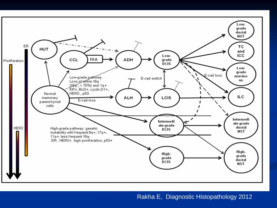
Rakha E, Diagnostic Histopathology 2012
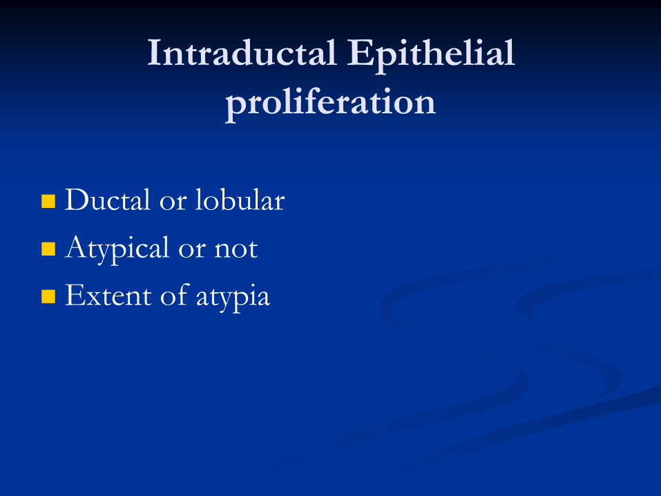
Intraductal Epithelial
proliferation
Ductal or lobular
Atypical or not
Extent of atypia
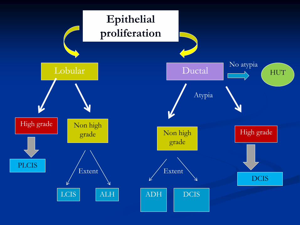
Epithelial
proliferation
High gradeNon high
grade
High grade Non high
grade
DCIS
DCISADH
Ductal Lobular HUT
PLCIS
ALHLCIS
No atypia
Atypia
Extent Extent
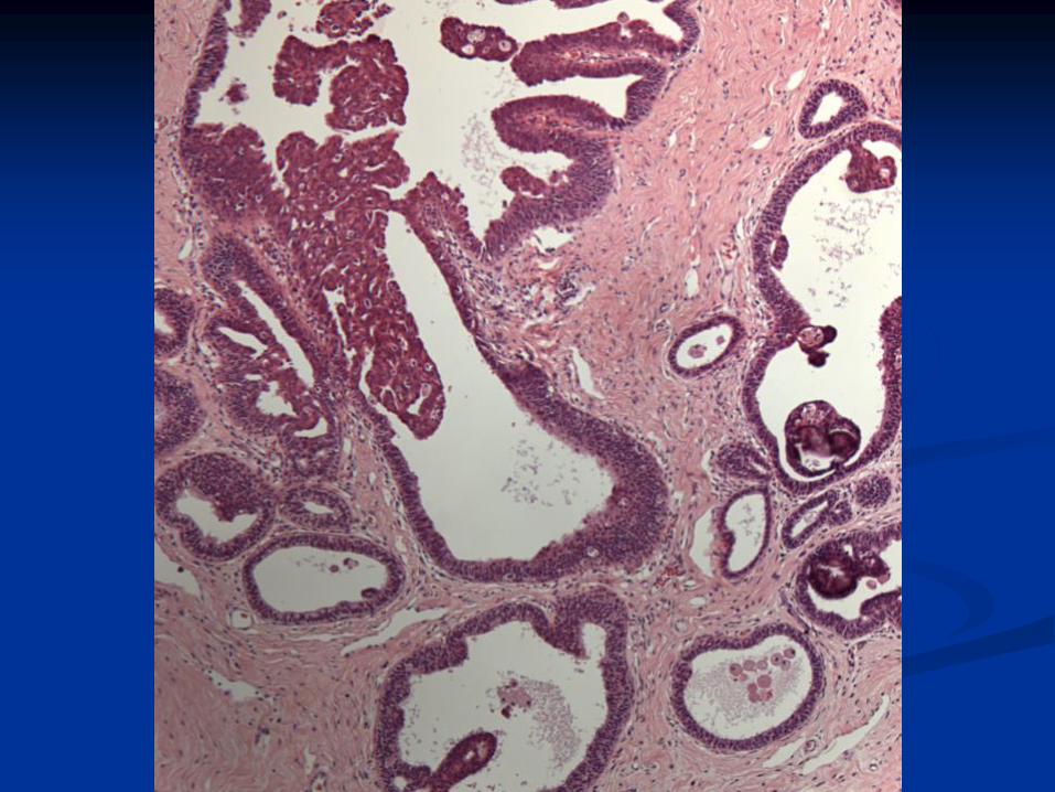
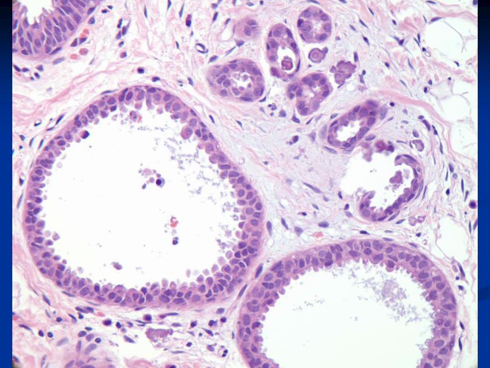
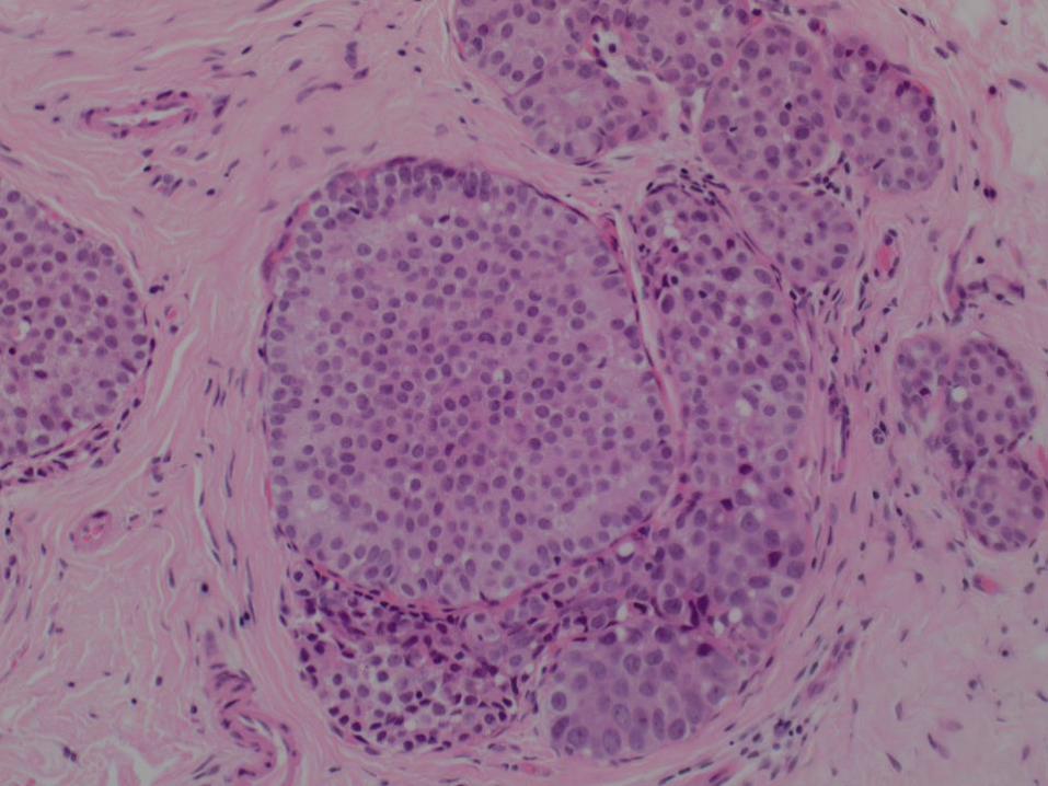
Non atypical ductal hyperplasia
(Epithelial hyperplasia of usual type, Usual ductal
hyperplasia)

Architecture: solid with peripheral slit like
spaces, streaming of nuclei.
Cytological: mixed population
Rounded and ovoid cells
Variation in size and shape


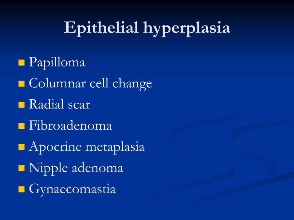
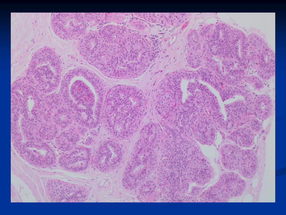
Epithelial hyperplasia
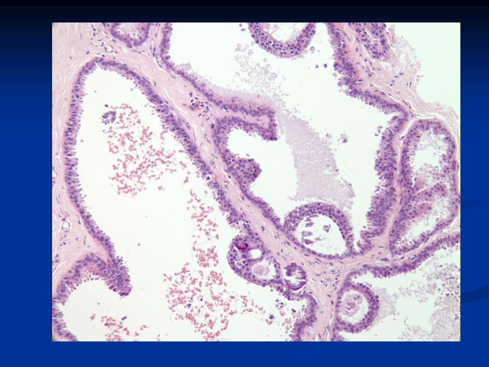
Papilloma
Columnar cell change
Radial scar
Fibroadenoma
Apocrine metaplasia
Nipple adenoma
Gynaecomastia

ADH
Quantitative diagnosis
Not high grade
If on core biopsy: designate as AIDEP
B-coding B3
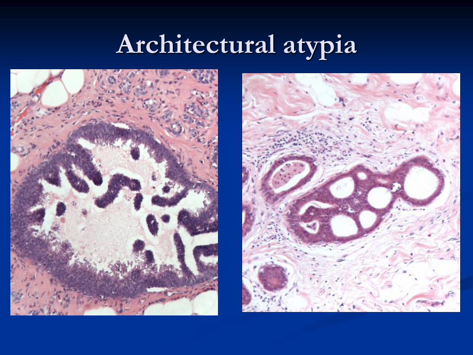
Architectural atypia


Fauna-form changes in the breast Shamonki et al Am J Surg Pathol 2006
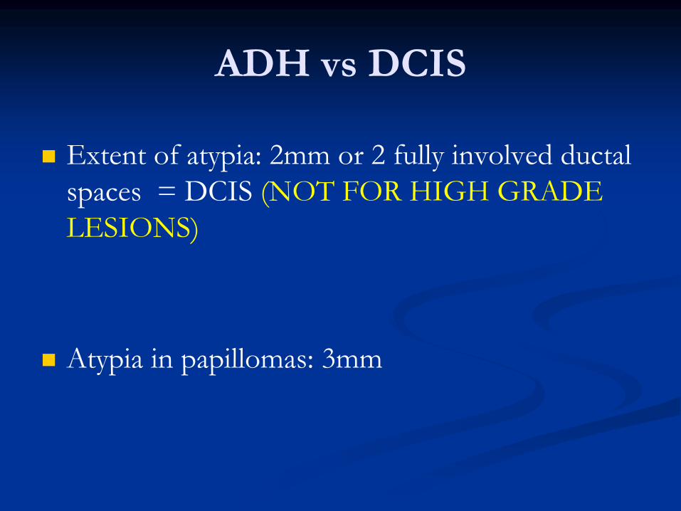
ADH vs DCIS
Extent of atypia: 2mm or 2 fully involved ductal
spaces = DCIS (NOT FOR HIGH GRADE
LESIONS)
Atypia in papillomas: 3mm


Useful Immunohistochemistry
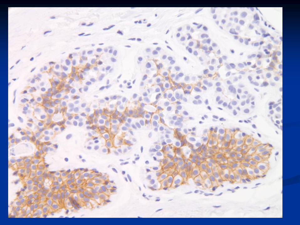
Hyperplasia ADH/DCIS
CK5 mixed Negative
ER Patchy positive Uniformly positive
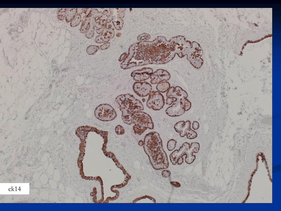
ck14
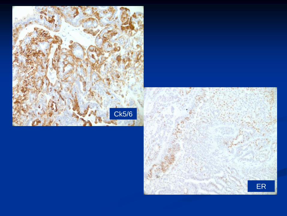
Ck5/6
ER
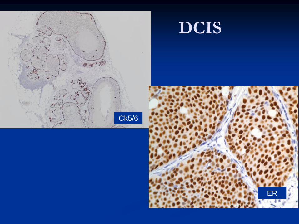
DCIS
Ck5/6
ER
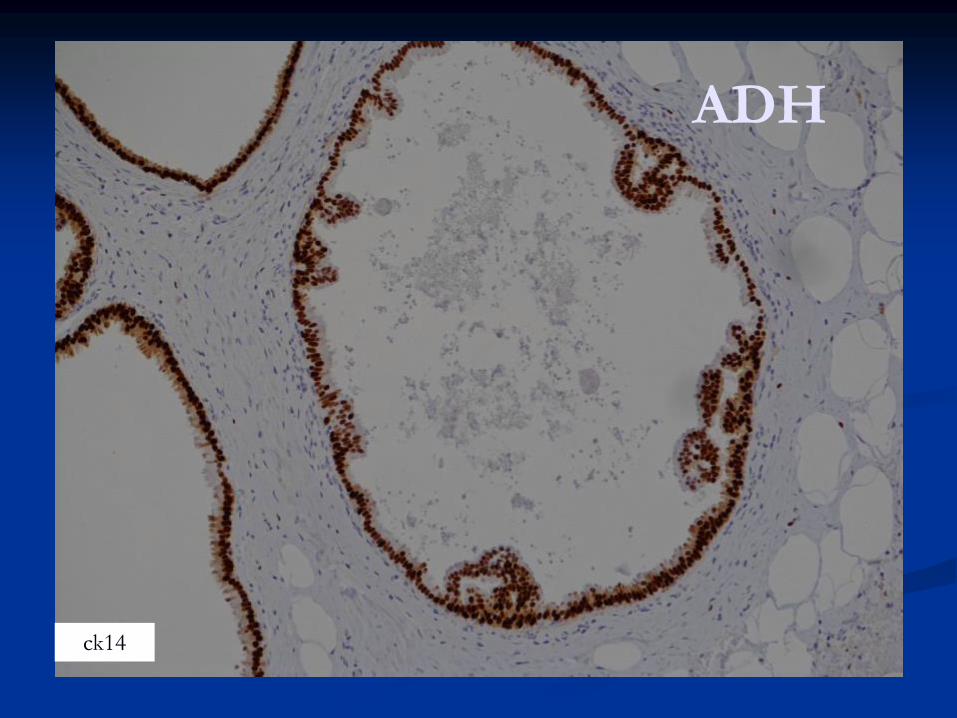
ck14
ADH
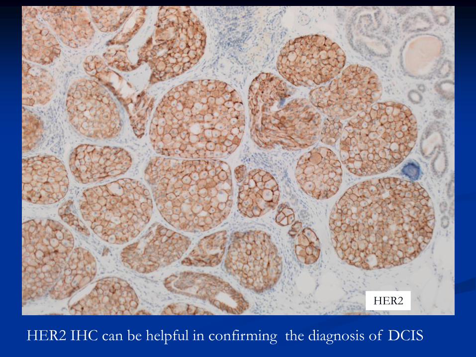
HER2
HER2 IHC can be helpful in confirming the diagnosis of DCIS
IHC Pitfalls
1. Apocrine lesions

Apocrine metaplasia
CK5
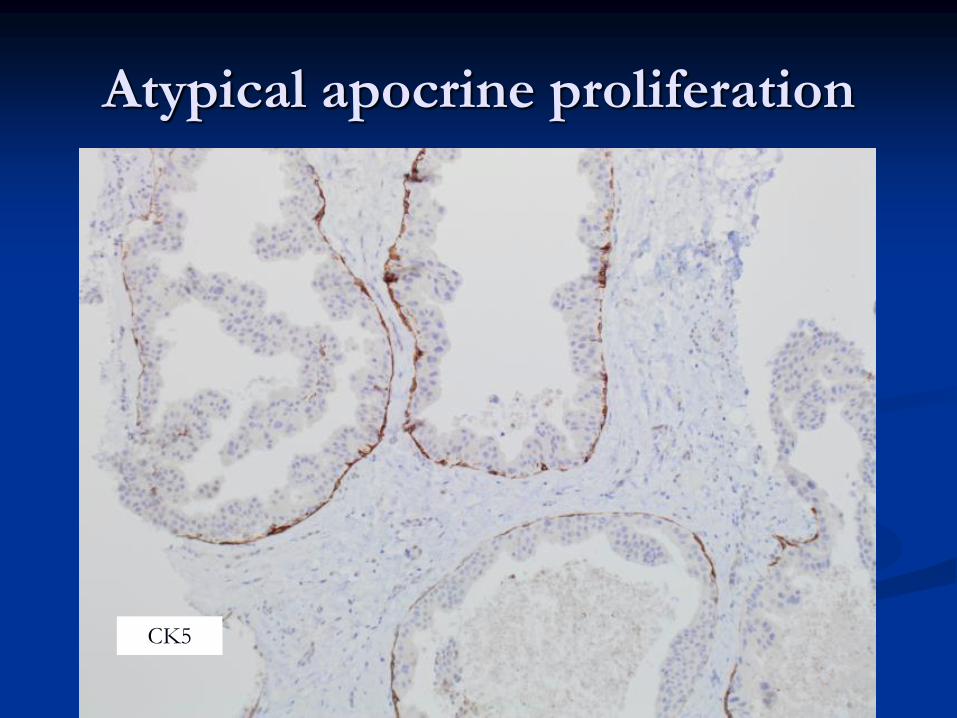
Atypical apocrine proliferation
CK5
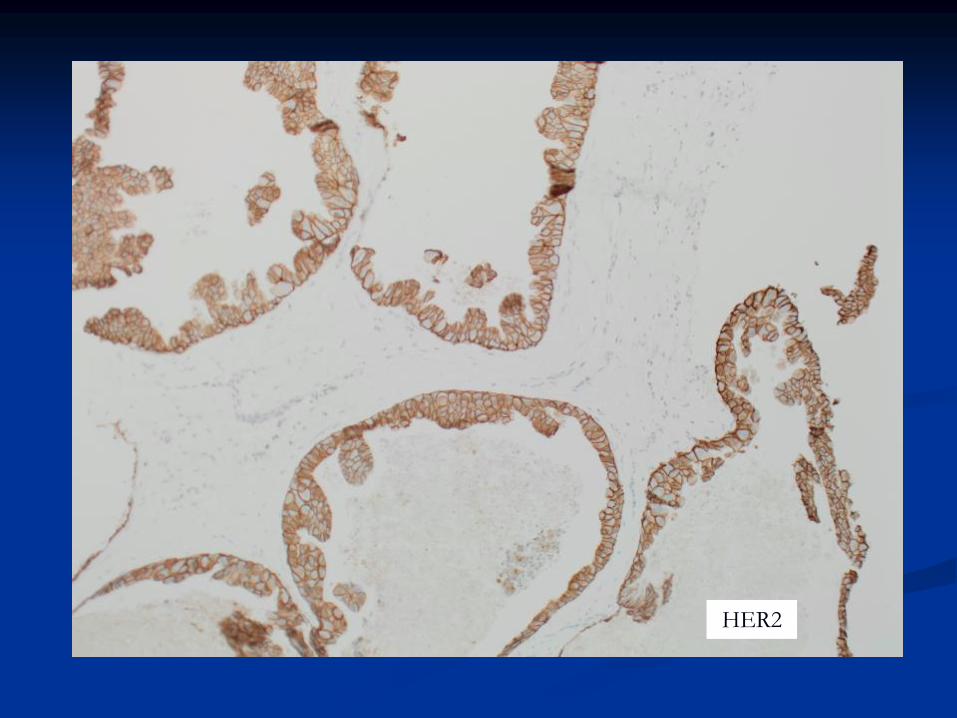
HER2

2. Basal phenotype DCIS
CK14
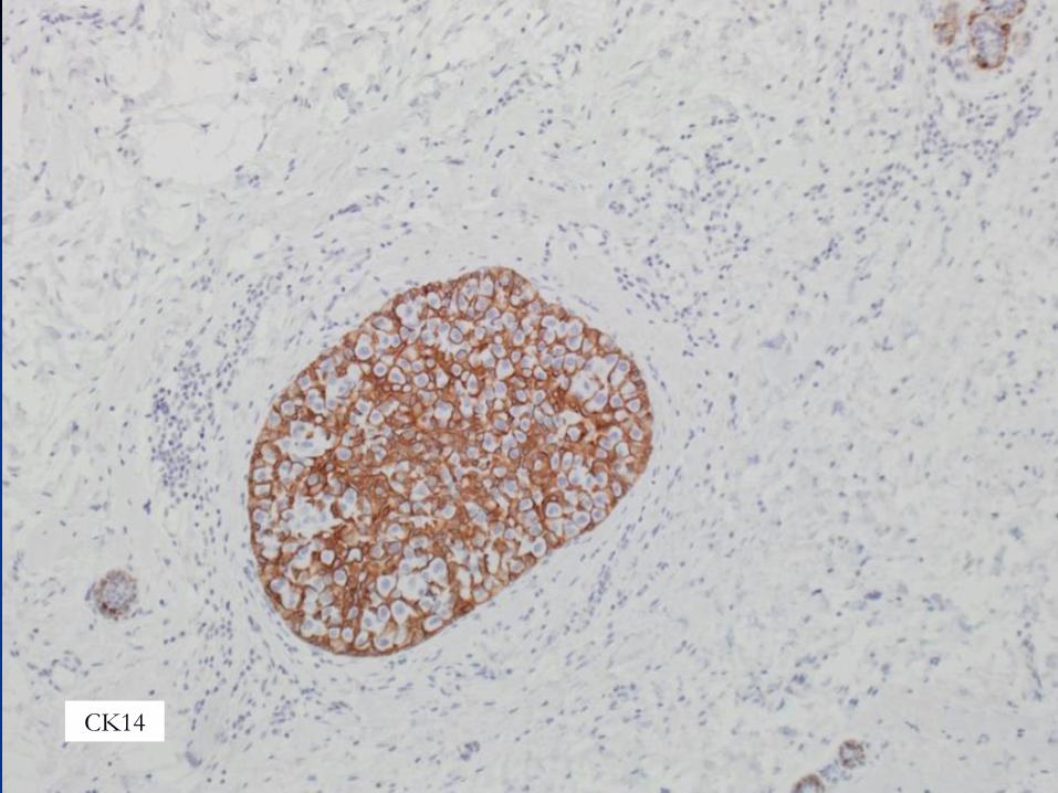
CK14
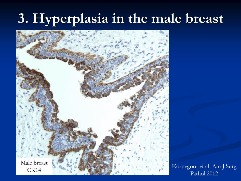
3. Hyperplasia in the male breast
Male breast
CK14 Kornegoor et al Am J Surg
Pathol 2012

Rakha et al 2016

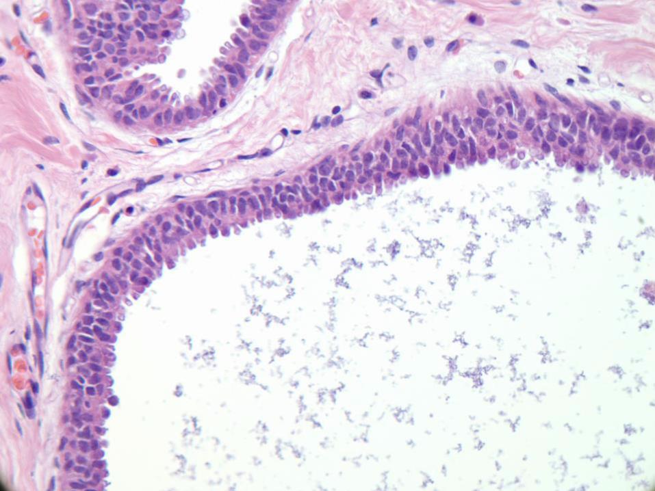
Flat epithelial atypia
Columnar cell lesions
Columnar cell change (without atypia):
columnar cell change/columnar cell
hyperplasia
Flat epithelial atypia
If high grade nuclei = flat DCIS
FEA: WHO 2012 Definition
A neoplastic proliferation of TDLUs
characterised by replacement of the native
epithelial cells by one to several layers of a
single epithelial cell type showing low grade
(monomorphic) cytological atypia

Previous terminology of FEA
Atypical lobule type A
Atypical columnar cell metaplasia
Atypical cystic lobules
Atypical cystic duct
Hypersecretory hyperplasia with atypia
Columnar cell change with atypia
DIN (DIN1a = FEA)
Clinging carcinoma (monomorphic type)
Monomorphic epithelial proliferation
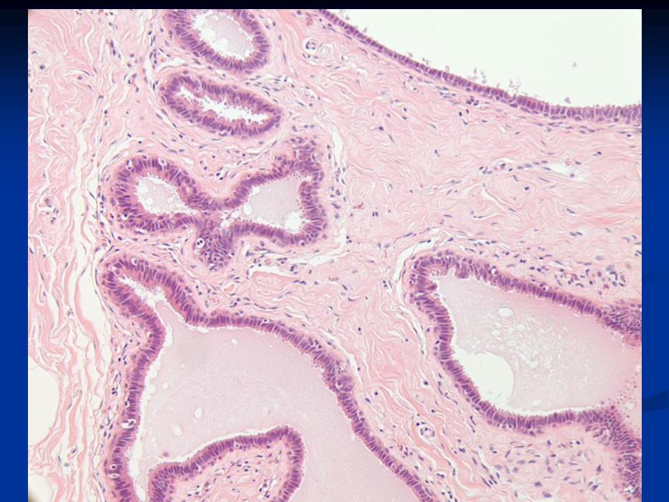
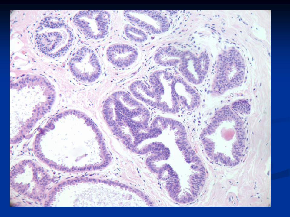
Columnar cell change
Dilated TDLU (oval/branching)
Columnar cells with polarity
Apical snouts
±Calcification
ER positive, CK5 negative

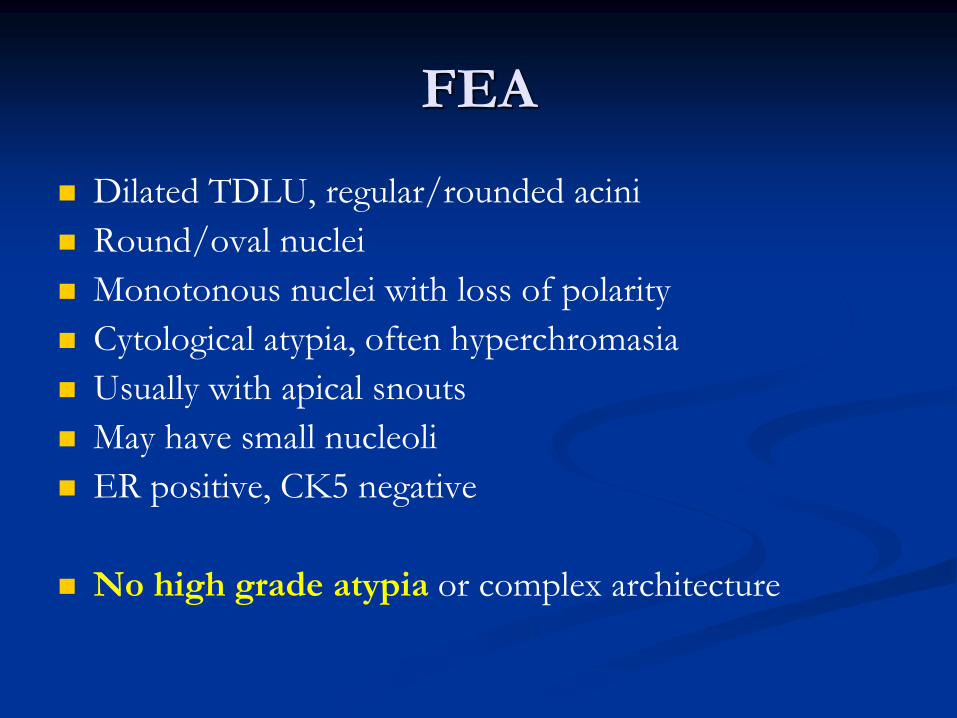
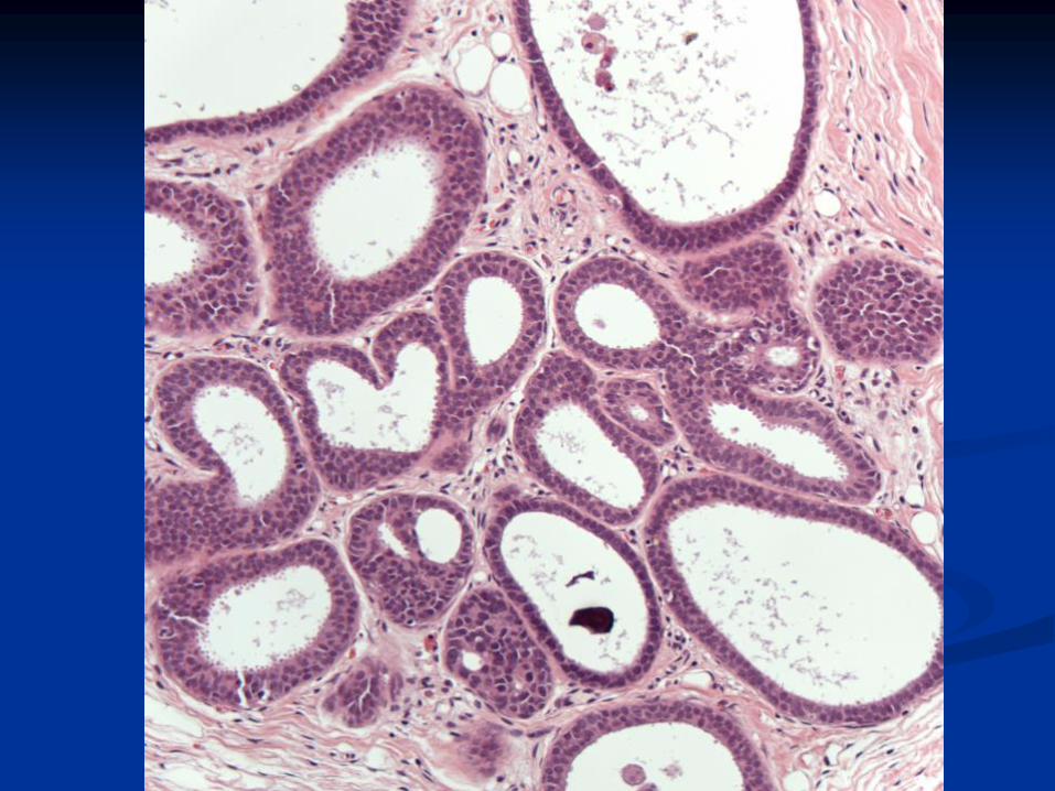
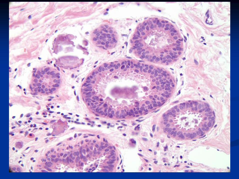
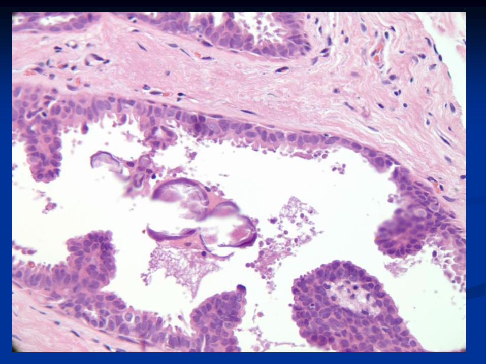
FEA
Dilated TDLU, regular/rounded acini
Round/oval nuclei
Monotonous nuclei with loss of polarity
Cytological atypia, often hyperchromasia
Usually with apical snouts
May have small nucleoli
ER positive, CK5 negative
No high grade atypia or complex architecture


FEA Immunohistochemical profile
ER, PR +, Bcl2+
CK5 –
Her 2 negative
Similar to ADH and low grade DCIS
FEA diagnosis is morphological

ER
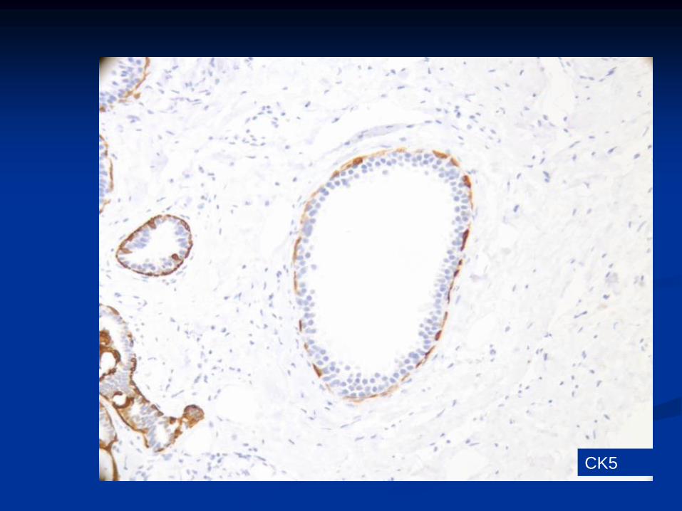
CK5
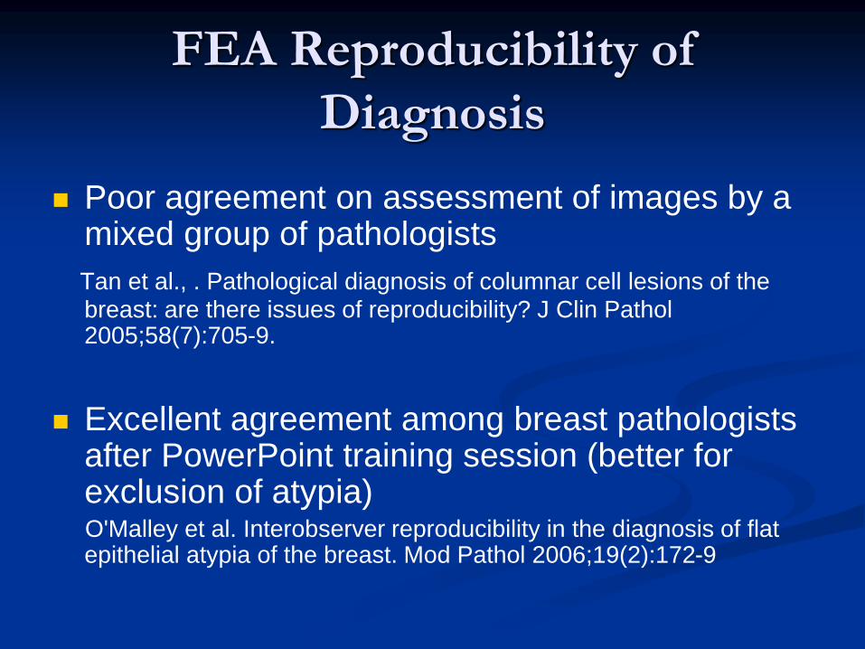
FEA Reproducibility of
Diagnosis
Poor agreement on assessment of images by a mixed group of pathologists
Tan et al., . Pathological diagnosis of columnar cell lesions of the breast: are there issues of reproducibility? J Clin Pathol 2005;58(7):705-9.
Excellent agreement among breast pathologists after PowerPoint training session (better for exclusion of atypia) O'Malley et al. Interobserver reproducibility in the diagnosis of flat epithelial atypia of the breast. Mod Pathol 2006;19(2):172-9

Tips
Do not over-diagnose atypia
Discuss with colleagues!
Rosen’s triad
Columnar cell lesions, lobular neoplasia,
tubular carcinoma

FEA and Lobular insitu
neoplasia
Associated lesions
Low nuclear grade neoplasia family
-Tubular, cribriform, grade 1 ductal NST, lobular, tubulo-lobular carcinoma
-CCC, FEA, ADH, low grade DCIS, lobular in situ neoplasia
-ER, bcl2, CK8, 18, 19 positive. CK5, 14, p53, HER2 neg
Abdel-Fatah et al, Am J Surg Pathol 2008
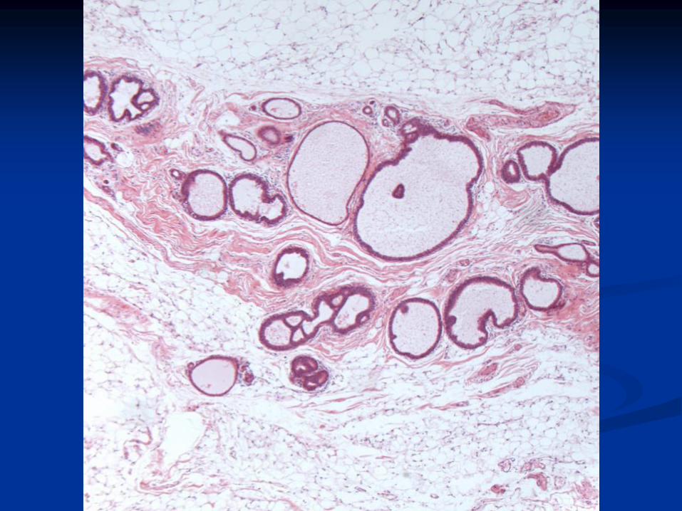
Lobular neoplasia
Encompasses atypical lobular hyperplasia
(ALH) and lobular in situ carcinoma
(LCIS).
LCIS: classical and variants

ALH vs LCIS
Depends on extent of lesion
LCIS: more than half of the acini are filled,
distended and distorted by the
dyscohesive lobular cells.

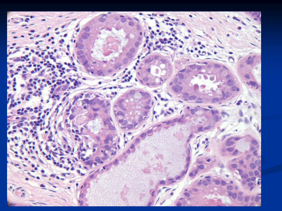
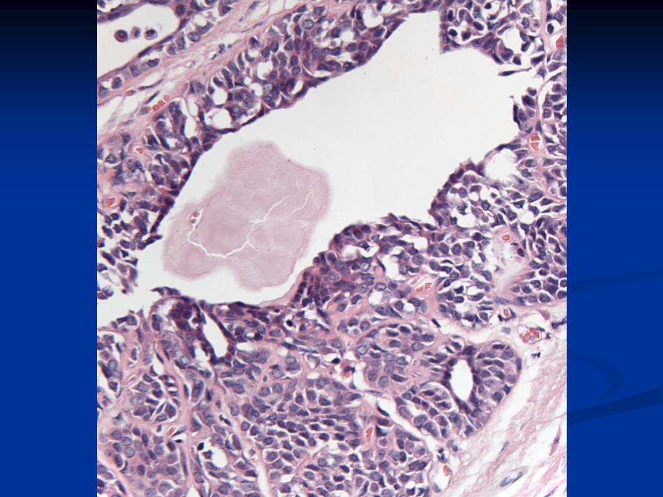
ALH
Histologically
A monomorphic proliferation within TDLU
of dyscohesive cells with uniform round
nuclei, indistinct nucleoli and scant
cytoplasm.
Intracytoplasmic lumina are often present
Pagetoid spread can be seen.

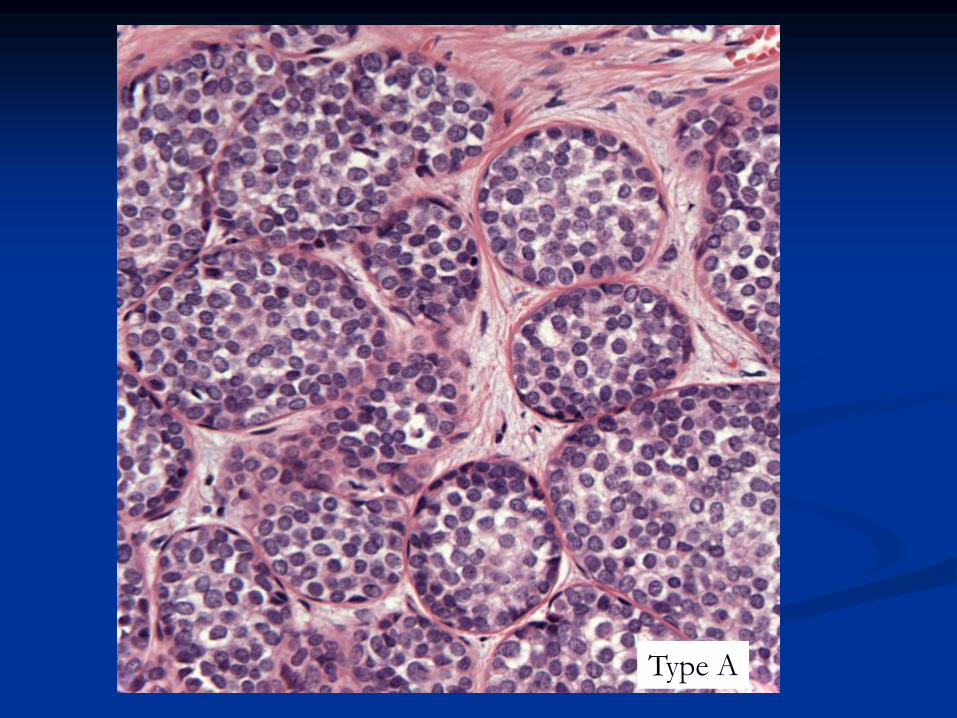
Type A cells: small uniform cells with
bland nuclei and scant cytoplasm
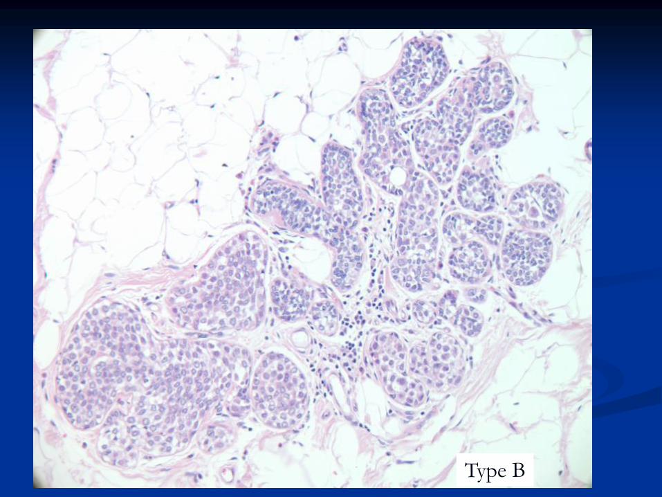
Type B cells: cells are larger, with
more cytoplasm and mild to moderate
atypia
Type A
Type B
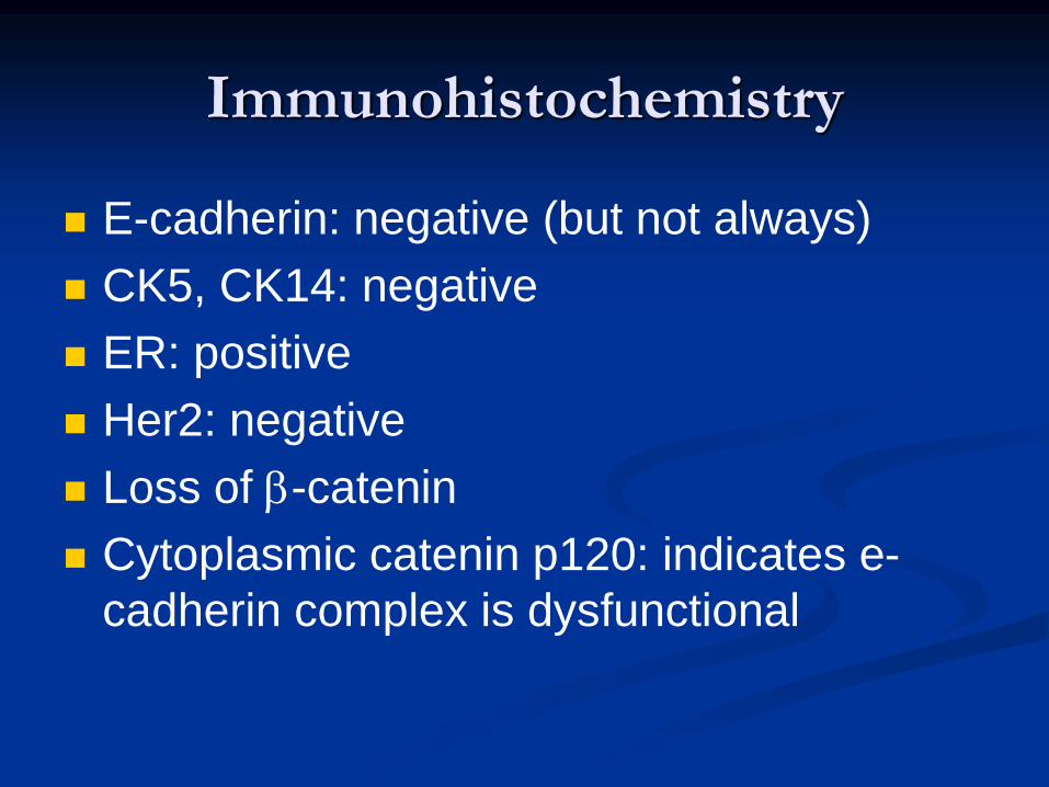
Immunohistochemistry
E-cadherin: negative (but not always)
CK5, CK14: negative
ER: positive
Her2: negative
Loss of -catenin
Cytoplasmic catenin p120: indicates e-
cadherin complex is dysfunctional

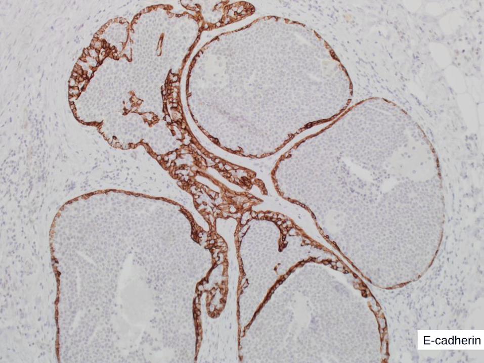
E-cadherin
E-cadherin
E-cadherin

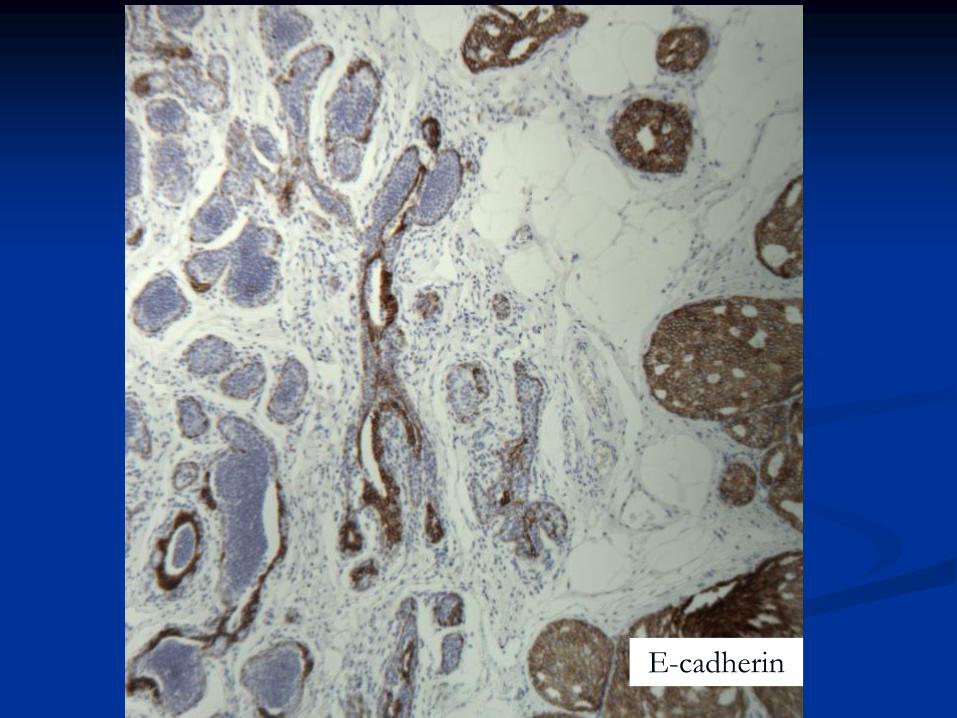
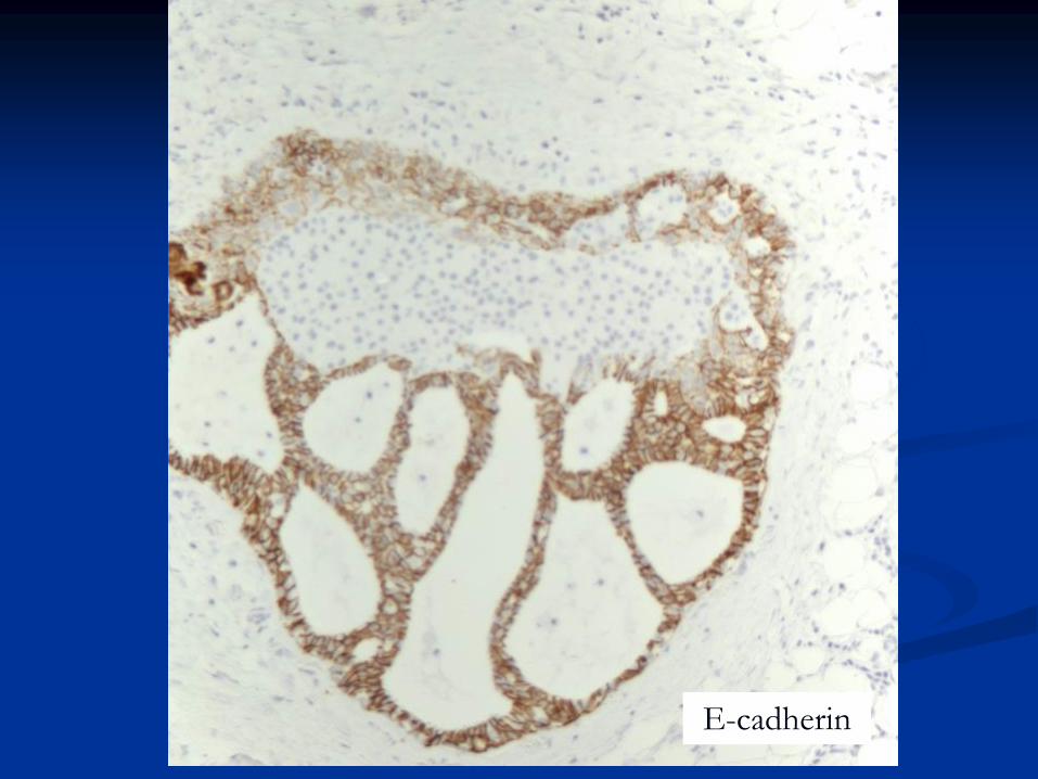
The diagnosis of lobular neoplasia is
morphological.
E-cadherin is often negative (but not always) in
invasive lobular carcinoma.
Expression may be diminished, aberrant or
heterogeneous.
Βcatenin (neg)and p120 (cytoplasmic) may be
helpful in difficult cases

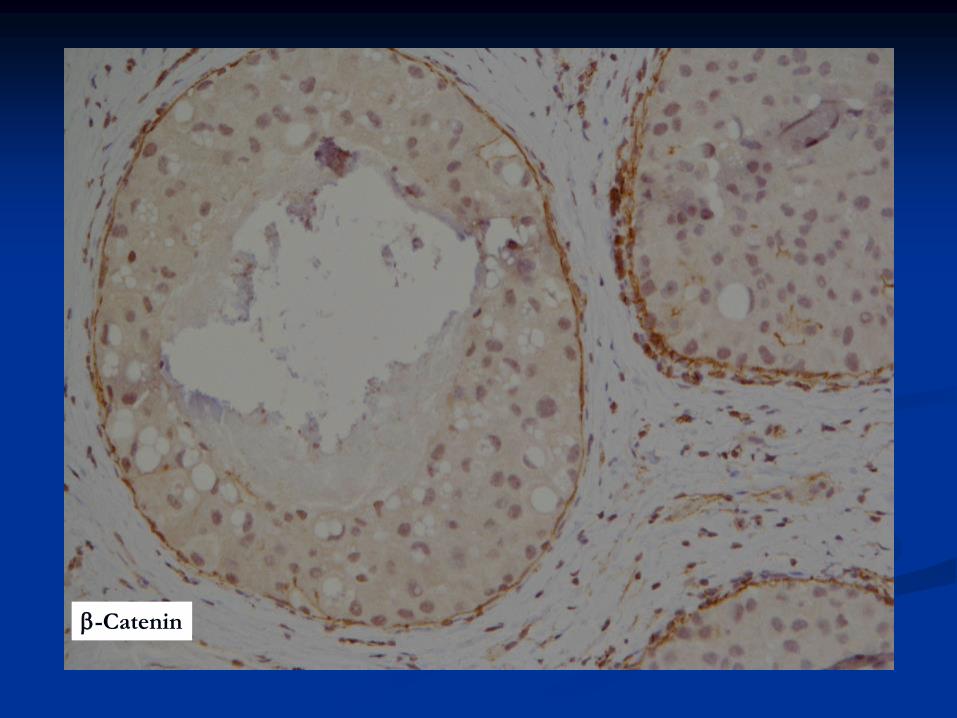
-Catenin
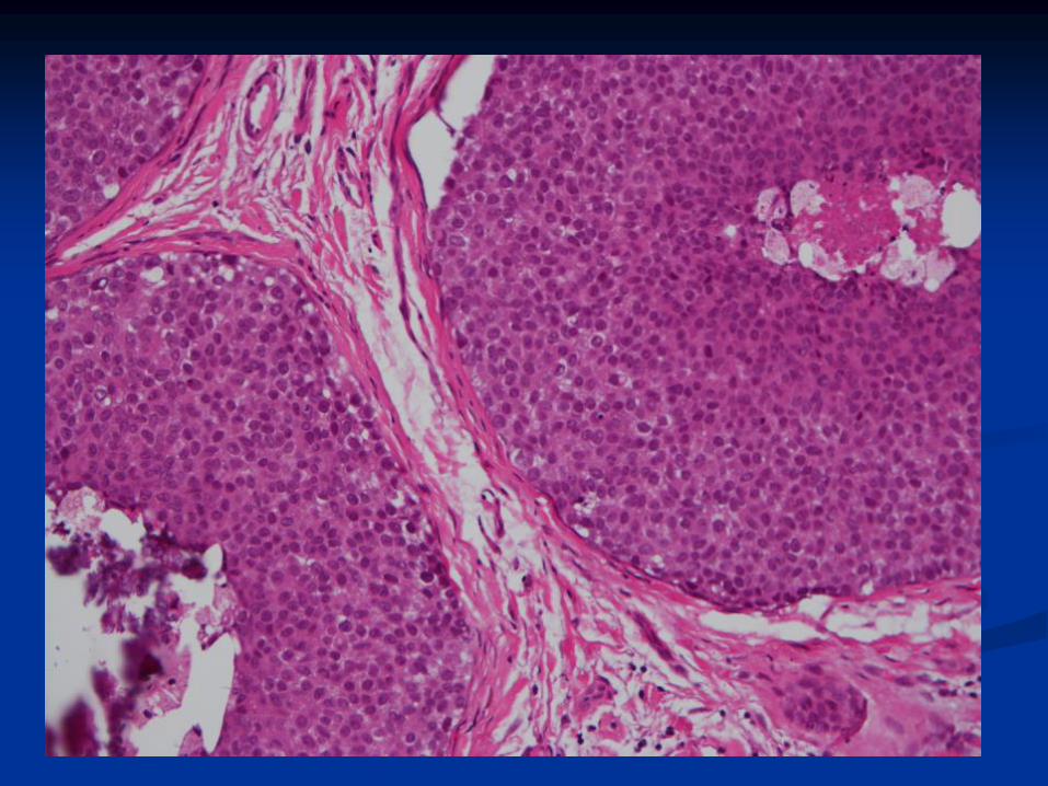
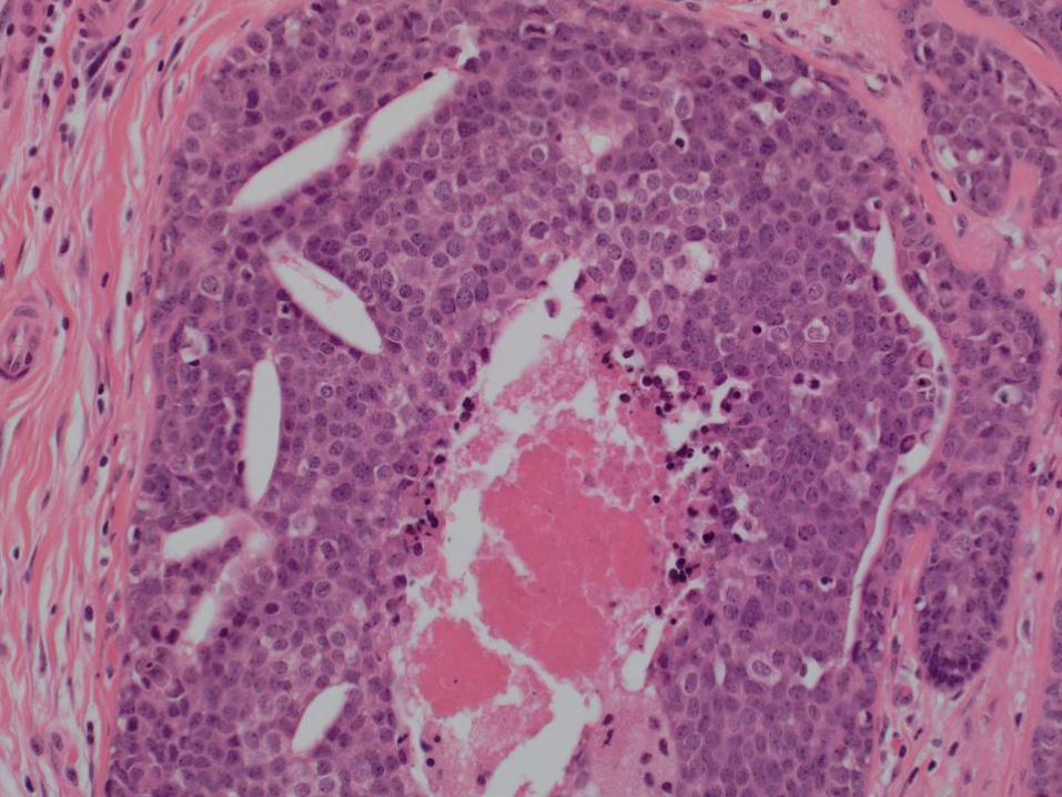
Variants of LCIS
Pleomorphic LCIS (PLCIS)
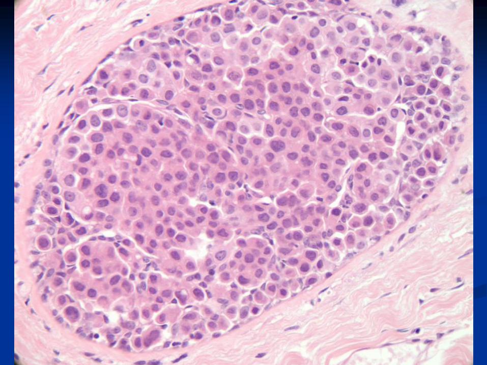
PLCIS
Definition
Similar to Rosen’s criteria for classic LCIS:
lobular units expanded by dyscohesive, pleomorphic cells with abundant eosinophilic cytoplasm, grade 3 nuclei, and prominent nucleoli (Chivukula et al 2008)

Recently recognized variant of
Lobular Carcinoma In Situ (LCIS)
May calcify hence present through
breast screening
Biology and natural history uncertain
Histologically: mimics high grade
DCIS
PLCIS


Pleomorphic apocrine LCIS
PAL-CIS
Chen et al 2005 (suppl) described 10
cases
Pleomorphic apocrine LCIS: LCIS with
myxoid, apocrine and pleomorphic
cytology
CGH: Loss of 16Q, gain at 1Q
DD apocrine DCIS with involvement of
lobules
Immunohistochemistry of PLCIS
E-CADHERIN NEGATIVE
OESTROGEN RECEPTOR may be POSITIVE OR NEGATIVE
GCDFP-15 OFTEN POSITIVE– may be helpful in histological diagnosis
HER-2 may be POSITIVE
P120 catenin: cytoplasmic staining in LCIS, membranous in ductal

E-cadherin
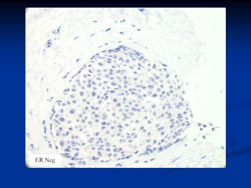
ER Neg
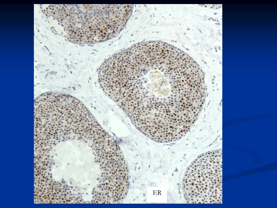
ER
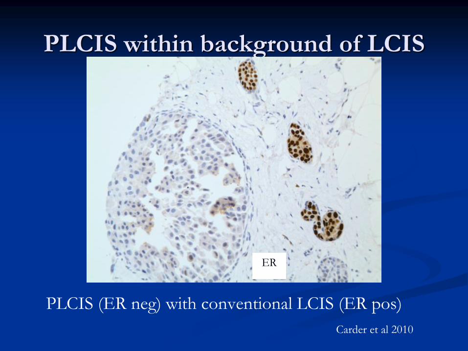
PLCIS within background of LCIS
PLCIS (ER neg) with conventional LCIS (ER pos)
Carder et al 2010
ER
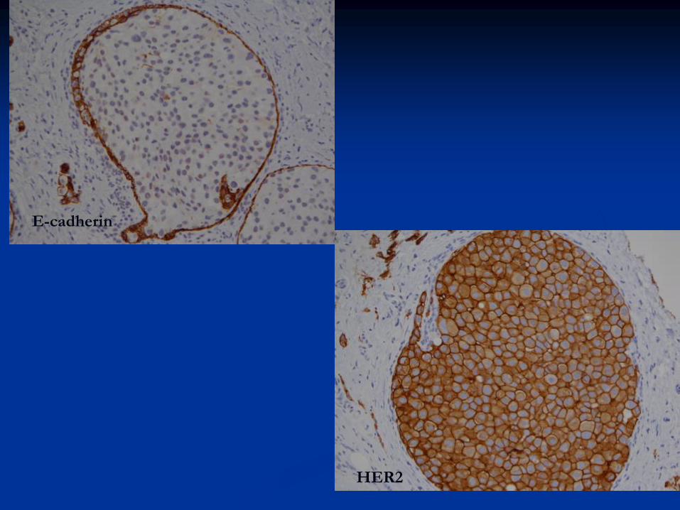
HER2
E-cadherin

Rare variant of LCIS
Classical LCIS with comedo necrosis
(Mass forming LCIS, Florid LCIS
E-cadherin

Sapino et al 2000 described 10 cases of LCIS
with necrosis, 4 of which associated with
invasive carcinoma
A study of 18 cases reported a strong association
with invasive cancer (67% of cases) Fadare et al
Am J Surg Pathol 2006 30:1445–1453
Chin et al 2013 examined the genetic profile of
20 cases of “Florid LCIS”. Lesions showed loss
of 16q (all cases) and 1q gain (80%)
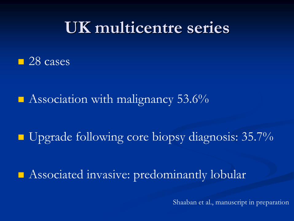
UK multicentre series
28 cases
Association with malignancy 53.6%
Upgrade following core biopsy diagnosis: 35.7%
Associated invasive: predominantly lobular
Shaaban et al., manuscript in preparation

Diagnostic challenges
Is the proliferation ductal or lobular?
( Low/intermediate grade DCS VS LCIS)
(High grade DCIS VS PLCIS)
Look for:
Cellular cohesion
Architectural pattern of DCIS
E-cadherin IHC can help
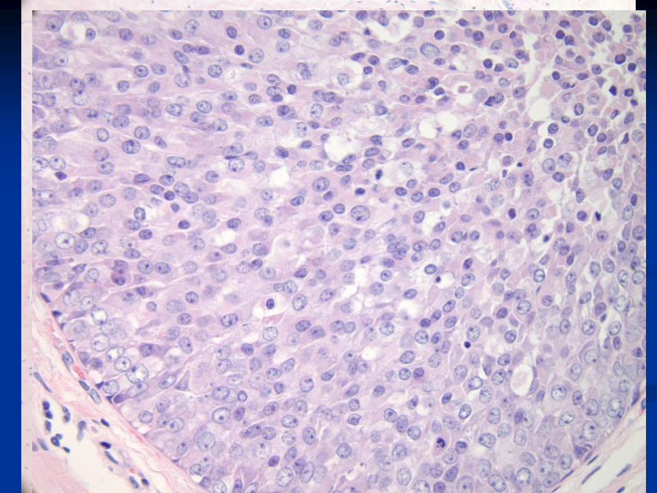
Are the nuclei pleomorphic enough to
designate as PLCIS?
Compare size with normal ductal epithelial cell.
IHC: ER, GCDFP-15, HER2 may help
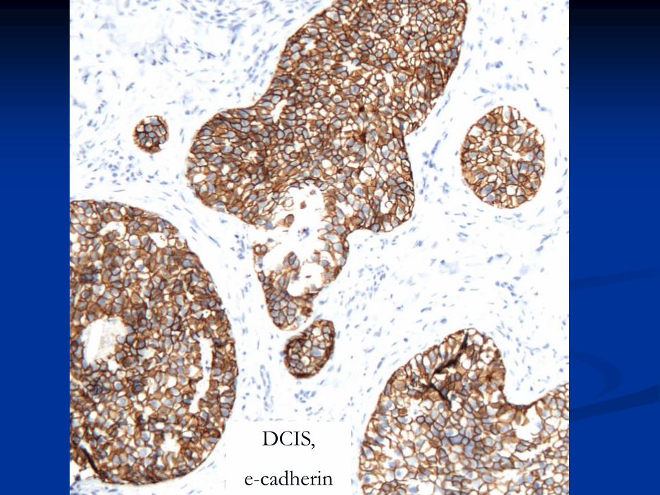
DCIS,
e-cadherin

Classical LCIS and
PLCIS: ER
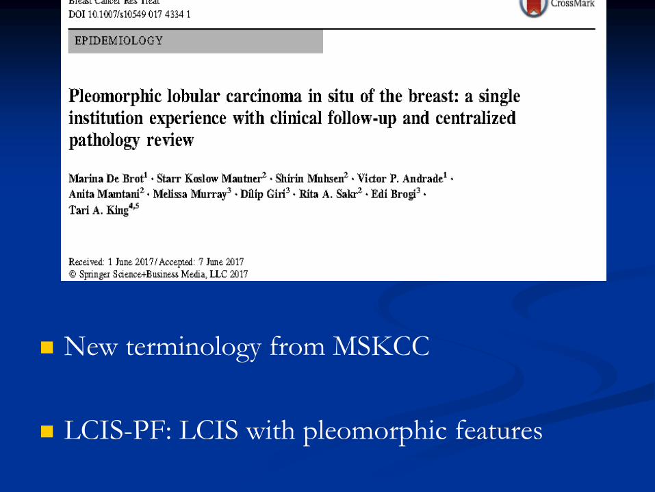
New terminology from MSKCC
LCIS-PF: LCIS with pleomorphic features

In situ carcinoma with mixed ductal
and lobular features
E cadherin IHC may show heterogeneous
staining (DD positive residual cells)
Schnitt group: Jacobs et al Carcinomas in situ of the
breast with indeterminate features: role of Ecadherin
staining in categorization. Am J Surg Pathol
2001;25:229–236.

Clinical relevance
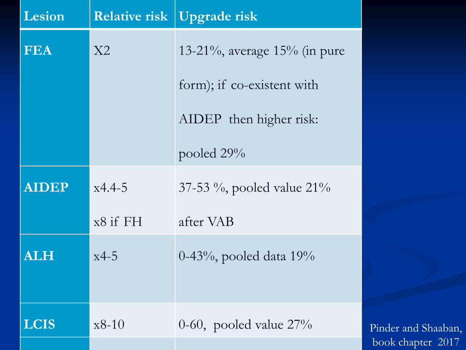
Lesion Relative risk Upgrade risk
FEA X2 13-21%, average 15% (in pure
form); if co-existent with
AIDEP then higher risk:
pooled 29%
AIDEP x4.4-5
x8 if FH
37-53 %, pooled value 21%
after VAB
ALH x4-5 0-43%, pooled data 19%
LCIS x8-10 0-60, pooled value 27%
PLCIS Not known 30-60%, pooled value 40%
Pinder and Shaaban,
book chapter 2017
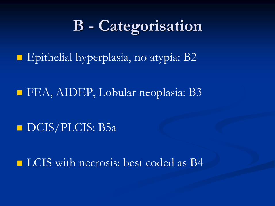
B - Categorisation
Epithelial hyperplasia, no atypia: B2
FEA, AIDEP, Lobular neoplasia: B3
DCIS/PLCIS: B5a
LCIS with necrosis: best coded as B4

Lobular neoplasia on core biopsy
ALH/Classical LCIS: code as B3 and
recommend second line VAB
PLCIS: code as B5a and manage as DCIS
Classical LCIS with necrosis: rare, best coded as
B4, recommend surgical excision.
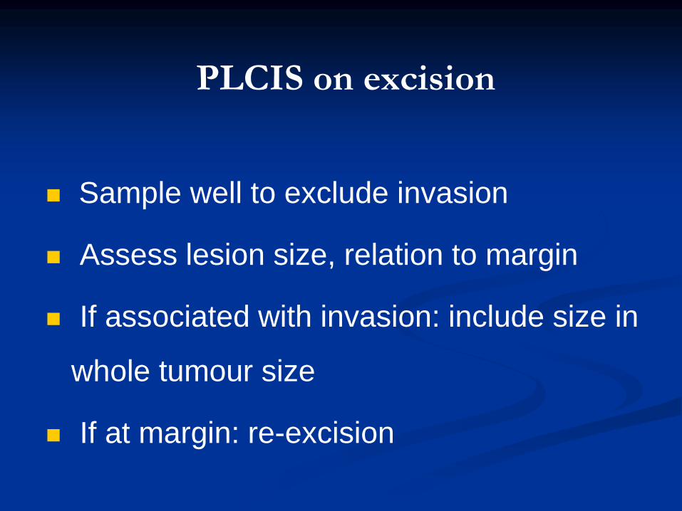
PLCIS on excision
Sample well to exclude invasion
Assess lesion size, relation to margin
If associated with invasion: include size in
whole tumour size
If at margin: re-excision


THANK YOU