hyperglycemia can delay left ventricular dysfunction but not autonomic damage after myocardial...
TRANSCRIPT
ORIGINAL INVESTIGATION Open Access
Hyperglycemia can delay left ventriculardysfunction but not autonomic damage aftermyocardial infarction in rodentsBruno Rodrigues1,2*, Kaleizu T Rosa2, Alessandra Medeiros3, Beatriz D Schaan4, Patricia C Brum5, Kátia De Angelis6
and Maria Cláudia Irigoyen2
Abstract
Background: Although clinical diabetes mellitus is obviously a high risk factor for myocardial infarction (MI), inexperimental studies disagreement exists about the sensitivity to ischemic injury of an infarcted myocardium.Recently, our group demonstrated that diabetic animals presented better cardiac function recovery and cellularresistance to ischemic injury than nondiabetics. In the present study, we evaluated the chronic effects of MI on leftventricular (LV) and autonomic functions in streptozotocin (STZ) diabetic rats.
Methods: Male Wistar rats were divided into 4 groups: control (C, n = 15), diabetes (D, n = 16), MI (I, n = 21), anddiabetes + MI (DI, n = 30). MI was induced 15 days after diabetes (STZ) induction. Ninety days after MI, LV andautonomic functions were evaluated (8 animals each group). Left ventricular homogenates were analyzed byWestern blotting to evaluate the expression of calcium handling proteins.
Results: MI area was similar in infarcted groups (~43%). Ejection fraction and +dP/dt were reduced in I comparedwith DI. End-diastolic pressure was additionally increased in I compared with DI. Compared with DI, I had increasedNa+-Ca2+ exchange and phospholamban expression (164%) and decreased phosphorylated phospholamban atserine16 (65%) and threonine17 (70%) expression. Nevertheless, diabetic groups had greater autonomic dysfunction,observed by baroreflex sensitivity and pulse interval variability reductions. Consequently, the mortality rate wasincreased in DI compared with I, D, and C groups.
Conclusions: LV dysfunction in diabetic animals was attenuated after 90 days of myocardial infarction and wasassociated with a better profile of calcium handling proteins. However, this positive adaptation was not able toreduce the mortality rate of DI animals, suggesting that autonomic dysfunction is associated with increasedmortality in this group. Therefore, it is possible that the better cardiac function has been transitory, and theautonomic dysfunction, more prominent in diabetic group, may lead, in the future, to the cardiovascular damage.
IntroductionDiabetes has been associated with an increased risk ofcardiovascular abnormalities and microvascular compli-cations. Although microvascular retinopathy and nephro-pathy are associated with a high degree of morbidity, theincreased mortality in patients with diabetes is primarilya consequence of cardiovascular disease [1]. In fact, clini-cal studies have demonstrated that diabetes, with conse-quent cardiomyopathy and cardiovascular neuropathy, is
an independent risk factor for cardiovascular disease andis associated with a 2- to 4-fold increased risk of coronaryheart disease [2].In this regard, experimental data have demonstrated
that hyperglycemia in diabetic animals leads to changesin the heart that contribute to injury during and follow-ing an ischemic event; however, the response of anuncontrolled hyperglycemic diabetic heart to ischemicinjury remains controversial [3-5]. Experimental studies[6-8] using an ischemia/reperfusion protocol have shownthat hearts from streptozotocin (STZ) diabetic rats thatundergo a period of no-flow ischemia have a reduced
* Correspondence: [email protected] Movement Laboratory, São Judas Tadeu University, São Paulo, BrazilFull list of author information is available at the end of the article
Rodrigues et al. Cardiovascular Diabetology 2011, 10:26http://www.cardiab.com/content/10/1/26
CARDIOVASCULAR DIABETOLOGY
© 2011 Rodrigues et al; licensee BioMed Central Ltd. This is an Open Access article distributed under the terms of the CreativeCommons Attribution License (http://creativecommons.org/licenses/by/2.0), which permits unrestricted use, distribution, andreproduction in any medium, provided the original work is properly cited.

myocardial infarction (MI) area and recovered ventricularfunction significantly better than nondiabetic hearts, indi-cating a possible cardioprotective role of hyperglycemia.In fact, the exposure to short periods of abnormallyhigher glucose medium or diabetes has been found toprotect the heart against a variety of pathological insults,including ischemia, hypoxia, and calcium overload [9,10].However, most studies reported in the literature have
been conducted using an ischemia/reperfusion model,thus it has not been possible to verify whether chronichyperglycemia can protect the diabetic heart againstpermanent coronary ligation. Recently, our group demon-strated that after 15 days of MI, diabetic animals hadreduced heart fibrosis, improved systolic function, andreduced infarct size. In accordance with this study, it ispossible that these data may have indicated the final path-way promoted by a positive balance in regulatory genesrelated to programmed cell survival, reduced inflammatorycytokines, and increased utilization of glucose as an energysubstrate [9]. However, other important risk factors, suchas cardiovascular autonomic neuropathy, and influenceson the mortality rate were not evaluated in this study.Changes in critical processes that regulate intracellular
calcium concentration and signaling have been a hall-mark of cardiomyopathy and heart failure. Alterations inthe expression and activity of cardiac proteins that parti-cipate in the calcium handling, (e.g., calcium pumpATPase of sarcoplasmic reticulum (SERCA2), depho-sphorylated phospholamban (PLN), which respectivelydecreases the affinity of SERCA2 for calcium, and sarco-lemmal sodium calcium exchanger (NCX), which med-iates calcium efflux from the cell [11] have been shownto occur in cardiomyopathy of STZ diabetic rats [12] andafter MI [13]. However, the impact of associationbetween STZ diabetes and MI in the expression of cal-cium handling proteins remains unknown. In this sense,the aim of the present study was to investigate the effectsof hyperglycemia induced by STZ diabetes in left ventri-cular (LV) dysfunction, net balance of regulatory proteinsinvolved in intracellular calcium homeostasis, autonomicdysfunction and mortality rate, in rats that underwent90 days of MI.
MethodsAnimals and GroupsExperiments were performed in adult male Wistar rats(~240 g) from the Animal House of the University of SãoPaulo, São Paulo, Brazil. Rats were fed standard laboratorychow and water ad libitum. The animals were housed incollective polycarbonate cages in a temperature-controlledroom (22°C) with a 12-hour dark-light cycle (light 07:00-19:00 h). The experimental protocol was approved bythe institutional animal care and use committee of the
Medical School of the University of São Paulo, and thisinvestigation was conducted in accordance with the pre-viously described [14]. Rats were randomly assigned tocontrol (C, n = 15), diabetes (D, n = 16), myocardialinfarction (I, n = 21), and diabetes + myocardial infarction(DI, n = 30).
Diabetes InductionExperimental diabetes was induced by intravenous injec-tion of 50 mg/kg STZ (Sigma Chemical Co., St. Louis,MO) dissolved in citrate buffer (pH 4.2). Rats werefasted overnight before STZ injection. Control rats wereinjected with buffer only (10 mM citrate buffer, pH 4.5).Forty-eight hours after STZ injection, diabetes was con-firmed by blood glucose levels above 200 mg/dL.
Myocardial InfarctionFifteen days after diabetes induction or buffer injection,anesthetized rats (80 mg/kg Ketamine and 12 mg/kg Xyla-zine, i.p.) underwent surgical occlusion of the left coronaryartery, which resulted in myocardial infarction, as pre-viously described elsewhere [9,13]. Briefly, a left intercostalthoracotomy was performed, the third intercostal spacedissected, and the heart exposed. The left anterior des-cending coronary artery was occluded with a single nylon(6.0) suture at approximately 1 mm distal to the left atrialappendage. The chest was closed with silk suture.The animals were maintained in the ventilator until
recovery. All rats received antibiotics (penicillin, 20,000 U)and Tramadol (20 mg/kg, every 6 h). Eight animals fromeach group were evaluated 90 days after myocardial infarc-tion induction (105 days after diabetes induction) to per-form the following experimental protocols. The wholesample (n = 75) was evaluated for mortality, an assessmentthat started after myocardial infarction surgery, excludingthe influence of anesthesia or surgical procedure stress.
Noninvasive Evaluation of Left Ventricular Function:Echocardiographic MeasurementsEchocardiography was performed by a double-blindedobserver, under the guidelines of the American Societyof Echocardiography, 2 days (initial evaluation) and90 days (final evaluation) after myocardial infarction.Rats were anesthetized (80 mg/kg Ketamine and12 mg/kg Xylazine), and images were obtained with a10-14 mHz linear transducer in a SEQUOIA 512(ACUSON Corporation, Mountain View, CA) for mea-surements of morphometric parameters: left ventricular(LV) mass (corrected by body weight) and LV end-diastolic diameter (LVEDD); systolic function para-meters: ejection fraction (EF) and velocity of circum-ferential fiber shortening (VCF); diastolic functionparameters: LV isovolumetric relaxation time (IVRT)
Rodrigues et al. Cardiovascular Diabetology 2011, 10:26http://www.cardiab.com/content/10/1/26
Page 2 of 11
and peak E deceleration time (EDT) corrected by thesquare root of R-R interval, because EDT and IVRTare HR dependent; and global function: myocardialperformance index (MPI). Echocardiographic para-meters were measured as described in detail elsewhere[9,13].The infarction area was delimited, which led us to
analyze the movement of the LV walls. Regions withsystolic shortening classified as absent or with paradoxi-cal movement were considered infarcted. The infarctedarea (in %) was thus measured as the ratio of theseregions by the total area of LV walls [9,13,15].
Autonomic EvaluationsOne day after the final echocardiographic evaluation, 2catheters filled with 0.06 mL of saline were implanted intothe femoral artery and femoral vein of the anesthetizedrats (80 mg/kg Ketamine and 12 mg/kg Xylazine, IP).Twenty-four hours later, the arterial cannula was con-nected to a strain-gauge transducer (Blood PressureXDCR, Kent Scientific, USA), and arterial pressure (AP)signals were recorded over a 30-minute period in con-scious animals by a microcomputer equipped with an ana-log-to-digital converter board (WinDaq, 2-kHz, DATAQ,Springfield, OH). The recorded data were analyzed on abeat-to-beat basis to quantify changes in mean AP (MAP)and heart rate (HR) [13,15].Sequential bolus injections (0.1 mL) of increasing doses
of phenylephrine (0.25 to 32 μg/kg) and sodium nitro-prusside (0.05 to 1.6 μg/kg) were given to induceincreases or decreases in MAP pressure responses (foreach drug), ranging from 5 to 40 mm Hg. Baroreflex sen-sitivity expressed as bradycardic (BR) and tachycardic(TR) responses in beats per minute per millimeter ofmercury, as described elsewhere. The overall variabilityof the pulse interval (PI) in the time domain was assessedby the standard deviation (SD) of the time series [13,15].
Invasive Evaluation of Left Ventricular FunctionOne day after the final autonomic evaluation, LV functionwas also measured invasively in anesthetized rats (pento-barbital sodium, 40 mg/kg). One catheter of PE-50 wasinserted into the right carotid artery and advanced intothe LV. Ventricular pressure signals were measured with atransducer and conditioner (Blood Pressure XDCR, Kent©
Scientific, USA) and digitally recorded (5 min) with a dataacquisition system (WinDaq, 2-kHz, DATAQ, Springfield,OH). The recorded data were analyzed on a beat-to-beatbasis to quantify changes in LV pressure. The followingindices were obtained: heart rate (HR), LV systolic pres-sure (LVSP), LV end-diastolic pressure (LVEDP), andmaximum rate of LV pressure rise and fall (+dP/dt and-dP/dt), as previously described [13,16].
Western Blot AnalysisLeft ventricular homogenates were analyzed by Westernblotting to evaluate the expression of sarcoplasmic reticu-lum calcium ATPase pump (SERCA2), phospholamban(PLN), phosphorylated - PLN at serine 16 (phospho-ser16-PLN), phosphorylated - PLN at threonine 17 (phospho-thr17-PLN), phosphatase protein 1 (PP1), and sodium cal-cium exchanger (NCX), as described elsewhere [17].Mouse monoclonal antibodies to SERCA2 (1:2500), PLN(1:500), and NCX (1:2000) were obtained from AffinityBioReagents (Golden, CO); rabbit polyclonal antibody toprotein phosphatase type 1 (PP1, 1:1000) and proteinphosphatase type 2 A (PP2A, 1:1000) were obtained fromUpstate (Lake Placid, NY); phospho-ser16-PLN (1:5000)and phospho-thr17-PLN (1:5000) were obtained fromBadrilla (Leeds, UK). Glyceraldehyde-3-phosphate dehy-drogenase (GAPDH, 1:2000) was obtained from AdvancedImmunochemical (Long Beach, CA). Targeted bands werenormalized to cardiac GAPDH.
Statistical AnalysisData are reported as means ± SEM. Two-way andrepeated measures ANOVA were used to comparegroups followed by the Student-Newman-Keuls post-test. The Kaplan-Meier method was used to determinethe survival curves, which were compared by using thelog-rank test. Pearson correlation was used to studyassociations between variables. Significance level wasestablished at p < 0.05.
ResultsAnimalsAt the beginning of the protocol, body weight was similarbetween the groups (~230 ± 15 g). After 90 days of MI,STZ-diabetic rats had a reduced body weight (D: 213 ±10 and DI: 211 ± 10 g) compared with normoglycemicrats (C: 508 ± 3 and I: 471 ± 9 g). Glycemia was increasedin STZ-diabetic rats (D: 400 ± 27 and DI: 371 ± 37 mg/dL) in comparison with normoglycemics (C: 90 ± 3 and I:96 ± 3 mg/dL).
Left Ventricular Function: Noninvasive and InvasiveEvaluationsThe myocardial infarction akinetic area (MI area) mea-sured by echocardiography was similar between infarctedgroups at the initial (I: 40 ± 3 and DI: 41 ± 3% of LVwall) and final (I: 47 ± 2 and DI: 40 ± 4% of LV wall) eva-luations. There was a correlation between the MI areaevaluated by echocardiography and Masson’s trichromestain histological method (r = 0.90, p < 0.0001).The noninvasive left ventricular function parameters
are shown in Table 1. Initial evaluation, performed 2 daysafter the myocardial infarction surgery, showed that LV
Rodrigues et al. Cardiovascular Diabetology 2011, 10:26http://www.cardiab.com/content/10/1/26
Page 3 of 11
mass, IVRT, EDT, and MPI were similar between theexperimental groups. In contrast, increased LV chamber(LVEDD), reduced EF, and VCF were observed in theinfarcted groups (I and DI) compared with noninfarcted(C and D) rats at the initial evaluation.Final echocardiographic evaluation (90 days after myo-
cardial infarction, 105 days after STZ induction) showedthat LV mass was not different in D rats in relation to theC; however, the I group had an increase compared withthe initial evaluation. Moreover, diastolic dysfunction waspresent in D, I, and DI compared with C rats and in thediabetic groups (D and DI) compared with that at theinitial evaluation. The D group had reduced systolic func-tion evaluated by EF and VCF in relation to the initial eva-luation and C group (VCF). Myocardial infarction groups(I and DI) demonstrated systolic dysfunction comparedwith the C group. However, DI animals showed attenua-tion of systolic dysfunction, as demonstrated by anincrease in EF and VCF compared with I rats at the finalevaluation. MPI was increased in the final evaluations ofthe D, I, and DI groups in comparison with the C group.Parallel with these results, DI had reduced MPI comparedwith the I group, indicating lower global ventricular dys-function in these animals.Invasive LV function data, performed after the final
echocardiographic evaluation, demonstrated that theLVSP was reduced in D, I, and DI groups compared withthat in the C group (92 ± 3, 113 ± 4, 93 ± 4 vs. 134 ±5 mmHg, respectively). Furthermore, STZ-diabetic
animals (D and DI) had additional impairment in LVSPcompared with I animals. As expected, LVEDP was mark-edly increased in both I (20 ± 2 mmHg) and DI (12 ±3 mmHg) rats compared with D and C rats (6 ± 1 and 5 ±0.3 mmHg, respectively). However, the DI group had atte-nuated LVEDP compared with the I group. Ventricularfunction was also estimated by +dP/dt (inotropic index)and -dP/dt (lusitropic index). Experimental groups (D, I,and DI) exhibited diastolic dysfunction evaluated by -dP/dt compared with the C group (D: -4315 ± 473, I: -3208 ±481, DI: -4030 ± 484 vs. C: -7186 ± 169 mmHg/sec). Simi-larly, D, I, and DI animals displayed a reduction in +dP/dtinotropic index (6567 ± 415, 4301 ± 457 and 5702 ±325 mmHg/sec, respectively) compared with C animals(9445 ± 420 mmHg/sec). However, it is important to pointout that the DI group had attenuated systolic dysfunction,also evidenced by invasive measurements (+dP/dt), com-pared with I rats.Invasive LV function data paralleled echocardiography
findings, as observed by the positive correlation obtainedbetween LVEDD and LVEDP (r = 0.85; p < 0.0005), sug-gesting that increased diastolic diameter was associatedwith higher LVEDP values. In addition, a positive corre-lation was also obtained between EF and +dP/dt (r =0.82; p = 0.0007).
Hemodynamic and Autonomic FunctionHemodynamic and autonomic evaluations are presentedin Table 2. Diabetic groups (D and DI) had a reduction
Table 1 Initial and final echocardiographic measurements in control (C), diabetes (D), myocardial infarction (I), anddiabetes + myocardial infarction (DI) groups
Parameters C D I DI
Morphometric
LV mass (g/kg) Initial 1.02 ± 0.02 0.95 ± 0.01 1.05 ± 0.04 1.04 ± 0.01
Final 1.11 ± 0.03 0.95 ± 0.02 1.18 ± 0.06# 1.16 ± 0.06
LVEDD (cm) Initial 0.65 ± 0.01 0.63 ± 0.01 0.75 ± 0.01*† 0.77 ± 0.03*†
Final 0.71 ± 0.02 0.71 ± 0.02 0.85 ± 0.03*† 0.86 ± 0.03*†
Systolic
EF (%) Initial 74 ± 2 80 ± 1 48 ± 3*† 51 ± 3*†
Final 71 ± 1 61 ± 2# 42 ± 3*† 55 ± 5*‡
VCF (circ/s) (10-4) Initial 56 ± 3 54 ± 2 38 ± 2* 38 ± 3*
Final 50 ± 3 38 ± 4#* 34 ± 2* 43 ± 1‡
Diastolic
IVRT (ms) Initial 1.81 ± 0.07 1.96 ± 0.05 1.81 ± 0.09 1.94 ± 0.08
Final 1.79 ± 0.05 2.07 ± 0.07* 2.03 ± 0.05* 2.16 ± 0.15*
EDT (ms) Initial 1.87 ± 0.11 1.95 ± 0.10 1.97 ± 0.10 1.83 ± 0.09
Final 1.75 ± 0.07 2.20 ± 0.09#* 2.13 ± 0.09* 2.45 ± 0.25#*
Global Function
MPI Initial 0.39 ± 0.01 0.41 ± 0.03 0.46 ± 0.03 0.44 ± 0.03
Final 0.34 ± 0.03 0.50 ± 0.02* 0.57 ± 0.04* 0.45 ± 0.01*‡
Values are expressed as mean ± SEM. LV mass - Left ventricular mass corrected by body weight; LVEDD - Left ventricular end-diameter during diastole; EF - Ejectionfraction; VCF - Velocity of circumferential fiber shortening; EDT- Peak E desacceleration time; IVRT- Left ventricular isovolumetric relaxation time; MPI - Myocardialperformance index. # p < 0.05 vs. initial evaluation; * p < 0.05 vs. C; † p < 0.05 vs. D; ‡ p < 0.05 vs. I (n = 8 for each group).
Rodrigues et al. Cardiovascular Diabetology 2011, 10:26http://www.cardiab.com/content/10/1/26
Page 4 of 11
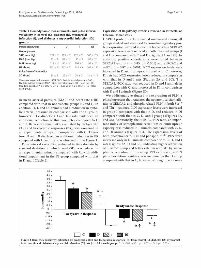
in mean arterial pressure (MAP) and heart rate (HR)compared with that in nondiabetic groups (C and I). Inaddition, D, I, and DI animals had a reduction in systo-lic arterial pressure in comparison with the C group;however, STZ-diabetic (D and DI) rats evidenced anadditional reduction of this parameter compared to Cand I. Baroreflex sensitivity, evaluated by tachycardic(TR) and bradycardic responses (BR), was worsened inall experimental groups in comparison with C. There-fore, D and DI displayed an additional reduction in BRcompared with C and I rats, as observed in the Figure 1.Pulse interval variability, evaluated in time domain by
standard deviation of pulse interval (SD), was reduced inall experimental animals compared with C, with addi-tional impairment in the DI group compared with thatin D and I (Table 2).
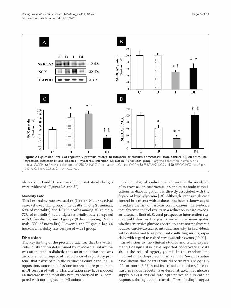
Expression of Regulatory Proteins Involved in IntracellularCalcium HomeostasisGAPDH protein levels remained unchanged among allgroups studied and were used to normalize regulatory pro-tein expression involved in calcium homeostasis. SERCA2expression levels were reduced in both infarcted groups (Iand DI) compared with C and D (Figures 2A and 2B). Inaddition, positive correlations were found betweenSERCA2 and EF (r = 0.85; p < 0.001) and SERCA2 and+dP/dt (r = 0.87; p < 0.001). NCX expression levels wereincreased in D and I groups compared with C; however,DI rats had NCX expression levels reduced in comparisonwith that in D and I rats (Figures 2A and 2C). TheSERCA2/NCX ratio was reduced in D and I animals incomparison with C, and increased in DI in comparisonwith D and I animals (Figure 2D).We additionally evaluated the expression of PLN, a
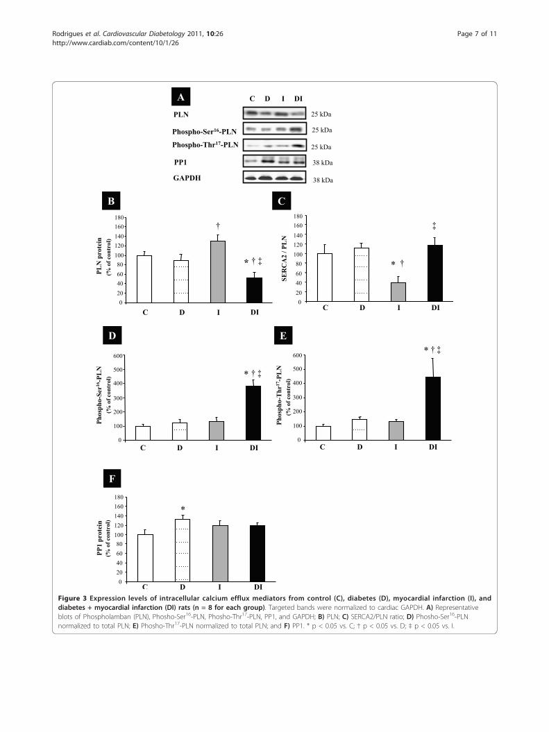
phosphoprotein that regulates the apparent calcium affi-nity of SERCA2, and phosphorylated PLN in both Ser16
and Thr17 residues. PLN expression levels were increasedin group I compared with that in D, and reduced in DIcompared with that in C, D, and I groups (Figures 3Aand 3B). Additionally, the SERCA2/PLN ratio, an impor-tant index of sarcoplasmic reticulum calcium uptakecapacity, was reduced in I animals compared with C, D,and DI animals (Figure 3C). The expression levels ofboth phospho-ser16-PLN and phospho-thr17-PLN wereincreased only in DI animals compared with C, D, and Irats (Figures 3A, D and 3E), indicating higher activationof SERCA2 pump and better calcium reuptake by sarco-plasmic reticulum in this group. PP1 expression, a PLNphosphorylation regulator, was increased in the D groupcompared with that in C; however, although the increase
Table 2 Hemodynamic measurements and pulse intervalvariability in control (C), diabetes (D), myocardialinfarction (I), and diabetes + myocardial infarction (DI)groups
Parameter/Group C D I DI
Hemodynamic
SAP (mm Hg) 129 ± 2 109 ± 3* 117 ± 3*† 106 ± 2*‡
DAP (mm Hg) 92 ± 2 83 ± 2* 90 ± 3 87 ± 2*
MAP (mm Hg) 111 ± 2 96 ± 3* 104 ± 3 99 ± 2*
HR (bpm) 352 ± 12 302 ± 7* 342 ± 6 307 ± 19*
Pulse Interval Variability
SD (bpm) 34 ± 3 22 ± 4* 24 ± 2* 13 ± 1*†‡
Values are expressed as mean ± SEM. SAP - Systolic arterial pressure; DAP -Diastolic arterial pressure; MAP - Mean arterial pressure; HR - Heart rate; SD -Standard deviation. * p < 0.05 vs. C; † p < 0.05 vs. D; ‡ p < 0.05 vs. I (n = 8 foreach group).
-2.5-2.0-1.5-1.0-0.5
00.51.01.52.02.53.03.54.04.55.0
Tachycardic Response
Bradycardic Response
bpm
/mm
Hg
C D I DI
** * †
** † * ‡
Figure 1 Baroreflex sensitivity estimated by bradycardic (BR) and tachycardic responses (TR) from control (C), diabetes (D), myocardialinfarction (I) and diabetes + myocardial infarction (DI) rats (n = 8 for each group). * p < 0.05 vs. C; † p < 0.05 vs. D; ‡ p < 0.05 vs. I.
Rodrigues et al. Cardiovascular Diabetology 2011, 10:26http://www.cardiab.com/content/10/1/26
Page 5 of 11
observed in I and DI was discrete, no statistical changeswere evidenced (Figures 3A and 3F).
Mortality RateTotal mortality rate evaluation (Kaplan-Meier survivalcurve) showed that groups I (13 deaths among 21 animals,62% of mortality) and DI (22 deaths among 30 animals,73% of mortality) had a higher mortality rate comparedwith C (no deaths) and D groups (8 deaths among 16 ani-mals, 50% of mortality). However, the DI group had anincreased mortality rate compared with I group.
DiscussionThe key finding of the present study was that the ventri-cular dysfunction determined by myocardial infarctionwas attenuated in diabetic rats, an attenuation that wasassociated with improved net balance of regulatory pro-teins that participate in the cardiac calcium handling. Inopposition, autonomic dysfunction was more prominentin DI compared with I. This alteration may have inducedan increase in the mortality rate, as observed in DI com-pared with normoglycemic MI animals.
Epidemiological studies have shown that the incidenceof microvascular, macrovascular, and autonomic compli-cations in diabetic patients is directly associated with thedegree of hyperglycemia [18]. Although intensive glucosecontrol in patients with diabetes has been acknowledgedto reduce the risk of vascular complications, the evidencethat glycemic control results in a reduction in cardiovascu-lar disease is limited. Several prospective intervention stu-dies published in the past 2 years have investigatedwhether intensive glucose control to near-normoglycemiareduces cardiovascular events and mortality in individualswith diabetes and have produced conflicting results, espe-cially with regard to risk of cardiovascular events [19-21].In addition to the clinical studies and trials, experi-
mental designs also have reported controversial dataabout the role of hyperglycemia in the mechanismsinvolved in cardioprotection in animals. Several studieshave shown that hearts from diabetic rats are equally[22] or more [5,23] sensitive to ischemic injury. In con-trast, previous reports have demonstrated that glucosesupply plays a critical cardioprotective role in cardiacresponses during acute ischemia. These findings suggest
0
20
40
60
80
100
120
C D I DI
SER
CA
2 pr
otei
n (%
of c
ontr
ol)
SERCA2
NCX
GAPDH
110 kDa
120 kDa
38 kDa
C D I DI
*
*†
†
NC
X p
rote
in
(% o
f con
trol
)
*
†
‡
SER
CA
2 / N
CX
*
† ‡
A B
C D
020406080
100120140160180200
C D I DI
*
†
0
20
40
60
80
100
120
C D I DI
* †
Figure 2 Expression levels of regulatory proteins related to intracellular calcium homeostasis from control (C), diabetes (D),myocardial infarction (I), and diabetes + myocardial infarction (DI) rats (n = 8 for each group). Targeted bands were normalized tocardiac GAPDH. A) Representative blots of SERCA2, Na+-Ca2+ exchanger (NCX) and GAPDH; B) SERCA2; C) NCX; and D) SERCA2/NCX ratio. * p <0.05 vs. C; † p < 0.05 vs. D; ‡ p < 0.05 vs. I.
Rodrigues et al. Cardiovascular Diabetology 2011, 10:26http://www.cardiab.com/content/10/1/26
Page 6 of 11
PLN
Phospho-Ser16-PLN
25 kDa
25 kDa
38 kDa
Phospho-Thr17-PLN 25 kDa
PP1
A C D I DI
GAPDH 38 kDa
0
100
200
300
400
500
600
C D I DI
Phos
pho-
Ser16
-PL
N(%
of c
ontr
ol) * † ‡
0
100
200
300
400
500
600
Phos
pho-
Thr
17-P
LN
(% o
f con
trol
)
C D I DI
† ‡*
B C
D E
020406080
100120140160180
C D I DI
PLN
pro
tein
(% o
f con
trol
)
‡†*
020406080
100120140160180
C D I DI
SER
CA
2 / P
LN
†
‡
*
F
†
PP1
prot
ein
(% o
f con
trol
)
020406080
100120140160180
C D I DI
*
Figure 3 Expression levels of intracellular calcium efflux mediators from control (C), diabetes (D), myocardial infarction (I), anddiabetes + myocardial infarction (DI) rats (n = 8 for each group). Targeted bands were normalized to cardiac GAPDH. A) Representativeblots of Phospholamban (PLN), Phosho-Ser16-PLN, Phosho-Thr17-PLN, PP1, and GAPDH; B) PLN; C) SERCA2/PLN ratio; D) Phosho-Ser16-PLNnormalized to total PLN; E) Phosho-Thr17-PLN normalized to total PLN; and F) PP1. * p < 0.05 vs. C; † p < 0.05 vs. D; ‡ p < 0.05 vs. I.
Rodrigues et al. Cardiovascular Diabetology 2011, 10:26http://www.cardiab.com/content/10/1/26
Page 7 of 11
that higher glucose delivery to the heart may improvetolerance to ischemic stress, leading to changes in sig-naling mechanisms involved in cardioprotection [24,25].Recently, Chu et al. [8] provided evidence that diabetesinduced by Aloxan and resultant hyperglycemia, thoughdetrimental to global cardiac function and associatedwith a poorer prognosis, was cardioprotective againstmyocardial ischemic-reperfusion injury in pigs in theshort-term. The authors observed a pronounced reduc-tion in the myocardial infarction area in diabetic ani-mals, likely being a result of increased availability anduse of glucose, the heart’s preferred energy substrate intimes of stress.In fact, some studies have demonstrated that the
reduction in myocardial infarction size [26] is deter-mined by the diminished number of dead myocytes indiabetic animals [27], and would be the key point to thehigher resistance of diabetic rats to ischemic injury. Incontrast with the results in the literature and data fromour group [9], the MI area was similar between I and DIgroups in the present study. This discrepancy in datamay be the result of the prolonged time of diabetes andMI investigated here. Thus, the higher resistance of thediabetic heart cannot be explained by the reduction inthe myocardial infarction akinetic area in the presentstudy.
Morphological and Functional Responses to IschemicInjuryMyocardial remodeling after MI is usually characterizedby compensatory hypertrophy of myocytes, ventriculardilatation and increased interstitial collagen depositionand fibrosis [5]. In our study, LVEDD was increased ininfarcted compared with noninfarcted groups; however Iand DI groups had similar results of LV mass andLVEDD, highlighting the fact that STZ-diabetes had noinfluence on cardiac remodeling after MI. To the con-trary, Bäcklund et al. [5] showed that 12 weeks after MIsurgery cardiomyocyte apoptosis was associated with LVenlargement and increased cardiac fibrosis in diabeticanimals. However, it is possible that the prolonged timeof STZ-diabetes (4 weeks) before the MI surgery, incontrast with our study (15 days), has been the responsi-ble for discrepancy in the results.To further investigate the mechanisms underlying the
improved tolerance to ischemia of diabetic rats, we eval-uated the cardiac function of the experimental groups.Our data indicate that diabetes partially attenuated theventricular dysfunction caused by myocardial ischemia(DI animals compared with I animals). In fact, DI ani-mals had improved systolic and diastolic functionindexes, paralleled by reduced MPI, an index that repre-sents global myocardial stress [16]. These data corrobo-rate previous findings of our group where 15 days after
MI, STZ-diabetic rats had attenuation of cardiac dys-function compared with the normoglycemic rats. Inter-estingly, this attenuation was associated with animproved net balance of inflammatory cytokines andregulatory genes that participate in cardiac cellularsurvival [9].
Molecular ChangesBecause cardiac dysfunction observed in cardiomyopathyand heart failure is strongly associated with the expres-sion profile of the cardiac proteins related to intracellularcalcium handling [11], we evaluated the SERCA2, PLN,phospho-ser16-PLN, phospho-thr17-PLN, PP1, and NCX.It was previously reported that in heart failure cardiacdysfunction is in part a consequence of changes in intra-cellular calcium homeostasis, which may be related to analtered expression, function, or regulation of SERCA2[11,28]. In the present study, although the MI groups(I and DI) displayed a reduction in SERCA2 expression,the DI group had a decrease in NCX and PLN proteins,resulting in increased SERCA2/NCX and SERCA2/PLNratios in comparison with those in the I group. This find-ing may indicate a relative increase in SERCA2 pumpfunction and increased available calcium in the cytoplasmin diabetic animals that undergo MI, if compared withnormoglycemic MI rats. Likewise, increased phosphoryla-tion of PLN in both serine16 and threonine17 in DI ratsreinforces the possibility that SERCA2 function is betterpreserved in these experimental animals. Indeed, thephosphorylation of PLN in both residues is involved inan increased calcium reuptake by sarcoplasmic reticulum,although the increase in PLN phosphorylation at Thr17
may be interpreted, in some cases, as one of the latestcompensatory mechanisms activated when cardiac func-tion is deteriorated, as observed in acidosis and reperfu-sion periods [29].In the present study, because the molecular analyses
were performed in all LV homogenates to quantify theexpression of proteins by Western blot, it is possible thatthe compensatory mechanisms involving calcium regula-tion in the noninfarcted area might have influenced theresults. However, it is necessary to consider that not onlythe myocardial infarction area, but also LV mass weresimilar between the infarcted groups (I and DI). Thesefindings suggest that experimental diabetes is associatedwith the activation of endogenous cardioprotectivemechanisms, which successfully remain after 90 days ofmyocardial ischemia attenuating the remaining LV dys-function in the DI group.
Autonomic Impairment and Mortality RateIndependently of the mechanism involved in the pre-served left ventricular function observed in DI rats, itwas expected that the mortality rate would be reduced
Rodrigues et al. Cardiovascular Diabetology 2011, 10:26http://www.cardiab.com/content/10/1/26
Page 8 of 11
in this group compared with the I group. However, DIanimals evidenced an increase in mortality rate 90 daysafter myocardial infarction compared with the I group,suggesting that other mechanisms and dysfunctionsmight be influencing the survival rate of the diabeticanimals. Possible mechanisms, all involving the meta-bolic characteristic of the diabetic state, have been pre-viously suggested: long duration of the diabetes, severityof hyperglycemia [30], high levels of circulating freefatty acids, and decreased glucose utilization by cardio-myocytes [31] which contribute to contractile derange-ment [32] and higher arrhythmia susceptibility [33]. Infact, van den Brom et al., [34], by using positron emis-sion tomography and echocardiography, evidencedincreases in myocardial fatty acid oxidation with a con-comitant decrease of insulin-mediated myocardial glu-cose utilization in early diabetic cardiomyopathy. Inaddition, the authors observed that these metabolicalterations were associated with impaired myocardialfunction.Cardiovascular autonomic neuropathy has been consid-
ered an important determinant of mortality in diabeticpatients independently of other known risk factors [35].In the present study, more prominent autonomic dys-function was observed in the DI group compared withthat in I animals. This evidence may indicate that auto-nomic dysfunction, previously described by us [36,37],could be influencing the mortality observed in D andmainly in DI rats. In accordance with these results, ourgroup previously demonstrated that sedentary STZ dia-betic rats had accentuated autonomic dysfunction andconsequently a higher mortality rate (~49%) comparedwith control rats. Additionally, 8 weeks of aerobic exer-cise training attenuated the autonomic dysfunction andreduced the mortality levels in the trained group to 16%[36]. For this reason, new therapeutic strategies havebeen studied in diabetes management. Shyu et al., [38]described the isolation of pancreatic endocrine precursorcells, from adult human pancreatic tissue, and the redif-ferentiation of insulin-secreting b-like cells from in vitro-cultured insulin-producing cell precursors. These dataemphasize that surgically resected pancreatic tissue mayrepresent an alternative source of functional insulin-producing cells and to minimize the cardiovasculardysfunctions of diabetes.
Study LimitationsSome possible limitations of the present investigationdeserve comment. First, because the MI animals (I andDI) started the protocol with similar values for MI areaand LF function and necropsy evaluations were not per-formed in the animals that died during the 90 days ofthe protocol, the prediction of survival (or mortality)
based on these parameters becomes difficult and is lim-ited to methods used in this study. Second, the hemody-namic and autonomic parameters were evaluated at theend of the protocol, and comparisons were made byincluding a control group in the experimental design.Indeed, the direct method for recording blood pressuredepends on the catheterization of arterial vessels thatare functional during a small time period. Consequently,the biological signals were recorded only at the end ofthe experimental period, leading to the lack of baselinevalues in the same animals at the start of the study.
ConclusionWe have demonstrated that the left ventricular dysfunc-tion in diabetic animals was attenuated after 90 days ofmyocardial infarction. This is probably a compensatorymechanism associated with metabolic and molecularadjustments of the cardiac tissue, where the end pointwas a better profile of calcium handling proteins. How-ever, this positive adaptation was not able to reduce themortality rate of DI animals. These data suggest thatother mechanisms, as well as cardiovascular autonomicdysfunction identified in this study, is associated withmortality rate in these animals. It is important to empha-size that in the present investigation, the time course ofdiabetes and MI was determined and well knows, in con-trast with that observed in clinical situations. Therefore,it is possible that the better cardiac function has beentransitory in DI rats, and the autonomic dysfunction,more prominent in diabetics, may lead, in the future, tocardiovascular damage, as observed in diabetic patientsafter an ischemic event.
List of abbreviationsBR: bradycardic response; DAP: diastolic arterial pressure; +dP/dt: maximumrate of left ventricular pressure rise; -dP/dt: maximum rate of left ventricularpressure fall; EDT: peak E wave deceleration time; EF: ejection fraction;GAPDH: glyceraldehyde-3-phosphate dehydrogenase; HR: heart rate; IVRT:left ventricular isovolumetric relaxation time; LV: left ventricle; LV mass: leftventricular mass corrected by body weight; LVEDD: left ventricular end-diastolic diameter; LVEDP: left ventricular end-diastolic pressure; LVSP: leftventricular systolic pressure; MAP: mean arterial pressure; MI: myocardialinfarction; MPI: myocardial performance index; NCX: sodium calciumexchanger; phospho-ser16-PLN: phosphorylated phospholamban at serine16; phospho-thr17-PLN: phosphorylated phospholamban at threonine 17; PI:pulse interval; PLN: dephosphorylated phospholamban; PP1: phosphataseprotein 1; SAP: systolic arterial pressure; SD: standard deviation; SEM:standard error of mean; SERCA2: calcium pump ATPase of sarcoplasmicreticulum; STZ: streptozotocin; TR: tachycardic response; VCF: velocity ofcircumferential fiber shortening.
Acknowledgements and fundingThis study was supported by Fundação de Amparo à Pesquisa do Estado deSão Paulo (FAPESP- 01/00009-0; 07/57595-5) and Fundaçao E.J. Zerbini. BRhas doctorate and postdoctorate scholarships from the Coordenação deAperfeiçoamento de Pessoal de Nível Superior (CAPES) and ConselhoNacional de Pesquisa e Desenvolvimento (CNPq), respectively. MCI, KDA, andPCB had a financial support from Conselho Nacional de Pesquisa eDesenvolvimento (CNPq-BPQ).
Rodrigues et al. Cardiovascular Diabetology 2011, 10:26http://www.cardiab.com/content/10/1/26
Page 9 of 11

Author details1Human Movement Laboratory, São Judas Tadeu University, São Paulo, Brazil.2Hypertension Unit, Heart Institute (InCor), Medical School of University ofSão Paulo, São Paulo, Brazil. 3Federal University of São Paulo, BiosciencesDepartment, Santos, SP, Brazil. 4Endocrine Division, Clinical Hospital of PortoAlegre, Federal University of Rio Grande do Sul, Porto Alegre, Brazil. 5Schoolof Physical Education and Sports, University of São Paulo, São Paulo, Brazil.6Nove de Julho University, São Paulo, Brazil.
Authors’ contributionsBR designed, performed, and coordinated the experiments, as well asprepared the manuscript. KTR carried out the echocardiographicmeasurements. AM participated in the Western blot analysis. BDS helped todraft the manuscript. PCB participated in the data discussion and helped todraft the manuscript. KDA participated in the design, data discussion andhelped to draft the manuscript. MCI conceived the study and participated inits design and coordination. All authors have read and approved the finalmanuscript.
Competing interestsThe authors declare that they have no competing interests.
Received: 28 February 2011 Accepted: 6 April 2011Published: 6 April 2011
References1. Roper NA, Bilous RW, Kelly WF, Unwin NC, Connolly VM: Excess mortality
in a population with diabetes and the impact of material deprivation:longitudinal, population based study. BMJ 2001, 322:1389-1393.
2. Haffner SM, Lehto S, Ronnemaa T, Pyorala K, Laakso M: Mortality fromcoronary heart disease in subjects with type 2 diabetes and innondiabetic subjects with and without prior myocardial infarction.N Engl J Med 1998, 339:229-234.
3. Feuvray D, Lopaschuk GD: Controversies on the sensitivity of the diabeticheart to ischemic injury: the sensitivity of the diabetic heart to ischemicinjury is decreased. Cardiovasc Res 1997, 34:113-120.
4. Ravingerova T, Neckar J, Kolar F, Stetka R, Volkovova K, Ziegelhöffer A,Styk J: Ventricular arrhythmias following coronary artery occlusion inrats: is the diabetic heart less or more sensitive to ischaemia? Basic ResCardiol 2001, 96(2):160-168.
5. Bäcklund T, Palojoki E, Saraste A, Eriksson A, Finckenberg P, Kytö V,Lakkisto P, Mervaala E, Voipio-Pulkki LM, Laine M, Tikkanen I: Sustainedcardiomyocyte apoptosis and left ventricular remodelling aftermyocardial infarction in experimental diabetes. Diabetologia 2004,47(2):325-330.
6. Liu Y, Thornton JD, Cohen MV, Downen JM, Schaffer SW: Streptozotocin-induced non-insulin dependent diabetes protect the heart frominfarction. Circulation 1993, 88:1273-1278.
7. Ravingerova T, Stetka R, Volkovova K, Pancza D, Dzurba A, Ziegelhöffer A,Styk J: Acute diabetes modulates response to ischemia in isolated ratheart. Mol Cell Biochem 2000, 210:143-151.
8. Chu LM, Osipov RM, Robich MP, Feng J, Oyamada S, Bianchi C, Sellke FW: Ishyperglycemia bad for the heart during acute ischemia? J ThoracCardiovasc Surg 2010, 140:1345-1352.
9. Malfitano C, Alba Loureiro TC, Rodrigues B, Sirvente R, Salemi VM,Rabechi NB, Lacchini S, Curi R, Irigoyen MC: Hyperglycaemia protects theheart after myocardial infarction: aspects of programmed cell survivaland cell death. Eur J Heart Fail 2010, 12(7):659-667.
10. Schaffer SW, Croft CB, Solodushko V: Cardioprotective effect of chronichyperglycemia: effect on hypoxia-induced apoptosis and necrosis. Am JPhysiol Heart Circ Physiol 2000, 278(6):H1948-54.
11. Periasamy M, Bhupathy P, Babu GJ: Regulation of sarcoplasmic reticulumCa2+ ATPase pump expression and its relevance to cardiac musclephysiology and pathology. Cardiovasc Res 2008, 77(2):265-273.
12. Choi KM, Zhong Y, Hoit BD, Grupp IL, Hahn H, Dilly KW, Guatimosim S,Lederer WJ, Matlib MA: Defective intracellular Ca(2+) signalingcontributes to cardiomyopathy in Type 1 diabetic rats. Am J Physiol HeartCirc Physiol 2002, 283(4):H1398-H1408.
13. Jorge L, Rodrigues B, Rosa KT, Malfitano C, Loureiro TC, Medeiros A, Curi R,Brum PC, Lacchini S, Montano N, De Angelis K, Irigoyen MC: Cardiac andperipheral adjustments induced by early exercise training intervention
were associated with autonomic improvement in infarcted rats: role infunctional capacity and mortality. Eur Heart J .
14. Kilkenny C, Browne WJ, Cuthill IC, Emerson M, Altman DG: Improvingbioscience research reporting: the ARRIVE guidelines for reportinganimal research. PLoS Biol 2010, 8(6):e1000412.
15. Mostarda C, Rodrigues B, Vane M, Moreira ED, Rosa KT, Moraes-Silva IC,Lacchini S, Casarini DE, De Angelis K, Irigoyen MC: Autonomic impairmentafter myocardial infarction: role in cardiac remodelling and mortality.Clin Exp Pharmacol Physiol 2010, 37(4):447-452.
16. Wichi R, Malfitano C, Rosa K, De Souza SB, Salemi V, Mostarda C, DeAngelis K, Irigoyen MC: Noninvasive and invasive evaluation of cardiacdysfunction in experimental diabetes in rodents. Cardiovasc Diabetol2007, 26:6-14.
17. Medeiros A, Rolim NP, Oliveira RS, Rosa KT, Mattos KC, Casarini DE,Irigoyen MC, Krieger EM, Krieger JE, Negrão CE, Brum PC: Exercise trainingdelays cardiac dysfunction and prevents calcium handling abnormalitiesin sympathetic hyperactivity-induced heart failure mice. J Appl Physiol2008, 104(1):103-109.
18. Selvin E, Marinopoulos S, Berkenblit G, Rami T, Brancati FL, Powe NR,Golden SH: Meta-analysis: glycosylated hemoglobin and cardiovasculardisease in diabetes mellitus. Ann Intern Med 2004, 141:421-431.
19. Action to Control Cardiovascular Risk in Diabetes Study Group, Gerstein HC,Miller ME, Byington RP, Goff DC Jr, Bigger JT, Buse JB, Cushman WC,Genuth S, Ismail-Beigi F, Grimm RH Jr, Probstfield JL, Simons-Morton DG,Friedewald WT: Effects of intensive glucose lowering in type 2 diabetes.N Engl J Med 2008, 358:2545-2559.
20. The ADVANCE Collaborative Group: Intensive blood glucose control andvascular outcomes in patients with type 2 diabetes. N Engl J Med 2008,358:2560-2572.
21. Duckworth W, Abraira C, Moritz T, Reda D, Emanuele N, Reaven PD,Zieve FJ, Marks J, Davis SN, Hayward R, Warren SR, Goldman S, McCarren M,Vitek ME, Henderson WG, Huang GD, VADT Investigators: Glucose controland vascular complications in veterans with type 2 diabetes. N Engl JMed 2009, 360:129-139.
22. Vogel WM, Apstein CS: Effects of alloxan-induced diabetes onischemia-reperfusion injury in rabbit hearts. Circ Res 1988,62(5):975-982.
23. Higuchi M, Ikema S, Sakanashi M: Correlation of contractile dysfunctionand abnormal tissue energy metabolism during hypoperfusion withnorepinephrine in isolated rat hearts: differences between normal anddiabetic hearts. J Mol Cell Cardiol 1992, 24(10):1125-1141.
24. Cave AC, Ingwall JS, Friedrich J, Liao R, Saupe KW, Apstein CS, Eberli FR:ATP synthesis during low-flow ischemia: influence of increased glycolyticsubstrate. Circulation 2000, 101(17):2090-2096.
25. King LM, Opie LH: Glucose and glycogen utilization in myocardialischemia - changes in metabolism and consequences for the myocyte.Mol Cell Biochem 1998, 180(1-2):3-26.
26. Ravingerova T, Neckar J, Kolar F: Ischemic tolerance of rat heart in acuteand chronic phases of experimental diabetes. Mol Cell Biochem 2003,249:167-174.
27. Xu G, Takashi E, Kudo M, Ishiwata T, Naito Z: Contradictory effects ofshort- and long-term hyperglycemias on ischemic injury of myocardiumvia intracellular signaling pathway. Exp Mol Pathol 2004, 76(1):57-65.
28. de Tombe PP: Altered contractile function in heart failure. Cardiovasc Res1998, 37:367-380.
29. Mattiazzi A, Mundiña-Weilenmann C, Guoxiang C, Vittone L, Kranias E: Roleof phospholamban phosphorylation on Thr17 in cardiac physiologicaland pathological conditions. Cardiovasc Res 2005, 68(3):366-375.
30. Tosaki A, Engelman DT, Engelman RM, Das DK: The evolution of diabeticresponse to ischemia/reperfusion and preconditioning in isolatedworking rat hearts. Cardiovasc Res 1996, 31(4):526-536.
31. Zorzano A, Sevilla L, Camps M, Becker C, Meyer J, Kammermeier H,Muñoz P, Gumà A, Testar X, Palacín M, Blasi J, Fischer Y: Regulation ofglucose transport, and glucose transporters expression and trafficking inthe heart: studies in cardiac myocytes. Am J Cardiol 1997, 80(3A):65A-76A.
32. Belke DD, Larsen TS, Gibbs EM, Severson DL: Altered metabolism causescardiac dysfunction in perfused hearts from diabetic (db/db) mice. Am JPhysiol: Endocrinol Metab 2000, 279(5):E1104-E1113.
33. DaTorre SD, Creer MH, Pogwizd SM, Corr PB: Amphipathic lipidmetabolites and their relation to arrhythmogenesis in the ischemicheart. J Mol Cell Cardiol 1991, 23(Suppl 1):11-22.
Rodrigues et al. Cardiovascular Diabetology 2011, 10:26http://www.cardiab.com/content/10/1/26
Page 10 of 11
34. van den Brom CE, Huisman MC, Vlasblom R, Boontje NM, Duijst S,Lubberink M, Molthoff CF, Lammertsma AA, van der Velden J, Boer C,Ouwens DM, Diamant M: Altered myocardial substrate metabolism isassociated with myocardial dysfunction in early diabeticcardiomyopathy in rats: studies using positron emission tomography.Cardiovasc Diabetol 2009, 22:8:39.
35. Maser RE, Mitchell BD, Vinik AI, Freeman R: The association betweencardiovascular autonomic neuropathy and mortality in individuals withdiabetes: a meta-analysis. Diabetes Care 2003, 26(6):1895-1901.
36. Mostarda C, Rogow A, Silva IC, De La Fuente RN, Jorge L, Rodrigues B,Heeren MV, Caldini EG, De Angelis K, Irigoyen MC: Benefits of exercisetraining in diabetic rats persist after three weeks of detraining. AutonNeurosci 2009, 145(1-2):11-16.
37. Dall’ago P, D’Agord Schaan B, da Silva VO, Werner J, da Silva Soares PP, deAngelis K, Irigoyen MC: Parasympathetic dysfunction is associated withbaroreflex and chemoreflex impairment in streptozotocin-induceddiabetes in rats. Auton Neurosci 2007, 131(1-2):28-35.
38. Shyu JF, Wang HS, Shyr YM, Wang SE, Chen CH, Tan JS, Lin MF, Hsieh PS,Sytwu HK, Chen TH: Alleviation of hyperglycemia in diabetic rats byintraportal injection of insulin-producing cells generated from surgicallyresected human pancreatic tissue. J Endocrinol 2011, 208(3):233-244.
doi:10.1186/1475-2840-10-26Cite this article as: Rodrigues et al.: Hyperglycemia can delay leftventricular dysfunction but not autonomic damage after myocardialinfarction in rodents. Cardiovascular Diabetology 2011 10:26.
Submit your next manuscript to BioMed Centraland take full advantage of:
• Convenient online submission
• Thorough peer review
• No space constraints or color figure charges
• Immediate publication on acceptance
• Inclusion in PubMed, CAS, Scopus and Google Scholar
• Research which is freely available for redistribution
Submit your manuscript at www.biomedcentral.com/submit
Rodrigues et al. Cardiovascular Diabetology 2011, 10:26http://www.cardiab.com/content/10/1/26
Page 11 of 11