hydrothermal synthesis and characterization of zno nano · pdf fileabstract—this study...
TRANSCRIPT
Abstract—This study reports on Zinc oxide (ZnO) nano crystals
prepared using zinc nitrate hexahydrate (Zn(NO3)2.6H2O) and
sodium hydroxide (NaOH) as the starting precursors in the molar
ratio’s of 1:2 and 1:10 through the hydrothermal method. The effects
of NaOH concentration on structural and optical properties of ZnO
nano crystals were investigated. The prepared samples were annealed
at 600 ˚C to obtain the ZnO nano crystals. The ZnO nanoparticles
were characterized with X-ray diffraction (XRD), Field Emission
Scanning Electron Microscopy (FE-SEM) and UV-vis absorption
spectroscopy. The hexagonal wurtzite structure of ZnO nanocrystals
was confirmed from XRD results. The Full Width at Half Maximum
(FWHM) of XRD peaks increased with increase of NaOH
concentration which indicates that the average crystallite size of ZnO
nano crystals decreased with increase of NaOH concentration. FE-
SEM pictures exhibited hexagonal shaped ZnO nanocrystals
comprising of cylindrical pores of diameters ranging from 9 nm to 12
nm. The number of pores as well as their diameters enhanced with
increasing concentration of NaOH. Absorption spectra of these ZnO
nano crystals showed an absorption peak positioned at 350 nm. This
is due to the excitonic absorption in the ZnO nano crystals. The
prepared porous ZnO samples using hydro thermal method may
reduce the required reflection losses in the front surface which is one
of the important desirable features in optoelectronic devices.
Keywords—ZnO nanocrystals, Hydrothermal method,
Optoelectronic devices.
I. INTRODUCTION
Semiconductors with dimensions in the nano meter realm
are important because of their optical and electrical properties
which can be tuned by changing the size of the nanoparticles.
The most common and very important property of these
semiconductor nanocrystals is that their band gap varies with
changes in particle size. This effect is called quantum
confinement effect. ZnO is one of the wide band gap
semiconductors which exhibit quantum confinement effects in
experimentally accessible conditions. Zinc Oxide (ZnO) is
having a wide and direct band gap of 3.37 eV and large
exciton binding energy of 60 meV at room temperature [1].
Hence, ZnO finds applications in various fields such as
antireflection coatings, transparent electrodes in solar cells,
ultraviolet (UV) light emitters, diode lasers, varistors,
piezoelectric devices, spintronics, surface acoustic wave
propagation, and also in sensing of gas [2]. Various chemical
synthetic methods have been developed to prepare such
nanoparticles. It has been widely used in near-UV emission,
A.Ramachandra Reddy, A. N. Mallika, K. Sowri Babu, and K. Venugopal
Reddy are with Department of Physics, Materials Science Laboratory,
National Institute of Technology Warangal-506 004, Telangana, India.
(corresponding author e-mail: [email protected],
gas sensors, transparent conductor and piezoelectric
application [3]. Moreover, ZnO is abundantly available
material, economical and it has many advantages over the GaN
semiconductor which is currently being used in optoelectronic
devices. However, it is very difficult to
prepare p-type ZnO. If it is done, ZnO is going to replace the
GaN in the near future. ZnO nanoparticles can be prepared on
a large scale at low cost by simple solution - based methods,
such as chemical precipitation, sol-gel synthesis and
solvothermal/ hydrothermal reaction [2]. Hydrothermal
technique is a promising alternative synthetic method because
of the low process temperature and very easy to control the
particle size. The hydrothermal process has several advantages
over other growth processes such as use of simple equipment,
catalyst-free growth, low cost, ease of large scale production,
eco-friendly and less hazardous [4]. The low reaction
temperatures make this method attractive for microelectronics
and plastic electronics [5]. This method has also been
successfully employed to prepare nano scale ZnO and other
luminescent materials. The hydro-thermal process in general
progresses in a closed system at a high autogeneous pressure.
By the benefit of the closed system with high pressure, the
required temperature for preparing ceramic powder can be
greatly reduced because of enhanced reactivity of reactive
species, and fine particles with high sinterability. In addition,
the evaporation of volatile species can be suppressed, and the
stoichiometry of ceramics can be maintained [6]. Ming Yang
et.al has studied the effect of different precursors on
hydrothermally synthesized 1-D ZnO [7]. The particle
properties such as morphology and size can be controlled via
the hydrothermal process by adjusting the reaction
temperature, time and concentration of precursors [8].
Porous ZnO has some specific advantages such as high
surface area, chemical and photochemical stability, uniformity
in pore size, shape selectivity, and rich surface chemistry [9].
The high surface area of porous ZnO makes its surface more
active. The highly active surface would increase the
probability of interaction of gases with the semiconductor,
which in turn increases the sensitivity of the material [10]. So,
the material has found a variety of promising applications such
as catalysts, nano-sieve filters, dye sensitized solar cells, bio-
and electrochemical sensors, bone-replacement materials and
also in gas sensors [9, 11, 12]. For example, for dye sensitized
solar cells, ZnO thin films should be porous and have high
specific surface area for exhibiting high conversion efficiency
of light into current [13].
The present study focuses on the hydrothermal synthesis of
ZnO nano crystals with different NaOH concentrations (1:2,
Hydrothermal Synthesis and Characterization of
Zno Nano Crystals
A.Ramachandra Reddy, A. N. Mallika, K. Sowri Babu, and K. Venugopal Reddy
International Journal of Mining, Metallurgy & Mechanical Engineering (IJMMME) Volume 3, Issue 2 (2015) ISSN 2320–4060 (Online)
52
and 1:10). The structural and optical properties of these ZnO
nanocrystals were studied.
II. EXPERIMENTAL SECTION
A. Synthesis
Analytical grade zinc nitrate hexahydrate (Zn (NO3)2. 6H2O,
98%) and sodium hydroxide (NaOH, 97 %) were used as the
starting chemicals. To prepare ZnO nano crystals, Zinc nitrate
hexahydrate and NaOH were taken in required quantities and
were dissolved in de-ionized water. An aqueous solution of 0.5
mol/L Zn(NO3)2.6H2O was mixed with the appropriate
amount of 1 and 5 mol/L NaOH solution under magnetic
continuous stirring to obtain the mole ratio of
Zn(NO3)2.6H2O:NaOH of 1:2 and 1:10. The final pH of the
mixed solutions was highly basic with pH of 14. The mixture
was put into a Teflon-lined-stainless steel autoclave unit for
hydrothermal reaction at 150 ˚C for 7h. After hydrothermal
reaction, the reactor was naturally cooled to room temperature.
The obtained product was filtered, washed with de-ionized
water till the pH of the final solution was 7.0. Finally the as-
prepared sample was calcined at 600˚ C in a programmable
muffle furnace at a rate of 2˚ C / min for 1 hr.
B. Characterization Techniques
The prepared samples were characterized with X-ray
diffractometer equipped with CuKα radiation (λ= 1.540598 Å,
Model: PANalytical) to investigate crystal structure, crystallite
size, strain and lattice parameters. The morphology and
particle size of ZnO nano crystals were analysed by Field
emission scanning electron microscope (Model: Carl Zeiss
Ultra 55) and the absorption of the samples was recorded on
UV-Vis spectrometer (Model: Varian, Cary 5000).
III. RESULTS AND DISCUSSIONS
A. XRD Analysis
The crystal structure of the samples was investigated by
analyzing the XRD data. The X-ray diffraction patterns of
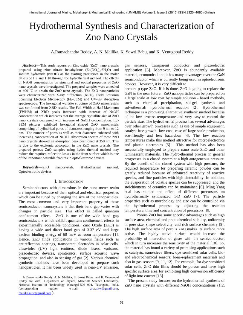
hydrothermal synthesized samples were shown in Fig.1. XRD
spectra depicts the characteristic peaks corresponding to
reflection planes (100), (002), (101), (102), (110), (103), (112)
and (201) of wurtzite structure of ZnO [2]. From the Fig.1, it is
clear that, the intensity of (100) reflection plane is higher than
the (101) reflection plane for 1:2 NaOH concentration. In
general, (101) reflection plane exhibits highest intensity for
hexagonal wurtzite structure of ZnO. However, the intensity of
(101) reflection peak became maximum for highest
concentrations of NaOH (1:10).
The high intensity of (100) reflection plane can be attributed to
the textured grain growth along (100) direction. FE-SEM
pictures shown below confirm the textured growth of ZnO
nanocrystals along (100) reflection plane. The similar kind of
enhancement in XRD peaks was also observed in Sn doped
ZnO thin films [14].
The average crystallite sizes were calculated using
Scherrer’s formula [15].
cos
9.0xrdd
Where,
2
0
2
FWHM
is the peak broadening after removing the
instrumental broadening, β (FWHM) is the full width half
maximum and β0 is the correction factor (0.007radians). The
FWHM values were also increased with increase of NaOH
concentrations indicating the decrease of the crystallite size.
The average crystallite size calculated for 1:2 and 1:10 of
NaOH concentrations are 45 nm and 43 nm respectively
(Table.1). The bond length )(l of Zn-O is calculated using the
following equation [16]. Table I
The average crystallite size and the other parameters derived
from XRD data.
2
22
2
1
3cu
al
Where u is the positional parameter which can be expressed
as
25.0
3 2
2
c
au
sin3a ,
sinc [17].
The calculated bond lengths were tabulated in Table.1.
From the table.1, it is clear that the bond length was enhanced
with increase of NaOH concentration.
Fig.1.The XRD patterns of ZnO with NaOH 1:2 and 1:10
concentrations.
The average crystallite size and strain in nano crystals were
also calculated from the spectral line shape based on
Sample
Code
D
(nm)
a
(Å)
c
(Å)
Bond
length(l)
(Å)
Strain(ɛ)X
10-4
1:2 45 0.2854 0.4944 0.17856 12.2
1:10
43
0.2855
0.4946
0.17861
12.8
International Journal of Mining, Metallurgy & Mechanical Engineering (IJMMME) Volume 3, Issue 2 (2015) ISSN 2320–4060 (Online)
53
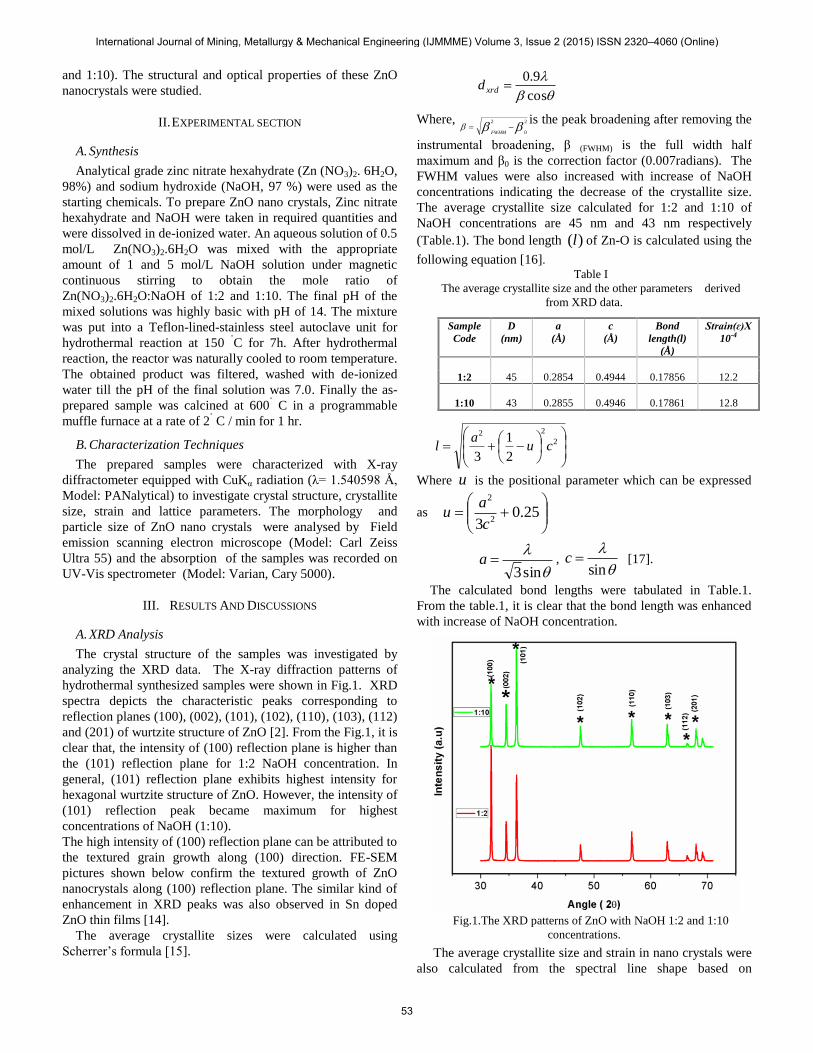
Williamson–Hall (W–H) plots and using the following
equation [2].
sin49.0cos
d
Where is the full width at half maximum, is the lattice
strain, is the Bragg angle, d is the average crystallite size
and is the wavelength of X-rays. A plot drawn between (β
cosθ)/λ and (4sinθ)/λ gave rise to a straight line graph with a
positive intercept as shown in Fig. 1(b). The strain is
calculated from the slope of the linear fit and the intercept
gives the inverse of crystallite size. It is observed from the
strain plots that the strain increased with increase of NaOH
concentration.
Fig.1 (b). The W-H plots for Hydrothermally synthesized ZnO
nanocrystals at 1:2 and 1:10 NaOH concentrations.
B. FE-SEM Analysis
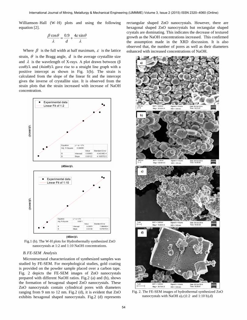
Microstructural characterization of synthesized samples was
studied by FE-SEM. For morphological studies, gold coating
is provided on the powder sample placed over a carbon tape.
Fig. 2 depicts the FE-SEM images of ZnO nanocrystals
prepared with different NaOH ratios. Fig.2 (a) and (b), shows
the formation of hexagonal shaped ZnO nanocrystals. These
ZnO nanocrystals contain cylindrical pores with diameters
ranging from 9 nm to 12 nm. Fig.2 (d), it is evident that ZnO
exhibits hexagonal shaped nanocrystals. Fig.2 (d) represents
rectangular shaped ZnO nanocrystals. However, there are
hexagonal shaped ZnO nanocrystals but rectangular shaped
crystals are dominating. This indicates the decrease of textured
growth as the NaOH concentrations increased. This confirmed
the assumption made in the XRD discussion. It is also
observed that, the number of pores as well as their diameters
enhanced with increased concentrations of NaOH.
Fig. 2. The FE-SEM images of hydrothermal synthesized ZnO
nanocrystals with NaOH a),c)1:2 and 1:10 b),d)
International Journal of Mining, Metallurgy & Mechanical Engineering (IJMMME) Volume 3, Issue 2 (2015) ISSN 2320–4060 (Online)
54

C. UV-Vis Analysis
The absorption spectra of the synthesised samples were
acquired using UV-Vis spectrophotometer in the wavelength
region of 200-600 nm. The UV-visible absorption spectra of
ZnO nano crystals taken by dispersing them in ethanol are
shown in Fig.3. The absorption depends on several factors
such as band gap, Oxygen deficiency, size and structure of the
nanoparticles, surface roughness and impurity centres [18].
It is observed from the figure that peak appearance in the
absorption curves positioned at 350nm may be attributed to the
fundamental absorption of exciton.
Fig.3 Absorption spectra of ZnO nanocrystals
IV. CONCLUSIONS
ZnO nano crystals were successfully synthesised by
hydrothermal method by varying the NaOH concentrations.
The structural and optical properties of ZnO nano crystals
prepared with varying NaOH concentrations was studied. The
hexagonal wurtzite structure of ZnO was confirmed from XRD
studies. The crystallite size decreased with increase of NaOH
concentrations. In addition to this, the (100) diffraction peak
exhibited a higher intensity for NaOH with 1:2 concentration
and it was assigned to the textured grain growth along (100)
direction. From FE-SEM studies, hexagonal plate like
morphology along with pores was observed. With increase of
NaOH concentrations, the number of pores as well as the pore
diameter was enhanced. The excitonic absorption was
observed at 350 nm. Thus the porous ZnO prepared using
hydrothermal method may reduce the required reflection losses
in the front surface of optoelectronic devices which is one of
the important desirable features.
ACKNOWLEDGMENT
The authors are thankful to Dr. S. Srinath, School of Physics,
UoH, for providing FE-SEM facility and also to the FE-SEM
technician Ms. Arundhathi for her help in taking the measurements.
REFERENCES
[1] M. K. Jayaraj, ―Synthesis of ZnO nanoparticles by hydrothermal
method‖, Proc. of SPIE Vol. 6639 66390J-1.
[2] A.N.Mallika, ―Structural and photoluminescence properties of Mg
substituted ZnO nanoparticles‖, Opt. Mater. Vol. 36 (2014) pp.879-884.
[3] S.J Pearton, ―Recent progress in processing and properties of ZnO‖,
Prog.Mater. Sci, 50(2005)293-340.
[4] H.Bai, ―Green hydrothermal synthesis and photoluminescence of ZnO2
nanoparticles‖, Mater.Letter.Vol.64 (2010) pp. 341-343.
[5] Lee. C.Y, ―Effect of phosphorus dopant on photoluminescence and
field-emission characteristics of Mg0.1 Zn0.9 O nanowires‖, J. Appl.
Phys. Vol. 99, pp.024303, 2006
[6] Chung-Hsin Lu*, Influence of hydrothermal conditions on the
morphology and particle size of zinc oxide powder, Ceramics
International 26 (2000) 351-357.
[7] Ming Yang, Hydrothermal synthesis of one-dimensional zinc oxides
with different precursors, Nanotechnology 17 (2006) 206–212
[8] HaiYan Xu, Hao Wang*, Hydrothermal synthesis of zinc oxide powders
with controllable Morphology, Ceramics International 30 (2004) 93–97
[9] Kılıç B, Gür E, Tüzemen S. Nanoporous ZnO photoelectrode for dye-
sensitized solar cell. J Nanomater 2012, DOI: 10.1155/2012/474656.
[10] Li B, Wang Y. Hierarchically assembled porous ZnO microstructures
and applications in a gas sensor. Superlattice Microst 2011, 49: 433–
440.
[11] Jeon SM, Kim MS, Cho MY, et al. Fabrication of porous ZnO nanorods
with nano-sized pores and their properties. J Korean Phys Soc 2010, 57:
1477–1481.
[12] Dai Z, Liu K, Tang Y, et al. A novel tetragonal pyramid-shaped porous
ZnO nanostructure and its application in the biosensing of horseradish
peroxidase. J Mater Chem 2008, 18: 1919–1926.
[13] Liu Z, Jin Z, Li W, et al. Preparation of porous ZnO plate crystal thin
films by electrochemical deposition using PS template assistant. Mater
Lett 2006, 60: 810–814.
[14] Deepu Thomas, ―Appraisal on Textured Grain Growth and
Photoconductivity of ZnO Thin Film SILAR‖, Hindawi Publishing
Corporation, Advances in Chemistry,Volume 2014, Article ID 549019,
5 pages.
[15] B.D. Cullity, Elements of X-ray Diffractions, Addison-Wesley, Reading,
MA, 1978. pp.102.
[16] X.S.Wang, Z.C.Wu, J. webz, G. liu, Ferroelectric and dielectric
properties of Li-doped ZnO thin films prepared by pulsed laser
deposition Appl. Phys. A 77(2003)561–565.
[17] F. K. Shan, B. I. Kim, G. X. Liu, Z. F. Liu, J. Y. Sohn, W. J. Lee, B. C.
Shin ,Y. S. Yu, Blue shift of near band edge emission in Mg doped ZnO
thin films and aging, J. Appl. Phys. 95(2004)4772-4775.
[18] Imran Khan, ―Structural and optical properties of Zr doped ZnO nano
particles‖, Opt mater. 35 (2013) 1189-1193
.
International Journal of Mining, Metallurgy & Mechanical Engineering (IJMMME) Volume 3, Issue 2 (2015) ISSN 2320–4060 (Online)
55