hydrocephalus management what’s new · hydrocephalus by preventing or reducing the rate of...
TRANSCRIPT

HYDROCEPHALUS MANAGEMENT WHAT’S NEW

Radiology • USG
• CT
- Ventricle size >9 mm at atrium(antenatal diagnosis) - Normal ventricles are slit like - IVH can be assessed, serial follow-up
Ø Dilatation of occipital and temporal horns(>2mm) Ø Evans Index(>0.3), Ø Sylvian, interhemispheric fissure and sulci not visible Ø Ballooning of Frontal horns, third ventricle, periventricular ooze

Radiology • MR Imaging
• Newer sequences:
• CISS –
- Upward bowing of corpus callosum, high intensity signals around frontal horns- transependymal migration of CSF, ballooning of 3rd ventricle downwards and posteriorly
(constructive interference in steady state)- for evaluation of ventriculocisternostomy

• Other Rapid MRI sequences- FIESTA- Pulsatile flow of CSF, HASTE- for HCP, ventricular size, No sedation, can be done in children
Cine phase contrast MRI
CARDIAC gated - Assess CSF pulsatile flow, - Aqueductal stroke volume(>18ml/min), - Flow through TV, - Posterior fossa cystic masses

Other imaging modalities
• SPECT- to look for cerebral perfusion and post operative monitoring of shunt function

Management
Medical Surgical
Endoscopic Shunts Temporary -EVD
-Ommaya - Spinal taps - VSG shunt

Medical Management Acetazolamide
- 8-30 mg/kg/d, up to 100 mg/kg used - -- carbonic anhydrase inhibitor, decreases CSF production, Aquaporin4 reversible inhibitor, - Safety in Children not established, ?teratogenic
Frusemide
- 1mg/kg/d - Loop diuretic, high concentration inhibit carbonic anhydrase

Expert Opin Pharmacother. 2005 Aug;6(9):1525-38.
Short-term medical management of hydrocephalus. Poca MA, Sahuquillo J. Department of Neurosurgery, Vall d'Hebron University Hospital, Autonomous University of Barcelona, Passeig Vall d'Hebron 119-129, 08035 Barcelona, Spain. [email protected]
The most suitable drug seems to be acetazolamide, alone or in combination with furosemide. At present, osmotic agents are no longer used in the treatment of hydrocephalus. Fibrinolytic
therapy administered directly into the ventricular system may not avoid the need for shunt placement, but may help in the management of
hydrocephalus by preventing or reducing the rate of catheter obstruction and accelerating clot resolution
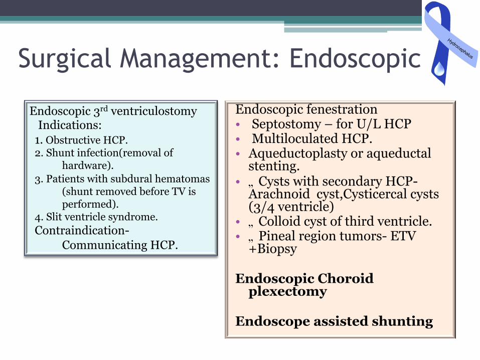
Surgical Management: Endoscopic
Endoscopic fenestration • Septostomy – for U/L HCP • Multiloculated HCP. • Aqueductoplasty or aqueductal
stenting. • �„ Cysts with secondary HCP-
Arachnoid cyst,Cysticercal cysts(3/4 ventricle)
• �„ Colloid cyst of third ventricle. • �„ Pineal region tumors- ETV
+Biopsy Endoscopic Choroid
plexectomy Endoscope assisted shunting
Endoscopic 3rd ventriculostomy Indications: 1. Obstructive HCP. 2. Shunt infection(removal of
hardware). 3. Patients with subdural hematomas
(shunt removed before TV is performed).
4. Slit ventricle syndrome. Contraindication-
Communicating HCP.

Endoscopic Third Ventriculostomy: Is it Safe and Cost Effective in Post Meningitic Hydrocephalus? Vivek Tandon; Ashish Suri; Sarat P. Chandra MCh; Ashok Kumar Mahapatra MD
In cases of post meningitic hydrocephalus, a trial of ETV can prove to be a viable and cost effective alternative to shunt surgery. It may also prove useful in treating patients with multiple shunt failures, Radiological improvement lags behind clinical improvement.
Endoscopic third ventriculostomy in infants YR Yadav, Sumeet Jaiswal, Nelson Adam, Abhijeet Basoor, Gaurav Jain Neurosurgery Unit, NSCB Medical College, Jabalpur, MP, Neurol India 2006;54:161-3
83.3% (45 cases) clinical success rate. Overall failure rate in our study was 16.7% (8 stoma blocks and 1 procedure abandoned). Low birth weight pre mature infants had higher failure rate (3 out of 5 infants 60%) compared to full term infants with normal birth weight (12.3%). Age did not have any impact on the success rate ( P >0.05). Success rates were not significantly different in patients with aqueductal stenosis (85.4%) and TBM (66.6%) (Fisher's exact test, P=0.3).
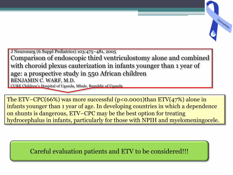
J Neurosurg (6 Suppl Pediatrics) 103:475–481, 2005
Comparison of endoscopic third ventriculostomy alone and combined with choroid plexus cauterization in infants younger than 1 year of age: a prospective study in 550 African children BENJAMIN C. WARF, M.D. CURE Children’s Hospital of Uganda, Mbale, Republic of Uganda
The ETV–CPC(66%) was more successful (p<0.0001)than ETV(47%) alone in infants younger than 1 year of age. In developing countries in which a dependence on shunts is dangerous, ETV–CPC may be the best option for treating hydrocephalus in infants, particularly for those with NPIH and myelomeningocele.
Careful evaluation patients and ETV to be considered!!!

Shunts • VP shunt
• VA shunt- distal catheter placement via facial vein/ seldinger technique
• Torkildsen shunt- ventricles to cisternal space.
• Miscellaneous –Ventriculopleural, gallbladder,ureter or bladder.
• LP/TP shunt
• Cyst or subdural shunt

• Shunt systems come in a variety of configurations and models but they have similar functional components: ▫ Valve Mechanisms – flow or differential ▫ Fixed, programmable, or variable settings ▫ Catheters
� Ventricular (proximal) � Peritoneal/Atria (distal)
▫ Accessories � Reservoirs, Siphon Devices � Connectors, Filters, Pumping Chambers
Shunt Systems

Shunt Infections -Prevention • Sterile surgical technique. • Use of double gloves (Kulkarni et al – loss of glove integrity-30% ), Prevent
CSF leakage to skin • Change glove when handling shunt.(Atiq ur Rehman et al, Journal of
Neurosurgery paediatrics Jun 2010 / Vol. 5 / No. 6 / Pages 569-572)
• Antibiotic impregnated shunt tubing. • Use of antibiotics before dental procedures, one piece system, biannual
screening, hypothermia during surgery.
Antibiotic prophylaxis for surgical introduction of intracranial ventricular shunts (Review) Ratilal BO , Costa J, Sampai O C The Cochrane Collaboration and published in The Cochrane Library 2009, Issue 1
Demonstrated a benefit of systemic prophylactic antibiotics for the first 24 hours postoperatively to prevent shunt infection, regardless of the patient’s age and the type of internal shunt used. The benefit of its use after this period remains uncertain. Evidence suggests that antibiotic-impregnated catheters reduce the incidence of shunt infection

Advancements in biomaterials • Antibiotic impregnated shunt tubings. • Coated silicone tubings for converting them into hydrophilic and
more lubricious material.- valve is not included, only proximal and distal catheter Antibiotic impregnated shunts (Codman)
» Ventricular and Distal Silicone Catheters
» Impregnated with Two Antibiotics -Rifampicin & Clindamycin
» And they are ORANGE!!!
» Drugs are trapped between matrix molecules and are eluted slowly, activated by CSF/water, » Antibiotics are eluted till 28 days » Do not soak in saline
Bioglide(Medtronics)
• BioGlide is a covalently-bonded hydrogel that aids with ease of insertion, reduces bacterial adhesion, and absorbs water-soluable antibiotic solutions
• Created to address the issue of “infection”
• Ease of use of convenient antibiotics
• Retains antibiotics up to 5 days
• ??difficulty in handling

Effect of Antibiotic-Impregnated Shunts on Infection Rate in Adult Hydrocephalus: A Single Institution’s Experience S. Harrison Farber, Scott L. Parker, Owoicho Adogwa, Matthew J. McGirt, Daniele Rigamonti Neurosurgery 69:625–629, 2011
Shunt infection incidence was decreased in AIS (1.2%) vs non-AIS (4.0%) cohorts (P = .0492). Staphylococcus epidermidis was the most common pathogen in AIS and non-AIS cohorts. Oxacillin resistance was not increased in the AIS cohort.
Acta Neurochir (Wien) (2004) 146: 603-610
Effect of hydrophilic coating on microorganism colonization in silicone tubing F. Cagavi', N. kalan, H. Celik, D. Gur, and B. iiq
With in-vivo experiments we can say that, coated material catheters are superior than the silicone catheters in respect to colonization but after the bacterial colonization has occurred, the amount of colonization did not differ

Shunt Fractures
• Most common in neck • Barium in shunt tubing leaches over time • Modifications: - Streak of barium along tubing - Another layer of silicone over barium coating

What's new: Shunt insertion • USG guided: use of separate burrhole/ larger burrhole,
in open fontanelle

Shunt Malposition: Shunt insertion • Neuroendoscope guided : to visualize ventricular
anatomy and choroid plexus J Neurosurg. 2003 Feb;98(2):284-90.
Lack of benefit of endoscopic ventriculoperitoneal shunt insertion: a multicenter randomized trial. Kestle JR, Drake JM, Cochrane DD, Milner R, Walker ML, Abbott R 3rd, Boop FA; Endoscopic Shunt Insertion Trial participants.
Ventricular catheters, which during surgery were thought to be situated away from the choroid plexus, were demonstrated to be in it on postoperative imaging in 67% of patients who had undergone endoscopic insertion and 61% of those who had undergone nonendoscopic shunt placements. The incidence of shunt failure at 1 year was 42% in the endoscopic insertion group and 34% in the nonendoscopic group

What's New: Shunt Insertion • Neuronavigation for shunt placement
J Neurosurg. 2010 Dec;113(6):1273-8. Epub 2010 Apr 16.
Effect of electromagnetic-navigated shunt placement on failure rates: a prospective multicenter study. Hayhurst C, Beems T, Jenkinson MD, Byrne P, Clark S, Kandasamy J, Goodden J, Nandoe Tewarie RD, Mallucci CL
postoperative CT in both groups using a 3-point scale developed for this study: (1) optimal position free-floating in CSF; (2) touching choroid or ventricular wall; or (3) intraparenchymal. 75 patients were included in the study, 41 with standard shunts and 34 with EM-navigated shunts. Seventy-four percent of navigated shunts were Grade 1 compared with 37% of the standard shunts (p=0.001, chi-square test). There were no Grade 3 placements in the navigated group, but 8 in the standard group, and 75% of these failed. Early shunt failure occurred in 9 patients in the standard group and in 2 in the navigated group, reducing the early revision rate from 22 to 5.9% (p=0.048, Fisher exact test)

Spinal Dysraphism
A Randomized Trial of Prenatal versus Postnatal Repair of Myelomeningocele N. Scott Adzick, M.D. Et al N Engl J Med 2011; 364:993-1004March 17, 2011
Recruitment of 183 of a planned 200 patients. The first primary outcome occurred in 68% of the infants in the prenatal-surgery group and in 98% of those in the postnatal-surgery group (relative risk, 0.70; 97.7% confidence interval [CI], 0.58 to 0.84; P<0.001). Actual rates of shunt placement were 40% in the prenatal-surgery group and 82% in the postnatal-surgery group (relative risk, 0.48; 97.7% CI, 0.36 to 0.64; P<0.001). Prenatal surgery also resulted in improvement in the composite score for mental development and motor function at 30 months (P=0.007) and in improvement in several secondary outcomes, including hindbrain herniation by 12 months and ambulation by 30 months. However, prenatal surgery was associated with an increased risk of preterm delivery and uterine dehiscence at delivery.

Shunts revisited � “Multicentre randomized trials of CSF shunt valve design
have failed to demonstrate any difference among the valves in cases of shunt failure.”
1. DRAKE Jm et al-RCT of CSF valve design in pediatric pts. Neurosurgery 43:294-305. 1999
2. Pollack et al- RCT of a programmable valve versus a conventional valve for patients with HCP. Neurosurgery 45:1399-1408,1999.
� Exception = Antibiotic impregnated shunt.

Indian Scenario
• “The inexpensive Chhabra shunt in comparison to Codman shunt had no statistically significant diff in outcome”(J Neurosurgery {peds 4}102:358-362,2005)
In third world countries shunt malfunction rates are higher specially in cases of post meningitic hydrocephalus. Expertise and equipment for performing ETV is scarcely available, however a patient must be carefuly evaluated as a potential candidate for ETV

Ideal Shunt • Reduce obstruction at the ventricular catheter tip and/or shunt
valve • Decrease the risk of overdrainage,. • Reduce the chance of mechanical failure or suboptimal shunt
operation • Minimize or eliminate bacterial biofilm and thrombus formation. • Compatible with diagnostic imaging technology • Diagnostic tools for use in a hospital or outpatient setting that
work in real-time to quantitatively determine shunt function. • Monitoring and diagnostic tools for the home setting to detect
shunt problems at the early stage • External monitoring tools or implantable sensors to detect
suboptimal shunt operation, • Feedback systems in which sensors monitor and then vary shunt
operation to maintain specific values for ventricular volume and pressure.

Thank You