hydraulic properties of zinnia elegans - wur
TRANSCRIPT

Hydraulic properties of Zinnia elegans
from cellular development in vitro to performance in planta
Peter Twumasi

Promotoren: Prof. Dr. A. M. C. Emons Hoogleraar Plantencelbiologie Wageningen Universiteit Prof. Dr. O. van Kooten Hoogleraar Tuinbouwproductieketens Wageningen Universiteit Co-promotoren: Dr. Ir. W. van Ieperen Universitair docent bij de leerstoelgroep Tuinbouwproductieketens Wageningen Universiteit Dr. J. H. N. Schel Universitair hoofddocent bij de leerstoelgroep Plantencelbiologie Wageningen Universiteit Promotiecommissie: Prof. Dr. C. Mariani (Radboud Universiteit, Nijmegen) Prof. Dr. L. H. W. van der Plas (Wageningen Universiteit) Dr. Ir. E.J. Woltering (Wageningen Universiteit en Research Centrum) Prof. Dr. M. De Proft (Katholieke Universiteit Leuven, België) Dit onderzoek is uitgevoerd binnen de C.T. de Wit Onderzoekschool ‘Production Ecology and Resource Conservation’.

Hydraulic properties of Zinnia elegans
from cellular development in vitro to performance in planta
Peter Twumasi
Proefschrift ter verkrijging van de graad van doctor
op gezag van de rector magnificus van Wageningen Universiteit,
Prof. Dr. M. J. Kropff in het openbaar te verdedigen
op dinsdag 8 mei 2007 des namiddags te 16:00 uur in de Aula

Peter Twumasi, 2007
Hydraulic properties of Zinnia elegans: from cellular development in vitro to performance in
planta
PhD Dissertation, Wageningen University, The Netherlands
With summaries in English and Dutch
Keywords: Apoptosis, average daily temperature, caspase, cell death, cysteine,
cytochemistry, differential day and night temperature, differentiation, DNA fragmentation,
electrical conductivity, in vitro culture, leaf osmolarity, light intensity, mesophyll cells,
osmolarity, osmotic potential, plant quality, programmed cell death, protease, suspension
culture, tracheary element, vase life, vessel element, water stress, xylem hydraulic
conductance, xylogenesis, Zinnia elegans
ISBN 90-8504-658-5

Contents Chapter 1 General introduction 9 Chapter 2 Modulation of xylem conduit dimensions by soil water availability
and the effect on the vase life of Zinnia elegans cut flowers 27 Chapter 3 Effect of temperature on xylem vessel and vessel element lengths in Zinnia elegans stems 45 Chapter 4 Establishing in vitro xylogenic Zinnia elegans suspension culture with improved efficiency in tracheary element differentiation 61 Chapter 5 Osmotic potential of Zinnia elegans plant material affects the yield and morphology of tracheary elements produced in vitro 81 Chapter 6 Effects of osmotic stress and timed hormone application on tracheary element differentiation in xylogenic cultures from two Zinnia elegans cultivars 99 Chapter 7 Manipulation of programmed cell death in xylogenic Zinnia elegans suspension cell cultures influences the kinetics and dimensions of tracheary elements produced 117 Chapter 8 General discussion 137
Summary in English 147 Summary in Dutch 150 Acknowledgements 153 About the author 155 Appendix 156 Education certificate of PE&RC 158

To my dear parents, Mr Kwame Ofori Atta, Sr (late) and Madam Adwoa Addai Pomaah
My wife, Agnes Twumasi
My sons, Jim O. Twumasi and Rodney Twumasi
My daughter, Miranda A. Twumasi

Selection of symbols and abbreviations used in this thesis Variable Description Units %TE Percentage of tracheary element differentiation ABA Abscisic acid ADT Average daily temperature oC BA Benzylaminopurine DAPI 4, 6-Diamidino-2-phenylindole DIC Differential interference contrast DIF Difference in day and night temperature oC DMSO Dimethyl sulfoxide DT Daytime temperature oC E-64 N-[N-(L-3-trans-carboxirane-2-carbonyl)-
L-leucyl]-agmatine EC Electrical conductivity dSm-1
FA Formaldehyde FDA Fluorescein-diacetate FTL First true leaves GA Glutaraldehyde Ht Harvest time for first true leaves (FTL) days Kh Stem hydraulic conductivity mgs-1kPa-1cm Lmax Maximum vessel length cm LO Leaf osmolarity mOsm NAA α-Naphthalene acetic acid NT Nighttime temperature oC OP Osmotic potential MPa PCD Programmed cell death TE Tracheary element TUNEL Terminal deoxynucleotidyl transferase
biotin-dUTP nick end labeling VE Vessel element VPE Vacuolar processing enzyme YVAD-AMC YVAD-7- amino-4-methylcoumarin Zasp Z-Asp-CH2-DCB (a general caspase inhibitor) τ Vessel half-length or median length cm


Chapter 1
General introduction

Chapter 1
Background
Plants, like other organisms, require a combination of essential genetic and
environmental factors for proper growth and development. Any adjustment in the correct
balance of interactions between these factors may seriously affect the development of a
particular phenotype with subsequent compromise of quality. Thus, for any set of genes, the
phenotype is strongly dependent on the prevailing environmental conditions. While many
studies have been devoted to the improvement of cut flower quality, such as increased flower
density and stem length, less have been focused on the structure and function of the xylem
vessels in the stem. Most especially, the hydraulic properties of the fundamental units of the
xylem, the tracheary elements (TEs), have been insufficiently studied. In order to gain insight
into the effect of xylem vessel dimensions on cut flower quality, this thesis deals with the
regulation of the xylem vessel structure and function in the cut flower Zinnia elegans. It
focuses on mechanisms involved in the development and regulation of hydraulic properties of
TEs at the cellular level, and of vessels and elements at the whole plant level. A further
consideration is given to the interaction of genetic and environmental factors with the
xylogenesis process.
Determinants of plant quality
A range of physiological and morphological traits are commonly used as a measure of
plant quality. In general, plant quality characteristics differ between species. In most plant
species, the shoot and internode lengths, or specific length and weight measurements, are
sufficient for quality determination. Quality determination requires further measurements,
such as the root growth potential, root and shoot electrolyte leakage, leaf properties, flower
density, vase life of cut flowers, and form, size and taste of the plant products. One parameter
as an important index for plant quality, especially in cut-flowers and many ornamentals, is the
stem length (Sysoyeva and Markovskaya, 2004). Occasionally, certain internal tissue
characteristics, such as the anatomy of different cell files in the stem, can be used to determine
quality. In cut flowers, for example, the xylem conduit dimensions (length and diameter) are
good indicators of vase life properties and for that matter the quality. Thus, each set of xylem
conduit dimensions dictates a specific hydraulic functioning of the xylem system that in effect
regulates the vase life properties of the cut flower. For instance, the restoration of water
uptake and turgidity at the start of the vase life of cut flowers is strongly affected by vascular
10

General introduction
hydraulic conductance and its recovery from embolism (Zimmermann and Jeje, 1981;
Comstock and Sperry, 2000; Nijsse et al., 2001; Twumasi et al., 2005).
The interaction between the genetic makeup of the plant and the conditions within
which the plant develops strongly affects any particular phenotype under study, for example,
in our case the xylem conduit dimensions (Gregorius, 1977; Cavalli-Sforza and Feldman,
1978). Thus, the environmental (E) and genotype (G) interaction can be mathematically
simplified as: P = f(E, G).
In a more complex set of genetic and environmental factors, the phenotype for the kth
observation on the ith genotype in the jth environment (Pijk) can be approached by the linear
equation: Pijk = Ej + Gi + (GE)ij + εijk
where, Ej is an environmental effect, Gi is a genotypic main-effect, (GE)ij is a genotype-by-
environment interaction effect, and εijk is a residual effect (Cooper et al., 2004). These
environmental and genetic interactions have over the years been harnessed to improve traits in
a particular organism. Environmental conditions, well established for plant quality
improvement, include the application of day and night temperature differences (DIF) during
plant growth and development (Erwin et al., 1994; Carvalho et al., 2002; Bachman and
McMahon, 2006), application of different photoperiods (Boyle and Stimart, 1983; Runkle et
al., 2001; Runkle and Heins, 2002), use of different light intensities and qualities (Runkle and
Heins, 2001), humidity (Mortensen and Gislerod, 1997), adjusted soil water (Nonami, 1998)
and plant nutrition (Rodriguez-Perez et al., 2001; Williamson et al., 2001).
The structure and function of the vascular system in general, and the xylem conduits in
particular, like the other plant characteristics mentioned above, are also susceptible to changes
in environmental conditions during plant growth and development (Lovisolo and Schubert,
1998; Nijsse, 2001). Therefore, a careful regulation of xylem structure and function by
environmental factors during growth could enhance plant quality.
Evolution of xylem conduit
Higher plants make use of a vascular system to achieve long distance transport of
water and mineral components from the soil to the leaves for transpiration, photosynthesis and
redistribution of photosynthetically manufactured foods and signaling compounds throughout
the plant (Sieburth and Deyholos, 2006). As a common phenomenon, all large and
multicellular organisms require this sort of system to ensure transport of water, food and
11
Chapter 1
signal molecules to and from tissue targets. Unlike animals that ensure continuous flow of
liquids in the vascular system by the pumping action of the heart, plants utilize increasing
gradients of negative pressure from the soil-plant-atmosphere continuum to maintain
continuous uptake of water against gravity (Pickard, 1981; Zimmermann, 1983; Tyree, 2003;
Zimmermann et al., 2004).
The development of the vascular system offered terrestrial plants a convenient
mechanism for water and nutrient transport as well as for mechanical strength (Comstock and
Sperry, 2000; Kozela and Regan, 2003; Bailey, 1953). Many of the other anatomical features,
needed to sustain complex land plants, evolved during the Ordovician and Silurian periods
(Edwards, 1996; Bateman et al., 1998; Edwards, 1993) including a water-resistant cuticle and
stomatal pores. So, the key to the evolution of larger, erect plant forms was the origin of
highly specialized vascular tissues permitting the long-distance transport of water. In the
upper Silurian we find the first appearance of xylem conduits in the form of tracheids
(Edwards and Davies, 1976): elongated single cells (lacking cytotoplasm at maturity)
specialized for transporting water under negative pressure. The walls of tracheids have
secondary walls with a banded pattern which avoids implosion, and are impregnated with the
stiffening agent lignin. Pit membranes, containing micropores for inter-vessel water transport
(Fig. 3), connects these bands. The lignified and thickened walls are both characteristics of
vascular plants (Doyle, 1998; Gross, 1980; Edwards, 1993); in terrestrial plants they are found
in a wide range of tissues, where they aid in protection and support as well as transport. The
earliest vascular plant fossils reveal thickened and lignified walls exclusively located in the
central conducting strands indicating their possible supporting roles in erect stems. Thus, the
lignin and secondary wall specializations made possible the development of xylem tissues that
are capable of the dual functions still performed by the tissue in extant species, namely 1)
transport of water under negative pressure in specialized conduits, and 2) withstanding the
compressional forces in the conduits (Comstock and Sperry, 2000).
Soil-plant-atmosphere continuum
The movement of water against gravity from the soil via the roots through the stem to
the leaves of the plant is achieved by the concerted effects of cohesion, adhesion and negative
pressure gradients along the soil-plant-atmosphere continuum. More than a century ago,
Dixon and Joly (1894) proposed that a pulling force was generated at the evaporative surface
12

General introduction
of leaves and that this force was transmitted downwards through water columns under tension
to lift water much like a rope under tension can lift a weight (Fig. 1). The cohesion–tension
theory (CTT), as it is known, supposes both adhesion of water to conduit walls and cohesion
of water molecules to each other (Tyree, 2003). The principal argument that arose over this
theory was the fact that, metastable water in vessels of nanometer lumen diameter will
cavitate under such a high negative force. Practical measurements have indeed indicated the
occurrence of a high rate of cavitations recorded in thousands of vessels compared to billions
or trillions of vessels present in a plant. Conduits are interconnected by multiple adjacent
conduits, providing redundancy in multiple pathways for water movement should one conduit
cavitate. The pore diameter in the pit membranes determines how negative the pressure can be
in a water-filled conduit before cavitations are seeded from an adjacent, embolized conduit
(Tyree, 2003). Therefore, any plant’s water status will be governed by the wall properties of
the xylem cells (also called tracheary elements (TEs) or vessel elements (VE’s)), the xylem
anatomy (number of vessels, diameter and length of vessels) as well as the driving force for
water movement throughout the plant directly or indirectly imposed and regulated by a set of
environmental conditions such as relative humidity, drought and light quantum. Zimmermann,
a renowned supporter of CTT, and colleagues showed that other forces come into operation
when exclusive tension fails to lift water against gravity due to environmental conditions
(Zimmermann et al., 2002). These authors have suggested longitudinal cellular and xylem
osmotic pressure gradients, axial potential gradients in the vessels as well as gel- and gas
bubble-supported interfacial gradients as possible candidate forces. This multiforce theory
(MFT), as they termed it, overcomes the problem of the CTT that life on earth depends on
water being in a highly metastable state.
13

Chapter 1
Fig. 1: Transpiration-Cohesion-Tension: a mechanism to pull xylem sap up the plant. Water ascends in the
xylem vessels by the interplay between cohesion forces between the water molecules and the adhesion of these
water molecules to the xylem walls creating “strands” of water supported by increasing upward negative
pressures. Disconnections within these water strands caused by excessive negative pressures or entry of gas
columns (embolism) lead to disruption of this metastability of water and thereby disfunction of the water
transporting xylem vessel. (Biological Sciences, 2nd ed. by Scott Freeman, copyright © 2005 by Pearson
Education, Inc.; reprinted with permission).
Xylem formation (xylogenesis)
Xylem cells develop from procambial or cambial initials but can also be induced to
develop from parenchyma cells when the plant is wounded, and in tissue culture conditions
(Emons et al., 1993; Mulder and Emons, 1993; Nishitani et al., 2002). This differentiation
process is a fundamental process during development of all higher plants that among many
other things relies upon long distance transport of water and nutrients. The xylem conduit
14
General introduction
formation begins with cell division in the procambial or vascular cambium cells upon receipt
of local cell signaling molecules, especially auxin. The individual cells then form TEs after
going through a series of processes involving cell elongation, secondary cell wall synthesis,
lignification and programmed cell death (PCD), collectively termed xylogenesis. The final
product is a hollow cell corpse with secondary thickened wall that may fuse at the later stages
of development with others to form the xylem vessels specialized for conducting water
(Fukuda, 1996; Roberts and McCann, 2000). The elements in the vessels or conduits are
vertically connected with perforations in the endwalls separating the adjacent elements, and
horizontally lying side-by-side with other conduits joined by the pores in their pit membranes
(Fig. 1 and 2). The only exceptions in endwall perforations are those two elements at ends of
the vessel having one of their endwalls imperforated to set limits of the length of the vessel.
The perforation of the endwalls is thought to be caused by the action of cellulases (released
during autolysis) on the endwalls that contain sparse cellulose microfibrils and are not
lignified (Ye, 2002).
The establishment of systems for in vitro differentiation of cells into tracheary
elements has provided valuable information to understand complex processes involved in
xylogenesis. Such studies are difficult to perform at whole plant level due to the complex
nature of the plant system. Induction of xylem cells has been achieved in callus, in
suspension-cultured cells, and in excised tissues and cells (Roberts et al., 1988; Fukuda,
1992). Until now, many plants have been studied for xylem developmental processes, at levels
ranging from molecular genetic screens to physiological and morphological studies. The
Zinnia elegans mesophyll suspension culture system dominates the various types of model
systems used for xylogenesis research.
The Zinnia cell culture system
The Zinnia system consists of mesophyll cells, preferably isolated from first true
leaves of 14-d old seedlings of Zinnia elegans, which are nourished in a specific liquid culture
essentially containing a correct balance of two plant growth regulators, auxin and cytokinin.
By maintaining appropriate culture conditions, about 80% of these photosynthetic mesophyll
cells are redifferentiated into TEs at approximately 96-h (Chapter 4 and 5, this thesis). The
differentiation process is characterized by extensive cellulose deposition, nuclear DNA
degradation, rapid disruption of the tonoplast and autolysis of the whole cell which is a typical
15

Chapter 1
hall mark of programmed cell death. The remainder is a dead and hollow cell with a
secondary thickened wall (De Jong et al., 2000; Orzaez et al., 2001; Woltering et al., 2002).
Fig. 2 shows a summary of events occurring during the tracheary element differentiation
process. The Zinnia system is simple and amenable to biochemical and molecular analyses,
and it offers a real hope for understanding the nature of the molecular machinery whereby
plant cells become specialized to carry out particular functions (Roberts and McCann, 2000).
Fig. 2: Differentiation and fusion of vessel elements during the formation of a xylem vessel. The VE initially
expands and then undergoes intense synthesis of secondary cell wall bands, followed by autolysis of the
differentiating cell via programmed cell death. The adjacent differentiated VE’s may fuse by forming
perforations in their transverse walls.
Xylem conduit diameter, length and hydraulic conductivity
The xylem conduit is composed of hundreds or thousands of vertically and
horizontally stacked individual vessel elements (VEs) or TEs. With the exception of the two
vessel members located at both ends of the vessel, top and bottom, the rest of the vessel
elements have their end walls perforated.
In general, xylem vessels are built from stacks of variable but finite number of TEs.
TE diameter is only influenced during the early phase of xylogenesis (i.e. cell expansion) due
to the robustness of secondary thickening of the matured elements. It has been found that the
cell diameter and length of individual Zinnia cells, differentiating into TEs in vitro, increased
rapidly until the first signs of secondary thickening (Cosgrove, 1998). Correlation between the
expression of two expansin or cell wall loosening genes (ZeExp1 and ZeExp2) and primary
16

General introduction
wall expansion and elongation of TEs in Zinnia cultures has been shown (Im et al., 2000).
Similar to properties of pipes, the flow of water in xylem conduits is governed by Hagen-
Poiseuille’s law, which states that the flow rate of a fluid in pipes is directly proportional to
the fourth power of the lumen diameter (Aloni and Zimmermann, 1983; Vogel, 1988; Tyree
and Ewers, 1991). The water flux (ψ ) in the stem can therefore be defined mathematically as:
ψ = πρD4/8η
where ψ, water flux (kg/s); D, conduit diameter (m); η, the dynamic viscosity of water
(MPa.s); ρ, the density of water (kg/m3). The lumen diameter of a vessel is therefore essential
for the hydraulic efficiency of the plant.
Xylem conduit length may be affected by either the length of individual TEs or the
number of TEs that fuse to form the vessel. The individual TE length is determined by cell
elongation of the xylem precursor cell during the early phase of TE development. To sustain
cell elongation, (a) new wall material has to be produced (Golgi apparatus), (b) delivered
constantly (cytoskeleton), (c) the existing expanding wall wall has to remain flexible and (d)
turgor pressure should be sufficient as a driving force. If one of the four conditions is lacking,
cell elongation stops. As in all plant cells, during individual TE differentiation, secondary cell
wall is deposited after cell elongation has stopped. In addition, in TEs wall thickenings are
formed, which initially contain pure cellulose.
A higher occurrence of fusion of individual TEs may give rise to longer conduits. It
has been speculated, however, that the number of TEs per vessel is determined by a mere
random distribution considering the fact that different types of xylem conduit lengths exist at
any time in any given plant (Zimmermann and Jeje, 1981). Until now, there has been no
experimental prove of any of the two possibilities responsible for determining the length of
xylem conduit.
In plants, the two hydraulic resistances that influence the efficiency of water uptake
via xylem conduits are: (1) resistance to flow in the lumen and (2) crossing the thin, but much
more restrictive, microporous pit membranes of the interconduit pits (Fig. 3). Therefore, a
longer conduit exhibits higher vertical interconduit flow via perforated endwalls compared to
shorter conduits that might move water between conduits through the horizontal pit
membranes (Comstock and Sperry, 2000). Thus, by enhancing the formation of longer and
wider conduits as opposed to shorter and narrower ones, one could improve the hydraulic
efficiency of the plant.
17

Chapter 1
Fig 3: Drawing of xylem vessels consisting of fused tracheary or vessel elements and showing the interconduit
structures through which water moves between adjacent vessels. The pits are sites lacking secondary cell wall,
and the pit membrane is the remaining primary cell wall sheet in the pits. The pit membrane contains
microscopic pores that allow passage of water but prevent air embolism. The rate of embolism and hydraulic
resistance are partly influenced by the strength and porosity of the pit membrane. (CS = cross section; VE =
vessel element).
Ideal xylem conduit
By measuring the following xylem conduit dimensions, it might be possible to predict
the hydraulic performance of the plant and its products, especially in cut flowers. The
following six properties of the xylem conduit are determinants of the hydraulic efficiency of
any plant: (1) vessel length, (2) number of elements per unit length of a vessel, (3) diameter of
vessels, (4) type of perforation plates (simple or compound), (5) size of interconduit pit
membranes pores, and (6) number of vessels per unit area of stem. For example, a cut flower
with conduits of longer length and wider diameter but at the same time having enough
intraconduit compound perforation plates and narrower pit membrane pores will be efficient
in water uptake and at the same time equipped with safety devices in dealing with cavitation
and embolism. Thus, the hydraulic performance of a plant will depend on the composition of
the xylem conduits having particular hydraulic properties as described above.
18

General introduction
Suitable plant models for studying xylogenesis and xylem hydraulic function
Until the early 1980s, a lot of studies had been done on development and
differentiation of the plant vascular system mostly at the whole plant level (Aloni, 1987; Ye,
2002). From 1980 onwards attention was shifted to the differentiation of the vascular system
at the single cell level especially in the case of in vitro differentiation of the xylem (Fukuda
and Komamine, 1980a). Most of these studies in vitro were focused on the fundamental
principles of the differentiation mechanism of the vascular system (Fukuda et al., 1993;
Fukuda, 1996). There are, however, just a handful of studies that have tried to mimic some of
the whole plant environmental conditions in vitro and just a few about the hydraulic properties
of elemental units (i.e., TEs) of the xylem vessels during vascular differentiation (Roberts and
Haigler, 1994; Kuriyama and Fukuda, 2001). There is therefore a wide gap left for complete
understanding of how closely the in vitro system for vascular differentiation studies relates to
the in planta situation and whether environmental factors that influence the whole plant
developmental processes can be perfectly mimicked in a well defined miniature in vitro
system.
Zinnia elegans is grown on a small scale as cut flowers. However, availability of its
stable and highly synchronous cell culture (producing over 60%TEs) for xylem differentiation
studies has made it more popular for study than other cut flowers (Fukuda and Komamine,
1980a; Church, 1993). Since the onset of this remarkable system, several studies using this
system ranging from simple cell physiological studies (Lee and Roberts, 2004) to genomics
(Demura et al., 2002; Pesquet et al., 2005), have provided us with valuable information, from
fundamental basis of cellulose microfibril deposition on the cellular level up to the formation
of the water-conducting xylem vessels over the entire plant (Roberts et al., 2004; Oda et al.,
2005).
The characteristic features of the Zinnia elegans cell culture system have tremendously
lifted this plant beyond its use as mere cut flower (sometimes called ‘killer’ cut flower due to
its fragile flower stalk) to a broader application in plant science. More interestingly, Zinnia
elegans due to its dual function, i.e., as a tool for fundamental cell and molecular research as
well as a whole plant in physiological and horticultural research, makes it the most preferred
model plant for research that aims to use molecular and cellular studies for the improvement
of the post-harvest quality of horticultural products such as cut flowers.
19

Chapter 1
Objectives and strategies
The main objective of this study was to investigate in vitro and in planta regulation of
xylogenesis in Zinnia elegans by growth conditions and genotype, and how the resultant
modification of the xylem conduit dimensions during development would influence the
hydraulic properties of the xylem, possibly leading to improvement of the post-harvest quality
of the cut flowers. Various post-harvest quality aspects of many horticultural products depend
on plant water relations during and after growth, which are directly influenced by the xylem
vascular system. Important post-harvest quality aspects of cut flowers (restoration of water
uptake and turgidity at the start of the vase life) strongly depend on the hydraulic conductance
of the vascular system and the ability to recover from embolisms in the base of the stem. As
mentioned earlier, these functional aspects of the xylem system depend on number and
dimensions (diameter and length) of individual xylem vessels which are formed in the pre-
harvest phase and which are influenced by the plant water status and genetic background.
Possibilities to improve the post-harvest quality of cut flowers, therefore, include (1) the
breeding of cultivars that form xylem vessels with dimensions that favour the post-harvest
quality, such as vase life, or (2) the actual control of xylem dimensions during growth by
environmental factors. The latter is considered in this thesis. To achieve this, mechanistic
knowledge about interactions between processes during vessel formation which influence
vessel diameter and length, environmental factors and genetic background, is required. We
therefore tried to find answers to the following questions:
- Do different genetic make-ups of the plant determine different dimensions of the
xylem conduits with consequent effect on the hydraulic properties that determine the
post-harvest quality (e.g. vase life) of a cut flower?
- Within a particular genotype, how do different environmental conditions, such as
water stress and DIF during growth periods, affect the dimensions of the xylem
conduits in planta and the vase life of these cut flowers?
- Can these environmental conditions be mimicked in vitro for the study of the
xylogenesis process at single cell and molecular level to understand how the
dimensions of a xylem vessel are determined and regulated?
- Are there possibilities for improvements of the established xylogenic cell culture for a
more extensive and reliable usage in the xylogenesis research?
20

General introduction
- Which processes are important in determining a particular type of vessel dimension at
cellular or molecular level? And could these conduit dimension-determining processes
evoke the same dimensional effect in the whole plant?
Outline of the thesis
Chapter 1: General introduction. This chapter covers the description of the problem of
plant water uptake, effects of xylem dimensions on the post harvest quality of cut flowers,
literature on general formation of xylem, general description of Zinnia elegans, and an outline
of the thesis.
Chapter 2: Zinnia elegans cultivars: drought responses and characterization. Here
we present results on the two pre-selected cultivars of Zinnia elegans grown in the greenhouse
at different water stress levels. The anatomical and functional analyses of the xylem conduits
in relation to the post-harvest quality of the cut flowers are as well presented in this chapter.
Chapter 3: Effects of differential day and night temperatures on hydraulic
properties of the two Zinnia elegans cultivars. This chapter focuses on the effects of
temperature on the whole plant morphology, xylem anatomy and hydraulic functioning of cut
flowers from the two Zinnia elegans cultivars. It also provides anatomical information on
vessel elements and discusses the mechanisms involved in the formation of a xylem conduit
of particular length.
Chapter 4: Technical aspects of xylogenic Zinnia suspension cell cultures. This
chapter covers technical details in setting up a Zinnia suspension cell culture system and the
subsequent differentiation into tracheary elements. Relevant assays, specific for tracking the
xylogenesis process are being described in this chapter.
Chapter 5: Leaf osmolarity and tracheary element differentiation in xylogenic Zinnia
elegans cell cultures. This chapter focuses on the performance of Zinnia cultures established
from plant material that had been osmotically preconditioned by use of hydroponic cultures
with varied electrical conductivities or plants grown in soil with varied light intensities.
Chapter 6: Osmotic stress effects on in vitro xylogenesis. This chapter describes in vitro
TE differentiation and the effects of osmotic stress and timing of hormone application on the
onset and percentage of TE formation, and morphology of the TEs.
Chapter 7: Manipulation of TE dimensions and differentiation through regulation of
caspase-controlled programmed cell death (PCD) in xylogenic Zinnia elegans cell
21
Chapter 1
cultures. In this chapter we present results on the use of caspase inhibitors on the onset of
PCD during TE differentiation. We discuss the potentials of the in vitro results to be applied
in planta for whole plant hydraulic conductivity improvements.
Chapter 8: General discussion. This chapter discusses the previous chapters with an
attempt to integrate the in planta and in vitro experimental results. It elaborates on the
potentials of the application of the research findings to improve the quality of plant
production, especially in cut flowers. It also highlights areas that need further research.
References
Aloni, R., and Zimmermann, M.H. (1983). The control of vessel size and density along the
plant axis - a new hypothesis. Differentiation 24, 203-208.
Aloni, R.R. (1987). Differentiation of vascular tissues. Annual review of plant physiology 38,
179-204.
Bachman, G.R., and McMahon, M.J. (2006). Day and night temperature differential (DIF)
or the absence of far-red Light alters cell elongation in ‘Celebrity White’ Petunia. J. Amer.
Soc. Hort. Sci 131, 309-312.
Bailey, I.W. ( 1953). Evolution of the tracheary tissue of land plants. Am J Bot 40, 4±8.
Bateman, R.M., Crane, P.R., DiMichele, W.A., Kenrick, P.R., Rowe, N.P., Speck, T., and
Stein, W.E. (1998). Early evolution of land plants: phylogeny, physiology, and ecology of the
primary terrestrial radiation Annual Review of Ecology and Systematics 29, 263±292.
Boyle, T.T.H., and Stimart, D.D.P. (1983). Developmental responses of Zinnia to
photoperiod. J Am Soc Hortic Sci 108, 1053-1059.
Carvalho, S.M.P., Heuvelink, E., Cascais, R., and Van Kooten, O. (2002). Effect of day
and night temperature on internode and stem length in chrysanthemum: Is everything
explained by DIF? Ann Bot-London 90, 111-118.
Cavalli-Sforza, L.L., and Feldman, M.W. (1978). The evolution of continuous variation.
III. Joint transmission of genotype, phenotype and environnment. Genetics 90, 391-425.
Church, D.L. (1993). Tracheary element differentiation in Zinnia mesophyll cell-cultures.
Plant Growth Regul 12, 179-188.
Comstock, J.P., and Sperry, J.S. (2000). Theoretical considerations of optimal conduit
length for water transport in vascular plants. New Phytologist 148, 195-218.
Cooper, M., Podlich, D., and Smith, S.O. (2004). Complex traits genetics and gene-to-
22

General introduction
phenotype models. 4th International Crop Science Congress, Published on CDROM.
Cosgrove, D.J. (1998). Molecular regulation of plant cell wall extensibility. Gravit Space
Biol Bull 11, 61-70.
De Jong, A.J., Hoeberichts, F.A., Yakimova, E.T., Maximova, E., and Woltering, E.J.
(2000). Chemical-induced apoptotic cell death in tomato cells: involvement of caspase-like
proteases. Planta 211, 656-662.
Demura, T., Tashiro, G., Horiguchi, G., Kishimoto, N., Kubo, M., Matsuoka, N.,
Minami, A., Nagata-Hiwatashi, M., Nakamura, K., Okamura, Y., Sassa, N., Suzuki, S.,
Yazaki, J., Kikuchi, S., and Fukuda, H. (2002). Visualization by comprehensive microarray
analysis of gene expression programs during transdifferentiation of mesophyll cells into
xylem cells. P Natl Acad Sci USA 99, 15794-15799.
Dixon, H.H., and Joly, J. (1894). On the ascent of sap. Philosophical Transactions of the
Royal Society London Series B 186, 563-576.
Doyle, J.A. (1998). Phylogeny of vascular plants. Annual Review of Ecology and Systematics
29, 567±599.
Edwards, D. (1996). New insights into early land ecosystems: a glimpse of a Lilliputian
world. Review of Palaeobotany and Palynology 90 159±174.
Edwards, D. ( 1993). Tansley Review No. 53. Cells and tissues in the vegetative sporophytes
of early land plants. New Phytologist 125, 225±247.
Edwards, D., and Davies, E.C.W. (1976). Oldest recorded in situ tracheids. Nature 263,
494±496.
Emons, A.M.C., Mulder, B.M., and Kieft, H. (1993). Pyrolysis mass spectrometry of
developmental stages of maize somatic embryos. Acta botanica Neerlandica 42, 319-339.
Erwin, J., Velguth, P., and Heins, R. (1994). Day/night temperature environment affects cell
elongation but not division in Lilium longiflorum Thunb. J. Exp. Bot. 45, 1019-1025.
Fukuda, H. (1992). Tracheary element formation as model system of cell differentiation. Int.
Rev. of Cytol 136, 289-332
Fukuda, H. (1996). Xylogenesis: Initiation, progression, and cell death. Annu Rev Plant Phys
47, 299-325.
Fukuda, H., and Komamine, A. (1980a). Establishment of an experimental system for the
study of tracheary element differentiation from single cells Isolated from the mesophyll of
zinnia elegans. Plant Physiol 65, 57-60.
23

Chapter 1
Fukuda, H., Sato, Y., Yoshimura, T., and Demura, T. (1993). Molecular mechanisms of
xylem differentiation. J Plant Res, 97-107.
Gregorius, H.R. (1977). The genotype x environment-to-phenotype relationship. TAG
Theoretical and Applied Genetics 49, 165-176.
Gross, G.G. ( 1980). The biochemistry of lignification. Advances in Botanical Research 8,
25±63.
Im, K.H., Cosgrove, D.T., and Jones, A.M. (2000). Subcellular localization of expansin
mRNA in xylem cells. Plant Physiol 123, 463-470.
Kozela, C., and Regan, S. (2003). How plants make tubes. Trends Plant Sci 8, 159-164.
Kuriyama, H., and Fukuda, H. (2001). Regulation of tracheary element differentiation. J
Plant Growth Regul 20, 35-51.
Lee, S., and Roberts, A.W. (2004). Tracheary element differentiation is correlated with
inhibition of cell expansion in xylogenic mesophyll suspension cultures. Plant Physiol Bioch
42, 43-48.
Lovisolo, C., and Schubert, A. (1998). Effects of water stress on vessel size and xylem
hydraulic conductivity in Vitis vinifera L. J Exp Bot 49, 693-700.
McCann, M.C., Domingo, C., Stacey, N.J., Milioni, D., and Roberts, K. (2000). Tracheary
element formation in an in vitro system. . (Oxford: BIOS Scientific Publishers Ltd).
Mortensen, L.M., and Gislerod, H.R. (1997). Effects of air humidity and air movement on
the growth and keeping quality of roses. Gartenbauwissenschaf 62, 273-277.
Mulder, B.B.M., and Emons, A.A.M.C. (1993). Cell wall development in maize somatic
embryos studied by pyrolysis—mass spectrometry. Journal of Analytical and Applied
Pyrolysis, 25, 255-264.
Nijsse, J. (2001). Functional anatomy of the water transport system in cut chrysantemum. . In
Horticultural Production Chains ( Wageningen University).
Nijsse, J., van der Heijden, G.W.A.M., van Ieperen, W., Keijzer, C.J., and van Meeteren,
U. (2001). Xylem hydraulic conductivity related to conduit dimensions along chrysanthemum
stems. J Exp Bot 52, 319-327.
Nishitani, C., Demura, T., and Fukuda, H. (2002). Analysis of early processes in wound-
induced vascular regeneration using TED3 and ZeHB3 as molecular markers. Plant Cell
Physiol 43, 79-90.
Nonami, H. (1998). Plant water relations and control of cell elongation at low water
24

General introduction
potentials. J Plant Res 111, 373-382.
Oda, Y., Mimura, T., and Hasezawa, S. (2005). Regulation of secondary cell wall
development by cortical microtubules during tracheary element differentiation in Arabidopsis
cell suspensions. Plant Physiol 137, 1027-1036.
Orzaez, D.D., Jong, A.A.J.d., and Woltering, E.E.J. (2001). A tomato homologue of the
human protein PIRIN is induced during programmed cell death. Plant Mol Biol 46, 459-468.
Pesquet, E., Ranocha, P., Legay, S., Digonnet, C., Barbier, O., Pichon, M., and Goffner,
D. (2005). Novel markers of xylogenesis in Zinnia are differentially regulated by auxin and
cytokinin. Plant Physiol 139, 1821-1839.
Pickard, W.F. (1981). The ascent of sap in plants. Progress in Biophysics and Molecular
Biology 37, 181-229.
Roberts, A.W., and Haigler, C.H. (1994). Cell expansion and tracheary element
differentiation are regulated by extracellular pH in mesophyll cultures of Zinnia elegans L.
Plant physiol. 105, 699-706.
Roberts, A.W., Frost, A.O., Roberts, E.M., and Haigler, C.H. (2004). Roles of
microtubules and cellulose microfibril assembly in the localization of secondary-cell-wall
deposition in developing tracheary elements. Protoplasma 224, 217-229.
Roberts, K., and McCann, M.C. (2000). Xylogenesis: the birth of a corpse. Curr Opin Plant
Biol 3, 517-522.
Roberts, L.W., Gahan, P.B., and Aloni, R. (1988). Vascular differentiation and plant
growth regulators. (Springer).
Rodriguez-Perez, J.A., Fernandez-Falcon, M., and Socorro-Monzon, A.R. (2001). The
effect of salinity on growth and nutrition of Leucospermum cordifolium. J Hortic Sci Biotech
76, 601-607.
Runkle, E.S., and Heins, R.D. (2001). Specific functions of red, far red, and blue light in
flowering and stem extension of long-day plants. J Am Soc Hortic Sci 126, 275-282.
Runkle, E.S., and Heins, R.D. (2002). Stem extension and subsequent flowering of seedlings
grown under a film creating a far-red deficient environment. Sci Hortic-Amsterdam 96, 257-
265.
Runkle, E.S., Heins, R.D., Cameron, A.C., and Carlson, W.H. (2001). Photocontrol of
flowering and stem extension of the intermediate-day plant Echinacea purpurea. Physiol
Plantarum 112, 433-441.
25

Chapter 1
Sieburth, L.E., and Deyholos, M.K. (2006). Vascular development: the long and winding
road. Curr Opin Plant Biol 9, 48-54.
Sysoyeva, M.I., and Markovskaya, E.F. ( 2004). Day and night temperature optimisation for
plant quality based on regression models. Acta Hort. (ISHS) 654, 55-62.
Twumasi, P., van Ieperen, W., Woltering, E.J., Emons, A.M.C., Schel, J.H.N., Meeteren,
U., and van Marwijk, D. (2005). Effects of water stress during growth on xylem anatomy,
xylem functioning and vase life in three Zinnia elegans cultivars. Acta Horticulturae 669, 303
- 312.
Tyree, M.M.T., and Ewers, F.F.W. (1991). The hydraulic architecture of trees and other
woody plants. The New phytologist 119, 345-360.
Tyree, M.T. (2003). Plant hydraulics: the ascent of water. Nature 423, 923.
Vogel, S. ( 1988). Life's devices (Princeton, NJ, USA.: Princetin University Press).
Williamson, L.C., Ribrioux, S.P.C.P., Fitter, A.H., and Leyser, H.M.O. (2001). Phosphate
availability regulates root system architecture in Arabidopsis. Plant Physiol 126, 875-882.
Woltering, E.J., van der Bent, A., and Hoeberichts, F.A. (2002). Do plant caspases exist?
Plant Physiol 130, 1764-1769.
Ye, Z.H. (2002). Vascular tissue differentiation and pattern formation in plants. Annu Rev
Plant Biol 53, 183-202.
Zimmermann, M.H. (1983). Xylem structure and the ascent of sap. (Berlin: Springer-
Verlag).
Zimmermann, M.H., and Jeje, A.A. (1981). Vessel-length distribution of some American
woody plants Can J Bot 59, 1882-1892.
Zimmermann, U., H., S., L.H., W., and A., H. (2004). Water ascent in tall trees: does
evolution of land plants rely on a highly metastable state? New Phytol 162.
Zimmermann, U., Schneider, H., Wegner, L.H., Wagner, H.J., Szimtenings, M., Haase,
A., and Bentrup, F.W. (2002). What are the driving forces for water lifting in the xylem
conduit? Physiol Plant 114, 327-335.
26

Chapter 2
Modulation of xylem conduit dimensions by soil water availability and the
effect on the vase life of Zinnia elegans cut flowers
Published in part as:
Twumasi, P., van Ieperen, W., Woltering, E.J., Emons, A.M.C., Schel, J.H.N., Meeteren,
U., and van Marwijk, D. (2005). Effects of water stress during growth on xylem anatomy,
xylem functioning and vase life in three Zinnia elegans cultivars. Acta Horticulturae 669, 303
- 312.

Chapter 2
Abstract
Long distance water transport from the roots to the leaves in angiosperm plants is
made possible by the evolutionary development of the xylem conduits with the additional
property of mechanical support for the plant. In all angiosperm plants, the xylem conduit
dimension (vessel length and diameter), and the vessel-to-vessel connections, greatly
influence the hydraulic conductivity of the plant. In this paper we describe the effects of water
stress levels measured as soil water content in the root environment on the xylem conduit
dimensions as well as the vase life of cut flowers from two Zinnia elegans cultivars, “Envy”
and “Purple Prince”. Three different soil water contents were set up for growing of the cut
flowers in the greenhouse: water-stress (20%, v/v soil water content); moderately-watered
(50%, v/v soil water content); and well-watered (70%, v/v soil water content). Morphological
analyses of the whole plants revealed an increasing plant size with decreasing water stress
level in both Zinnia cultivars. Comparatively, the Purple Prince plants were bigger than the
Envy plants. Generally, the hydraulic conductivity of the stems increased with increasing soil
water content in the root environment. The xylem conduit number per stem cross section was
the same in both cultivars from the three different water treatments. Anatomical
measurements showed significant differences in xylem vessel length only in Envy at different
water treatments. Large statistical differences were found in the proportion of large-diameter
xylem vessels in plants from the three soil water contents. Vessel density increased by
between 80 and 130% in the water-stressed plants from both Zinnia cultivars as compared to
the well-watered plants. The vase life of the cut flowers increased with increasing water stress
in both Zinnia cultivars. We therefore conclude that higher efficiency in embolism recovery
associated with the narrower xylem conduits is important for longer vase life in Zinnia cut
flowers.
Keywords: Plant quality, vase life, water stress, xylogenesis, xylem hydraulic conductance,
Zinnia elegans
Abbreviations: ABA, abscisic acid; EC, electrical conductivity; Kh, stem hydraulic
conductivity; Lmax, maximum vessel length; τ, half-length or median length of vessels (length
at which 50% of the vessels terminate)
28

Modulation of xylem conduit dimensions
Introduction
The evolution of the vascular system in terrestrial plants offered them a convenient
mechanism for distant water, nutrient and hormone transport with additional mechanical
support (Bailey, 1953; Pickard, 1981; Kozela and Regan, 2003; Sieburth and Deyholos,
2006). Despite this successful evolution, hydraulic properties of the xylem vessels are
affected by a number of factors during development. An important one is the dimension of the
xylem conduit (i.e., length and diameter) and the conduit density at cross sectional area of the
stem (Nijsse et al., 2001). Such properties of the xylem vessels greatly influence the water
status of the plant and thereby become a general plant quality-determining factor. For
example, it has been shown that the ability of the plant to overcome cavitations and embolism
strongly depends on these xylem properties (Ellerby and Ennos, 1998; Lovisolo and Schubert,
1998; Vogt, 2001; van Ieperen et al., 2002)
Also in cut flowers, the xylem vessel dimensions are important in determining their
quality. The post harvest quality of cut flowers is commonly quantified
as the vase performance in terms of their ability to maintain a high capacity of water transport
combined with their ability to overcome embolism. This means removal of embolism in the
xylem conduits that might have occurred during transport or by excessive transpiration (van
Ieperen et al., 2002). Although other factors, such as microbial growth, formation of tyloses
and deposition of materials in the lumen of xylem vessels, also contribute to restricted water
transport (Van Doorn, 1997), the structural and functional performance of the xylem vessels
is essential for maintaining harvest quality in many aspects of cut flowers. In theory, the
hydraulic capacity of longer and wider xylem vessels will potentially exceed that of shorter
and narrower vessels. However, in practice most plants develop a combination of shorter,
longer, narrower and wider vessels to meet the uncertain conditions that often take toll on the
survival and quality of the plant. For example, it has been shown that despite the inefficient
water transport capacity of the narrower vessels, such vessels are very important safety
devices in dealing with ever increasing cavitation and embolism imposed by harsher
environmental conditions such as drought and frost (Comstock and Sperry, 2000; Tyree,
2003). During development of the xylem, a right balance is made between making xylem
vessels more efficient for water uptake and also maintaining properties capable of
withstanding harsher environmental conditions, such as drought and frost, to prevent vessel
dysfunction through cavitation and embolism. How these plants achieve this balance is not yet
29

Chapter 2
understood, but such information will be greatly useful in production of plants with desired
qualities within any given set of plant growth conditions.
In this study, we aimed at modulation of the xylem conduit dimensions by applying
different levels of water stress during the growth of two Zinnia elegans cultivars, “Envy” and
“Purple Prince”. Zinnia elegans was used in this work because of two reasons: 1) it is a
horticultural product - a so-called cut-flower; and 2) cell suspension culture systems with a
highly synchronized mesophyll differentiation into xylem tracheary elements (TEs) exist.
This fulfils our future objectives to study cell-water relations and xylem vessel modifications
at cellular level.
Materials and methods
Plant material
Seeds of two Zinnia elegans cultivars, “Envy” and “Purple Prince” (Muller
Bloemzaden BV, Lisse, Netherlands) were sown in peat-based commercial potting compost
(Lentse Potgrond nr. 4; 85% peat, 15% clay, Lentse Potgrond, Lent, The Netherlands). After
10 days a homogeneous set of seedlings per cultivar was selected and transplanted to 10 litre
pots filled with perlite (Agra-perlite: No 1; grading 0.6-1.5 mm). Each pot contained 3 plants
of the same cultivar. Plants were placed in the Unifarm greenhouse at Wageningen
University, The Netherlands and grown at 70% RH and 18/22 oC night/day temperature (set-
points). Three constant levels of volumetric water content (Θ v/v%) were continuously
maintained in the pots during the whole growth period using ECH2O probes (Model EC-20;
Decagon Devices Inc, Pullman, Washington, USA) in combination with an automatic drip
irrigation system. The pots were subjected to the following water treatments throughout the
growth period: well-watered (70% v/v water content), moderately-watered (50% v/v water
content), and water-stressed (20% v/v water content). In all, three sets of 8 randomly selected
cut flowers in each treatment were used for the following analyses: 1) xylem function and
conduit diameters; 2) xylem conduit length; and 3) vase life in each of the treatments. The
experiment was performed in duplo.
Xylem hydraulic conductivity measurement
At approximately 35 days after transplanting, a period just before flower opening, two
sets of eight randomly selected plants per treatment were transported to the laboratory for
30

Modulation of xylem conduit dimensions
hydraulic conductance measurements and vessel length determination (see below). Flower
stems were harvested under water (to prevent air entrance) at the root-shoot junction and re-
cut (under water) just above the first leaf-pair and 20-25 cm higher to get stem samples of
sufficient length (longer than expected maximal vessel length). Hydraulic conductance of
these stem segments was determined from pressure-flow relationships as described by van
Ieperen et al. (2003). Hydraulic conductivity (Kh) was calculated from the measured hydraulic
conductance and the length of each sample.
Xylem anatomy (number of vessels, diameter- and length distributions)
After measuring the hydraulic conductivity a 2-3 cm long piece was cut from the
middle of the first internode of each stem sample and stored in 75% ethanol for further
anatomical analysis. 10-30µm thick stem cross-sections were made with a Reichert-Jung 2050
microtome from pieces of stem sections tightly held in styrofoam sandwich (1cm x 1cm x
2cm). The digital images of the sections were made using a Nikon DXM-1200 camera on a
Leica Aristoplan microscope. Images from whole cross-sections (magnification 10x) and all
individual vascular bundles per cross-section (80x) were made. Diameter analysis was done
on images with individual vascular bundles (resolution ≈1 µm per pixel) and vessel counting
was done on images of whole cross-sections using the UTHSCSA ImageTool program
(developed at the University of Texas Health Science Center, San Antonio, Texas). Vessel
number and diameters of all individual vessels at cut stem surface were established for all
samples per cultivar per watering-treatment (n=8). The vessel length distribution was
determined in both cultivars × watering treatment combinations (n=8) using the latex
perfusion method (Zimmermann and Jeje, 1981) and analysed according to Nijsse et al.
(2001).
Vase life
After all plants started to flower (no difference in timing between cultivars and watering
treatments) 4 pots with plants per cultivar x watering treatment combination were transported
to the laboratory. Flower stems were harvested 1cm above the root/shoot junction and re-cut
by 0.5 cm using a sharp razor blade. Lower leaves were removed up to 10-15 cm from the
stem base. To regain full turgidity and remove any embolisms in the vascular system of the
flower stems, they were placed in buckets with ice/water mixture (3:1) and stored over night
31
Chapter 2
at 4 oC in darkness (van Meeteren, 1992). After re-hydration the flower stems were re-cut by
another 3 cm (razor blade) to remove any type of blockage from the base of the flower stems.
Before starting the vase period all flowers were dehydrated to approximately 95% of their
initial full turgor fresh weight. Dehydration was done by placing the cut flower stems in a
room at 20 oC, 60% RH and a light intensity of 14 µmol.m-2.s-1 (Philips, TLD 50W/84HF).
Weight loss of individual cut flower stems was monitored and the time to reach the 95% level
of their initial weight (full turgor) was recorded (no statistical significant differences between
cultivars and watering treatments). After reaching 95% of their initial fresh weight the cut
flowers were placed in vases (500 ml Erlenmeyer flasks) filled with tap water in the same
room (light period 12 h.). The base of the flower stem was free from the bottom of the flask
and between 1-7 cm below the water surface (depth changed due to water uptake during vase
period). Weight of all flower stems was measured at 20, 44 and 70 h after the start of the
dehydration period and expressed as percentage of the weight at full turgor. After each
measurement the flasks were refilled with tap water. After finishing the vase period
experiment, average leaf area per cut flower was measured (LI-3100, LI-COR Inc, Lincoln,
USA).
Statistics
Analyses of variance for the effects of different volumetric water content in the root
environment as well as for the cultivar on the plant height, plant stem diameter at first
internode, xylem vessel number, vessel length and vessel diameter were performed by Prism
v.4 (GraphPad Software, Inc.). For each analysis a significance level of 5% was maintained.
Results
In general, the different levels of soil water during plant growth induced significant
changes in morphological characteristics (e.g. plant height and stem diameter), xylem
anatomy, hydraulic functioning of the xylem vessels, and the vase life of the cut flowers from
both Zinnia elegans cultivars.
Morphological characteristics
In both Envy and Purple Prince, the three different water availability levels imposed at
the root environments during the growth period induced significant differences in plant height
32
Modulation of xylem conduit dimensions
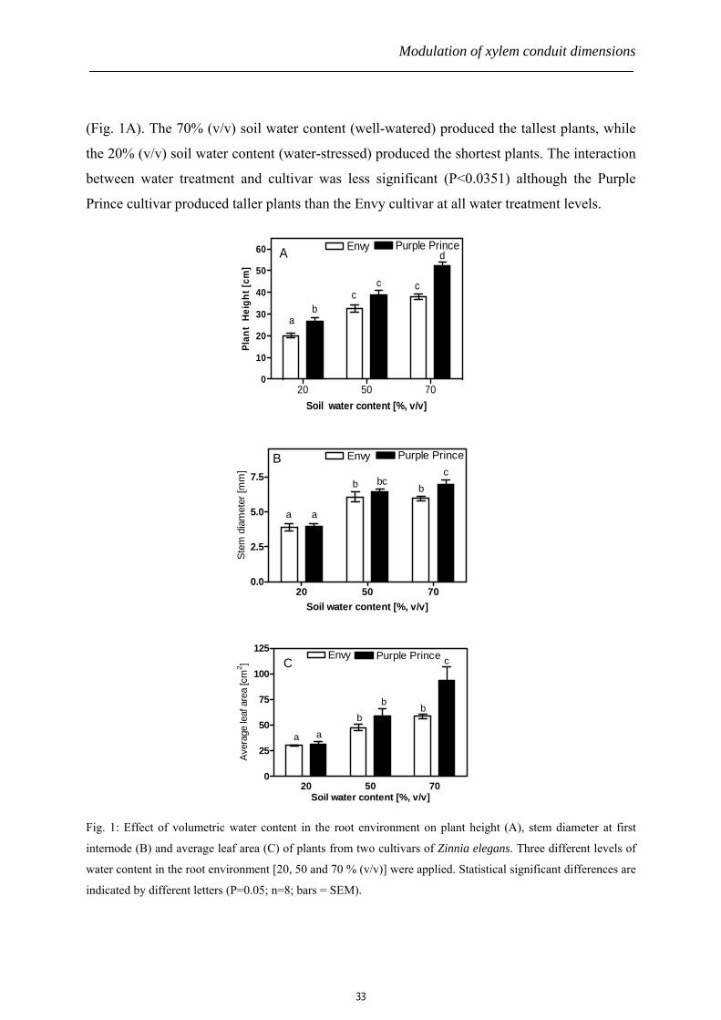
(Fig. 1A). The 70% (v/v) soil water content (well-watered) produced the tallest plants, while
the 20% (v/v) soil water content (water-stressed) produced the shortest plants. The interaction
between water treatment and cultivar was less significant (P<0.0351) although the Purple
Prince cultivar produced taller plants than the Envy cultivar at all water treatment levels.
20 50 700
10
20
30
40
50
60 Envy Purple PrinceA
ab
cc c
d
Soil water content [%, v/v]
Plan
t H
eigh
t [cm
]
20 50 700.0
2.5
5.0
7.5
Envy Purple PrinceB
a a
b bcb
c
Soil water content [%, v/v]
Stem
dia
met
er [m
m]
20 50 700
25
50
75
100
125 Envy Purple Prince
a a
bb b
cC
Soil water content [%, v/v]
Aver
age
leaf
are
a [c
m2 ]
Fig. 1: Effect of volumetric water content in the root environment on plant height (A), stem diameter at first
internode (B) and average leaf area (C) of plants from two cultivars of Zinnia elegans. Three different levels of
water content in the root environment [20, 50 and 70 % (v/v)] were applied. Statistical significant differences are
indicated by different letters (P=0.05; n=8; bars = SEM).
33

Chapter 2
The stem diameter at the first internode was also statistically different especially between
20% and 70% (v/v) water treatments in both cultivars (Fig. 1B). The 50% and 70% (v/v) soil
water contents, however, achieved similar responses in the stem diameter in both cultivars.
We also observed significantly larger leaf area in well- and moderate-watered plants as
compared with the water-stressed plants from both Zinnia cultivars (Fig. 1C).
Xylem functioning
The hydraulic conductivity (Kh) in the lower part of cut flower stems differed
significantly between different watering conditions. The Kh was lowest in the water-stressed
plants and highest in the well-watered plants. By increasing soil water content from 20% to
70% (v/v) during growth, a 96% and a 100% rise in stem water transport in plants from Envy
and Purple Prince cultivars respectively were achieved (Fig. 2). Thus, the results show a
positive correlation between level of water content in the root environment and the Kh. This
correlation was conserved in both Zinnia cultivars, although Purple Prince recorded higher Kh
at 70% water content as compared to the Envy cultivar.
20 50 700.0
0.5
1.0
1.5
2.0
2.5Envy Purple Prince
a
a ab ab b
c
Soil water content [%, v/v]
Kh
[mgs
-1kP
a-1cm
]
Fig. 2: Effect of different volumetric water content [20, 50 and 70% (v/v)] in the root environment on the
hydraulic conductivity in stems of two Zinnia elegans cultivars. Statistical significant differences are indicated
by different letters (P=0.05; n=8; bars = SEM).
Xylem anatomy
In general, the number of xylem vessels at the cut surface of the stem cross sections at
first internode did not differ significantly from the three water treatments. In addition, the
Zinnia cultivar type did not affect the vessel number (Fig. 3A). Calculation of the xylem
vessel density or ‘wood’ density at first internode showed a large increase (87% in Envy and
34

Modulation of xylem conduit dimensions
133% in Purple Prince) in the water-stressed plants as compared to the well-watered plants
(fig. 3B). The vessel density in moderate- and well-watered plants, however, did not differ
statistically in either of the cultivars.
2000
20 50 700
25
50
75
b bb
b
Soil water content [%, v/v]Xy
lem
ves
sel d
ensi
t [v
esse
ls.m
m-2
]y
100B a
a Envy Purple Prince
20 50 700
1000
Envy Purple PrinceA
aa
aa
aa
Soil water content [%, v/v]
Num
ber o
f ves
sels
per
stem
cro
ss s
ectio
n
Fig. 3: Graphs showing the vessel number (A) and the mean vessel density (B) at the cut surface of stems at first
internode from two Zinnia elegans cultivars grown with three different water availability levels in the soil (20,
50 and 70%, v/v). Statistical significant differences are indicated by different letters (P=0.05; n=8; bars = SEM).
The maximum vessel length (Lmax) and the vessel half-length or median length (τ)
were not significantly different between the three different soil water treatments in the Purple
Prince cultivar. In the Envy cultivar, however, both Lmax and τ of water-stressed plants were
significantly lower as compared to the moderate-watered (Lmax, P=0.0069; τ, P=0.0024) and
well-watered (Lmax, P=0.0325; τ, P=0.0060) plants (Fig. 4A and B). No difference was found
in Lmax and τ between plants from Envy cultivar grown with moderate- and well-watered
plants (Lmax, P=0.7791; τ, P=0.6500).
20 50 700
10
20
30
40Envy Purple PrinceB
aa
b
aca
bc
Soil water content [%, v/v]
τ [m
m]
20 50 700
100
200
300Envy Purple PrinceA
a ab
ab acbc
Soil water content [%, v/v]
L max
(mm
)
Fig. 4: Effect of different volumetric water contents [20, 50 and 70% (v/v)] in the root environment on: A,
maximum xylem vessel length (Lmax); and B, xylem vessel half- or median length (τ) in stems of two Zinnia
elegans cultivars. Statistical significant differences are indicated by different letters (P=0.05; n=8; bars = SEM).
35

Chapter 2
The xylem vessel diameter distribution analysis indicated a shift of vessels towards a
larger vessel diameter class with increasing levels of soil water content around the root
environment. In both cultivars, the well-watered plants produced the highest proportion of
larger vessel diameter classes. Thus, the vessel diameter distribution skews to the right (larger
diameter classes) in the well-watered plants while it skews to the left (smaller diameter
classes) in the water-stressed plants (Fig. 5).
-56-1
011
-1516
-2021
-2526
-3031
-3536
-4041
-4546
-5051
-5556
-600
10
20
3070%20% 50%Purple Prince
A
Diameter class (µm)
perc
enta
ge o
f ves
sels
[%]
>5 6-10
11-15
16-20
21-25
26-30
31-35
36-40
41-45
46-50
51-55
56-60
61-65
66-70
0
10
20
3070%50%20%Envy
B
Diameter class [µm]
Perc
enta
ge o
f ves
sels
[%]
Fig. 5: Effect of different volumetric water contents [20, 50 and 70% (v/v)] in the root environment on the xylem
vessel diameter distribution in stems of two Zinnia elegans cultivars, Purple Prince (A) and Envy (B). (n=8
stems; bars = SEM).
Plants from all levels of water treatments, and in both cultivars, produced a mixture of small
and large xylem vessels, with a lumen diameter ranging from 5 to 65 µm. However, there was
36

Modulation of xylem conduit dimensions
a clear difference in the proportion of each of these vessel diameter classes per plant per
treatment. The mean vessel diameter decreased with increasing drought in the root
environment, resulting in larger mean vessel diameters in these cut flower plants at 70% (v/v)
soil water content compared with those at 20% (v/v) soil water content in both cultivars (Fig.
6).
20 50 700
10
20
30
40
ab c
cd c
Envy Purple Prince
Soil water content [%, v/v]
Mea
n ve
ssel
dia
met
er [µ
m]
Fig. 6: The effect of different soil water contents around the root environment on the mean diameters of the
xylem vessels from two cultivars of Zinnia elegans. Statistical significant differences are indicated by different
letters (P=0.05; n=8; bars = SEM).
Vase life
Generally, the cut flowers from both Zinnia elegans cultivars produced from
moderate- and well-watered plants did not fully regain their original water uptake during the
early phase of their vase life (Fig. 7). This resulted in severe leaf wilting and associated loss
of the entire ornamental value within 1-2 days. Water-stressed cut flowers from both Envy
and Purple Prince regained water uptake and increased fresh weight during vase life. Envy cut
flowers from the three water treatments showed statistical differences between each other. In
Purple Prince, however, the cut flowers from the moderate- and well-watered plants recorded
no differences in the vase life. Analysis of the vase life also showed no significant interaction
between cultivar and water treatment. Although statistically less significant, the water-
stressed Envy cut flowers performed better in the vase than the Purple Prince counterparts.
The reverse of this was observed in cut flowers from the 70% soil water content.
37

Chapter 2
0 25 50 7580
90
100
110
120
E70%
E50%PP20
P70%
E20%
P50%
Vase period [h]
Fres
h w
eigh
t [%
]
Fig. 7: Time course of fresh weight changes of Zinnia elegans cut flowers from two cultivars, Envy (open
symbols) and Purple Prince (closed symbols), during vase life after 1h desiccation. Cut flowers were grown at
three different water availability levels around the root environments. E=Envy, P=Purple Prince. (n=4; bars =
SEM).
Discussion
The morphological analyses of the cut flowers from both Zinnia elegans cultivars
showed large differences between the water-stressed and moderate- or well-watered plants
(Fig. 1 and 2). The shorter plant height, thin stems and smaller leaf area of the Zinnia elegans
cut flowers caused by the water-stressed conditions in the root environment show a long-term
regulatory mechanism for water balance in the plant. A short-term regulation of such water
balance is seen in, for example, stomatal closure. These mechanisms regulate water flow in
the soil-plant-atmosphere continuum through lowering of the stomatal conductivity during
transpiration to maintain an adequate amount of water for the plant’s biological processes
(Moreshet et al., 1990; Meinzer et al., 1996; Brodribb and Holbrook, 2005). In our study, we
found no statistical differences neither in the number of xylem vessels per cross sectional area
of the cut flower stems between the three different water treatments used, nor between the
cultivars. Generally, the maximum vessel length (Lmax) and the vessel median length (τ) did
not differ between water treatments in both Zinnia elegans cultivars (Fig. 4). The only
exception was in the Envy cultivar where differences were found in Lmax and τ between the
38

Modulation of xylem conduit dimensions
water-stressed and moderate- or well-watered plants. Despite the insignificant effect on the
vessel number and slight effect on the xylem vessel length caused by the water treatment, the
hydraulic conductivity of stems from both Zinnia cultivars increased by 100% when soil
water content increased from 20% v/v (water-stressed) to 70% v/v (well-watered) (Fig. 2).
These large differences in the hydraulic conductivity between the water-stressed and well-
watered plants could, therefore, not be attributed to either the number of the vessels per stem
section or the length of the xylem vessel since these dimensions were generally unaffected by
the water treatments.
Further xylem anatomical examinations of the stems showed relatively higher
proportion of vessels with larger lumens in the well-watered cut flower stems as compared to
the water-stressed cut flower stems from both cultivars (Fig. 5). Since hydraulic conductivity
of a conduit is proportional to the fourth power of the lumen diameter according to the Hagen-
Poiseuille’s equation (Aloni and Zimmermann, 1983; Tyree, 2003), the xylem vessel diameter
is an important parameter in regulating the plant’s water status. Thus, the proportion of large-
diameter vessels is most important in determining the water uptake efficiency of the plant. By
calculating the theoretical conductance (Σd4), a clear indication of the greater contribution of
the larger vessels to the total hydraulic conductance in the stem could be recognized (Choat et
al., 2005). Although longer vessels are efficient in both horizontal (intra-vessel) and vertical
(inter-vessel) water transport due to the reduction in the high interconduit flow resistance in
the pit membrane (Lancashire and Ennos, 2002; Choat et al., 2006), shorter and narrower
vessels are more efficient in dealing with embolism especially caused by harsher
environments such as drought. Thus, longer vessels once embolized can transport the air
column over longer distances that might affect many other neighbouring vessels (Comstock
and Sperry, 2000). Therefore by choosing wider vessels and maintaining vessel length, plants
can increase water transport capacity and at the same time manage spread of embolism. In a
theoretical calculation based on the Hagen-Poiseuille’s equation, a 1.4× increment in lumen
diameter of a conduit will cause approximately 4× increase in water flow capacity. It is
therefore not surprising that in our results, although with minor change in vessel length, the
large change in vessel diameter induced an effective change in hydraulic conductivity of the
Zinnia elegans stems produced with different water stress conditions.
It has long been proposed that there exists a positive correlation between angiosperm
xylem vessel length and diameter (Zimmermann and Jeje, 1981). This assumption is, in part,
39

Chapter 2
in agreement with our observations in the Envy cultivar which showed positive correlations
between the vessel diameter and vessel length in response to the different soil water contents.
However, we also observed no correlation between the vessel length and diameter in the
Purple Prince cultivar, implying that the length-to-diameter relationship in the xylem conduit
is not constant. An independent regulation of the vessel length and vessel diameter by sets of
different environmental conditions or different genetic backgrounds may be responsible for
this non-linear length-to-diameter relationship in xylem vessels from plants of different
species or the same species grown under different environmental conditions. Our results also
confirmed the inverse relationship between water content in the soil and the xylem vessel (or
wood) density (Searson et al., 2004). Vessel density increased by between 80 and 130% in the
water-stressed plants from both Zinnia cultivars as compared to the well-watered plants.
The vase life of cut flowers varies greatly from species to species. However, in almost
all species, the vase life of cut flowers basically depends on the water balance of the cut
flower. Therefore the vase life is influenced by factors determining water transport as well as
transpiration (performance of the stomata). In our experiments, we found a negative
correlation between the vase life and the proportion of large-lumen vessels in the cut flower
stems. The water-stressed cut flowers from both Zinnia elegans cultivars (having low density
large-lumen vessels) achieved longer vase life compared to the well-watered and moderate-
watered cut flowers (Fig. 5 and 6). The well-watered and moderate-watered cut flowers did
not recover from the 5% initial water loss during the first 24-h of vase life with a greater loss
in fresh weight followed by severe leaf wilting. The water-stressed plants did recover faster
from the initial water loss and also maintained the initial ornamental quality over several
days. Since the well-watered and moderate-watered cut flowers recorded higher hydraulic
conductivity compared with the water-stressed plant, conductance may not be responsible for
the shorter vase life. Such differences in the vase life between the water-stressed and the well-
watered cut flowers may be due to the poor recovery of larger vessels from embolism (not
measured) that was induced by the 5% weight loss at the beginning of the vase life
experiment. In general, severe water stress may cause cavitations and air embolism (Schultz
and Matthews, 1988) resulting in reduced function of the xylem in transporting water. It has
been previously observed that large-diameter vessels could not remove the emboli during the
vase life of Chrysanthemum cut flowers (van Ieperen et al., 2002). Thus, by increasing the
proportion of the large-diameter vessels in the stem of cut flowers, the vase life is negatively
40

Modulation of xylem conduit dimensions
affected as these types of vessels are most vulnerable to embolism.
Although the actual mechanism behind the control of the xylem conduit diameter is
not clearly understood, it has been proposed that phytohormones, whether up-regulated or
down-regulated, affect the dimensions of the xylem vessels. How these regulations of the
genes are affected by water stress is also not clearly understood. It has been reported that
water stress increases the production of abscisic acid (ABA) and the degradation of cytokinin
(Itai and Vaadia, 1971; Hare et al., 1997). Since ABA has been shown not to have an effect on
xylem hydraulic conductivity (Lovisolo et al., 2002; Aasamaa et al., 2004), the negative effect
of water stress on hydraulic conductivity in Zinnia may be caused by lower cytokinin levels.
Another candidate phytohormone in xylem anatomical regulation is auxin. Auxin has long
been shown to be involved in the differentiation of the xylem cells (Baum et al., 1991; Aloni,
1992; Paponov et al., 2005). It has long been proposed that the increase in the xylem conduit
size from distal to basal regions of the stem is due to 1) the length of basipetal transport of
auxin from the leaves, and 2) the auxin concentration gradient (Aloni and Zimmermann,
1983; Tuominen et al., 1997; Uggla et al., 1998). The longer it takes for auxin to arrive at the
site of xylem differentiation, combined with a possible decrease in auxin concentrations in the
lower regions of the plant, the larger the size and lower the density of the xylem vessels that
are formed. Thus, any effect of the amount of water in the root environment on the auxin
production and transport may indirectly affect the dimensions of the xylem vessels. Brandl
and Lindow (1997) reported an enhanced expression of ipdC, a plant inducible gene involved
in indoleacetic acid (IAA) biosynthesis in Erwinia herbicola on bean plants (Phaseolus
vulgaris cv. Bush Blue Lake 274) growing with low water availability. Such enhancement of
auxin production by water-stress conditions supports our observation of smaller vessels in the
water-stressed Zinnia plants. Direct measurement of the effect of water-stress on the auxin
production and transport in the Zinnia plant tissues would offer more information about the
control of the xylem vessel dimensions during differentiation. Application of this information
and use of molecular tools, such as over-expressing auxin genes in whole plants in
combination with different soil water contents may lead to knowledge needed for production
of relatively larger plants with well controlled xylem dimensions necessary for high
ornamental quality.
We conclude from this study that different levels of soil water content affect
differently the xylem anatomy and function. Water stress affects Zinnia cut flowers and
41

Chapter 2
results in smaller plants, narrower xylem vessels, and lower stem hydraulic conductivity. In
general, the increase in stem hydraulic conductivity by high soil water content was caused by
the increase in xylem vessel diameter according to the Poiseuille equation. It is also evident
from this study that xylem vessel diameter and length are independently regulated by different
genetic and environmental conditions. About the vase performance of the cut flowers, we
found that water-stressed plants containing narrower vessels are superior to well-watered
plants with larger vessels. The vulnerability of these larger vessels to embolism is responsible
for this shorter vase life in the well-watered cut flowers from both Zinnia cultivars.
References
Aasamaa, K., Sober, A., Hartung, W., and Niinemets, U. (2004). Drought acclimation of
two deciduous tree species of different layers in a temperate forest canopy. Trees-Struct Funct
18, 93-101.
Aloni, R. (1992). The control of vascular differentiation. Int J Plant Sci 153, S90-S92.
Aloni, R., and Zimmermann, M.H. (1983). The control of vessel size and density along the
plant axis - a new hypothesis. Differentiation 24, 203-208.
Bailey, I.W. ( 1953). Evolution of the tracheary tissue of land plants. Am J Bot 40, 4±8.
Baum, S.F., Aloni, R., and Peterson, C.A. (1991). Role of cytokinin in vessel regeneration
in wounded Coleus internodes. Ann Bot-London 67, 543-548.
Brandl, M.T., and Lindow, S.E. (1997). Environmental signals modulate the expression of
an indole-3-acetic acid biosynthetic gene in Erwinia herbicola. Mol. Plant-Microbe Interact.
10, 499-505.
Brodribb, T.J., and Holbrook, N.M. (2005). Water stress deforms tracheids peripheral to
the leaf vein of a tropical conifer. Plant Physiol 137, 1139-1146.
Choat, B., Brodie, T.W., Cobb, A.R., Zwieniecki, M.A., and Holbrook, N.M. (2006).
Direct measurements of intervessel pit membrane hydraulic resistance in two angiosperm tree
species. Am. J. Bot. 93, 993-1000.
Choat, B.B., Ball, M.M.C., Luly, J.J.G., and Holtum, J.J.A.M. (2005). Hydraulic
architecture of deciduous and evergreen dry rainforest tree species from north-eastern
Australia. Trees 19, 305-311.
Comstock, J.P., and Sperry, J.S. (2000). Theoretical considerations of optimal conduit
length for water transport in vascular plants. New Phytologist 148, 195-218.
42

Modulation of xylem conduit dimensions
Ellerby, D.J., and Ennos, A.R. (1998). Resistances to fluid flow of model xylem vessels
with simple and scalariform perforation plates. J Exp Bot 49, 979-985.
Hare, P.D., Cress, W.A., and van Staden, J. (1997). The involvement of cytokinins in plant
responses to environmental stress. Plant Growth Regul 23, 79-103.
Itai, C., and Vaadia, Y. (1971). Cytokinin activity in water-stressed shoots. Plant Physiol 47,
87-90.
Kozela, C., and Regan, S. (2003). How plants make tubes. Trends Plant Sci 8, 159-164.
Lancashire, J.J.R., and Ennos, A.A.R. (2002). Modelling the hydrodynamic resistance of
bordered pits. J Exp Bot 53, 1485-1493.
Lovisolo, C., and Schubert, A. (1998). Effects of water stress on vessel size and xylem
hydraulic conductivity in Vitis vinifera L. J Exp Bot 49, 693-700.
Lovisolo, C., Schubert, A., and Sorce, C. (2002). Are xylem radial development and
hydraulic conductivity in downwardly-growing grapevine shoots influenced by perturbed
auxin metabolism? New Phytol 156, 65-74.
Meinzer, F.F.C., Fownes, J.J.H., and Harrington, R.R.A. (1996). Growth indices and
stomatal control of transpiration in Acacia koa stands planted at different densities. Tree
Physiol 16, 607-615.
Moreshet, S.S., Cohen, Y.Y., Green, G.G.C., and Fuchs, M.M. (1990). The partitioning of
hydraulic conductances within mature orange trees. J Exp Bot 41, 833-839.
Nijsse, J., van der Heijden, G.W.A.M., van Ieperen, W., Keijzer, C.J., and van
Meeteren, U. (2001). Xylem hydraulic conductivity related to conduit dimensions along
chrysanthemum stems. J Exp Bot 52, 319-327.
Paponov, I.A., Teale, W.D., Trebar, M., Blilou, I., and Palme, K. (2005). The PIN auxin
efflux facilitators: evolutionary and functional perspectives. Trends Plant Sci 10, 170-177.
Pickard, W.F. (1981). The ascent of sap in plants. Progress in Biophysics and Molecular
Biology 37, 181-229.
Sahin, A., and Abokhodair, A. (1998). Geostatistical approach in design of sampling
patterns for Jabal-Sayid sulfide deposit, Western Saudi-Arabia. Journal of African Earth
Sciences and the Middle East 8, 40-42.
Schultz, H.R., and Matthews, M.A. (1988). Resistance to water transport in shoots of Vitis
vinifera L. Plant Physiol. 88, 718-724.
Searson, M.J., Thomas, D.S., Montagu, K.D., and Conroy, J.P. (2004). Wood density and
43

Chapter 2
anatomy of water-limited eucalypts. Tree Physiol 24, 1295-1302.
Sieburth, L.E., and Deyholos, M.K. (2006). Vascular development: the long and winding
road. Curr Opin Plant Biol 9, 48-54.
Tuominen, H., Puech, L., Fink, S., and Sundberg, B. (1997). A radial concentration
gradient of indole-3-acetic acid is related to secondary xylem development in hybrid Aspen.
Plant Physiol 115, 577-585.
Tyree, M.T. (2003). Plant hydraulics: the ascent of water. Nature 423, 923.
Uggla, C., Mellerowicz, E.J., and Sundberg, B. (1998). Indole-3-acetic acid controls
cambial growth in scots pine by positional signaling. Plant Physiol 117, 113-121.
Van Doorn, W.G. (1997). Water relations of cut flowers. Horticultural-Reviews. 18, 1-85.
van Ieperen, W., van Meeteren, U., and Nijsse, J. (2002). Embolism repair in cut flower
stems: a physical approach. Postharvest Biol Tec 25, 1-14.
Van Ieperen, W., Volkov, V.S., and Van Meeteren, U. (2003). Distribution of xylem
hydraulic resistance in fruiting truss of tomato influenced by water stress. J Exp Bot 54, 317-
324.
van Meeteren, U. (1992). Role of air embolism and low water temperature in water balance
of cut chrysanthemum flowers. Sci Hortic-Amsterdam 51, 275-284.
Vogt, U.K. (2001). Hydraulic vulnerability, vessel refilling, and seasonal courses of stem
water potential of Sorbus aucuparia L. and Sambucus nigra L. J Exp Bot 52, 1527-1536.
Zimmermann, M.H., and Jeje, A.A. (1981). Vessel-length distribution of some American
woody plants Can J Bot 59, 1882-1892.
44

Chapter 3
Effect of temperature on xylem vessel and vessel element lengths in Zinnia
elegans stems
P. Twumasi1, 2, W. van Ieperen1, J. Oosterkamp1, J. H. N. Schel2 and O. van Kooten1
(2007). Effect of temperature on xylem vessel and vessel element lengths in Zinnia elegans
stems
1Department of Plant Sciences, Horticultural Production Chains Group, Wageningen
University, Marijkeweg 22, 6709 PG Wageningen, The Netherlands 2Department of Plant Sciences, Laboratory of Plant Cell Biology, Wageningen University,
Arboretumlaan 4, 6703 BD Wageningen, The Netherlands

Chapter 3
Abstract
The hydraulic performance of the xylem system is influenced by the properties of the
vessel elements such as the length and the diameter. Although it has been shown that certain
factors, e.g. growth conditions, influence the length of vessels in the plant, it has not been
proven whether longer vessels result from cell elongation or higher rate of vessel elements
fusion. In this study, we investigated the effect of temperature on whole vessel length and the
individual vessel element length in the stems of two Zinnia elegans cultivars, ‘Purple Prince’
and ‘Envy’. An increase in the average daily temperature (ADT) during growth correlated
positively with the whole plant length, the whole vessel length and the length of individual
vessel elements in both Zinnia cultivars. The temperature effect on these characteristics was,
however, more pronounced in the cv Purple Prince as compared to the cv Envy. Differential
day and night temperature during growth (DIF) influenced plant height, but vessel length
remained unaffected. The percentile change in the total vessel length by temperature was
significantly higher as compared to the percentile change in length of the individual vessel
members caused by same effect. It can therefore be deduced from these results that, although
the individual vessel elements increase in length when the whole vessel length is increased by
temperature, a greater contribution to this increase in length is caused by an increased rate of
fusion of the individual vessel elements.
Keywords: Average daily temperature, differential day and night temperature, vessel
element, xylogenesis, Zinnia elegans
Abbreviations: ADT, average daily temperature; DIC, differential interference contrast; DIF,
difference in day and night temperature; DT, daytime temperature; Lmax, maximum vessel
length; τ, median or half-length of vessels (length at which 50% of the vessels terminate); NT,
nighttime temperature
Introduction
In vascular plants, long distance water transport from roots to leaves occurs via xylem
vessels: capillary tubes composed of many vessel elements, stacked in series. The individual
vessel elements within a xylem vessel have perforated end walls, which can be completely
perforated or scalariform. At both ends of a vessel, the outer vessel elements lack a perforated
46

Effect of temperature on xylem vessels
end wall and vessels are often tapered. Although vessels can be rather long (up to several
meters in tall tree’s (e.g. Quercus rubra) and subtropical Lianas (Zimmermann and Jeje,
1981; Ewers and Fisher, 1989b) they usually have a length that is much shorter than the total
length of the water transport path. Consequently, water has to move between subsequent
vessels during its way up from roots to transpiring leaves. This lateral vessel-to-vessel water
transport occurs at places where distal and proximal ends of subsequent vessels run side by
side and are interconnected via bordered pits.
Hydraulic conductance of xylem in plants depends on several factors, among which
are the number of functional vessels, the structural network design of the interconnected
vessels, and the morphology of individual vessel elements and whole vessels (Dimond, 1966;
Comstock and Sperry, 2000). Besides these characteristics, hydraulic efficiency is also
affected by the nature of perforation plates between vessel elements within a vessel (Ellerby
and Ennos, 1998; Zwieniecki et al., 2001) and by the number and hydraulic properties of
bordered pits, which determine the resistance for vessel-to-vessel water transport (van
Ieperen et al., 2000; Zwieniecki et al., 2001; Hacke et al., 2006).
One of the most important determinants of variation in hydraulic conductance in
xylem has been the dependence of vessel lumen resistance on the fourth power of vessel
radius (Tyree and Zimmermann, 2002). Extensive earlier work on xylem anatomical studies
in different plant species has shown a correlation between hydraulic conductance and xylem
vessel dimensions, in particular number and diameter (Lovisolo and Schubert, 1998; Nijsse et
al., 2001; Solla and Gil, 2002; Twumasi et al., 2005). However, the relationship between
vessel radius and xylem hydraulic resistance is often less clear. Even measurements on single
vessels with no vessel endings have given variable results (Giordano et al., 1978), possibly
because slight deviations of xylem vessel radius from the ideal capillary radius will be
magnified at least 4-fold in calculated hydraulic resistance. In water conducting xylem tissue
as a whole, also vessel length and bordered pit resistance appears to have strong effects on
overall hydraulic conductance (Comstock and Sperry, 2000; Zwieniecki et al., 2001). The
exact contribution of vessel length to overall xylem hydraulic resistance is complicated but
expected to be important: long vessels decrease the relative contribution of vessel-to-vessel
transport through bordered pits to overall xylem hydraulic resistance (Comstock and Sperry,
2000; van Ieperen et al., 2000; Nijsse, 2001). Unfortunately, evaluation of the importance of
vessel length for overall hydraulic conductance is scarce, due to technical barriers and
47

Chapter 3
especially the laborious methods to determine vessel length distributions (Comstock and
Sperry, 2000). It is virtually impossible to measure the length of xylem vessels directly
because of the enormous length, ranging from a few centimeters in small plants, such as
Arabidopsis thaliana, to several meters in tropical trees. However, whether long or short, the
xylem vessels possess microscopic diameters. All present methods to measure vessel length
are indirect and based on determining vessel length distributions by counting the number of
continuous vessels at varying lengths of a stem segment (Skene and Balodis, 1968;
Zimmermann and Jeje, 1981; Ewers and Fisher, 1989a; Nijsse, 2001; Tyree and
Zimmermann, 2002; Nijsse, 2004). Environmental effects on vessel length distributions have
hardly been topic of research, probably due to difficulties to measure their sizes and shapes
(Comstock and Sperry, 2000). In Acer rubrum early- and latewood vessels have different
length distributions (Zimmermann and Potter, 1982). In Zinnia elegans reduced water
availability during growth changed vessel length distributions towards shorter vessels
(Chapter 2, this thesis).
Since it is obvious that besides vessel diameter also lateral flow resistance (governed
by bordered pits at vessel-to-vessel connections) importantly influences overall xylem
hydraulic conductance, vessel length distributions can be expected to be an important factor
determining the overall efficiency for water transport in a fully functional xylem system.
The aim of this work was to investigate the effect of temperature on vessel length
distribution in stems of two Zinnia elegans cultivars, which differ in general growth responses
to environmental conditions (Twumasi et al., 2005). Effects of average daily temperatures
(ADT’s) as well as thermoperiodicity (different day and night temperatures at similar average
temperature (DIF)) on plant size and vessel length distributions are investigated, as well as the
effect of temperature on vessel element size. DIF is calculated as difference between daytime
temperature (DT) and nighttime temperature (NT), i.e., DIF=DT-NT. DIF treatments are of
particular interest because different DIF-treatments (positive or negative) can influence plant
size at similar ADT (Langton and Cockshull, 1997).
Materials and methods
Plant material
Seedlings of two cultivars of Zinnia elegans, “Purple Prince” and “Envy”, were
generated from seeds sown in 40x60 cm trays with commercial potting soil (Lentse potgrond
48

Effect of temperature on xylem vessels
nr 4; 85% peat, 15% clay) in the Unifarm greenhouse of Wageningen University and
Research Centre, The Netherlands. The seedlings were raised under natural light conditions
(light screen above 200 µmol m-2 s-1 photosynthetic active radiation), 22/18 oC (Day/Night)
temperature, 70% Relative Humidity (setpoints) and ambient CO2 (350 ppm)). After two
weeks, seedlings of homogeneous characteristics were selected, transplanted to 13 cm
diameter pots, and moved to one of three artificially lit growth chambers (17 /21 /25oC with
equal VPD’s (0.55 kPa) and ambient CO2). Light intensity was approximately 200 µmol m-2
s-1, supplied by a mixture of SON-T and High Pressure Sodium lamps for a period of 12h per
day (light on at 8:00am). Plants were harvested for measurements after flowers were fully
opened.
Temperature treatments
Nine different temperature treatments were applied: 3 constant temperatures (17, 21
and 25oC) and 6 differential day and night temperatures (DIFs) in all possible combinations of
17, 21 and 25oC). These combinations resulted in 5 different average daily temperatures
(ADT: 17,19,21,23 and 25oC). The DIF treatments were applied by moving the plants two
times each day (8:00 and 20:00) according to their day- and night temperature combinations.
The whole experiment was replicated in time. 12 plants were grown per temperature treatment
x cultivar combination in two trays of 6 pots each, which did not shadow each other. After
each plant movement (twice a day) all trays (including the constant temperature treatments)
were randomly repositioned within their growth chamber. The extreme ADT treatments (17
and 25oC) were replicated again (triplicate) to collect material for length measurements on
individual vessel elements. Watering the plants once a day was enough to maintain adequate
level of soil water.
Plant measurements
After harvest, total stem length, number of internodes and individual lengths of all
internodes and of the flower stalk (above last leaf pair) were measured in 10 plants per
cultivar x temperature treatment combination. Per stem total length of all internodes (except
flower stalk) was calculated from the individual internode lengths and average internode
length from the total length of all internodes and number of internodes per individual plant.
49

Chapter 3
Vessel length distribution measurements
Vessel length distributions were measured in five of the applied temperature
treatments (17/17, 17/25, 21/21, 25/17, and 25/25oC (DT/NT)). Six randomly chosen plants
per cultivar x temperature treatment were used for determination of their vessel length
distribution using the latex paint particle method (as described by Nijsse, 2001). Stems (35-40
cm long) were cut under water from fully hydrated plants with the distal cut through the
middle of the second internode. The stem segments were connected with their proximal cut
end to a pressure system and forced to take up a latex paint solution via their distal cut end for
approximately 24h at a pulling pressure of 50-100 kPa. Latex paint suspension fills xylem
vessels from their open cut-end inwards, but cannot pass bordered pit membranes at vessel-to-
vessel connections. Consequently, only vessels that are continuously open from the cut-end
fill with colored paint particles. Latex paint never reached the upper cut surface of a stem
segment, indicating that all stem segments were longer than the maximal vessel length. After
impregnation with latex paint the stem segments were repeatedly cut back with a razor blade
from the distal cut-end upwards, in 5 mm increments while the number of paint filled vessels
per cross-sectional cut was counted using a binocular microscope (Leica MZ8). As previously
observed in many plant species (Nijsse, 2004), also in Zinnia elegans stem, the relation
between the distance from the distal cut surface and number of colored vessels follows an
exponential decay. The vessel length distribution was deduced from this decay and described
by the half-length or median length (τ; 50% of the vessels are shorter than this length) and the
maximal vessel length (Lmax) according to Nijsse (2004).
Determination of individual vessel element length
Individual vessel element length was determined from the 3rd internode of 4 randomly
chosen plants per cultivar which were grown at continuous 17 oC or 25oC (daytime- (DT) and
nighttime (NT) temperature). These two temperature treatments were selected from all
applied temperature treatments because they showed the most distinct difference in stem
length and vessel length distribution. Three stem sections of 1 cm length were cut from the
base, middle and top locations of the internode. The stem pieces were each dissected
longitudinally into two halves and fixed overnight in 4% paraformaldehyde. They were
dehydrated through an ethanol series, and then embedded in Technovit 7100 resin (Kuroiwa
et al., 1990). The blocks were trimmed and sectioned longitudinally at 5-10 µm with
50

Effect of temperature on xylem vessels
disposable glass blades prepared with LKB 2078 Histoknifemaker (LKB-Produkter AB,
Bromma, Sweden) on a rotary microtome (Reichert-Jung 2050, Germany). The thin sections
were stretched on degassed water at 35 oC and transferred onto glass slides. Drying and
sticking of the sections to the glass slides was achieved by baking for 2 min on a hot plate at
80 oC. The sections on the slides were stained with 1% toluidine blue for 3 min, rinsed in
distilled water and enclosed in 99% glycerol.
The sections were imaged for the xylem vessels elements with 10x or 20x DIC (differential
interference contrast) objectives on an inverted microscope (Axiovert 200, Carl Zeiss)
equipped with a Zeiss AxioCam HR digital camera controlled by Axiovision digital imaging
software. The images were analyzed with Image J software (Wayne Rasband National
Institutes of Health, USA). In each cultivar x treatment combination more than 300 vessel
elements were analyzed.
Statistical design and analysis
The experimental setup was a complete randomized block design with 2 or 3 (only 17
and 25oC ADT) replications in time. Each replication consisted of 12 plants per
treatment/cultivar combination. 12 plants per treatment × cultivar were analyzed for whole
plant characteristics and xylem vessel length distributions. 4 plants per treatment × cultivar
were analyzed for length of the individual vessel elements. Over 250 individual vessel
elements were analyzed per treatment × cultivar combination. Analyses of variance
(ANOVA) were conducted and treatment and cultivar effects tested at 5% probability level
using the statistical package Genstat 8 (IACR, Rothamsted, UK).
Results
Morphological characteristics and plant development rate The temperature during growth influenced plant development rate: plants grown at
17oC average daily temperature (ADT) were harvested one week later than plants that were
grown at 25oC ADT. Plants were harvested once the flowers had fully opened, and this period
was influenced by both ADT and DIF. Total growth duration was 60 days at 17oC ADT in
both cultivars. Plant size (stem length including flower stalk) was clearly influenced by
temperature treatments and cultivar type. Both cultivars reacted qualitatively similar on all
temperature treatments (Fig.1), but cv Purple Prince was always larger (P<0.001).
51

Chapter 3
40
4550
5560
6570
7580
85
15 17 19 21 23 25 27
Average Day Temperature [oC]
Plan
t Len
gth
[cm
]
-8
-4
0
4
8
DIFA
40
4550
55
6065
70
7580
85
15 17 19 21 23 25 27
Average Day Temperature [oC]
Plan
t Len
gth
[cm
]
-8
-4
0
4
8
DIFB
C
0
10
20
30
40
50
6070
80
90
-10 -5 0 5 10DIF [oC]
Plan
t Len
ght [
cm]
Envy
Purple Prince
Fig. 1: Effect of ADT and DIF temperature-treatments on final plant size in two Zinnia elegans cultivars: Envy
(A) and Purple Prince (B). The effect of DIF on plant length in the two Zinnia cultivars at an average
temperature of 21 oC is shown in detail in C. Points represent averages and bars indicate the standard error of
mean.
Plant length increased with ADT between 17 and 25oC (P<0.001). Opposite DIF treatments
resulted in different plant length at 4 and 8oC absolute DIF (P<0.05). Treatment effects on
total plant length were caused by qualitatively similar effects on total stem and flower stalk
length (above the last leaf pair). In general, the number of internodes was higher and the
average internode length larger in cv Purple Prince as compared with cv Envy. The number of
52
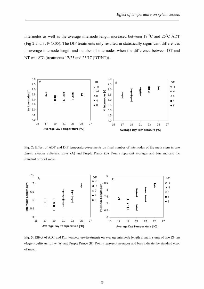
Effect of temperature on xylem vessels
internodes as well as the average internode length increased between 17 oC and 25oC ADT
(Fig 2 and 3; P<0.05). The DIF treatments only resulted in statistically significant differences
in average internode length and number of internodes when the difference between DT and
NT was 8oC (treatments 17/25 and 25/17 (DT/NT)).
4.0
4.5
5.0
5.5
6.0
6.5
7.0
7.5
8.0
15 17 19 21 23 25 27
Average Day Temperature [oC]
Nr I
nter
node
s [-
]
-8
-4
0
4
8
DIFB
4.0
4.5
5.0
5.5
6.0
6.5
7.0
7.5
8.0
15 17 19 21 23 25 27
Average Day Temperature [oC]
Nr I
nter
node
s [-
]
-8
-4
0
4
8
DIFA
Fig. 2: Effect of ADT and DIF temperature-treatments on final number of internodes of the main stem in two
Zinnia elegans cultivars: Envy (A) and Purple Prince (B). Points represent averages and bars indicate the
standard error of mean.
5
5.5
6
6.5
7
7.5
15 17 19 21 23 25 27
Average Day Temperature [oC]
Inte
rnod
e Le
ngth
[cm
] -8
-4
0
4
8
DIFA
6
6.5
7
7.5
8
8.5
9
15 17 19 21 23 25 27
Average Day Temperature [oC]
Inte
rnod
e Le
ngth
[cm
] -8
-4
0
4
8
DIFB
Fig. 3: Effect of ADT and DIF temperature-treatments on average internode length in main stems of two Zinnia
elegans cultivars: Envy (A) and Purple Prince (B). Points represent averages and bars indicate the standard error
of mean.
53

Chapter 3
54
Vessel length distribution and vessel element length
An exponential vessel length distribution was observed in all examined stems of both
Zinnia cultivars. The half-length or median value of the vessel length distribution (τ; 50% of
all vessels is shorter than this half-length) and the estimated maximal vessel length (Lmax)
increased between 17 oC and 25oC ADT in both cultivars (Fig. 4A and 5A; P<0.001).
Between the two cultivars, statistically significant differences in Lmax and τ were only
observed at 25oC ADT (P<0.001). In both cultivars, opposite DIF-treatments (17/25 and
25/17 (DT/NT)) did not produce a statistically significant difference in the vessel length
distributions (Fig 4B and 5B). A positive correlation was observed between the vessel length
distribution parameters Lmax and τ and temperature during the light (daytime) period as well as
with temperature during the dark (nighttime) period (Figs. 4 CD and 5 CD).
0.0
0.5
1.0
1.5
2.0
2.5
3.0
3.5
15 17 19 21 23 25 27
ADT [oC]
half
leng
th [c
m]
Envy
Purple Prince
A
0.0
0.5
1.0
1.5
2.0
2.5
3.0
3.5
-10 -5 0 5 10
DIF [oC]
half
le
nc
gth
[
m]
Envy
Purple Prince
B
0.0
0.5
1.0
1.5
2.0
2.5
3.0
3.5
15 17 19 21 23 25 2
DT[oC]
half
leng
th [c
m]
Envy
Purple Prince
C
0.0
0.5
1.0
1.5
2.0
2.5
3.0
3.5
15 17 19 21 23 25 27
NT[oC]
half
leng
th [c
m]
Envy
Purple Prince
D
Fig. 4: Effect of temperature during growth on the half-length (τ) of the vessel length distribution in main stems
of the two Zinnia elegans cultivars: Envy and Purple Prince. A: Effect of ADT, B: effect of DIF, C: effect of
daytime temperature (DT), D: effect of nighttime temperature (NT). Points represent averages and bars indicate
the standard error of mean.

Effect of temperature on xylem vessels
55
0
510
1520
25
3035
40
15 17 19 21 23 25 27
ADT [oC]
L max
[cm
]
Envy
Purple Prince
A
0
5
10
15
20
25
30
35
-10 -5 0 5 10
DIF [oC]
L max
[cm
]
Envy
Purple Prince
B
0
5
10
15
20
25
30
35
15 17 19 21 23 25 27
DT[oC]
L max
[cm
]
Envy
Purple Prince
C
0.0
5.0
10.0
15.0
20.0
25.0
30.0
35.0
15 17 19 21 23 25 27
NT[oC]
L max
[cm
]
Envy
Purple Prince
D
Fig. 5: Effect of temperature during growth on the maximal vessel length (Lmax) of the vessel length distribution
in main stems of the two Zinnia elegans cultivars: Envy and Purple Prince. A: Effect of ADT, B: effect of DIF,
C: effect of daytime temperature (DT), D: effect of nighttime temperature (NT). Points represent averages and
bars indicate the standard error of mean.
Temperature influenced the vessel element length in both cultivars: vessel elements
were longer in stems grown at 25oC compared to 17oC ADT. Cultivar differences in vessel
element length were only observed at 25oC: at this temperature vessel elements were longer in
stems of cv Purple Prince as compared to cv Envy (Fig. 6).
Chapter 3
250260270280290300310320330340350
17 25
Temperature [oC]
Vess
el E
lem
ent L
engt
h [ µ
m]
EnvyPurple Prince
Fig. 6: Effect of (constant) temperature during growth on the average length of individual vessel elements in
stems of two Zinnia elegans cultivars: Envy and Purple Prince. Averages ± Standard Error of Means.
Discussion
The performance of plants during growth in general, but also during post-harvest as is
the case in cut flowers, is greatly influenced by anatomical structure and function of the
xylem vessels (Nijsse et al., 2001). This trait becomes even more important among plants
growing in limited water supply or water stressed conditions (Twumasi et al., 2005; Chapter
2, this thesis). Long and wide vessels usually result in a large capacity for water transport in
the plant, which might reduce the probability for water stress in upper plant parts at
circumstances of high evaporative demand and sufficient water availability. On the other
hand, short and small vessels reduce the risk of embolism occurrence and proliferation in case
of drought, frost, or mechanical damage, because embolisms cannot pass bordered pits
(Comstock and Sperry, 2000; Tyree and Zimmermann, 2002). Especially little is known about
vessel length responses to environmental stress and its specific effects on xylem hydraulic
conductance, and as far as we know, the specific effect of temperature on vessel length
distribution within a species has not been investigated before. Many comparisons of vessel
length distributions have been made between species (Zimmermann and Jeje, 1981; Hacke et
al., 2006), but little is known about the specific effects of environmental factors on vessel
length within species. Whether, for instance, a shorter vessel is formed by fusion of less
numbers of vessel elements per vessel or due to shorter individual vessel elements or to a
combination of both, is unknown.
Increasing average daily temperature (ADT) induced longer vessels in both Zinnia
56

Effect of temperature on xylem vessels
elegans cultivars. The larger vessels are only partly due to longer vessel elements. The
increase in average vessel element length was only 5 – 15 % (for Envy and Purple Prince
respectively), while the increase in τ and Lmax of the vessel length distribution appeared to be
a multiple of that (110-130%). The total vessel length increase due to temperature may
therefore not only be attributed to the length increase of the individual vessel members.
Consequently, also the number of fused vessel elements per xylem vessel must have been
increased with temperature because the number of stacked vessel elements per vessel and
individual vessel element lengths together determines the total vessel length. It appears that
the contribution of increased vessel element length to vessel length increase was larger in cv
Purple Prince than in cv Envy. As far as we know the relative roles of vessel element length
and number of fused vessels per vessel on vessel length have never been investigated before.
Due to the laborious work involved, we analyzed the individual vessel element lengths
in only four stems out of the 12 Zinnia stems from the various DIF treatments used for the
whole vessel length determination. The only work on number of stacked cells per internode as
affected by DIF was done on the epidermal cells, and indicated a conserved number of cells
per internode independent of length of the internode as induced by DIF (Erwin et al., 1994).
Our new results show that, although DIF caused small differences in final stem length in both
Zinnia cultivars, no such differences were measured in vessel length distributions. This
indicates that average daily temperature (ADT) rather than specifically day or night
temperature (DIF) influences vessel length. The plant length response to DIF was partly due
to an effect on the length of the flower stalk above the last leaf pair and an effect on both
internode number and average internode length. Because temperature affected both stem
length and vessel length differently in response to DIF, DIF-treatments might be a method to
separate plant size from vessel length within a species. This could be interesting for research
purposes as well as to control xylem dimensions during the production of for instance cut
flowers to influence post-harvest quality.
In summary: (1) Average daily temperature during growth (ADT) increased plant
length and xylem vessel length in a similar way. (2) Differential day and night temperature
during growth (DIF) did not influence xylem vessel length, while large DIF during growth
clearly changed plant size and average internode length. (3) Temperature during growth
influenced average length of individual vessel elements in the same direction as total vessel
length. The effect was more pronounced in cv Purple Prince compared to cv Envy. (4) The
57

Chapter 3
percentile increase in vessel length was much larger than the percentile increase in average
vessel element length, which implies that temperature also influences the number of fused
vessel members per xylem vessel. The contribution of change in vessel element length to
vessel length was larger in cv Purple Prince than in Envy.
We therefore conclude from this study that an increase in temperature during plant
growth induces longer xylem vessels, and that this increase is prominently caused by higher
rate of vessel element fusion rather than elongation of the individual vessel elements.
References
Comstock, J.P., and Sperry, J.S. (2000). Theoretical considerations of optimal conduit
length for water transport in vascular plants. New Phytologist 148, 195-218.
Dimond, A.E. (1966). Pressure and flow relations in vascular bundles of the tomato plant.
Plant Physiol Biochem 41, 119-131
Ellerby, D.J., and Ennos, A.R. (1998). Resistances to fluid flow of model xylem vessels
with simple and scalariform perforation plates. J Exp Bot 49, 979-985.
Erwin, J., Velguth, P., and Heins, R. (1994). Day/night temperature environment affects
cell elongation but not division in Lilium longiflorum Thunb. J. Exp. Bot. 45, 1019-1025.
Ewers, F.W., and Fisher, J.B. (1989a). Techniques for measuring vessel lengths and
diameters in stems of woody plants. American Journal of Botany 76, 645±656.
Ewers, F.W., and Fisher, J.B. (1989b). Variation in vessel length and diameter in stems of
six tropical and subtropical lianas. Am J Bot 76, 1452±1459.
Giordano, R., Salleo, A., Salleo, S., and Wanderlingh, F. (1978). Flow in xylem vessels
and Poiseuille's law. Canadian Journal of Botany 56, 333-338.
Hacke, U.G., Sperry, J.S., Wheeler, J.K., and Castro, L. (2006). Scaling of angiosperm
xylem structure with safety and efficiency. Tree Physiol 26 689-701.
Kuroiwa, T., Kuroiwa, H., Mita, T., and Fujie, M. (1990). Fluorescence microscopic study
of the formation of giant mitochondrial nuclei in the young ovules of Pelargonium zonale.
Protoplasma V158, 191-194.
Langton, F.A., and Cockshull, K.E. (1997). Is stem extension determined by DIF or by
absolute day and night temperatures? Sci Hortic-Amsterdam 69, 229-237.
Lovisolo, C., and Schubert, A. (1998). Effects of water stress on vessel size and xylem
hydraulic conductivity in Vitis vinifera L. J Exp Bot 49, 693-700.
58

Effect of temperature on xylem vessels
Nijsse, J. (2001). Functional anatomy of the water transport system in cut chrysanthemum.
PhD Thesis: In Horticultural Production Chains ( Wageningen University).
Nijsse, J. (2004). On the mechanism of xylem vessel length regulation. Plant Physiol. 134,
32-34.
Nijsse, J., van der Heijden, G.W.A.M., van Ieperen, W., Keijzer, C.J., and van
Meeteren, U. (2001). Xylem hydraulic conductivity related to conduit dimensions along
chrysanthemum stems. J Exp Bot 52, 319-327.
Skene, D.S., and Balodis, V. (1968). A study of vessel length in Eucalyptus obliqua L’He´rit.
J Exp Bot 19, 825-830.
Solla, A., and Gil, L. (2002). Xylem vessel diameter as a factor in resistance of Ulmus minor
to Ophiostoma novo-ulmi. Forest Pathol 32, 123-134.
Twumasi, P., van Ieperen, W., Woltering, E.J., Emons, A.M.C., Schel, J.H.N., Meeteren,
U., and van Marwijk, D. (2005). Effects of water stress during growth on xylem anatomy,
xylem functioning and vase life in three Zinnia elegans cultivars. Acta Horticulturae 669, 303
- 312.
Tyree, M.T., and Zimmermann, M.H. (2002). Xylem structure and the ascent of sap.
Springer, Berlin.
van Ieperen, W., van Meeteren, U., and van Gelder, H. (2000). Fluid ionic composition
influences hydraulic conductance of xylem conduits. J Exp Bot 51, 769-776.
Zimmermann, M.H., and Jeje, A.A. (1981). Vessel-length distribution of some American
woody plants Can J Bot 59, 1882-1892.
Zimmermann, M.H., and Potter, D. (1982). Vessel-length distribution in branches, stem,
and roots of Acer rubrum. International Association of Wood Anatomists (IAWA): 1 Bulletin
3, 03±109.
Zwieniecki, M.A., Melcher, P.J., and Holbrook, N.M. (2001). Hydraulic properties of
individual xylem vessels of Fraxinus americana. J Exp Bot 52, 257-264.
59


Chapter 4
Establishing an in vitro xylogenic Zinnia elegans suspension culture with
improved efficiency in tracheary element differentiation
P. Twumasi1, 2, J. H. N. Schel1 and A. M. C. Emons1 (2007). Establishing an in vitro
xylogenic Zinnia elegans suspension culture with efficient tracheary element differentiation
1Department of Plant Sciences, Laboratory of Plant Cell Biology, Wageningen University,
Arboretumlaan 4, 6703 BD Wageningen, The Netherlands 2Department of Plant Sciences, Horticultural Production Chains Group, Wageningen
University, Marijkeweg 22, 6709 PG Wageningen, The Netherlands

Chapter 4
Abstract
The Zinnia elegans mesophyll cell culture is a useful system for xylogenesis studies.
The xylogenic Zinnia system is associated with a highly synchronous tracheary element (TE)
differentiation, making it more suitable for molecular studies requiring larger amounts of
molecular isolates, such as mRNA and proteins. There is, however, the problem of non-
uniformity in the reported percentages of TEs (%TE), sometimes with very low values. One
possible cause of these instabilities in the %TE in an in vitro Zinnia cell culture may be due to
the lack of a well standardized experimental procedure for establishing the Zinnia system. In
this study, we describe a revised and concise procedure for setting up a xylogenic Zinnia
system for highly synchronous TE differentiation. In addition, a number of cytological and
biochemical methods for fast and reliable tracking of developmental changes during TE
differentiation, such as nuclear and vacuole integrity, secondary cell wall synthesis, esterase
activity measurement for viability count and lignification, are being described
Keywords: Cytochemistry, in vitro culture, tracheary element, xylogenesis, Zinnia elegans
Abbreviations: BA, benzylaminopurine; DAPI, 4, 6-diamidino-2-phenylindole; DIC,
differentital interference contrast; FDA, fluorescein-diacetate; NAA, α-naphthalene acetic
acid; PCD, programmed cell death; TE, tracheary element; TUNEL, Terminal
deoxynucleotidyl Transferase Biotin-dUTP Nick End Labeling; VE, vessel element
Introduction
Xylem vessels and tracheids are important structures in higher plants due to their
water conducting abilities and mechanical support (Tyree, 2003). The xylem cells originate
from the root and shoot procambium during the early developmental stages of the plant and
also from the vascular cambium during the secondary growth periods of the plant. For over a
century, extensive work has been done to unravel the complex mechanisms involved in xylem
formation and its hydraulic function in the plant (Aloni, 1987; Chaffey, 1999; Dengler, 2001).
In just over two decades, our knowledge about xylogenesis both at cellular and molecular
levels has increased more than ever before (McCann et al., 2001). For instance, there is a
great deal of studies involving xylem formation focusing on understanding the mechanism of
cellulose synthesis (Haigler et al., 2001; Mellerowicz et al., 2001). The availability of
62

In vitro xylogenic Zinnia elegans system
xylogenic cell culture systems, such as Zinnia, Arabidopsis and Populus, has provided
essential tools for in an attempt to provide an in-depth understanding of the xylogenesis
process (Ye, 2002). Programmed cell death (PCD) is essential in all organisms including
animals and plants during the formation of certain functional structures, including the xylem
vessels. It is also involved in defense mechanisms to ward off infection and its spread, for
example, in the hypersensitive response (HR). In animals this type of self-induced cell death
is referred to as apoptosis, a term relating to the apoptotic bodies (or membrane-bound
structures) resulting from the breakdown of the membrane at the end of the death process
(Yang et al., 1999; Ranganath and Nagashree, 2001; Sanmartin et al., 2005). In animals, these
apoptotic bodies are later engulfed through phagocytotic activity in the organism. Such
apoptotic bodies have, however, not been found in plant cells during programmed cell death.
Because of the involvement of the PCD in TE differentiation in the xylem (Fukuda, 1996;
Roberts and McCann, 2000), more attention is being focused on the use of this simpler
xylogenic system to study regulation of the PCD in plants.
The in vitro xylogenic cell culture is suitable for the study of xylem development and
differentiation studies due to the easy accessibility for manipulation, microscopic analyses
and production of one simple cell type isolated from the complexity of the tissue. On the other
hand, the whole plant xylogenic process is preserved. For example, all the different types of
xylem vessel element patterning, the protoxylem (annular and spiral vessels) and the
metaxylem (scalariform, reticulate and pitted vessels), are all produced in the in vitro
xylogenic cell cultures (Fig. 1). Since many molecular studies require extraction of ample
amounts of the molecule under study, a highly efficient and synchronous TE differentiating
cell culture would be necessary. The Zinnia elegans xylogenic cell culture, although
introduced long ago (Fukuda and Komamine, 1980a), continues to show the highest
efficiency and synchrony in TE differentiation better than any of the recently introduced
xylogenic cultures (Arabidopsis: Oda et al., 2005). At the time of Zinnia xylogenic cell
culture discovery, the yield of TE was just around 30% (Fukuda and Komamine, 1980a).
More recently, there have been records of TE differentiation as high as 60% (Church, 1993;
Fukuda, 1996; Chapter 5 in this thesis).
Many authors, however, report relatively low %TE in the same xylogenic Zinnia cell
culture (Gabaldon et al., 2005; Tokunaga et al., 2005; Oda and Hasezawa, 2006).
Inconsistencies in the procedure for setting up the xylogenic Zinnia culture may be partly
63

Chapter 4
responsible for such low TE differentiation. We have therefore optimized the procedure (e.g.,
by using right stage of leaf development in reduced microbial environment, high viability in
isolated mesophyll cells, correct hormone and cell concentrations and precise time for
hormone application) for establishing highly efficient and synchronous TE differentiation in
the xylogenic Zinnia elegans mesophyll cell suspension culture. Fast and reliable cytological
and biochemical methods for monitoring progress of TE differentiation have also been
described in this paper.
Materials, methods and results
Plant material
60 seeds of the Zinnia elegans cultivar “Envy”(similar results were obtained with the
“Purple Prince” cultivar) (Muller Bloemzaden BV, Lisse, Netherlands) were germinated in
30x40x7 cm trays filled with peat-based commercial potting compost (Lentse Potgrond nr. 4;
85% peat, 15% clay, Lentse Potgrond, Lent, The Netherlands) in the Unifarm greenhouse of
Wageningen University, The Netherlands. 16-h day light of 200 µmol.m-2.s-1 at 25 oC, and 8-
h darkness at 20 oC and a relative humidity of 70% were used. Regular watering was applied
carefully to the base of the plants to prevent excessive microbial contamination of the leaves
predominantly caused by contact with the soil. Effective reduction of microbial load on the
stem and leaf material proved to be essential for producing a sterile cell suspension.
Isolation and culture of mesophyll cells
Isolation of mesophyll cells from the leaf of Zinnia elegans plants is a critical step as it
influences greatly the efficiency of TE differentiation in the culture upon induction. An initial
culture of mesophyll cells with cell viability of 60% or more is suitable for TE differentiation.
Cultures that have cell viability below 40% either do not differentiate or dedifferentiate with
very low TE numbers. There is therefore the need to set a right balance between obtaining
high yields of mesophyll cells during isolation and maintaining high viability in the isolated
mesophyll cells. Below are important steps strictly to be followed to ensure isolation of
healthy and high amounts of mesophyll cells from the leaves of Zinnia elegans. A schematic
representation of these steps is shown in Fig. 2A. It must be noted that the following steps,
except for the first two, must be carried out in a sterile airflow cabinet with all table surfaces
made sterilized with 70% ethanol for 15 min before operation, to ensure maintenance of an
64

In vitro xylogenic Zinnia elegans system
aseptic condition throughout the cell culture preparation. It is also recommended that sterile
hand gloves be worn throughout the cell isolation period.
1. 30 to 50 sets of first true leaves of Zinnia elegans seedlings of approximately 14 days old
are harvested by cutting the stalks connecting leaves to stems. The right stage for the
seedlings is when the first true leaves have just fully expanded and the primordium of the
second set of leaves has just initiated (Fig. 2A). This stage is critical because older leaves
tend to produce lower rates of TEs.
2. Surface sterilization of the leaves is accomplished in a 500 ml beaker containing 300 ml
of cold 0.15% NaOCl and 0.001% Triton X-100 solution for 10 minutes. By using a
smooth glass rod, the leaves are stirred gently in the solution to ensure complete contact of
all the leaf surfaces with the disinfectant. Caution must be taken to avoid causing
scratches on the leaf surfaces. Similarly, the sterilization process must be stopped even
before the 10 min incubation in the disinfectant solution when green spots appear on the
leaf surfaces. Failure to do so may results in lower cell viability in the final cell
suspension due to the deleterious action of the hypochlorite on the cells.
3. The sterile leaves are then rinsed three times in sterile Milli-Q water.
4. Gentle maceration of the sterile and washed leaves in a cold 30 ml non-inductive Zinnia
culture medium (without hormones; Table 1) is achieved by using mortar and pestle. This
mechanical isolation of the cells is also a critical step because application of too high
pressure results in high yield of mesophyll cells, but with low cell viability. Typically,
Zinnia mesophyll cells are loosely positioned in the leaf tissue between the epidermal
layers. Therefore, a gentle abrasion of the surfaces of the leaves is enough to cause
exudation of the mesophyll cells into the surrounding culture medium.
5. The mixture is filtered through an ‘autoclave sterile’ 50µm nylon mesh, and the filtrate
containing the mesophyll cells is collected in a sterile 50 ml beaker.
6. The filtrate is then transferred to 10 ml centrifuge tubes and spun to pellet the mesophyll
cells at 200x g or 1100 rpm for 1 min.
7. The supernatant is removed by gentle decantation or by using a sterile pipette. The pellet
made up of the mesophyll cells are resuspended in fresh non-inductive medium. The tubes
are centrifuged again at the same settings (above). This step is repeated twice. The
washing removes floating cell organelles, such as the plastids. A cleaned mesophyll
suspension should have a colorless and transparent supernatant after centrifugation.
65
Chapter 4
8. Finally, the cleaned mesophyll cells are resuspended in 10 ml of a non-inductive Zinnia
culture medium (without hormone). A small portion, about 200µl, of the cell suspension is
transferred to a fresh Eppendorf tube to be used for determination of cell density using a
haemocytometer, and for viability count with fluorescein-diacetate (FDA) (see below).
The cell density is then corrected to 1.0x10⁵ cells/ml in the final suspension. The isolated
mesophyll cells are single cells, completely separated from one another. It is only during
culturing that some of these cells would divide. The daughter cells remain fused as
columns of elongated cells with cell walls in between the cells and multiple nuclei visible
when stained with 4, 6-diamidino-2-phenylindole (DAPI). In our hands, 30-50 sets of
Zinnia elegans leaves can yield 300-500 ml of mesophyll cell suspension at 105 cells/ml
cell density.
9. The mesophyll cell suspension can be split into various portions for immediate treatments
before culturing (e.g., by adding inductive hormones: auxin and cytokinin) or treatments
to be done after culturing in non-inductive medium for a certain period of time. Cultures
in 6-well plates of 6ml capacity each is found to be most suitable probably since the
shaking is very effective during incubation. A 3ml suspension in each well of 6-well
plates and sealed with parafilm, is incubated in the dark on a shaker with a speed of 100
rpm and at 25 0C.
Zinnia culture medium composition
The basic nutrient composition of the Zinnia culture medium originally constituted by
Fukuda and Komamine (1980a) has not been changed over the years. However, the inductive
hormonal components and/or their concentrations and the time of application to the culture
medium are usually varied in one experiment to another and/or from one laboratory to
another. Different combinations of auxin and cytokinin concentrations usually ranging from
0.1 to 2 mg/L have been reported as effective balance for TE differentiation. The presence of
only one of these two hormones does not induce TE differentiation in the Zinnia mesophyll
cultures (Milioni et al., 2001). We have obtained the best results by using equimolar
concentrations of α-naphthalene acetic acid (NAA) and benzylaminopurine (BA) (1mg/l each)
and have achieved as high as 74% TE differentiation in the Zinnia mesophyll cell culture.
These hormones must be filter-sterile as their activities may be affected by heat during
autoclaving. The various chemical components and their concentrations in the Zinnia culture
66

In vitro xylogenic Zinnia elegans system
medium are presented in Table 1.
Table 1: Medium for Zinnia elegans mesophyll cell culture
Medium components
Concentration (mg/L)
Macroelements KNO3NH4Cl MgSO4.7H2O CaCl2.2H2O KH2PO4 Microelements MnSO4.4H2O H3BO3ZnSO4.7H2O Na2MoO4.2H2O CuSO4.5H2O Na2EDTA FeSO4.7H2O Organic growth factors Glycine myo-inositol Nicotinic acid Pyridoxine-HCl Thiamin-HCl Biotin Folic Acid Phytohormones Auxin (NAA) Cytokinin Sucrose D-mannitol pH
2 020 54 247 147 68 25 10 10 0.25 0.025 370 28 20 100 50 0.5 0.5 0.05 0.05 1 1 10 000 36 400 5.5
Sequence of events during TE differentiation
Mechanically isolated Zinnia mesophyll cells have definite shapes, usually cylindrical
but with one end larger in diameter than the other. Thus, whole mesophyll cells are isolated
not protoplasts (plant cells without cell walls). These cells elongate and expand during the
67

Chapter 4
culture period. We have found that the optimal period for application of phytohormones to
the cell culture is after 24-h. A more representative initial viability count by FDA is obtained
at this time and allows for exclusion of those cells that give false positives but have been
mechanically damaged during the isolation procedure and therefore die prematurely in the
course of incubation in the medium. Another advantage for applying the phytohormones after
24-h is to allow the cells enough time to achieve homogeneous osmolarities compared to the
vast differences that exist in freshly isolated leaf mesophyll cells (Lee and Roberts, 2004;
Chapter 5 in this thesis). It must, however, be noted that the viability changes in the non-
inductive culture are insignificant within 5-7 days. The high deterioration which occurs when
the culture is kept longer than this period is a result of exhaustion of the nutrients and
accumulation of toxic waste in the culture (Fig. 4). Refreshment of the medium was a remedy
for this problem, but there is an associatetd reduction in TE differentiation in such aged
cultures.
Although there is some degree of cell expansion in the negative control cell cultures
(without phytohormones), an initial extensive cell expansion is observed within the first 36-h
of the hormone-treated cultures (containing 1mg/l NAA and 1 mg/l BA). Cell expansion is
arrested around 48h after application of the hormones. The cells in the negative control
cultures do not undergo cell division (Fig. 5). It is at this 48h period that the first signs of TE
differentiation become visible in the inductive cultures (with hormones). These signs include
rapid overall secondary cell wall synthesis, nuclear condensation, and DNA laddering (Fig.
3). At 60-72 h the banded pattern of cellulose thickening appears together with signs of
tonoplast rupture (Fig. 3H). These bands, however, do not contain lignin at this stage of
development (Hosokawa et al., 2001). Intense autolysis and pronounced cellulose bands are
observed between 72 and 96h after application of the hormones. The first sign of lignification
occurs around 96 h and lignification continues until 120h even after autolysis of the TEs. The
lignification is easily observed by autofluorescence of the lignin when excited with UV.
Within the same differentiating mesophyll culture, TEs with different cellulose band
patterning - spiral, reticulate, scaliform and pitted – could be found (Fig. 1). The sequence of
events that occur during TE differentiation in an in vitro xylogenic Zinnia mesophyll cell
culture is schematically summarized in Fig. 6.
68

In vitro xylogenic Zinnia elegans system
(A) Annular (B) Spiral (C) Reticulate (D) Pitted
Fig. 1: Drawings (upper panel, (Karam, 2005)) and micrographs (lower panel) showing the different secondary wall thickening patterns of the tracheary or vessel elements (TEs or VEs) in whole plants also present in the in vitro xylogenic Zinnia elegans mesophyll cell suspension cultures. The wall thickenings of the elements can be annular, spiral, reticulate or pitted, or a mixture of two or more.
The xylogenic Zinnia mesophyll cell culture unlike non-xylogenic suspension cultures,
such as tobacco bright yellow (BY-2) cells, has to be initiated from freshly isolated leaf
mesophyll cells of 14-d old Zinnia seedlings, and no procedure has been found for
subculturing. The mechanical isolation usually results in death of a substantial number of
cells. It has been realized that the TE differentiation process is influenced by high proportions
of dead mesophyll cells. We have found in our study that an initial cell viability of 60% or
higher is more efficient for TE differentiation in the xylogenic Zinnia cell cultures. To be able
to compare TE differentiation efficiency of two or more cultures with different initial cell
viabilities, a correction factor needs to be included in the equation for calculating the %TE to
eliminate bias. Thus,
where %TEactual is the true yield of TE caused by only the initially viable mesophyll cells in βγα100% =actualTE
69

Chapter 4
the culture; α is the number of TEs counted in a representative fraction of the culture; β is the
total number of differentiated and non-differentiated cells in the representative sample
analyzed; and γ is the starting fractional viability of the culture.
To quantify the yield of TEs at any point during the progress of the culture, the cells are
counted with a haemocytometer in a light microscope. The differentiated TEs are clearly
visible among the cell population due to the banded patterning on the surface which may
range from a simple spiral to complex pitted structure. In advanced TE-differentiating
cultures, such as at 96h or longer after application of the hormones, the TEs can be observed
in a fluorescence microscope with UV filter (420nm) due to the autofluorescence of the
lignified secondary cell wall thickenings (Fig. 3).
Cytological techniques for monitoring the progress of TE differentiation
Similar to developmental processes of many other cells, the TE differentiation in
xylogenic in vitro Zinnia elegans mesophyll cell culture can be subjected to many cytological
and biochemical measurements through staining and microscopical techniques. Some of these
techniques applied during TE differentiation include vital staining, cellulose detection and
lignification measurements, nuclear and vacuole integrity measurements, DNA laddering and
TE anatomical measurements. The following are some of the fast and reliable techniques
employed to measure progress of TE differentiation in a xylogenic Zinnia suspension culture.
Vital staining with FDA
The viability of the Zinnia elegans mesophyll culture is very important both at the
beginning of the culture and during the progress of TE differentiation. It has been observed
that high levels of dead mesophyll cells in the culture completely inhibit or reduce TE
differentiation. Moreover, since mature TEs are dead and hollow cells with secondary
thickening, the TE differentiation itself eventually results in lowering of the initial viability. It
is therefore imperative to know beforehand the viability of the mesophyll culture to be able to
determine the efficiency of the culture differentiating into TEs upon hormone application.
Fluorescein-diacetate (FDA) vital staining is based on the detection of the esterase
enzyme activity which is exclusively associated with living cells. These esterases lose their
activity once the cell dies. Hydrolysis of FDA by the esterases from living cells produces an
acetate moiety and a yellow florescein. The percent viability of the cells is determined by a
70

In vitro xylogenic Zinnia elegans system
1:1 mixture of cell suspension and 0.01% FDA on a slide (giving final FDA concentration of
0.005%). Observation is done with a fluorescence microscope using excitation wavelength of
488nm. With the exception of the vacuole, the entire living cell lights up yellow or green (Fig.
3G). This negative staining of the vacuole in living cells is an indication that the tonoplast is
intact. Therefore, FDA staining can in addition be used to determine the collapse of the
vacuole during the TE differentiation (see below). Dividing the yellow stained cells by the
total cells (live and dead) gives the viability expressed as percentage.
Nuclear staining with DAPI
Another important marker of TE differentiation is the change that occur in the nucleus
of the mesophyll cell. During differentiation and around 48-h after application of inductive
hormones, the nucleus condenses and assumes a non-spherical shape. Such a change is one of
the earliest signs that indicates the new structural and functional role of that individual cell. At
this point the vacuole and plasma membrane are intact. Useful information from the nucleus
is the time of autolysis. All autolysed TEs loose their organelles including the nucleus.
Therefore the number of TEs with nucleus relative to the total number of TEs is recorded as
index of extent of autolytic differentiation (Sugiyama and Fukuda, 1995). To visualize the
nucleus, the cells are stained with 1 µg/ml 4, 6-diamidino-2-phenylindole (DAPI) in 0.1%
Triton X-100 (pH 4-6). The observations are made with a fluorescence microscope with
excitation wavelength of 365nm and emission wavelength of 420nm (Fig. 3IJ). The nucleus
appears as a white-blue stained object. The DAPI test is also useful for measuring rate of cell
division in the culture as multiple spots per cell strand indicate cell division (Fig. 5).
DNA laddering detection with TUNEL
Although nuclear shrinkage is used as a marker for detecting the onset of TE
differentiation, the result is sometimes difficult to interpret and therefore misleading. This is
because even in non-differentiating cultures, some necrotic cells may have their nuclei
condensed and assuming a non-spherical shape. To overcome this difficulty, a method that is
based on detection of breaks (laddering) in the DNA strands called TUNEL (Terminal
deoxynucleotidyl Transferase Biotin-dUTP Nick End Labeling) has been introduced (Groover
et al., 1997). During TE differentiation, certain specific proteases called caspases become
activated. This brings about a cascade of activation of several proteases and DNases that
71

Chapter 4
eventually process the organelles leading to autolysis of the cell. In the nucleus and at very
early stages of TE differentiation, these DNases introduce nicks in the DNA strands. In the
TUNEL assay the terminal deoxynucleotidyl transferase (TdT) transfers biotin-dUTP to these
strand breaks of cleaved DNA. The biotin-labeled cleavage sites are then amplified and
detected with a fluorescence microscope (Chapter 7, this thesis).
Staining of the cell wall
The TE differentiation is mainly characterized by deposition of ample amounts of
cellulose microfibrils as the main part of the secondary cell wall. The spatial deposition of this
polymer results in a banded pattern inward from the cell surface giving the TE a characteristic
wall patterning that eventually becomes lignified. Detection of cellulose in the cell wall is
done by staining with 0.0001% Calcofluor White. This is an organic compound that only
shows fluorescence upon exposure to 365 nm when bound to specific polysaccharides, i.e.,
cellulose or chitin (Ohlsson et al., 2006). The Calcofluor white emits at 410 nm when bound
to cellulose and is visible as white-blue (Fig. 2B and 3DEF). The binding of Calcofluor white
to cellulose and the sensitivity are high and the fluorescence is very stable. Care must be
taken in order not to contaminate the cells with fungal hyphae or remnants from tissue paper
on the slide as they contribute to the background noise.
Lignin detection
Lignin, a macromolecular constituent of plant cell walls, is synthesized and
accumulated in the cell walls of vascular tissues such as vessels and tracheids that engage in
the conductance of water and also the mechanical support of the plant body. Peroxidase
enzymes have been found to be involved in polymerization of monolignols into lignin
(Boerjan et al., 2003). The lignin is not deposited against existing walls as a extra layer, but
impregnates existing walls intercalating between existing polymers and thus replacing water
(Cano-Delgado et al., 2003). During in vitro TE differentiation, lignification of TEs starts 12-
h after visible secondary wall thickening (Hosokawa et al., 2001), while collapse of the
central vacuole containing various breakdown enzymes of TEs as the trigger of cell death
occurs 6-h after visible banded secondary wall thickening (Groover et al., 1997; Obara et al.,
2001). Thus the the lignification process is maintained in the TEs even after cell autolysis or
cell death.
72

In vitro xylogenic Zinnia elegans system
Lignified TEs can be detected by staining the lignin component with phloroglucinol-
HCl (Siegel, 1953). Phloroglucinol is dissolved in 20% HCl, about 1% (w/v). Then a 1:1
mixture of the cell suspension and this solution are incubated on a glass slide for 10 min at
room temperature. This staining procedure stains lignin of the cell wall of TEs reddish purple.
Alternatively, lignified TEs can be observed by blue-white autofluorescence of the lignified
secondary thickening using an excitation wavelength of around 400nm (Fig. 3C).
Graph showing TE differentiation in inductive culture
14-day old Zinnia elegans first true leaves harvested, surface sterilized and mesophyll cells mechanically isolated
Mesophyll cells cultured in inductive medium for tracheary element (TE) differentiation. B= 20 µm
ar
96h 0h 48h
Fig. 2A: Schematic representation of the processes and time course during TE differentiation in a Zinnia elegans
mesophyll cell suspension culture. The differentiation process starts around 48-60 h and completes around 96h.
73

Chapter 4
C D
BA
Fig. 2B: DIC (A, C) and fluorescence (B, D) images of differentiating tracheary elements at 48h (A, B) and 96h
(C, D) showing bands of secondary cellulose after induction in xylogenic Zinnia elegans cell culture. The TEs
were stained with 0.001% Calcofluor white for the fluorescent imaging. (Bar=15µm). (See Appendix for detailed
colour figure).
Vacuolar staining
The vacuole plays an important role in the final phase of PCD. The rupture of the
tonoplast causing the release of many proteases, DNases and other hydrolytic enzymes from
the vacuole is an irreversible process that completes the TE differentiation process. These
enzymes once released digest all the organelles including the plasma membrane leading to
autolysis of the differentiating TE. The remainder is a dead and hollow TE with secondary
wall thickening. The intact vacuoles can be distinguished by staining with dyes that can enter
the cytoplasm but are excluded from the vacuole. Among these dyes are the vital dye
fluorescein diacetate (FDA) and FM 4-64. Addition of FDA and FM 4-64 dyes to the cell
suspension, to a final concentration of 0.1 and 1 µg/l respectively, is sufficient to observe the
state of the vacuole. Staining with FDA shows clear separation of cytoplasm and vacuole in
TEs or cells with intact tonoplast, and a uniform diffusion of the dye in TEs with collapsed
vacuole (Fig. 3GH). On the other hand, tonoplasts are stained red with FM4-64 when excited
by 520 nm (Groover and Jones, 1999; Kuriyama, 1999; Fukuda et al., 2004). A problem with
74

In vitro xylogenic Zinnia elegans system
FM 4-64 staining in the Zinnia mesophyll cells is the intense autofluorescence of the
chloroplasts which remain present throughout the culture period, though with gradual
reduction in the pigmentation.
E
Fig. 3: Micrographs showing characteristic developments during tracheary element differentiation in a xylogenic
Zinnia elegans mesophyll culture. A, DIC image of freshly isolated mesophyll cells; B, DIC image of fully
differentiated TE 96h after induction; C, Autofluorescence of lignin in walls of differentiated TE at 96h
(Excitation = 420 nm); D, E, F, Fluorescence images of mesophyll cells at 0-h, 72 and 96h respectively after
hormone application upon staining with 0.0001% Calcofluor white (Excitation = 365nm, emission = 410nm); G,
H, Fluorescein diacetate (FDA) stain (0.005%) showing intact vacuoles at 48h and collapsed vacuoles at 72h
respectively (Excitation = 488nm, emission 510nm); I, J, Nuclear staining with DAPI (1mg/ml) at 0-h and 48h
after induction respectively (Excitation = 420nm). Bar = 15µm. (See Appendix for detailed colour figure).
75
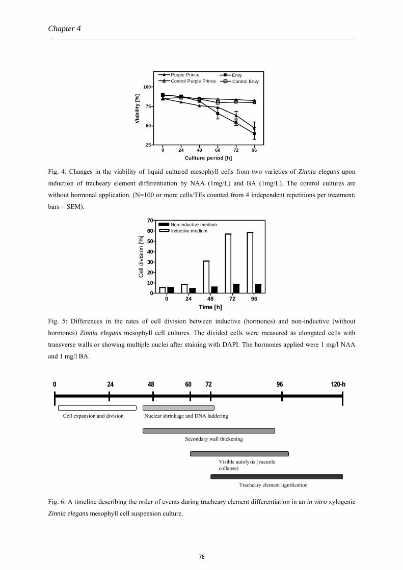
Chapter 4
0 24 48 60 72 9625
50
75
100
EnvyPurple PrinceControl EnvyControl Purple Prince
Cullture period [h]
Viab
ility
[%]
Fig. 4: Changes in the viability of liquid cultured mesophyll cells from two varieties of Zinnia elegans upon
induction of tracheary element differentiation by NAA (1mg/L) and BA (1mg/L). The control cultures are
without hormonal application. (N=100 or more cells/TEs counted from 4 independent repetitions per treatment;
bars = SEM).
0 24 48 72 960
10
20
30
40
50
60
70
Inductive mediumNon-inductive medium
Time [h]
Cel
l div
isio
n [%
]
Fig. 5: Differences in the rates of cell division between inductive (hormones) and non-inductive (without
hormones) Zinnia elegans mesophyll cell cultures. The divided cells were measured as elongated cells with
transverse walls or showing multiple nuclei after staining with DAPI. The hormones applied were 1 mg/l NAA
and 1 mg/l BA.
Nuclear shrinkage and DNA laddering
Visible autolysis (vacuole collapse)
Secondary wall thickening
Cell expansion and division
120-h 96 60 72 48 24 0
Tracheary element lignification
Fig. 6: A timeline describing the order of events during tracheary element differentiation in an in vitro xylogenic
Zinnia elegans mesophyll cell suspension culture.
76

In vitro xylogenic Zinnia elegans system
Discussion
The xylogenic Zinnia suspension culture is a pliable cell culture system that continues
to dominate all the other xylogenic cultures, such as Arabidopsis and Populus, due to its
broad utilization in cytological, molecular and physiological studies involving xylem
formation. The associated high frequency and synchrony in TE differentiation of the Zinnia
system, measuring over 60%, makes it useful especially for molecular studies involving
isolation of ample amounts of molecular markers. Additionally, the Zinnia culture, from leaf
mesophyll origin, is a perfect system for studying the mechanism of the cell-autonomous
programmed cell death process in plants, involved in the development of the vasculature.
Another interesting feature of the Zinnia system is its high reproducibility in TE
differentiation and the relatively short time necessary for the differentiation process.
Nevertheless, the critical steps involved in setting up the culture must be carefully followed to
realize these potentials. Also the monitoring techniques for the TE differentiation process are
mostly straight forward and less time consuming making it possible to track the
developmental processes involved.
One limitation associated with this system is the abrupt reduction of TE yield or
complete inhibition of TE formation in older cultures. Thus it is difficult, if not impossible, to
maintain a mother culture that can be sub-cultured over a longer period of time and still
maintains high TE differentiation capacity. This means that every experiment requires
preparation of freshly isolated mesophyll cells to initiate the xylogenic culture. Probably the
high level of synchronous TE differentiation in fresh Zinnia cultures is associated with
production of wound or stress signaling molecules. It is already known that wounds inflicted
on vascular bundle cells cause local accumulation of signal molecules such as auxin to initiate
vascular regeneration through transdifferentiation of localized parenchyma cells into new
tracheary or vessel elements (TEs or VE’s) and sieve elements to connect ends of the severed
bundle (Aloni, 1992; Nishitani et al., 2002). It is also likely that the increase in stress in the
isolated mesophyll cells caused by reactive oxygen species, exogenous cytokinin and other
potential stress-inducing compounds in the Zinnia culture are required for TE differentiation
in the freshly formed cultures. Thus, once this stress effect subsides in older cultures, the TE
differentiation is either reduced or not achieved at all. It would, therefore, be interesting
applying some stress-inducing compounds, such as oxidizing agents (e.g. H2O2), to older
Zinnia mesophyll cell cultures in vitro to regenerate or boost the lost or reduced TE
77

Chapter 4
differentiation activity in the cell culture. It would also be interesting to investigate in planta
the role of these stress-inducing factors on TE differentiation and the eventual vessel
formation in the xylem.
A timeline ordering the series of events that occur during in vitro TE differentiation is
presented in Fig. 6. This timeline supports the assumption that the collapse of the vacuole
brings the differentiation process to an end, although the lignification of the already
synthesized cellulose bands continues after vacuole collapse. The xylogenic Zinnia system is
at the moment the most suitable cell culture tool for xylogenesis studies. It is, however,
expected that the recent creation of a xylogenic culture from Arabidopsis thaliana, with its
molecular genetic possibilities, although with a lower TE differentiation at the moment (Oda
et al., 2005), would in the future complement the Zinnia system for extensive research into the
xylogenesis process in vitro and with subsequent in planta applications for improvement of
hydraulic properties of the plant.
References
Aloni, R. (1992). The control of vascular differentiation. Int J Plant Sci 153, S90-S92.
Aloni, R.R. (1987). Differentiation of vascular tissues. Annual review of plant physiology 38,
179-204.
Boerjan, W., Ralph, J., and Baucher, M. (2003). Lignin biosynthesis. Annu Rev Plant Biol
54, 519-546.
Cano-Delgado, A., Penfield, S., Smith, C., Catley, M., and Bevan, M. (2003). Reduced
cellulose synthesis invokes lignification and defense responses in Arabidopsis thaliana. Plant
J 34, 351-362.
Chaffey, N. (1999). Cambium: old challenges - new opportunities. Trees-Struct Funct 13,
138-151.
Church, D.L. (1993). Tracheary element differentiation in Zinnia mesophyll cell-cultures.
Plant Growth Regul 12, 179-188.
Dengler, N.G. (2001). Regulation of vascular development. J Plant Growth Regul 20, 1-13.
Fukuda, A., Nakamura, A., Tagiri, A., Tanaka, H., Miyao, A., Hirochika, H., and
Tanaka, Y. (2004). Function, intracellular localization and the importance in salt tolerance of
a vacuolar Na+/H+ antiporter from rice. Plant Cell Physiol. 45, 146-159.
Fukuda, H. (1996). Xylogenesis: Initiation, progression, and cell death. Annu Rev Plant Phys
78

In vitro xylogenic Zinnia elegans system
47, 299-325.
Fukuda, H., and Komamine, A. (1980a). Establishment of an experimental system for the
study of tracheary element differentiation from single cells Isolated from the mesophyll of
Zinnia elegans. Plant Physiol 65, 57-60.
Gabaldon, C., Ros, L.V.G., Pedreno, M.A., and Barcelo, A.R. (2005). Nitric oxide
production by the differentiating xylem of Zinnia elegans. New Phytologist 165, 121-130.
Groover, A., and Jones, A.M. (1999). Tracheary element differentiation uses a novel
mechanism coordinating programmed cell death and secondary cell wall synthesis. Plant
Physiol 119, 375-384.
Groover, A., DeWitt, N., Heidel, A., and Jones, A. (1997). Programmed cell death of plant
tracheary elements differentiating in vitro. Protoplasma 196, 197-211.
Haigler, C.H., Ivanova-Datcheva, M.,Hogan, P.S., Salnikov, V.V.,Hwang, S., Martin, K.,
and Delmer, D.P. (2001).Carbon partitioning to cellulose synthesis.Plant Mol Biol 47, 29-51.
Hosokawa, M., Suzuki, S., Umezawa, T., and Sato, Y. (2001). Progress of lignification
mediated by intercellular transportation of monolignols during tracheary element
differentiation of isolated Zinnia mesophyll cells. Plant Cell Physiol 42, 959-968.
Karam, G.N. (2005). Biomechanical model of the xylem vessels in vascular plants. Ann Bot
(Lond) 95, 1179-1186.
Kuriyama, H. (1999). Loss of tonoplast integrity programmed in tracheary element
differentiation. Plant Physiol 121, 763-774.
Lee, S., and Roberts, A.W. (2004). Tracheary element differentiation is correlated with
inhibition of cell expansion in xylogenic mesophyll suspension cultures. Plant Physiol Bioch
42, 43-48.
McCann, M.C., Stacey, N.J., Dahiya, P., Milioni, D., Sado, P.E., and Roberts, K. (2001).
Zinnia. Everybody needs good neighbors. Plant Physiol 127, 1380-1382.
Mellerowicz, E.J., Baucher, M., Sundberg, B., and Boerjan, W. (2001). Unravelling cell
wall formation in the woody dicot stem. Plant Mol Biol 47, 239-274.
Milioni, D., Sado, P.E., Stacey, N.J., Domingo, C., Roberts, K., and McCann, M.C.
(2001). Differential expression of cell-wall-related genes during the formation of tracheary
elements in the Zinnia mesophyll cell system. Plant Mol Biol 47, 221-238.
Nishitani, C., Demura, T., and Fukuda, H. (2002). Analysis of early processes in wound-
induced vascular regeneration using TED3 and ZeHB3 as molecular markers. Plant Cell
79

Chapter 4
Physiol 43, 79-90.
Obara, K., Kuriyama, H., and Fukuda, H. (2001). Direct evidence of active and rapid
nuclear degradation triggered by vacuole rupture during programmed cell death in Zinnia.
Plant Physiol 125, 615-626.
Oda, Y., and Hasezawa, S. (2006). Cytoskeletal organization during xylem cell
differentiation. J Plant Res 119, 167-177.
Oda, Y., Mimura, T., and Hasezawa, S. (2005). Regulation of secondary cell wall
development by cortical microtubules during tracheary element differentiation in Arabidopsis
cell suspensions. Plant Physiol 137, 1027-1036.
Ohlsson, A.B., Djerbi, S., Winzell, A., Bessueille, L., Ståldal, V., Li, X., Blomqvist, K.,
Bulone, V., Teeri, T.T., and Berglund, T. (2006). Cell suspension cultures of Populus
tremula × P. tremuloides exhibit a high level of cellulose synthase gene expression that
coincides with increased in vitro cellulose synthase activity. Protoplasma V228, 221-229.
Ranganath, R.M., and Nagashree, N.R. (2001). Role of programmed cell death in
development. International Review of Cytology- a Survey of Cell Biology, Vol. 202 202,
159-242.
Roberts, K., and McCann, M.C. (2000). Xylogenesis: the birth of a corpse. Curr Opin Plant
Biol 3, 517-522.
Sanmartin, M., Jaroszewski, L., Raikhel, N.V., and Rojo, E. (2005). Caspases. Regulating
death since the origin of life. Plant Physiol. 137, 841-847.
Siegel, S.M. (1953). On the biosynthesis of lignins. Physiol Plant 6, 134-139.
Sugiyama, M., and Fukuda, H. (1995 ). Zinnia mesophyll culture system to study
xylogenesis. In: Plant Tissue Culture Manual, Supplement 5: pp. H21-15. Kluwer Academic
Publishers, Dordrecht.
Tokunaga, N., Sakakibara, N., Umezawa, T., Ito, Y., Fukuda, H., and Sato, Y. (2005).
Involvement of extracellular dilignols in lignification during tracheary element differentiation
of isolated Zinnia mesophyll cells. Plant Cell Physiol 46, 224-232.
Tyree, M.T. (2003). Plant hydraulics: the ascent of water. Nature 423, 923.
Yang, Z., Cai, C.L., and Song, Y.C. (1999). Recent progress of plant apoptosis. Prog
Biochem Biophys 26, 439-443.
Ye, Z.H. (2002). Vascular tissue differentiation and pattern formation in plants. Annu Rev
Plant Biol 53, 183-202.
80
Chapter 5
Osmotic potential of Zinnia elegans plant material affects the yield and
morphology of tracheary elements produced in vitro
P. Twumasi1, 2, V. Flores Fukuda1, 2, J. H. N. Schel1, W. van Ieperen2 and A. M. C.
Emons1 (2007). Osmotic potential of Zinnia elegans plant material affects the yield and
morphology of tracheary elements produced in vitro
1Department of Plant Sciences, Laboratory of Plant Cell Biology, Wageningen University,
Arboretumlaan 4, 6703 BD Wageningen, The Netherlands 2Department of Plant Sciences, Horticultural Production Chains Group, Wageningen
University, Marijkeweg 22, 6709 PG Wageningen, The Netherlands

Chapter 5
Abstract
The Zinnia elegans cell suspension culture is excellent for xylogenesis studies at the
cellular and molecular level, due to the high and synchronous in vitro differentiation of
tracheary elements (TEs). The percentage TE differentiation (%TE) in the culture is, however,
influenced by a number of factors before and during cell differentiation. One of the factors
that is potentially important but has not got much attention is the initial osmolarity of the plant
material. To examine whether the growth conditions that determine leaf osmolarity (LO)
affect the final %TE, we used three light intensities (50, 70 and 100 µmol.m-2s-1), and three
electrical conductivity (EC) levels (EC 2, 4 and 6 dS.m-1 ) in hydroponic systems to induce
different osmolarities in leaf materials from two cultivars (cvs) of Zinnia elegans, Envy and
Purple Prince. The isolated leaf mesophyll cells were subsequently cultured in a liquid
medium (300 mOsm extracellular osmolarity) containing NAA (1 mg.l-1) and BA (1 mg.l-1).
The LO increased in both Zinnia cvs with increasing light intensity and increasing EC during
growth. Mesophyll cell size correlated negatively with EC, but the correlation was positive
with light intensity in both Zinnia cvs. There was an overall positive correlation between
%TE and LO although the degree of %TE change versus LO differed between light and EC
treatments, and also between the two Zinnia cvs. Envy cv is the best known Zinnia elegans cv
for establishing xylogenic cultures. However, it turned out that by subjecting the plants to
different growth conditions, the Purple Prince cv produces a higher %TE as compared to the
Envy cv. At EC4 the TE differentiation for the Purple Prince cv was 75%, a level that is 25-
60% higher than those earlier reported. We conclude that light intensity and EC of the root
environment affect the LO of Zinnia elegans which in turn influence the development and
therefore dimensions of TEs in an in vitro xylogenic culture. Thus, proper optimization of the
growth conditions for the Zinnia plants prior to establishment of xylogenic cultures leads to
enhancement of in vitro TE formation.
Keywords: Electrical conductivity, in vitro culture, leaf osmolarity, light intensity, osmotic
potential, tracheary element, xylogenesis, Zinnia elegans
Abbreviations: BA, benzylaminopurine; cv, cultivar; DIC, differential interference contrast;
EC, electrical conductivity; FTL, first true leaves; Ht, harvest time for FTL; LO, leaf
osmolarity; NAA, α-naphthalene acetic acid; OP, osmotic potential; PCD, programmed cell
82

Leaf osmolarity affects tracheary element differentiation
death; TE, tracheary element; %TE percentage TE differentiation
Introduction
Xylogenesis is an important research area in plant science, due to the significance of
the vascular development in plant performance (Fukuda, 1996). All higher plants rely on the
vascular system for long distance transport of water and minerals, and the photosynthetically
manufactured sugars. The xylem is an important component of the vascular system, composed
mainly of conduits built from stacks of tracheary elements (TEs). These TEs are differentiated
cambium cells consisting of dead, hollow and lignified cellulose cell walls with local
secondary thickenings. Fusion of these TEs into conduits allow both horizontal and vertical
water conductance necessary for transport of water and minerals from the soil to the leaves
(Comstock and Sperry, 2000). The involvement of programmed cell death (PCD), a part of
the xylogenesis process, is under intense study to understand the well orchestrated death of
cells necessary for development of a specific functional structure. In xylogenesis, PCD is the
last process that facilitates the onset of autolysis, a final stage of TE differentiation in the
xylem (Fukuda, 1996; McCann, 1997; Fukuda, 2000; Roberts and McCann, 2000).
For decades, xylem formation has been vigorously studied in many plants
(Mellerowicz et al., 2001; Ye, 2002). Due to the complexity of the whole plant system, in
vitro techniques have been employed to unravel the physiology and biochemistry of
secondary cell wall and lignin biosynthesis at the cellular level (Pesquet et al., 2003). In the
past 50 years, many callus and single cell cultures from several plant species have been
adopted for xylogenesis studies (Yen and Yen, 1999). Introduction of the in vitro xylogenic
Zinnia elegans mesophyll cell suspension culture approximately two decades ago has
revolutionalized our understanding of the xylogenesis process (Fukuda and Komamine,
1980a; Pesquet et al., 2003). A comparable xylogenic culture has been established from
Arabidopsis thaliana (Oda et al., 2005). However, the Arabidopsis culture requires further
technical optimization to improve the efficiency (currently %TE of 30 is possible) and the
degree of synchronization during TE differentiation. Therefore, the xylogenic Zinnia elegans
mesophyll culture, for now, remains the best alternative for xylogenesis studies. It has a
higher degree of synchronicity and also efficiency in TE differentiation (Church and Galston,
1988; see also Chapter 4 of this thesis). Such a system is not only suitable for cell biological
studies but also for molecular genetic characterization of these cellular processes. At the
83

Chapter 5
moment a large genetic library/ESTs from the xylogenic Zinnia culture is available (Pesquet
et al., 2005).
In spite of the superiority of the Zinnia elegans culture system, there have been huge
inconsistencies in the final yield of TEs as reported by different authors (Church and Galston,
1988; Gabaldon et al., 2005a; Tokunaga et al., 2005; Oda and Hasezawa, 2006). These
differences appear to be partly caused by the extracellular osmolarity of the culture medium in
relation to that of the mesophyll cells or the leaf material (Lee and Roberts, 2004). One other
potentially important factor to consider is the initial intracellular osmotic potential (OP) or
leaf osmolarity (LO) of the leaf material that is used for establishing the xylogenic culture.
Osmotic preconditioning of the plant material by certain environmental conditions during the
growth period of the Zinnia plants may affect the osmoregulation of the isolated mesophyll
cells in the culture with effect on the differentiation of the TEs. This could mean that, the
initial LO or leaf cell sap osmolarity must be well controlled to maximize the degree of
synchronicity of TE differentiation with enhancement of dimensions of the TEs formed.
The intracellular OP of a plant is largely affected by plant water status, which is
influenced by transpiration and physical water availability and salinity in the root
environment (soil EC). The accumulation of the soil water salts (enhanced by transpiration)
and photosynthetically synthesized organic compounds (sugars and proteins) in the plant
tissues increase the local or systemic OP of the plant (Vaadia et al., 1961; Barlow et al., 1977;
Meyer and Boyer, 1981; Morgan, 1984; Boyer and Silk, 2004). Thus, application of different
soil ECs and/or imposition of different transpirational pressures via light intensities on the
growing plants will have consequent effects on tissue osmolarities in the plant tissues.
In this paper, we present the results of the effect of different LO’s, as induced by three
different light treatments in soil-grown plants, and three different EC treatments in
hydroponic plants of two Zinnia elegans L. cultivars (cvs), Envy and Purple Prince, on the in
vitro differentiation of TEs in suspension cell cultures.
Materials and methods
Plant material
Light treatment: Seedlings of the two Zinnia elegans cvs, Envy and Purple Prince,
were germinated and allowed to grow in peat-based commercial potting compost (Lentse
Potgrond nr. 4; 85% peat, 15% clay, Lentse Potgrond, Lent, The Netherlands) in four
84

Leaf osmolarity affects tracheary element differentiation
different compartments in climate rooms at Horticultural Production Chain Group of
Wageningen University and Research Centre, The Netherlands, set at 16-h light and 8-h
darkness with 25/20 oC temperature respectively. The relative humidity was maintained at
70% throughout the growth period. Each of the three compartments was given different light
levels of 50, 70 and 100µmol.m-2.s-1 respectively. Higher light intensity could not be used due
to the burning effects on the seedlings. Constant amount of water was supplied to maintain the
soil water content at 70% (v/v). Fully expanded first set of leaves from the seedlings was
harvested between 13 and 15 days after sowing for leaf sap osmolarity measurements and
mesophyll cell isolation to establish the cell culture.
EC treatments: Seeds of the two Zinnia elegans cvs were sown in cotton plugs in a
hydroponic system in a climate room. The system consisted of 30cm x 50cm x 10cm trays
containing rockwool plugs, partly (0.5 cm depth) immersed in (or floating on) the liquid
growth medium set at the different ECs ( 2, 4 and 6 dS/m). Further conditions were a 16-h
light and 8-h darkness period, d/n temperatures of 25 oC and 20 oC, 70% relative humidity
(RH) and a liquid medium pH of 5.5. Application of a water pump (250-300 L/h) ensured
even distribution of the solutes in the growth medium. The first true leaves (FTL) were
harvested at Ht (harvest-time at which only FTL are present and fully expanded) for LO
measurements and mesophyll isolation.
Leaf osmolarity measurements
Eight, randomly selected pairs of FTL of seedlings per cv per treatment (from both
light and hydroponic treatments) were sampled for the LO measurements using a Vapro®
Vapor Pressure Osmometer (model 5520, Wescor Inc., Logan, UT, USA). Each set of leaves
was separately folded into a 4x4 cm nylon mesh of pore size 10 µm. By using a Zwick Press
machine (Zwicki 1120, Zwick, Germany), the leaves in the nylon mesh were pressed
individually against the bottom of an Eppendorf tube. The sap from the pressed leaves was
collected in a new Eppendorf tube, and from this two samples of 10µl sap extract were
independently measured in the osmometer and averaged.
Leaf mesophyll isolation and culture
Approximately 30 pairs of FTL were harvested and surface sterilized in cold 0.15%
NaOCl solution containing 0.001% Triton X-100 for 10 min. The mesophyll cells were
85

Chapter 5
isolated mechanically and purified as described by Sugiyama and Fukuda (1995 ), with few
modifications (Chapter 4, this thesis). The osmolarity of the culture medium was adjusted to
300 mOsm by addition of D-mannitol. The cell density in the final culture was adjusted to
105cells/ml in the culture medium. Four samples of 3 ml each of the cell suspension per
treatment were cultured in six-well culture plates (Sigma; n=4 independently cultured wells).
Induction of TE differentiation in the cultures was achieved by exogenous application of
NAA (1mg/l) and BA (1 mg/l) at the onset of the mesophyll cell isolation (t = 0 h). The
control experiments were devoid of these growth regulators. The cultures were incubated in
the dark at 25 oC on a rotary shaker at 100 rpm speed.
Microscopy of TE differentiation and size measurements
The viability of the mesophyll cells was determined by staining with 0.005% (final
concentration) of fluorescein diacetate (FDA). Observation of the yellow fluorescent cells was
done by the use of a Diaphot-Nikon fluorescent microscope (Zeiss, The Netherlands)
equipped with a UV lamp, 488nm filter and a LCD Kappa DX 20L camera. In addition, a
Nikon Diaphot TMD 200 inverted microscope, equipped with differential interference
contrast (DIC) optics and a Sony 3CCD color video camera, was used for the direct
monitoring of cellulose band formation in the differentiating cells. Image J software (Wayne
Rasband National Institutes of Health, USA) was used for processing and measuring the
physical parameters (length and area) of the differentiated TEs from the microscopic images.
The yield of TEs or %TE was calculated as the percentage of mesophyll cells that
differentiated at 120-h after induction with growth regulators, with a correction factor for the
initial dead cells in the culture. Thus, %TE was calculated as:
βγα100% =actualTE
where %TEactual is the true yield of TEs caused by only the viable mesophyll cells in the initial
culture, α is the number of TEs counted in a representative fraction of the culture, β is the
total number of differentiated and non-differentiated (dead or live) cells in the sample
fraction, and γ is the initial fractional viability of the culture sample.
Statistical analysis
All experiments were accomplished in triplicate. For every cell culture, a minimum of
86

Leaf osmolarity affects tracheary element differentiation
30 cells or tracheary elements that were randomly chosen were used for calculations.
Analyses of variance for the effects of different light, EC, and cv (genetic) on the final yield
of TEs in the suspension cultures were performed by Prism v.4 (GraphPad Software, Inc.).
For each analysis, a significance level of 5% was maintained.
Results
Electrical conductivity and light intensity effects on plant morphology and leaf osmolarity
Ht increased with decreasing light intensity, ranging from 3-5 days earlier in
100µmol.m-2.s-1 light intensity compared with the 50 or 70 µmol.m-2.s-1 light intensities (Fig.
1A). Purple Prince plants were larger than Envy at all the three light treatments. In the EC
treatments, Ht increased with increasing EC in the root environment (Fig. 1B). Generally, the
leaf area increased with decreasing EC in the hydroponic media, and the plants growing in
EC2 (2dS/m) medium were harvested 1-5 days earlier than the plants growing in EC4 or EC5
growth medium. Plants from the Purple Prince cv grew slightly faster and larger than the
Envy cv at all the three EC treatments. A minimal change in mesophyll cell size by
differences in EC of the growth media was observed in both Zinnia cvs (Fig. 2A and B).
Significant differences in LO within cv were found only between EC6 and EC2 or EC4 in
both Zinnia cvs (Fig. 2C). LOs were different between the two cv at ECs 2 and 6 (dS/m)
where in each case Envy cv recorded higher LO over the Purple Prince cv. An increase of
medium EC from 2 to 6 caused 32% and 23% increase in LO of Envy cv and Purple Prince cv
respectively.
50 70 1000
10
20
30Envy Purple Prince
aa
b b c c
A
Lighth intensity [µmol.m-2.s-1]
FTL
harv
est t
ime,
Ht [
days
]
EC2 EC4 EC60
5
10
15
20
25Envy Purple Prince
a b cd
e eB
EC level [dS.m-1]
FTL
harv
est t
ime,
Ht [
days
]
Fig. 1: The effects of different (A) light intensities and (B) root medium electrical conductivities on the harvest
time (Ht) for the expanded FTL in two Zinnia elegans cvs. Different letters indicate statistically significant
differences. (N = 60 seedling x 3 repetitions; bar = SEM).
87

Chapter 5
2 4 60
25
50
75
100 Envy Purple Prince Aa a
ba a a
EC Levels [dS/m]
Mes
ophy
ll ce
ll le
ngth
[ µm
]
2 4 60
1000
2000
3000Envy Purple Prince B
a ab
aa c
EC levels [dS/m]
Mes
ophy
ll are
a [µ
m2 ]
2 4 6200
250
300
350
400
450Purple princeEnvy
ab a ab
c
d
C
EC level [dSm-1]
Leaf
osm
olar
ity [m
Osm
]
Fig. 2: Effect of EC in the root environment on the leaf mesophyll size (A, length; B, area) and LO (C) in two
cvs of Zinnia elegans seedlings at Ht. Different letters indicate statistically significant differences. [LO, leaf
osmolarity; Ht = harvest time for expanded FTL without second set of leaves, bar = SEM, N= 8 for LO
measurements; N=30 for mesophyll cell size measurements].
The effect of light intensity on LO and mesophyll cell size (length and area) is shown
in Fig.3. The Envy cv acquired the highest mean mesophyll cell size in plant materials
obtained from the 70 µmol.m-2.s-1 illumination, while in Purple Prince cv the highest mean
mesophyll cell size was associated with the 100 µmol.m-2.s-1 illumination (Fig. 3A and B).
The mean LO was highest in leaf materials from 100 µmol.m-2.s-1 illumination (Envy, 303.0
88

Leaf osmolarity affects tracheary element differentiation
mOsm; Purple Prince, 276.5 mOsm) and lowest in 50 µmol.m-2.s-1 illumination (Envy, 261.4
mOsm; Purple Prince, 260.1) in both Zinnia cvs (Fig. 3C). Again, the Envy cv recorded
higher mean LO at 100 µmol.m-2.s-1 illumination as compared with the Purple Prince cv. An
increase of light intensity from 50 to 100 µmol.m-2.s-1 caused the LO to increase by 17% and
3% in Envy cv and Purple Prince cv respectively.
50 70 1000
25
50
75
100Envy Purple Prince A
a acb
a a
Light intensity [µmol.m-2s-1]
Mes
ophy
ll ce
ll le
ngth
[ µm
]
50 70 1000
500
1000
1500
2000Envy Purple Prince B
a a
b
a a
c
Light intensity [µmol.m-2.s-1]
Mes
ophy
ll ce
ll ar
ea [µ
m2 ]
50 70 100200
250
300
350
400Envy Purple Prince
a ba
ab b
c
C
Lighth intensity [µmol.m-2s-1]
Leaf
osm
olar
ity [m
Osm
]
Fig 3: Effect of different light intensities on the leaf mesophyll size (A, length; B, area) and LO (C) in two cvs of
Zinnia elegans seedlings at Ht. Different letters indicate statistically significant differences. (Bar = SEM, N= 8
for LO measurements; N=30 for mesophyll cell size measurements).
89

Chapter 5
Tracheary element differentiation and dimensions
EC treatment: DIC images of the differentiated TEs at 120h from EC2 and EC6
treatments in both Zinnia elegans cvs are shown in Fig. 4A. In both cvs, the TE size increased
proportionally with the level of EC in the root environment (Fig. 4B-II and III). There were
generally no differences in TE sizes (length and area) between Envy and Purple Prince at the
three EC treatments. However, significant differences in %TE were found between Envy and
Purple Prince at EC 2 and 4 (Fig. 4B-I). At EC 4, the Purple Prince cv gave the highest %TE
of 75% (Fig. 4B-I). It was also found that an increase in LO osmolarity by 32% in Envy and
23% in Purple Prince with EC ranging from 2 to 6 dS/cm caused an increase of about 68%
and 66% TE differentiation respectively. There was a strong interaction between the genotype
of Zinnia elegans and the EC treatments with respect to TE differentiation.
d
cba
Fig. 4A: DIC images of differentiated TEs in suspension cell cultures from plants of two Zinnia elegans L. cvs,
Envy (a, d) and Purple prince (b, c) grown with media with EC 2 (a, b) and EC 6 (c, d). (Bar = 30 µm).
90

Leaf osmolarity affects tracheary element differentiation
2 4 60
25
50
75
100Purple PrinceEnvy
a
bb
c
bb
(I)
EC level (dS.m-1]
TE (%
)
2 4 60
25
50
75
100
125 Envy Purple Prince
ab
c ccd d
EC level [dS.m-1]
TE le
ngth
[ µm
]
(II)
2 4 60
1000
2000
3000
4000Envy Purple Prince
aa
b b bb
EC level [dS.m-1]
TE a
rea
[µm
2 ]
(III)
Fig. 4B: Effect of different EC in the root environment during growth periods of two Zinnia elegans L. cvs on
the (I) final percentage TE differentiation, (II) TE length, and (III) TE area in an in vitro mesophyll cell cultures
measured at 120h after TE induction with NAA (1mg/L) and BA (1mg/L). Values represent the mean of 3
replicates. Different letters indicate statistically significant differences. [Bar =SEM, N =100 or more TEs from 3
independent culture repetitions].
Light treatment: The DIC images of differentiated TEs at 120 h are shown in Fig. 5A.
Higher %TEs were observed in the xylogenic mesophyll cultures derived from plant materials
grown with 100 µmol.m-2-s-1 illumination as compared with the 50 or 70 µmol.m-2-s-1 in both
91

Chapter 5
Zinnia elegans cvs (Fig. 5B-I). Thus a positive correlation is found between light intensity
provided during growth of the plants and the final %TE in suspension cultures from both
Zinnia elegans cvs. It was found that an increase in LO osmolarity by 17 % in Envy and 3 %
in Purple Prince with light caused an increase of about116% and 130% TE differentiation
respectively. There was, however, no interaction between the Zinnia cvs (genetic background)
and the light intensity during growth in relation to the in vitro %TE results. Anatomical
measurements indicated a positive correlation between TE size (area and length) and light
intensities used in growing plants of the two Zinnia elegans cvs from which in vitro cultures
were established (Fig. 5B-II and III). Furthermore, visual observations during in vitro TE
differentiation indicated an earlier and more rapid differentiation in mesophyll cell cultures
originating from leaf materials of LO equal or slightly above the 300 mOsm extracellular
medium osmolarity. Generally, the %TE increased with increasing LO above that of the
medium, irrespective of the method used for inducing this higher LO in both Zinnia elegans
cvs. At all light and EC levels, the LO induction was more effective in the Envy cv than in the
Purple Prince cv.
d
cb a
Figure 5A: DIC images of differentiated TEs in suspension cell cultures from plants of two Zinnia elegans L.
cvs, Envy (a, d) and Purple Prince (b, c) grown with 50 µmol.m-2.s-1 (c, d) and 100 µmol.m-2.s-1 (a, b) of light
intensity in climate rooms. (Bar = 30µm).
92

Leaf osmolarity affects tracheary element differentiation
50 70 1000
10
20
30
40
50
60
70
80 Envy Purple Prince
a aa
a
bb
(I)
Light intensity [µmoles m-2 s-1]
TE (%
)
50 70 1000
50
100
150Envy Purple Prince
a ab b
bc(II)
Light intensities [µmol.m-2.s-1]
TE le
ngth
[µm
]
50 70 1000
1000
2000
3000
4000Envy Purple Prince
ab b b b
c(III)
Light intensity [µmol.m-2s-1]
TE a
rea
(µm
2 )
Fig. 5B: Effect of different light intensities during growth periods of seedlings of two Zinnia elegans L. cvs on
(I) final percentage TE differentiation, (II) TE length, and (III.) TE area in in vitro mesophyll cell cultures
measured at 120-h after TE induction with NAA (1mg/L) and BA (1mg/L). Values represent the mean of 3
replicates. Different letters indicate statistically significant differences. [Error bars =SEM, N>100 total cells
from 3 independent wells].
Discussion
Due to the suitability of the xylogenic Zinnia elegans cell suspension culture for
cytological and molecular research, lots of efforts have been focused on improvements in
establishing this system for various applications (Lee et al., 2000; McCann et al., 2001; Lee
93

Chapter 5
and Roberts, 2004; Shinohara et al., 2006; Tokunaga et al., 2006). The obvious expectation
from such improvements is to achieve higher percentage and more synchronous tracheary
element (TE) differentiation (Chapter 4, this thesis). Unfortunately, the majority of these
improvement techniques are being focused on the xylogenic cell suspension culture itself, i.e.,
processes occurring after isolation of the leaf mesophyll cells. Almost none of these
improvements involve preparation of the plant materials. For instance, there was no work
about the effect of growth conditions during plant development on the performance of the
xylogenic culture established from the mesophyll cells. A unique property of the xylogenic
Zinnia suspension cell culture is the lack of a mother culture from which an adequate TE
differentiation can be maintained. Thus, the originally developed mesophyll cells isolated
from the leaves are directly differentiated into the TEs in the culture. This implies that, any
influence of the developmental process of the mesophyll cells in planta will have
consequences on the performance of the xylogenic Zinnia culture established from these cells.
A factor with potential effect on the performance of the xylogenic culture is the OP or the LO
of the plant material. Lee and Roberts (2004) have shown that, due to the differences in
internal osmolarity of the isolated mesophyll cells, high heterogeneity in TE differentiation
exists in xylogenic Zinnia cultures. To prevent these inequalities in OP, a choice of growth
conditions aimed at adjusting the LO is necessary.
We found in our study that growing plants with low light intensity delays the harvest
time for the leaves required for mesophyll cell isolation. A similar effect was found when the
level of EC in the growth medium was increased. Apart from the reduction in leaf area caused
by an increasing EC, the leaf mesophyll cell viability, shape and size were neither affected by
light nor EC treatment in both Zinnia elegans cvs. This is an especially important
consideration since the quality of mesophyll cells influence the TE differentiation process
(Gabaldon et al., 2005b). Even with such low light levels, a small increase in intensity was
enough to induce higher LO. Similarly, the LO increased with increasing EC in the growth
medium in both Envy cv and Purple Prince cv. However, in the LO measurements, the
response of Envy cv to LO change caused by changes in the growth conditions was higher as
compared to the Purple Prince cv. By increasing light intensity, transpiration of the plant is
increased as the stomatal conductance increases (Morgan, 1984; Wei et al., 1999). Two
processes could be identified for this increase in LO by the light intensity: (1) the
photosynthetic rate that correlates with light intensity leads to synthesis and accumulation of
94

Leaf osmolarity affects tracheary element differentiation
sugars in the plant tissues especially in the leaf tissue causing elevation of the intracellular OP
(Flesch et al., 1991); (2) transpiration rate is dependent on light intensity. Thus, a higher light
intensity means an increase in the uptake of water together with dissolved minerals. While the
water continuously evaporates into the atmosphere via the stomata, the mineral components
accumulate in tissues resulting in higher intracellular OP (Vaadia et al., 1961).
In both Zinnia elegans cvs, the increase in LO by either light or EC, resulted in an
increase in the percentage of TEs formed in the culture after induction with the
phytohormones (1mg/l NAA and 1mg/l BA). In the Envy cv, an increase in the light intensity
from 50 to 100 µmol.m-2.s-1 caused 17% increase in LO which resulted in about 116%
increase in TE differentiation. In Purple Prince, however, only 3% increase in LO was
achieved, but such a minor osmotic change caused the %TE to rise by about 130%. Also, in
the EC treatment, an increase from 2 to 6 dS/m achieved 32% and 23 % increase in LO in
Envy and in Purple Prince respectively. The corresponding increase in %TE was 68% and
66% in Envy cv and Purple Prince cv respectively. Thus, although the LO achieved by the
two sets of growth conditions (i.e., light intensity and EC) were lower in Purple Prince as
compared to the Envy, the corresponding increase in TE differentiation was even higher in the
Purple Prince cv. It is intriguing to see that despite the differences in LO in response to
changes in the growth conditions in the two cvs of Zinnia elegans, a similar change was
observed in the final %TE in the culture. Thus, an increase in the overall LO osmolarity does
not necessarily determine the final TE differentiation. This suggests that other cellular
processes or factors with effects on TE differentiation might be influenced by these growth
conditions.
The observed increase in final %TE by the preconditioning of the materials might be a
result of establishing osmotic equilibrium across the cell membrane, ie, intracellular
environment in relation to the extracellular medium OP, with an associated minimal cell
expansion that can delay TE differentiation (Lee and Roberts, 2004). On the other hand, these
treatments applied during plant development eliminated the osmotic differences commonly
found between cells especially at different developmental stages or in different locations in
the same tissue (Molz and Boyer, 1978). The highest %TE was from cv Purple Prince at EC 4
(75%). This is 25-60% higher than those earlier reported (Lopez-Serrano et al., 2004;
Tokunaga et al., 2005; Oda and Hasezawa, 2006). Why this could not be improved further, for
example at EC 6 dS/cm, might be a result of the cellular homeostasis which prevents extreme
95

Chapter 5
OP (Romero, 2004). Although our results show that an increase in OP in the cells using
growth conditions improves %TE differentiation, specific growth conditions are necessary for
each kind of Zinnia genotype.
Comparison of the length and area measurements in TEs from various cultures
established from leaf mesophyll cells at different OPs showed a positive correlation between
LO and TE size. This suggests that TE initials with higher OP initially undergo extensive
expansion to reach a new OP which is equal to the OP of the xylogenic medium (extracellular
OP) before the phytohormones could induce TE differentiation. Extreme growth conditions
were avoided in our experiments, but it is expected that large changes in OPs of the
mesophyll cells as compared to the extracellular osmolarity of the growth media will
negatively affect the performance of the xylogenic culture. Expected excessive explosions or
plasmolysis of the mesophyll cells in such a culture could counter the positive effect of
growth conditions on TE differentiation. It is, therefore, of importance to balance the
intracellular and extracellular OPs before initiating a xylogenic culture.
We conclude from this study that plant growth conditions have effects on TE
differentiation in an in vitro xylogenic Zinnia elegans suspension culture, and that initial LO
(or OP of the isolated mesophyll cells) is an important factor for TE differentiation.
References
Barlow, E.W.R., Munns, R., Scott, N.S., and Reisner, A.H. (1977). Water potential,
growth, and polyribosome content of the stressed wheat apex. J. Exp. Bot. 28, 909-916.
Boyer, J.S., and Silk, W.K. (2004). Hydraulics of plant growth. Funct Plant Biol 31,761-773.
Church, D.L., and Galston, A.W. (1988). Kinetics of determination in the differentiation of
isolated mesophyll cells of Zinnia elegans to tracheary elements. Plant Physiol. 88, 92-96.
Comstock, J.P., and Sperry, J.S. (2000). Theoretical considerations of optimal conduit
length for water transport in vascular plants. New Phytologist 148, 195-218.
Flesch, V., Jacques, M., Cosson, L., Petiard, V., and Balz, J.P. (1991). Effects of light and
temperature on growth of Ginkgo-biloba cultivated under controlled long day conditions. Ann
Sci Forest 48, 133-147.
Fukuda, H. (1996). Xylogenesis: Initiation, progression, and cell death. Annu Rev Plant Phys
47, 299-325.
Fukuda, H. (2000). Programmed cell death of tracheary elements as a paradigm in plants.
96

Leaf osmolarity affects tracheary element differentiation
Plant Mol Biol 44, 245-253.
Fukuda, H., and Komamine, A. (1980a). Establishment of an experimental system for the
study of tracheary element differentiation from single cells isolated from the mesophyll of
Zinnia elegans. Plant Physiol 65, 57-60.
Gabaldon, C., Ros, L.V.G., Pedreno, M.A., and Barcelo, A.R. (2005a). Nitric oxide
production by the differentiating xylem of Zinnia elegans. New Phytologist 165, 121-130.
Gabaldon, C., Gomez Ros, L.V., Pedreno, M.A., and Ros Barcelo, A. (2005b). Nitric
oxide production by the differentiating xylem of Zinnia elegans. New Phytol 165, 121-130.
Jacobsen, A.L., Ewers, F.W., Pratt, R.B., Paddock, W.A., III, and Davis, S.D. (2005). Do
xylem fibers affect vessel cavitation resistance? Plant Physiol. 139, 546-556.
Lee, S., and Roberts, A.W. (2004). Tracheary element differentiation is correlated with
inhibition of cell expansion in xylogenic mesophyll suspension cultures. Plant Physiol Bioch
42, 43-48.
Lee, S., Woffenden, B.J., Beers, E.P., and Roberts, A.W. (2000). Expansion of cultured
Zinnia mesophyll cells in response to hormones and light. Physiol Plantarum 108, 216-222.
Lopez-Serrano, M., Fernandez, M.D., Pomar, F., Pedreno, M.A., and Barcelo, A.R.
(2004). Zinnia elegans uses the same peroxidase isoenzyme complement for cell wall
lignification in both single-cell tracheary elements and xylem vessels. J Exp Bot 55, 423-431.
McCann, M. (1997). Tracheary element formation: building up to a dead end. Trends Plant
Sci 2, 333-338.
McCann, M.C., Stacey, N.J., Dahiya, P., Milioni, D., Sado, P.E., and Roberts, K. (2001).
Zinnia. Everybody needs good neighbors. Plant Physiol 127, 1380-1382.
Mellerowicz, E.J., Baucher, M., Sundberg, B., and Boerjan, W. (2001). Unravelling cell
wall formation in the woody dicot stem. Plant Mol Biol 47, 239-274.
Meyer, R.F., and Boyer, J.S. (1981). Osmoregulation, solute distribution, and growth in
soybean seedlings having low water potentials. Planta 151, 482-489.
Molz, F.J., and Boyer, J.S. (1978). Growth-induced water potentials in plant cells and
tissues. Plant Physiol. 62, 423-429.
Morgan, J.M. (1984). Osmoregulation and water stress in higher plants. Annual Review of
Plant Physiology 35, 299-319.
Oda, Y., and Hasezawa, S. (2006). Cytoskeletal organization during xylem cell
differentiation. J Plant Res 119, 167-177.
97

Chapter 5
Oda, Y., Mimura, T., and Hasezawa, S. (2005). Regulation of secondary cell wall
development by cortical microtubules during tracheary element differentiation in Arabidopsis
cell suspensions. Plant Physiol 137, 1027-1036.
Pesquet, E., Jauneau, A., Digonnet, C., Boudet, A.M., Pichon, M., and Goffner, D.
(2003). Zinnia elegans: the missing link from in vitro tracheary elements to xylem. Physiol
Plantarum 119, 463-468.
Pesquet, E., Ranocha, P., Legay, S., Digonnet, C., Barbier, O., Pichon, M., and Goffner,
D. (2005). Novel markers of xylogenesis in Zinnia are differentially regulated by auxin and
cytokinin. Plant Physiol 139, 1821-1839.
Roberts, K., and McCann, M.C. (2000). Xylogenesis: the birth of a corpse. Curr Opin Plant
Biol 3, 517-522.
Romero, M.F. (2004). In the beginning, there was the cell: cellular homeostasis. Advan.
Physiol. Edu. 28, 135-138.
Shinohara, N., Sugiyama, M., and Fukuda, H. (2006). Higher extracellular pH suppresses
tracheary element differentiation by affecting auxin uptake. Planta, 1-11.
Sugiyama, M., and Fukuda, H. (1995 ). Zinnia mesophyll culture system to study xylogenesis. In:
Plant Tissue Culture Manual, Supplement 5: pp. H21-15. Kluwer Academic Publishers, Dordrecht.
Tokunaga, N., Uchimura, N., and Sato, Y. (2006). Involvement of gibberellin in tracheary
element differentiation and lignification in Zinnia elegans xylogenic culture. Protoplasma
V228, 179-187.
Tokunaga, N., Sakakibara, N., Umezawa, T., Ito, Y., Fukuda, H., and Sato, Y. (2005).
Involvement of extracellular dilignols in lignification during tracheary element differentiation
of isolated Zinnia mesophyll cells. Plant Cell Physiol 46, 224-232.
Vaadia, Y., Raney, F.C., and Hagan, R.M. (1961). Water relations - plant water deficits and
physiological processes. Annu Rev Plant Phys 12, 265-&.
Wei, C., Steudle, E., and Tyree, M.T. (1999). Water ascent in plants: do ongoing
controversies have a sound basis? Trends Plant Sci 4, 372-375.
Ye, Z.H. (2002). Vascular tissue differentiation and pattern formation in plants. Annu Rev
Plant Biol 53, 183-202.
Yen, H.C.E., and Yen, S.K. (1999). Effect of high salinity on tracheary element
differentiation in light-grown callus of Mesembryanthemum crystallinum. Plant Cell Tiss Org
58, 59-65.
98

Chapter 6
Effects of osmotic stress and timed hormone application on tracheary
element differentiation in xylogenic cultures from two Zinnia elegans
cultivars
P. Twumasi1, 2, J. H. N. Schel1, W. van Ieperen2 and A. M. C. Emons1 (2007). Effects of
osmotic stress and timed hormone application on tracheary element differentiation in
xylogenic cultures from two Zinnia elegans cultivars
1Department of Plant Sciences, Laboratory of Plant Cell Biology, Wageningen University,
Arboretumlaan 4, 6703 BD Wageningen, The Netherlands 2Department of Plant Sciences, Horticultural Production Chains Group, Wageningen
University, Marijkeweg 22, 6709 PG Wageningen, The Netherlands

Chapter 6
Abstract
The effects of osmotic stress and timed application of NAA and BA on in vitro
tracheary element (TE) differentiation were studied in xylogenic mesophyll cell cultures from
two Zinnia elegans ccultivars (cvs), ‘Envy’ and ‘Purple Prince’. Xylogenic culture media of
different osmotic stress, ranging from 100-700 mOsm, were used for TE differentiation in
both Zinnia cvs. In another set of experiments, TE-inductive hormones were applied to the
cultures at different times during progress of the culture. The percentage of TE differentiation
(%TE) increased with increasing extracellular osmolarity until it reached a maximum value
around 300 mOsm and then decreased further with any increase in extracellular osmolarity.
Early TE differentiation was observed in cultures with higher osmotic stress as compared to
the lower ones in reference to the 300 mOsm standard culture medium. The size of TEs
increased whilst %TE decreased with decreasing medium osmotic stress. The Envy cv was
found to be less prone to high osmotic stress than the Purple Prince cv, as shown by the
increasing amounts of TEs formed when osmotic stress in the culture increased till 300-400
mOsm. In the experiments in which hormones were applied at various times after culture
initiation, the induction of TEs showed a decreasing %TE with a delay in application of
hormones to the culture. However, the late-induced xylogenic cultures, as observed in both
Zinnia cvs, formed larger TEs than earlier-induced cultures. In this study, the NAA/BA–
treated cultures had extensive cell division that correlated positively with the rate of TE
formation as compared to the controlled cultures (without phytohormones) which recorded
0% cell division. We conclude from these studies that the duration of TE differentiation, and
the size and %TE in xylogenic Zinnia cultures are affected by the level of extracellular
osmotic stress as well as the time of hormone application.
Keywords: Differentiation, in vitro culture, mesophyll cell, osmolarity, tracheary element,
Zinnia elegans
Abbreviations: BA, benzylaminopurine; cv, cultivar; DIC, differential interference contrast;
DAPI, 4, 6-diamidino-2-phenylindole; FDA, fluorescein-diacetate; mOsm, milliosmoles; LO,
leaf osmolarity; NAA, α-naphthalene acetic acid; OP, osmotic potential; TE, tracheary
element; %TE, percentage TE differentiation; TUNEL, Terminal deoxynucleotidyl
Transferase Biotin-dUTP Nick End Labeling; VE, vessel element
100

Osmotic stress effect on tracheary element dimensions
Introduction
Long distance water transport in higher plants is achieved via the xylem system
(Anfodillo et al., 2006). The anatomical characteristics of xylem, particularly the vessels, are
very important for plants growing in environments with varying water availability (Lovisolo
and Schubert, 1998). Modified xylem characteristics have the potential to influence both the
hydraulic efficiency (Lovisolo et al., 2002) and the safe hydraulic performance of the vessels
(Hacke et al., 2006) of the xylem system. These xylem characteristics are not only important
for water transport during growth and development, but also necessary for preserving the
quality of plant products, such as cut flowers, by maintaining an efficient post-harvest water
transport. Thus, xylem vessel dimensions (vessel length, density and diameter) become
important determinants for the post-harvest quality of many plant products especially
horticultural products (van Ieperen et al., 2002). Work by Twumasi et al (2005) has shown
that cut flowers from Zinnia elegans having narrower and shorter xylem vessels tend to
perform better during vase life as compared with cut flowers having large-diameter and long
xylem vessels. Apart from these two major characteristics of the xylem conduits, i.e. vessel
diameter and vessel length (Nijsse et al., 2001), other properties such as end wall perforations
(Ellerby and Ennos, 1998) and inter-vessel pit membranes (Choat et al., 2006) are important
for regulating the efficiency and embolism protection of the hydraulic conductivity of the
stems. Thus, the developmental influence of the xylogenesis process leading to the
modification of one or more of the above mentioned xylem vessel characteristics will, to a
certain degree, have an influence on the final xylem hydraulic performance with
consequences for the quality and industrial performance of the plant.
The formation of xylem vessels of particular dimensions might be dictated by the
mechanism to ensure continued functional vessels in the midst of potential stress conditions
such as water deficit which facilitates cavitations and embolism (Solla and Gil, 2002). Due to
the difficulty in understanding the complex xylogenesis process and especially the unique
modifications in various pathways, resulting in particular types of xylem vessels for specific
ecological environments, a much simpler system for studying mechanisms that determine
xylem dimensions during xylogenesis is necessary. Many isolated plant cells have for a long
time been known to be capable of forming tracheary elements (TEs) or xylem vessels
(Phillips, 1980). However, xylem differentiation in most of these single cell systems is
incapable of forming a high percentage TE differentiation (%TE), and is highly asynchronous
101

Chapter 6
as well. Fortunately, the xylogenic Zinnia elegans system escapes these two bottlenecks
(Kohlenbach and Schmidt 1975; Fukuda and Komamine, 1980a). This xylogenic culture
system is known for its high percentage and synchrony in TE differentiation. Over 60% of the
initial mesophyll cells in the culture are differentiated 72h after induction (Church and
Galston, 1988; Fukuda, 1996; Roberts and McCann, 2000).The Zinnia elegans xylogenic
mesophyll cell culture is most pliable and remains a potential tool to dissect the complexity of
the xylogenesis process at cellular and molecular levels. Furthermore, Zinnia elegans is a
horticultural plant grown as cut flowers or pot plants. This makes it a suitable model for
research aiming at the modification of the hydraulic properties of the xylem to improve post
harvest quality of cut flowers in particular, and many other crop plants in general.
In this work we studied the influence of the in vitro extracellular osmotic potential
(OP), via mimicking of osmotic or drought stress in the extracellular culture medium, on the
differentiation pattern and morphology of the TEs (length and area) produced in xylogenic
mesophyll cell cultures from two cvs of Zinnia elegans: Purple Prince and Envy. The reason
for the choice of these two cvs follows from earlier in planta studies which showed large
differences between the two cvs in whole plant morphology and xylem vessel anatomy after
treatment with various levels of soil water stress and temperature (this thesis, Chapter 2 and
3). The Purple Prince cv is a bigger plant as compared to the Envy cv. We also studied similar
effects caused by delayed application of phytohormones (auxin and cytokinin) that trigger the
xylogenesis process in the in vitro xylogenic Zinnia elegans cultures.
Materials and methods
Plant material
Plants of two cvs of Zinnia elegans, “Envy” and “Purple Prince”, aged 14- and 16-d
old, were generated in 40x60 cm trays containing peat-based commercial potting compost
(Lentse Potgrond nr. 4; 85% peat, 15% clay, Lentse Potgrond, Lent, The Netherlands) in the
Unifarm greenhouse of Wageningen University and Research Centre, The Netherlands. The
growth conditions were set at 70% RH, 16-h photoperiod of 200 µmol.m-2.s-1 light intensity
and 22/18 oC day and night temperatures.
Leaf osmolarity measurements
Randomly selected eight pairs of first sets of true leaves per cv were sampled for the
102

Osmotic stress effect on tracheary element dimensions
leaf osmolarity (LO) measurements prior to mesophyll cell isolation by using a Vapro®
Vapor Pressure Osmometer (model 5520, Wescor Inc., Logan, UT, USA). Each of the leaf
sets was separately crashed and sap extracted by pressing with a Zwick Press machine
(Zwicki 1120, Zwick, Germany) through a 4x4 cm nylon mesh of 10µm pore size. The sap
was collected in a fresh Eppendorf tube, and three times 10 µl sap extracts were
independently measured per sample and averaged in the Vapor Osmometer.
Culture medium and mesophyll cells isolation
With the exception of the different concentrations of the phytohormones and D-
mannitol used, all the xylogenic Zinnia culture media contained all the chemical components
in the same concentrations as described by Sugiyama and Fukuda (1995). All the inductive
culture media contained 1mg/L NAA and 1mg/L BA. These growth regulators were absent in
the non-inductive culture media (control). The mesophyll cells were mechanically isolated
from the first true leaves of the Zinnia plants according to the method described in Chapter 4
of this thesis. All the steps requiring the use of culture media prior to experimental treatment
were done aseptically with non-inductive xylogenic medium at 300 mOsm (or 300 mmol/kg).
Drought or osmotic stress treatments in culture
The isolated and two-day old mesophyll cell cultures (in non-inductive medium) were
adjusted to a cell density of 0.5-1.0x10⁻⁵ cells/ml, and recultured in a series of freshly
prepared xylogenic culture media at different extracellular osmolarities ranging from 100 to
700 mOsm. The osmotic stress in the culture increases as the extracellular osmolarity goes up.
All the inductive media were simultaneously supplied with the two phytohormones (NAA and
BA) in equal concentrations at 1.0 mg/L. These osmolarity differences were achieved by the
application of an appropriate amount of D-mannitol to the culture media, and measured with a
cryo-osmometer (Advanced Model 3MOplus Micro-Osmometer; Advanced Instruments,
Norwood, MA, U.S.A.). In all, 4 repetitions of 3 ml cell suspension per treatment were
cultured in 6-well culture plates with the following incubation conditions: 100 rpm shaking
speed at 25 oC in the dark. The cultures were maintained between 5-10 days, while many
other cytological and biochemical measurements were performed at regular intervals to
determine the progress and the consequences of the various treatments on TE differentiation.
The viability was determined by staining with 0.005% (final concentration) of fluorescein
103

Chapter 6
diacetate (FDA) and observed with a fluorescence microscope (see below). To visualize the
nuclei as an index of cell division, the cells were stained with 1 µg/ml 4, 6-diamidino-2-
phenylindole (DAPI) in 0.1% Triton X-100 (pH 4-6). The rate of cell division was calculated
based on the count of nuclei per single aggregate of mesophyll cells or TEs or both.
Pulse application of phytohormones
Mesophyll cells from the two Zinnia elegans cvs were independently cultured in
xylogenic medium at 300 mOsm. Periodically, the two hormones, NAA and BA, in amounts
of 1mg/L each were exogenously applied to the growing mesophyll cell culture, followed by
cytological and biochemical measurements upon TE differentiation. The rates of TE
differentiation, cell viability, cell division rate and other cellular and morphological changes
(e.g. length, area and banded pattern of cellulose) were analyzed. TEs were calculated as the
number of differentiated cells per total number of cells and the TEs counted with a correction
factor for the dead cells prior to hormone application (this thesis, Chapter 4).
Microscopy and image processing
A Nikon Microphot fluorescence microscope, equipped with differential interference
contrast (DIC) objective lenses and cooled CCD camera, was used for both bright field and
fluorescence imaging of the differentiating TEs in the Zinnia mesophyll cell culture. The
images were processed with ImageJ (imaging software by Wayne Rasband, National Institute
of Health, USA).
Statistical analysis
All experiments were repeated three to four times and ANOVA was performed using
GraphPad Prism version 4.0 (San Diego California, USA, www.graphpad.com).
Results
Plant morphology and leaf osmolarity measurements
The harvesting of a fully expanded set of first true leaves without the second set of
leaves for mesophyll cell isolation was two days earlier in Purple Prince cv as compared to
the Envy cv. The average LO differed significantly between Envy and Purple Prince (Tab. 1).
Envy cv possessed a higher LO than Purple Prince cv, but for both cvs the values were about
104

Osmotic stress effect on tracheary element dimensions
50-70 mOsm higher as compared to the extracellular osmolarity of the standard Zinnia
xylogenic culture medium of 300 mOsm.
Tab. 1: Leaf osmolarity (LO) measurements of the first true leaves of 14-16 d old plants (used for mesophyll cell
isolations) from two cultivars of Zinnia elegans L. - Envy and Purple Prince. Different letters indicate significant
difference between the measurements. (N=8)
Zinnia elegans cv Leaf osmolarity (mOsm)
Envy 365.35a
Purple Prince 346.82 b
Osmotic effect on time course for tracheary element differentiation and morphology
The choice of the range of the various medium extracellular osmolarities was based on
the viability as well as the efficiency of inducing TEs. From our experiments we found 100
and 700 mOsm to be the minimum and maximum medium osmotic stress levels respectively,
that the Zinnia mesophyll cells could tolerate. Beyond these limits the cells either continue to
divide and expand without TE differentiation (<100mOsm), or else the cells plasmolyze
(>700mOsm). The two extremes, too high or too low osmotic stress, or extracellular
osmolarity, resulted in high records of cell viability loss. Fig. 1 shows the effects of different
extracellular osmolarities on the time course for TE differentiation in mesophyll cell cultures
from the two cvs of Zinnia elegans, Envy (A) and Purple Prince (B). In both cvs, TE
differentiation commences earlier in cultures with higher osmotic stress, and much later in
cultures with lower osmotic stress. The %TE at 120h after hormone addition (%TE120)
increases with increasing extracellular osmolarity till it reaches the optimum (near the
intracellular osmolarity) and then decreases with further increase in extracellular osmolarity.
This trend is observed in cultures from both Zinnia cvs used in the experiments. However, the
mesophyll cells from Envy cv were less affected by higher extracellular osmotic stress as
compared to Purple Prince cv (Fig. 2).
105

Chapter 6
0 24 48 72 96 120 144-5
5
15
25
35
45
55
65
75Envy A
Culture period after hormone application [h]
Trac
hear
y el
emen
t, [%
]
0 24 48 72 96 120 144-5
5
15
25
35
45
55100
200
300
400
500
600
700
Purple Prince B
Culture period after hormone application [h]
Trac
hear
y el
emen
t [%
]
Fig. 1: Time course for tracheary element (TE) differentiation of mesophyll cells from two Zinnia elegans cvs -
Envy (A) and Purple Prince (B)- in xylogenic culture media at different extracellular osmolarities, 100 -700
mOsm. (N = 4 x means of 100 or more cells/TEs; bar = SEM).
100 200 300 400 500 600 7000
25
50
75Purple PrinceEnvy
Extracellular osmolarity [mOsm]
TE12
0 [%
]
Fig. 2: Percentage tracheary element differentiation of mesophyll cells at 120 h, from two Zinnia elegans cvs,
cultured in inductive culture media (each containing 1mg/l NAA and 1mg/L BA) at different extracellular
osmolarities. (N = 4 x means of 100 or more cells/TEs; bar = SEM).
TE length and area decreased with increasing extracellular osmolarity in both cvs (Fig. 3; Fig.
4A and B). A more rapid change in TE size per unit change in extracellular osmolarity was
observed in media with extracellular osmolarities below the LO (approximately 300 mOsm)
as compared with those above this level. Thus, the difference in TE size was barely
significant between cultures having osmotic stress in the range of 400 and 700 mOsm. By
combining Fig. 2 and 4A, for example, into Fig. 4C, an equilibration of TE size and %TE
over the workable range of extracellular OP is indicated around 350 mOsm in both Zinnia
cvs.
106

Osmotic stress effect on tracheary element dimensions
B
A C
Fig. 3: DIC images of tracheary elements (TEs) differentiated from mesophyll cultures of Zinnia elegans cv
Envy in culture media of A) 100 mOsm, B) 300 mOm and C) 700 mOsm extracellular osmolarities. (Bar = 30
µm).
100 200 300 400 500 600 70070
95
120
145
170Envy
Purple PrinceA
Extracellular osmolarity [mOsm]
TE le
ngth
[µm
]
100 200 300 400 500 600 7001000
2000
3000
4000
5000
6000 EnvyPurple Prince
B
Extracellular osmolarity [mOsm]
TE a
rea
[ µm
2 ]
Figure 4: Length (A) and area (B) measurements of tracheary elements (TEs) from two cvs of Zinnia elegans
produced in xylogenic culture media at different extracellular osmolarities. (N = 40 TEs; bar = SEM).
107

Chapter 6
100 200 300 400 500 600 70070
95
120
145
170TE length -EN
TE length -PP
%TE -EN%TE -PP
0
25
50
Extracellular osmolarity [mOsm]
TE le
ngth
[µm
]T
E [%
]
Fig. 4C: Effect of different medium osmotic stresses on both %TE (broken line) and TE length (solid line) - EN,
Envy (closed symbols); and PP, Purple Prince (open symbols) - affected by extracellular osmolarity in culture
medium. (Bars=SEM).
Culture behaviour after hormone application
We observed a substantial change in the viability of the mesophyll cells in xylogenic
cultures from both cvs upon challenge with the TE-inductive phytohormones, NAA and BA,
in the 300mOsm culture medium (Fig. 5). This was expected to occur. Although less
significant, the cell viability in non-inductive control cultures (without hormones) was higher
in Purple Prince cultures as compared with Envy several days after culture initiation (Fig 5).
In the xylogenenesis induced cultures, the cell viability change became more pronounced 48-
h after exogenous application of the phytohormones. Although the viability in the control
cultures (without the phytohormones) decreased a bit, the change was insignificant as
compared with those induced with the hormones. It was also obvious that the application of
the hormones resulted in higher increase in cell division among the non-differentiating cell
population (‘feeder’ xylem parenchyma cells), which became more evident 24-h after the
exogenous application of the hormones (Fig. 6A). A positive correlation between cell division
and TE differentiation was observed, especially between 48 and 96-h in the inductive cultures
(Fig. 6B). In the control cultures, no increase in cell division was observed.
Application of the phytohormones to mesophyll cell cultures from the two Zinnia
elegans cvs at different time intervals, ranging from 0h to 120h, gave significant differences
in %TE and TE size at 96h after induction of TE differentiation (Fig. 7 and 8). The %TE was
higher in cultures earlier induced with the hormones as compared with those later induced. In
all, Purple Prince cv achieved highest TE differentiation at t = 0h, while for Envy cv it was at
t = 48h. With the exception of t = 48h, the Purple Prince cultures produced relatively higher
108

Osmotic stress effect on tracheary element dimensions
%TE differentiation at all time points of hormone application compared with the Envy
cultures. This might be a result of the relatively higher cell viability in cell cultures from the
Purple Prince cv as compared to the Envy cv (Fig. 5). Generally the %TE decreased while the
TE size increased with late application of the xylogenesis inducing hormones to the cell
cultures from both Zinnia elegans cvs. But in comparison the %TE and TE size were higher
in cultures from Purple Prince cv with different hormone application times as compared with
the Envy cv.
0 24 48 60 72 9625
50
75
100
EnvyPurple PrinceControl EnvyControl Purple Prince
Culture period [h]
Via
bilit
y [%
]
Fig. 5: Changes in the viability of liquid cultured mesophyll cells from two varieties of Zinnia elegans – Envy
and Purple Prince- upon induction of tracheary element differentiation by NAA (1 mg/L) and BA (1mg/L). The
control cultures are without hormonal application. (N=100 or more cells/TEs counted from 4 independent
repetitions per treatment; bars = SEM).
0 24 48 72 960
25
50
75Inductive mediumNon-inductive medium
a a b a
c
d d
a a a
A
Culture period [h]
Cel
l div
isio
n [%
]
0 24 48 72 960
25
50
75TE Cell division B
Culture period [h]
%
Fig. 6: Comparisons of (A) the rate of cell division in inductive cultures (with 1 mg/L NAA and 1mg/L BA) and
non-inductive cultures (without the hormones); and (B) rate of cell division and TE formation in inductive
culture of xylogenic Zinnia elegans cell suspension. Different letters indicate statistically significant difference.
(N = 100 or higher numbers of cells/TEs from 4 independent repetitions per treatment).
109

Chapter 6
0 24 48 72 96 1200
10
20
30
40
50
60
70Envy Purple Prince
Time of hormone application, t [h]
Trac
hear
y el
emen
ts [%
]
Fig. 7: Effect of delayed exogenous hormone application (1mg/L NAA, 1mg/L BA) in mesophyll cell cultures
for two cvs of Zinnia elegans, on the %TE measured 96-h after application of the inductive hormones. (N=100
cells/TEs counted from 3 independent repetitions per treatment; bars = SEM).
FC C 0-h 24-h 48-h0
50
100
150 Envy Purple Prince
a a
b b
a a
c
d
c
d
Time of hormone application [h]
Cel
l or T
E le
ngth
[µm
]
Fig. 8: Effect of delayed exogenous hormone application (1mg/L NAA, 1mg/L BA) in differentiating mesophyll
cell cultures, established from two cvs of Zinnia elegans, on tracheary element (TE) size measured 96h after
application of the hormones. FC, Length of freshly isolated mesophyll cells; C, length of mesophyll cells
cultured 96h in a non-inductive medium (without hormones); 0h, 24h and 48h are length measurements of TEs
from cultures induced with hormones at 0, 24 and 48h after mesophyll cell isolation from leaves of the two
Zinnia elegans cvs – Envy and Purple Prince. (N = 100 or more cells/TEs counted from 4 independent
repetitions per treatment; different letters indicate statistical significance at 95% confidence).
Discussion
The xylogenic Zinnia elegans cell culture has been extensively studied for the
development of TEs. It is the model system for studying TE developmental and cell-cell
interactions during this development. Without such a suspension culture, such cell-cell
interaction studies would have been difficult to approach, considering the complexity of the
whole plant system (McCann et al., 2001). Environmental effects on the in planta
110

Osmotic stress effect on tracheary element dimensions
development of the xylem, for example, during drought or osmotic stress conditions, are
critical for the efficient maintenance of the plant water status necessary for the entire
developmental processes in the plant (Nonami, 1998; Tang and Boyer, 2002; Tyree, 2003).
Plants still maintain adequate water uptake at different environmental conditions by
embarking a long term modification of the xylem vessel dimensions (Aloni, 1987; Twumasi
et al., 2005).
To elucidate the role of extracellular osmolarity and the effect of application of
hormones at different time intervals on the rate of differentiation and dimensions of TEs, we
used in vitro xylogenic mesophyll cell suspension cultures from two cvs of Zinnia elegans,
Envy and Purple Prince (this thesis Chapter 4). The choice of seven different culture media
osmolarities, ranging from 100 to 700 mOsm, was based on the cell viability in the culture
medium, and the osmolarities were achieved by addition of correct amounts of D-mannitol to
the culture medium. In the experiments on the influence of the timing of hormone treatment,
1mg/l NAA and 1mg/l BA were applied to the cell cultures at six different time points, from
0h to 120h, during the culture period. This hormonal balance exerted most efficient TE
induction in the xylogenic culture. The cell viability did not change at these different time
points.
Although TE differentiation in culture from both cvs was highest at 300 mOsm (Envy:
65%; Purple Prince: 45%), the cultures from the Purple Prince cv differentiated earlier and
more rapidly in the 300mOsm or higher as compared to the Envy cv (Fig. 1). The higher
intracellular osmolarity of the mesophyll cells from Envy cv is responmsible for this delay in
the differentiation process (Tab.1; also see Chapter 5 in this thesis). It is known that TE
differentiation is inhibited by cell elongation (Roberts and Haigler, 1994; Lee and Roberts,
2004). Thus, cell differentiation will occur only after the cell has achieved an osmotic balance
between the outer and internal environments. A tilt of this balance offsets the differentiation
process and the size of TEs attained (Fig. 3 and 5). High extracellular osmolarity inhibits cell
elongation with an accelerated TE differentiation. However, the %TE achieved is regulated by
the plasmolyic effect of the high osmolarity medium. In a medium with lower OP in reference
to the cell’s internal OP, water moves into the cell and causes an increased turgor pressure.
This is followed by a cell wall loosening as protons (H+) are pumped by membrane proteins to
the apoplast resulting in acidification and loosening of the cell wall for cell expansion to take
place (Rober-Kleber et al., 2003). Although the direct effect of OP differences across the cell
111

Chapter 6
is on the turgor pressure (MacRobbie, 2006), there is also a long term effect on the expression
of genes involved in the proton channels in the cell (Zorb et al., 2005). The difference in
intracellular osmolarity between the mesophyll cells from the two Zinnia cvs might not be the
only factor accounting for the large difference in the final TE differentiation (%TE) of 20% at
300 mOsm (Fig. 2). Differences in expression of proteins involved in cell wall loosening,
such as H+ ATPase and expansins (Cosgrove, 1998; Zorb et al., 2005), between the two
Zinnia elegans cvs will influence differently the cell expansion and TE differentiation.
Additionally, the direct effects of the different extracellular osmolarities on the cell wall
loosening and cell elongation mechanisms between the two cvs may partly account for these
large differences in %TE. It is therefore worthwhile investigating gene expression profiles for
cell wall loosening proteins in xylogenic Zinnia elegans cultures developing in different
extracellular osmolarities.
Delayed application of the TE differentiation-inducing hormones, NAA and BA,
caused an extensive suppression of TE differentiation. The size of the TEs, however,
correlated positively with the time of hormone application. For a long time auxin has been
shown to induce cell elongation, division and differentiation (Cleland and Rayle, 1978;
Cosgrove, 1993; Chen, 2001; Campanoni and Nick, 2005). It has already been shown that
exogenous auxin can cause rapid increases in steady state transcript levels of a small set of
genes, alters ion flux at the plasma membrane, and stimulates changes in the cell wall (Blatt
and Thiel, 1994; Abel et al., 1995; Andreev et al., 1996). Both loss of responsiveness of the
mesophyll cells to hormones as the culture ages, and the induction of high extensibility in the
cell wall prior to cell expansion, are possible causes of reduction in the %TE with an
associated increase in TE size. It has been shown in whole plants that, an increase in xylem
vessel size, especially in diameter, and a decrease in xylem density from the shoot to the root
are as a result of the basipetal auxin gradient (Aloni and Zimmermann, 1983; Saks and Aloni,
1985; Tuominen et al., 1997; Uggla et al., 1998). Such an observation in planta is in
agreement with results from the timed in vitro hormone application experiment. We found the
rate of TE differentiation to coincide with the rate of cell division in the xylogenic Zinnia
cultures (Fig. 6A). Other researchers have earlier shown with the same Zinnia suspension
culture that a number of chemical compounds, such as mono- and di-lignols, are being
produced during TE differentiation (Tokunaga et al., 2005). Production of some of these
112

Osmotic stress effect on tracheary element dimensions
molecules by the proliferating (or non-differentiating) cells may be affected by the
extracellular osmolarity with a subsequent effect on TE differentiation rate and dimensions.
We conclude from this study that both TE differentiation and TE dimensions (length
and area) are strongly influenced by the level of extracellular osmolarity. Also, the size of TEs
in an in vitro xylogenic culture Zinnia is regulated by the time of the hormone application.
References
Abel, S., Nguyen, M.D., and Theologis, A. (1995). The PS-IAA4/5-like family of early
auxin-inducible mRNAs in Arabidopsis thaliana. J Mol Biol 251, 533-549.
Aloni, R., and Zimmermann, M.H. (1983). The control of vessel size and density along the
plant axis - a new hypothesis. Differentiation 24, 203-208.
Aloni, R.R. (1987). Differentiation of vascular tissues. Annual review of plant physiology 38,
179-204.
Andreev, I.M., Timonina, V.N., Ermakova, S.A., and Sorokin, E.M. (1996). Changes in
the ultrastructure of maize coleoptile cells related to the stimulation of cell elongation by
exogenous auxin. Russ J Plant Physl+ 43, 515-523.
Anfodillo, T., Carraro, V., Carrer, M., Fior, C., and Rossi, S. (2006). Convergent tapering
of xylem conduits in different woody species. New Phytol 169, 279-290.
Blatt, M.R., and Thiel, G. (1994). K+ channels of stomatal guard cells: bimodal control of
the K+ inward-rectifier evoked by auxin. Plant J 5, 55-68.
Campanoni, P., and Nick, P. (2005). Auxin-dependent cell division and cell elongation. 1-
Naphthaleneacetic acid and 2,4-dichlorophenoxyacetic acid activate different pathways. Plant
Physiol 137, 939-948.
Chen, J.G. (2001). Dual auxin signaling pathways control cell elongation and division. J
Plant Growth Regul 20, 255-264.
Choat, B., Brodie, T.W., Cobb, A.R., Zwieniecki, M.A., and Holbrook, N.M. (2006).
Direct measurements of intervessel pit membrane hydraulic resistance in two angiosperm tree
species. Am. J. Bot. 93, 993-1000.
Church, D.L., and Galston, A.W. (1988). Kinetics of determination in the differentiation of
isolated mesophyll cells of Zinnia elegans to tracheary elements. Plant Physiol. 88, 92-96.
Cleland, R.E., and Rayle, D.L. (1978). Auxin, H+-excretion and cell elongation. Bot Mag
Tokyo, 125-139.
113

Chapter 6
Cosgrove, D.J. (1993). How do plant cell walls extend? Plant Physiol 102, 1-6.
Cosgrove, D.J. (1998). Molecular regulation of plant cell wall extensibility. Gravit Space
Biol Bull 11, 61-70.
Ellerby, D.J., and Ennos, A.R. (1998). Resistances to fluid flow of model xylem vessels
with simple and scalariform perforation plates. J Exp Bot 49, 979-985.
Fukuda, H. (1996). Xylogenesis: Initiation, progression, and cell death. Annu Rev Plant Phys
47, 299-325.
Fukuda, H., and Komamine, A. (1980a). Establishment of an experimental system for the
study of tracheary element differentiation from single cells Isolated from the mesophyll of
Zinnia elegans. Plant Physiol 65, 57-60.
Hacke, U.G., Sperry, J.S., Wheeler, J.K., and Castro, L. (2006). Scaling of angiosperm
xylem structure with safety and efficiency. Tree Physiol 26 689-701.
Kohlenbach , H.W., and Schmidt , B. (1975). Cytodifferentiation in the mode of direct
transformation of isolated mesophyll cell to tracheids. Zeit. Pflanzenphysiol. 75, 369-374.
Lee, S., and Roberts, A.W. (2004). Tracheary element differentiation is correlated with
inhibition of cell expansion in xylogenic mesophyll suspension cultures. Plant Physiol Bioch
42, 43-48.
Lovisolo, C., and Schubert, A. (1998). Effects of water stress on vessel size and xylem
hydraulic conductivity in Vitis vinifera L. J Exp Bot 49, 693-700.
Lovisolo, C., Schubert, A., and Sorce, C. (2002). Are xylem radial development and
hydraulic conductivity in downwardly-growing grapevine shoots influenced by perturbed
auxin metabolism? New Phytol 156, 65-74.
MacRobbie, E.A. (2006). Osmotic effects on vacuolar ion release in guard cells. Proc Natl
Acad Sci U S A 103, 1135-1140.
McCann, M.C., Stacey, N.J., Dahiya, P., Milioni, D., Sado, P.E., and Roberts, K. (2001).
Zinnia. Everybody needs good neighbors. Plant Physiol 127, 1380-1382.
Nijsse, J., van der Heijden, G.W.A.M., van Ieperen, W., Keijzer, C.J., and van
Meeteren, U. (2001). Xylem hydraulic conductivity related to conduit dimensions along
chrysanthemum stems. J Exp Bot 52, 319-327.
Nonami, H. (1998). Plant water relations and control of cell elongation at low water
potentials. J Plant Res 111, 373-382.
Phillips, R. (1980 ). Cytodifferentiation. Int. Rev. of Cytol suppl. 11A, 55-70.
114

Osmotic stress effect on tracheary element dimensions
Rober-Kleber, N., Albrechtova, J.T.P., Fleig, S., Huck, N., Michalke, W., Wagner, E.,
Speth, V., Neuhaus, G., and Fischer-Iglesias, C. (2003). Plasma membrane H+-ATPase is
involved in auxin-mediated cell elongation during wheat embryo development. Plant Physiol
131, 1302-1312.
Roberts, A.W., and Haigler, C.H. (1994). Cell expansion and tracheary element
differentiation are regulated by extracellular pH in mesophyll cultures of Zinnia elegans L.
Plant physiol. 105, 699-706.
Roberts, K., and McCann, M.C. (2000). Xylogenesis: the birth of a corpse. Curr Opin Plant
Biol 3, 517-522.
Saks, Y., and Aloni, R. (1985). Polar gradients of tracheid number and diameter during
primary and secondary xylem development in young seedlings of Pinus pinea. Ann Bot-
London 56, 771-778.
Solla, A., and Gil, L. (2002). Xylem vessel diameter as a factor in resistance of Ulmus minor
to Ophiostoma novo-ulmi. Forest Pathol 32, 123-134.
Sugiyama, M., and Fukuda, H. (1995). Zinnia mesophyll culture system to study
xylogenesis. In: Plant Tissue Culture Manual, Supplement 5: pp. H21-15. Kluwer Academic
Publishers, Dordrecht.
Tang, A.C., and Boyer, J.S. (2002). Growth-induced water potentials and the growth of
maize leaves. J Exp Bot 53, 489-503.
Tokunaga, N., Sakakibara, N., Umezawa, T., Ito, Y., Fukuda, H., and Sato, Y. (2005).
Involvement of extracellular dilignols in lignification during tracheary element differentiation
of isolated Zinnia mesophyll cells. Plant Cell Physiol 46, 224-232.
Tuominen, H., Puech, L., Fink, S., and Sundberg, B. (1997). A radial concentration
gradient of indole-3-acetic acid is related to secondary xylem development in hybrid Aspen.
Plant Physiol 115, 577-585.
Twumasi, P., van Ieperen, W., Woltering, E.J., Emons, A.M.C., Schel, J.H.N., Meeteren,
U., and van Marwijk, D. (2005). Effects of water stress during growth on xylem anatomy,
xylem functioning and vase life in three Zinnia elegans cultivars. Acta Horticulturae 669, 303
- 312.
Tyree, M.T. (2003). Plant hydraulics: the ascent of water. Nature 423, 923.
Uggla, C., Mellerowicz, E.J., and Sundberg, B. (1998). Indole-3-acetic acid controls
cambial growth in scots pine by positional signaling. Plant Physiol 117, 113-121.
115

Chapter 6
van Ieperen, W., van Meeteren, U., and Nijsse, J. (2002). Embolism repair in cut flower
stems: a physical approach. Postharvest Biol Tec 25, 1-14.
Zorb, C., Stracke, B., Tramnitz, B., Denter, D., Sumer, A., Muling, K.H., Yan, F., and
Schubert, S. (2005). Does H+, pumping by plasmalemma ATPase limit leaf growth of maize
(Zea mays) during the first phase of salt stress? J Plant Nutr Soil Sc 168, 550-557.
116

Chapter 7
Manipulation of programmed cell death in xylogenic Zinnia elegans
suspension cell cultures influences the kinetics and dimensions of tracheary
elements produced
P. Twumasi1, 2, T. Qian1, 2, J. H. N. Schel1, W. van Ieperen2, A. M. C. Emons1, O. van
Kooten2 and E. J. Woltering3 (2007). Manipulation of programmed cell death in xylogenic
Zinnia elegans suspension cell cultures influences the kinetics and dimensions of tracheary
elements produced
1Department of Plant Sciences, Laboratory of Plant Cell Biology, Wageningen University,
Arboretumlaan 4, 6703 BD Wageningen, The Netherlands 2Department of Plant Sciences, Horticultural Production Chains Group, Wageningen
University, Marijkeweg 22, 6709 PG Wageningen, The Netherlands 3Agrotechnology and Food innovations (A&F), Wageningen University and Research Center,
P. O. Box 17, 6700 AA Wageningen, The Netherlands

Chapter 7
Abstract
Programmed cell death (PCD) plays an important role during developmental processes
and defence reactions in plants. One specific developmental process is the formation of xylem
vessels which mainly consists of fused dead, hollow cell remnants called vessel or tracheary
elements (TEs). These elements are composed of lignified cellulose walls of fused xylem
cells. Anatomical dimensions of these xylem vessels, especially the length and diameter,
greatly influence water status of the plant. Manipulation of TE dimensions by e.g.
environmental conditions affects the plant's water status and affects agronomical
characteristics and performance. PCD is an integral part of the TE differentiation process and
we investigated if manipulation of PCD affects the kinetics of TE differentiation and the final
TE dimensions during in vitro differentiation of xylogenic cell cultures from two cultivars of
Zinnia elegans - Envy and Purple Prince. Application of E64 (a general protease inhibitor)
and Ac-Z-asp-CH2-DCB (a broad-range human caspase inhibitor), both well established
inhibitors of PCD processes in plants, to the xylogenic cultures resulted in a significant delay
in the timing of cell death and the formation of secondary wall thickenings. Also, they
affected the final TE dimensions (length and area) significantly, through postponement of the
usual growth arrest upon induction of TE differentiation with the hormones. We found the
effects to be concentration dependent within the non-lethal dose range of the drugs (<1000
nM). Both inhibitors were effective at 10nM concentration in the cell suspension cultures.
Additionally, at the effective concentration, 10nM or higher, each of the protease inhibitors
independently prevented the occurrence of the usual two peaks in TE formation - at 72h and
120h- after induction of TE differentiation by the phytohormones in the xylogenic Zinnia cell
cultures. The timing and role of PCD in relation to the formation of secondary wall
thickenings are discussed.
Keywords: Apoptosis, caspase, cell death, cysteine, differentiation, DNA fragmentation,
programmed cell death, protease, suspension culture, tracheary element, Zinnia elegans
Abbreviations: BA, benzylamino-purine; DIC, differential interference contrast; DMSO,
dimethyl sulfoxide; E-64, N-[N-(L-3-trans-carboxirane-2-carbonyl)-L-leucyl]-agmatine; FA,
formaldehyde; FDA, fluorescein diacetate; GA, glutaraldehyde; NAA, α-naphthalene-acetic
acid; PCD, programmed cell death; TE, tracheary element; TUNEL, Terminal
118

Programmed cell death in xylogenic Zinnia system
deoxynucleotidyl Transferase Biotin-dUTP Nick End Labeling); VPE, vacuolar processing
enzyme; YVAD-AMC, YVAD-7- amino-4-methylcoumarin; Zasp, Z-Asp-CH2-DCB
Introduction
The organismic levels of biological organization, homeostasis and defence require a
well coordinated and active cellular death process called programmed cell death (PCD). PCD
aids in establishment of functional structures in the body and in resistance to (spread of)
pathogens. Developmental and defence mechanisms in animals that employ PCD include
early formation of digits in vertebrates, metamorphosis in some insects and reptiles and
curtailment of cancerous spread. The hallmarks of PCD, mediated by caspases in mammalian
cells, include chromatin condensation, cell shrinkage, fragmentation of nuclear DNA
(laddering) and complete cellular disintegration into apoptotic bodies that are taken up and
digested by other cells. This type of PCD is commonly referred to as apoptosis (Woltering et
al., 2002). Although plant cells have not been shown to form apoptotic bodies, which is also
not expected because of the existence of a cell wall and consequent absence of phagocytosis, a
number of other morphological and biochemical features of animal PCD has been identified in
plant cells (Groover et al., 1997; Geitmann et al., 2004; Sanmartin et al., 2005; Gladish et al.,
2006). In plants, xylogenesis, senescence, self-incompatibility (Thomas and Franklin-Tong,
2004, Geitmann et al. 2004) and hypersensitive response are the most common examples of
biological processes that engage PCD (Heath, 2000; Lam, et al 2001). Cell death fulfils
several essential functions in plant development: (a) cell death during senescence is
accompanied by recycling of carbon, nitrogen, and phosphorus; (b) cell death is important in
sculpting tissues such as the formation of lysogenous aerenchyma, flower primordia during
floral abortion, and in determination of leaf shape and leaf perforations; (c) cell death as part
of a hypersensitive response is important for pathogen resistance; (d) cell death also occurs
during terminal differentiation and the classic example is the formation of vessel members and
tracheids, collectively termed tracheary elements (TEs); and (e) cell death occurs when the
metabolism of cells is perturbed either by coping with abiotic stresses imposed upon it or by
bioengineering (Jones, 2001; Geitmann et al., 2004). Perhaps the most dramatic example of
PCD in plants is that which occurs to form the water and nutrient conducting tubes that form
the vascular system. Both xylem (the water conducting cells) and phloem (the nutrient-
conducting cells) undergo autolysis as they differentiate and mature (Greenberg, 1996). Only
119

Chapter 7
a partial autolysis occurs in the phloem, unlike the xylem in which the differentiating cells or
TEs undergo complete autolysis leading to cell death (Fukuda, 2000). The establishment of
the xylogenic Zinnia elegans suspension cultures to study xylem differentiation has the
potential in revolutionalizing the study of the PCD mechanisms in the plant system (Fukuda,
1996; Roberts and McCann, 2000).
Since the nature of the xylem system influences the hydraulic and mechanical
properties of the plant, it is expected that any change in the signal transduction pathway
responsible for the execution of the xylogenesis process will alter these properties of the plant
(Israelsson et al., 2003). One of the important features of xylogenesis is PCD which occurs at
the last stage of xylem cell differentiation into dead and hollow tracheary or vessel elements
with secondary thickened walls. These elements form the conducting xylem vessels or
conduits after a fusion of a number of these elements end-to-end. The PCD process is a
potential step for the regulation of the dimensions of the vessels being produced. However,
such a regulation may only be effective in influencing the dimensions of the TEs or the
vessels when it occurs at early stages of the xylogenesis when the cell is still capable of
expansion. In the later stage of the xylogenesis, the more mature cells, endwalls of
neighbouring individual TEs become perforated or completely fuse with each other, thus
producing the xylem conduits.
The signaling molecules involved in the process of differentiation are not completely
characterized. It has, however, been reported that in the in vitro Zinnia cell cultures, a
portion of the cells do not differentiate but serve as nurse feeder cells to supply substrates
such as mono- and di-lignols, without which differentiation of the others will not occur
(Hosokawa et al., 2001; Tokunaga et al., 2005). It has long been shown that wounded plant
tissues with severed vascular tissue stimulate the accumulation of plant growth hormones such
as auxin and cytokinin possibly from the dying cells to facilitate local regeneration of the
vascular tissue (Aloni, 1992, 1993; Nishitani et al., 2002). In vitro xylogenic Zinnia
suspension cultures have provided clear understanding that chemical signals collectively
called xylogens play a role in cell-to-cell communication during xylogenesis (Groover and
Jones, 1999; Kuriyama and Fukuda, 2001; Motose et al., 2004).
Although lots of achievements have been made to unravel involvement of many other
signaling molecules in xylogenic suspension cell systems and in planta, responsible for
regulating xylogenesis, there still remain several unresolved questions (Dengler, 2001). In this
120

Programmed cell death in xylogenic Zinnia system
paper, we describe the effects of manipulation of PCD in xylogenic Zinnia elegans mesophyll
cell suspension culture on the timing, morphogenesis and differentiation of TEs.
Materials and methods
Plants, chemicals and cell culture
Seedlings of two Zinnia elegans cultivars, Envy and Purple Prince (Muller
Bloemzaden BV, Lisse, Netherlands), were raised in peat-based commercial potting compost
(Lentse Potgrond nr. 4; 85% peat, 15% clay, Lentse Potgrond, Lent, The Netherlands) at
Unifarm greenhouse of Wageningen University and Research Centre, The Netherlands. The
following growth conditions were used: 16-h light and 8-h darkness; 25/20 oC day and night
temperatures; 70% RH. Mesophyll cells were isolated from first true leaves of 14-day old
plants separately from the two Zinnia cultivars. The leaves were sterilized for 10 min in
0.15% sodium hypochlorite containing 0.01% Triton X-100, and then rinsed in sterile
Millipore water three times. The isolation of mesophyll cells was done mechanically by pestle
and mortar followed by filtration in 50µm nylon mesh in a Zinnia culture medium as
described in Chapter 4 of this thesis. Also see Fukuda and Komamine (1980a). The cells were
washed and pelleted three times at 150 x g in a centrifuge. The viability of the cells was
determined by staining with 0.005% (final concentration) of fluorescein diacetate (FDA). Cell
density was adjusted with the culture medium to 1x 10⁵ cells/ml and 3 ml portions were
cultured in 6-well culture plates. The non-inductive culture (without hormone) served as a
general negative control. The inductive culture [1mg/l α-naphthalene-acetic acid (NAA),
1mg/l benzylamino-purine (BA)] was separately treated with E64 (a general protease
inhibitor) and Ac-Z-asp-DCB (Zasp) (a broad-range human caspase inhibitor) both at three
different concentrations (1, 10 and 100nM) with a control set up having neither the caspase
inhibitor nor the protease inhibitor. In general, the cysteine protease inhibitor (E64) and the
broad-range caspase inhibitor were independently used to establish their effectiveness in the
delay of TE formation in xylogenic mesophyll cultures from the two genotypes of Zinnia
elegans. Unless mentioned, most biochemical characterization of the regulation of PCD was
accomplished by the application of only Zasp in the Envy culture. Dimethyl sulfoxide
(DMSO) was used as solvent for the drugs and the concentration in the culture did not exceed
0.1% which was found to have no influence on the performance of the cell culture (data not
shown). The cultures were incubated in the dark at 25oC on a shaker with rotating speed of
121

Chapter 7
100 rpm.
Cell and tracheary element morphology
Morphological changes in the Zinnia suspension cultures such as cell expansion, cell division,
cellulose deposition (stained with 0.0001% w/v Calcofluor white), lignin synthesis, cell
viability and percentage yield of TE (%TE), and appearance and patterning of cellulose bands
were measured at 12 or 24h time intervals by fluorescence and differential interference
contrast (DIC) microscopy. Image processing and measurements were done using Image J
software (Wayne Rasband, National Institutes of Health, USA).
Nuclear condensation, DNA labelling by TUNEL and gel electrophoresis
The morphology of the nuclei in control cells and in the differentiating TEs in relation
to the different drug treatments was studied at regular time intervals. Cells were first fixed in
4% formaldehyde (FA) solution containing 0.025% glutaraldehyde (GA), followed by
staining with 1ng/ml 4,6-diamidino-2-phenylindole (DAPI) in 0.1% Triton X-100 (pH 4-6).
The nuclei were examined with a Nikon Diaphot fluorescence microscope.
For in situ detection of nDNA fragmentation in cells undergoing xylogenesis, samples
were collected at various time points and fixed in MSB buffer (100mM PIPES, 2.5mM
EGTA, 2.5mM MgSO4.7H2O, pH 7.4) containing 4% FA and 0.025% GA for 1 hour at room
temperature. The cells were immobilized on glass slides coated with poly-L-lysine and dried
at room temperature. The fixed, dried cells were incubated for 20 min at 37°C in a
permeabilization solution containing proteinase K (20µl /mg proteinase K in 50nM Tris-HCl,
PH 7.5, 1mg BSA). This was followed by air drying the area around immobilized cells that
had been rinsed twice in PBS (200nM NaCl, 50nM Na2HPO4, 50nM NaH2PO4) (De Jong et
al. 2000). TUNEL labelling was done by applying 50µl reaction solution to cells and
incubating the slide in a humidified atmosphere at 37°C for 1h in the dark according to the
manufacturer’s instruction manual for suspension cells (Roche Applied Science, Germany).
This TUNEL kit requires no further amplification of the signal (label). The samples were
immersed in fresh MSB buffer (pH 6.9) and observed immediately with a Nikon fluorescence
microscope at 510nm.
Isolation of nDNA from Zinnia suspension culture at various time points for DNA
laddering detection with gel electrophoresis was performed according to De Jong et al. (2000)
122

Programmed cell death in xylogenic Zinnia system
with slight modifications. About 1ml of pelleted and frozen mesophyll cells/TEs were ground
into powder in liquid nitrogen. The powder was mixed with 15ml 65°C extraction buffer (0.1
M Tris, 50Mm EDTA, 50mM NaCl) which was freshly mixed with 15 µl β-mercaptoethanol
and 1ml 20% SDS and incubated at 65°C for 20min. Then 5ml 5M K-acetate was added to the
mixture, kept in ice for 30min and centrifuged at 4°C, 3500rpm for 30min. The supernatant
was filtered through a tissue paper and collected in a new tube, mixed with one equal volume
of isopropanol, and immediately spun down at 4°C for 5 min. The pellet was briefly dried,
dissolved in 300µl buffer (0.2M Tris PH=7.5, 50Mm EDTA, 2M NaCl, 2% cety-N,N,N
triethyl ammonium bromide) and incubated at 65°C for 15min. The sample was extracted with
one equal volume of chloroform. The supernatant was mixed with one equal volume of
isopropanol in a new tube and centrifuged at 4°C for 5 min. The pellet was dried and
dissolved in 15 µl TE buffer (10mM Tris pH=8.0, 1Mm EDTA). After measurement of DNA
concentration, 0.1µg µl-1 RNase and 1.5µl loading buffer were added. Agarose gel (1.8%
agarose) electrophoresis was performed with 15 µg DNA per lane.
Measurement of vacuolar collapse
The collapse of the vacuole during xylogenesis triggers the release of many degrading
enzymes, such as proteases, cellulases and pectinases, which cause the autolysis of the cell
(Ye, 2002). Therefore, the integrity of the tonoplast at different time points during TE
differentiation and in various treatments will differ. Cell or TE samples were stained with
0.005% FDA. Cells or TEs in which the vacuole had collapsed showed greenish-yellow
fluorescence in the entire cell (vacuole and cytosol) upon observation with a fluorescent
microscope (Nikon Diphot) at excitation wavelength of 488 nm. In cells with an intact
vacuole, the dye was excluded from the vacuole giving it a darker colour than the cytosol.
Statistics
All experiments were repeated three times, and 100 or more cells/TEs used for measurement
in each experiment. ANOVA of the effects of the treatments was performed on the means at
95% confidence interval using Prism v.4 (GraphPad Software, Inc.)
123

Chapter 7
Results
Effect of protease inhibitors on TE morphology and differentiation
The effect of both the caspase inhibitor Zasp and the protease inhibitor E64 on the
morphology and differentiation of TEs in Zinnia elegans mesophyll cell cultures was very
similar. Both Zasp and E64 caused a significant delay in TE differentiation, detected by the
production of local cell wall thickening, in cultures from both cultivars. Within the non-lethal
concentrations of the two inhibitors (< 1000nM), this delay in TE differentiation was
concentration dependent (Figs. 1 and 4). About 24-48 h delay in TE differentiation was
recorded in cultures containing 10nM or more of either of the inhibitors.
DC
A B
Fig.1: Bright field images showing the effect of the caspase inhibitor Zasp on differentiation of TEs 120h after
induction in xylogenic suspension culture of Zinnia elegans cv Envy. A, non-inductive culture; B, inductive
culture; C, inductive culture containing 10 nM Zasp; D, inductive culture containing 100 nM Zasp. The inductive
cultures contain 1mg/l NAA and 1mg/l BA. Bar=30µm.
The final percentage of TE (%TE) produced upon induction decreased steadily with
increasing inhibitor concentration in the xylogenic culture (Fig. 5). The %TE produced at the
end of the differentiation process did not differ between the control (without inhibitors) and
the treatments with 1nM E64 or 1nM Zasp. A significant difference in %TE, however, was
124

Programmed cell death in xylogenic Zinnia system
observed between the control and the cultures treated with 10nM or 100nM of either E64 or
Zasp. In terms of the TE dimensions (length and area), it was found that larger TEs were
produced in the cultures with applied inhibitors (Fig. 1 and 6). This disturbance of the PCD
resulted in a delay and reduction of TE differentiation but with an increase in the mean TE
size.
Effect of caspase inhibitor (Zasp) on biochemical changes during TE formation
Plant cells undergoing PCD are characterized by self-destruction of organelles such as
DNA laddering, nuclear condensation, vacuole collapse and the eventual autolysis of the
differentiating cell. In xylogenic Zinnia mesophyll cell cultures, these manifestations are
recognisable when the cells are differentiated into TEs by application of auxin and cytokinin
(Fukuda, 2000). These hallmarks of PCD were studied in cultures supplied with different
concentrations of the general caspase inhibitor, Zasp. A pronounced effect of the application
of the inhibitor was observed in the time required for initiation of cellulose bands biosynthesis
that later became lignified. Although the inhibitors used at different concentrations did not
eliminate the cytological and biochemical processes involved in xylogenesis (Fig 2), all the
processes (including onset of banded cellulose synthesis and lignification) were delayed by 2-
3 days. The biochemical and cytological markers representing the hallmarks of PCD such as
nuclear condensation, DNA breakdown and vacuole collapse occurred much later and at lower
amounts in inhibitor-treated cell cultures compared to the control (Fig. 3ABC). A negative
correlation was observed between the amount of TUNEL positive nuclei (nuclei with
degraded DNA) and the concentration of the caspase inhibitor (Zasp) applied in the xylogenic
Zinnia culture (Fig 3A). In the gel analysis of the DNA derived from 48-h old xylogenic
cultures, the fragmentation of the DNA observed in non-treated cultures was effectively
inhibited by 100nM Zasp (Fig. 3B). Measurements of cell viability and the amount of TUNEL
positive nuclei in differentiating cultures showed that the viability in the differentiating
mesophyll cell culture correlates positively with the inhibitor concentration. An opposite trend
was observed for the amount of TUNEL-positive nuclei. There is also a positive correlation
between the concentration of the caspase inhibitor and the percentage of cells with intact
vacuoles especially at 72-h of TE differentiation (Fig. 3C-III). Both Zasp and E64 affected the
TE differentiation and dimensions (Fig. 4 and 6).
125

Chapter 7
Fig. 2: Characteristic changes during TE differentiation in an in vitro xylogenic Zinnia elegans cell suspension cultures. A, DIC image of freshly isolated mesophyll cells; B, fluorescence image of DAPI stained nuclei of freshly isolated mesophyll cells; C, intact vacuoles (v) shown by staining with FDA at 0 h; D, fluorescence image showing collapse of the vacuole after 48 h; E, DAPI stained nuclei after 48h of TE induction; F, fluorescence image of Calcofluor white stained cell wall at 48h; G, DIC image of TE after 96h. H, I, autofluorescent TEs showing reticulate and pitted banding respectively after 96 h of differentiation. (v=vacuole). Bar = 15µm. (See Appendix for detailed colour figure).
D F
G H I
E
A B v
v v
C
0h
48h
96h
Fig. 3A: DNA fragmentation in nuclei as detected by the TUNEL assay in 72 h Zinnia elegans mesophyll cells during TE differentiation upon treatment with human caspase inhibitor, Zasp. A, negative control from non-inductive culture; B, positive control of an inductive culture without inhibitor; C, inductive culture with 10 nM Zasp; and D, inductive culture containing 100 nM Zasp. The fragmentation of the DNA in the nuclei is detected by the TUNEL assay visible as greenish fluorescence at 525 nm wavelength. Bar=30µm. (See Appendix for detailed colour figure).
126

Programmed cell death in xylogenic Zinnia system
bp
• • • •
800 400 200 100
Fig. 3B: Inhibition of DNA fragmentation in the xylogenic Zinnia mesophyll cell cultures by the caspase-specific
peptide inhibitor, Zasp. The 48h TE-inductive Zinnia cultures were treated with 100nM Zasp and negative
(without TE induction) and positive (TE induced but without inhibitor) controls. The DNA was isolated,
fractionated on the gel and visualised with ethidium bromide as described in Materials and methods. (NC,
negative control; C, positive control; bp, base pairs).
Caspase inhibitor Zasp disturbs the common two-peak in vitro TE formation
We have observed that the TE differentiation in xylogenic Zinnia cell cultures
normally occur at two distinct peaks (Fig. 7A). The first peak, also referred to as primary TE
differentiation, involves the morphogenesis that results from smaller average TE size as
compared to the secondary TE differentiation that immediately follows the first. The average
size of the TEs appearing in the secondary differentiation peak was higher than the size of
those in the primary differentiation peak (Fig. 7B and C). In our caspase experiments, there
was only a single peak representing the primary TE differentiation in the 10 and 100nM
caspase inhibitor-treated cultures as compared to the controls (Fig.7A). The TEs
differentiating in cultures containing inhibitors (10nM and 100nM) were monitored over
several hours without observing the appearance of the second differentiation peak.
127

Chapter 7
0 50 100 1500
20
40
60
c-h
c+h
1nM
10nM
100nM
I
Time(h)
Viab
ility
[%]
C 0 1 10 1000
10
20
30
40
50a
bbc
c
II
Conc. of Zasp [nM]
TUN
EL p
ositiv
e nu
clei
[%]
C 1 10 1000
10
20
30
40
50
60 48-h 72-h
aa
b
ac
aa
b
d
III
Conc. of Zasp [nM]
Cel
ls w
ith in
tact
vac
uole
[%]
Fig. 3C: Effect of different concentrations of the caspase inhibitor, Zasp, on cell viability (I), nuclear DNA
laddering measured as TUNEL positive nuclei (II), and vacuolar collapse (III), 48h (open bars) and 72h (close
bars) after induction of TE differentiation in xylogenic Zinnia elegans cv Envy mesophyll cell culture.
Statistically significant differences are indicated by different letters. Bars=SEM.
128

Programmed cell death in xylogenic Zinnia system
0
10
20
301nM 10nM 100nM0
E64 -purple prince%
TE
0 24 48 72 96 120 1680
10
20
Z-Asp-DCB Purple Prince
Time [h]
%TE
A
B
0
10
20
30
40
50E64 -Envy
%TE
0 24 48 72 96 120 1680
10
20
30
40
Z-Asp-DCB -Envy
Time [h]
%TE
C
D
Figure 4: Time course and %TE in two protease-inhibitor treated Zinnia elegans mesophyll suspension cultures
from the two cultivars (Envy and Purple Prince) measured at 24h intervals till completion of TE differentiation.
(Closed triangle, 0 nM, open square, 1 nM; closed circles, 10 nM; closed square, 100 nM of inhibitors). Bar =
SEM.
0 1 10 100
10
20
30
40
50en/E64
pp/E64en/Zasp
pp/Zasp
Inhibitor conc. [nM]
TE [%
]
Fig. 5: Effect of different independent concentrations of two protease inhibitors, E64 (broken line) and Zasp
(solid line), on the percentage differentiation of TEs in mesophyll cell suspension cultures from two cultivars of
Zinnia elegans, Envy (circle) and Purple Prince (square), measured at the end of the differentiation process. (E,
Envy cv; P, Purple Prince cv). Bars = SEM.
129

Chapter 7
0
50
100
150 Purple prince
a a
bb
TE le
ngth
[µm
]
0
50
100
150 Envy
a aa
b
TE le
ngth
(µm
)
0
1000
2000
3000
4000 Envy
a a
bb
TE a
rea
[ µm
2 ]
0 1 10 1000
2000
4000
6000Purple Prince
a b
c
d
E64 concentration [nM]
TE a
rea
[ µm
2 ]
a
b
c
d
0
50
100
150Envy
a a
bb
TE le
ngth
[ µm
]0
2000
4000
Envy
aa
bb
TE a
rea
[ µm
2 ]
0
50
100
150Purple Prince
a a
b b
TE le
ngth
[ µm
]
0 1 10 1000
2000
4000 Purple Prince
a a
bb
Z-asp-DCB concentration [nM]
TE a
rea
[ µm
2 ]
e
f
g
h
Figure 6: Effect of different concentrations of two protease inhibitors, E64 (left panel) and Zasp (right panel), on
the dimensions (length and area) of TEs produced in the xylogenic cell cultures from two cultivars of Zinnia
elegans -Envy (upper pannel) and Purple Prince (lower panel). Dimensions of TEs were measured at the end of
the differentiation process. a, e, c and g are TE length measurements; b, f, d and h are the TE area measurements.
Statistically significant differences are indicated by different letters. Bars = SEM.
130

Programmed cell death in xylogenic Zinnia system
48 72 96 120 168
10
20
300 nM1 nM
10 nM
A
100 nM
Time [h]
%TE
0
50
100
15072-h 168-h
TE le
ngth
[µm
]
B
0 1 10 1000
2000
4000
Z-asp-DCB cocentration [nM]
TE a
rea
[µm
2 ]
C
Fig. 7: Interference of the normal two-peak TE differentiation in xylogenic Zinnia elegans suspension cell
cultures by the general human caspase inhibitor, Zasp (A); and the effects of the inhibitor on the mean length (B)
and area (C) of TEs formed during the second peak of TE differentiation. Peaks are indicated by the arrows.
Bars=SEM.
Discussion
It has earlier been established that PCD is an integral part of the differentiation process
of TEs (Groover et al., 1997). Here we have shown that application of the general cysteine
protease, E64, or the broad range human caspase inhibitor, Zasp, to the xylogenic Zinnia
culture is able to delay the TE differentiation process. Application of the inhibitors
significantly decreased the incidence of DNA degradation as detected by TUNEL and by gel
electrophoresis. In addition, the application of these inhibitors delayed collapse of the central
vacuole thought to be the key process in plant PCD.
Programmed plant cell death is regulated by several types of proteases and, in many
plant systems both the human caspase inhibitors, as well as general cysteine protease
inhibitors have been shown to inhibit cell death (Woltering et al., 2000; Rotari et al 2005).
Strikingly, the proteases responsible for the observed caspase-like activity in plant extracts are
mostly not responsive to general cysteine inhibitors. This has led to the hypothesis that both
proteases with functional homology to human caspases and other proteases act together in the
131

Chapter 7
protease activation cascade to induce PCD in plants (Woltering et al., 2002). The observation
that the caspase specific inhibitor (Zasp) delays PCD in Zinnia xylogenic cultures is in line
with this hypothesis. The nature of the plant caspase-like proteases has been a matter of
ongoing debate. Recent reports indicate that at least two types of proteases, vacuolar
processing enzymes (VPEs) and subtilisin-like proteases are present in plants that show
caspase-like activity and are implicated in PCD (Woltering, 2004).
VPE is a plant legumain (asparaginyl-specific cysteine proteases) and shows
significant structural homology to animal caspases. VPEs play a role in post-translational
modification (maturation) of a variety of vacuolar proteases. Apart from cleavage adjacent to
an asparaginyl (Asn) residue, the VPE also shows activity towards Asp residues, and the
human caspase 1 substrate YVAD-AMC is cleaved by plant and mammalian legumains
(Woltering, 2004). Several authors have shown that VPE has Asp-specific cleavage activity
and that it is a target of caspase inhibitors but not general cysteine inhibitors (Hiraiwa et al.,
1999; Rotari et al., 2001). Other proteases with caspase-like activity are subtilisin-like
proteases. These are serine endopeptidases considered to be involved in the generation of
bioactive peptides by proteolytic processing of inactive precursors (Beers et al., 2000; Coffeen
and Wolpert, 2004). These proteases have been shown to be involved in fungal toxin
(victorin)-induced cell death and caspase inhibitors have been shown to target these proteases.
It is expected that other caspase-like proteases are present in plants but these have not yet
been identified (Woltering, 2004).
The striking effect of the caspase inhibitor on cell death in the xylogenic Zinnia
culture indicates that either a VPE, subtilisin-like proteases or other caspase-like proteases are
involved. Delaying PCD resulted in a significant delay in TE formation. The initiation of TE
formation occurred later and the final percentage of cells undergoing differentiation was less
in the cultures treated with the inhibitors compared to control cultures. In addition, inhibitor
treatment resulted in bigger TEs. This shows that manipulation of PCD delays cell growth
arrest and subsequent banded secondary cell wall formation, thereby significantly affecting
the amount and dimensions of TEs. This means that also in planta the timing and extent of cell
death may greatly affect final TE and vessel dimensions.
It has earlier been reported that the suppression of mesophyll cell expansion by the
application of different extracellular osmolarities during in vitro TE differentiation leads to
the formation of TEs of different sizes and, in addition, affects the density of the TEs formed
132

Programmed cell death in xylogenic Zinnia system
(Chapter 6 in this thesis). In this study, we have shown that the determination of the final
dimensions of TEs, as well as the percentage, is determined by the onset of PCD. Although an
interaction between extracellular osmolarity and the initiation of PCD during TE
differentiation has not been established, the effect of osmolarity may similarly act through an
effect on the timing of PCD.
The protease inhibitors caused significant delays in the time required for the onset of
the nuclear condensation, appearance of cellulose bands, DNA laddering, vacuole collapse
and the autolysis of the differentiating cells. The fact that these inhibitors in the in vitro
differentiation of TEs did not affect the order of events during xylogenesis but only caused
delay in the processes involved including termination of cell elongation suggests a
requirement for a threshold of signal molecules, for example caspases, to trigger the complete
execution of the xylogenesis process. Depending upon the prevailing environmental
conditions, such as drought or frost, plants are able to meet their water needs by adjusting the
dimensions of their xylem vessels for greater efficiency in conducting water including
properties to possible vessel dysfunction, such as embolism (Nijsse et al., 2001; Tyree, 2003).
This implies that a delayed PCD in the xylem during vessel formation may lead to larger
vessels efficient for hydraulic conductivity. Although the in vitro culture is much simpler
compared to the whole plant, making it difficult to translate the results to the whole plant
level, methods to induce similar modifications in xylem cells in planta might be useful for
altering the hydraulic system of the plant.
Another interesting finding in our experiments is the disappearance of the second peak
of TE formation caused by the application of the PCD inhibitors in the xylogenic Zinnia
cultures at a concentration above 10 nM (Fig. 7). In the control cultures, a rapid TE
differentiation occurred between 48 and 72 h after exogenous application of NAA and BA.
The second peak of TE differentiation emerges 96 h after the induction, and mostly consists of
larger TEs. The two most likely explanations for this observation are that: (1) the inequalities
in the internal osmolarities of the initially isolated mesophyll cells may result in some cells
having greater expansion over longer periods than others (Lee and Roberts, 2004). Once such
differences exist, the cells whose expansion has been arrested will undergo TE differentiation
to form the first peak of TE differentiation while the other expanding cells will only
differentiate after achieving the same osmolarity as the extracellular culture medium before
entering into the differentiation process; (2) the cells undergoing TE differentiation may
133

Chapter 7
produce other molecular compounds in the medium that repress the xylogenesis in other TE
initials. After release of the effector molecule(s), TE differentiation is delayed among a
portion of the cells playing a similar role as feeder cells. For a long time, it has been observed
in planta that in the xylem the secondary vessels are larger than the primary vessels, and also
the size of secondary xylem vessels (usually measured in terms of length, diameter or area)
increases with time of arrival (Tuominen et al., 1997). This difference in size has not been
fully addressed. The hypothesis that the size of the late-arrived xylem vessels is partly
influenced by the differentiation events of the early-formed vessels fits well with our
observation.
We conclude from this study that the xylogenic Zinnia elegans cell suspension is
suitable for studying PCD in plants and that suppression of PCD by either caspase or cysteine
protease inhibitors causes delays and reduction in TE differentiation, with an accompanied 20-
50% increase in the length and area of individual TEs formed in vitro.
References
Aloni, R. (1992). The control of vascular differentiation. Int J Plant Sci 153, S90-S92.
Aloni, R. (1993). The role of dcytokinin in organized differentiation of vascular tissues. Aust
J Plant Physiol 20, 601-608.
Beers, E.P., Woffenden, B.J., and Zhao, C. (2000). Plant proteolytic enzymes: possible
roles during programmed cell death. Plant Mol Biol 44, 399-415.
Coffeen, W.C., and Wolpert, T.J. (2004). Purification and characterization of serine
proteases that exhibit caspase-like activity and are associated with programmed cell death in
Avena sativa. Plant Cell 16, 857-873.
De Jong, A.J., Hoeberichts, F.A., Yakimova, E.T., Maximova, E., and Woltering, E.J.
(2000). Chemical-induced apoptotic cell death in tomato cells: involvement of caspase-like
proteases. Planta 211, 656-662.
Dengler, N.G. (2001). Regulation of vascular development. J Plant Growth Regul 20, 1-13.
Fukuda, H. (1996). Xylogenesis: Initiation, progression, and cell death. Annu Rev Plant Phys
47, 299-325.
Fukuda, H. (2000). Programmed cell death of tracheary elements as a paradigm in plants.
Plant Mol Biol 44, 245-253.
Fukuda, H., and Komamine, A. (1980a). Establishment of an experimental system for the
134
Programmed cell death in xylogenic Zinnia system
study of tracheary element differentiation from single cells Isolated from the mesophyll of
Zinnia elegans. Plant Physiol 65, 57-60.
Geitmann, A., Franklin-Tong, V.E., and Emons, A.C. (2004). The self-incompatibility
response in Papaver rhoeas pollen causes early and striking alterations to organelles. Cell
Death Differ 11, 812-822.
Gladish, D.K., Xu, J., and Niki, T. (2006). Apoptosis-like programmed cell death occurs in
procambium and ground meristem of Pea (Pisum sativum) root tips exposed to sudden
flooding. Ann Bot (Lond).
Greenberg, J.T. (1996). Programmed cell death: A way of life for plants. PNAS 93, 12094-
12097.
Groover, A., and Jones, A.M. (1999). Tracheary element differentiation uses a novel
mechanism coordinating programmed cell death and secondary cell wall synthesis. Plant
Physiol 119, 375-384.
Groover, A., DeWitt, N., Heidel, A., and Jones, A. (1997). Programmed cell death of plant
tracheary elements differentiating in vitro. Protoplasma 196, 197-211.
Hiraiwa, N., Nishimura, M., and Hara-Nishimura, I. (1999). Vacuolar processing enzyme
is self-catalytically activated by sequential removal of the C-terminal and N-terminal
propeptides. Febs Lett 447, 213-216.
Hosokawa, M., Suzuki, S., Umezawa, T., and Sato, Y. (2001). Progress of lignification
mediated by intercellular transportation of monolignols during tracheary element
differentiation of isolated Zinnia mesophyll cells. Plant Cell Physiol 42, 959-968.
Israelsson, M., Eriksson, M.E., Hertzberg, M., Aspeborg, H., Nilsson, P., and Moritz, T.
(2003). Changes in gene expression in the wood-forming tissue of transgenic hybrid aspen
with increased secondary growth. Plant Mol Biol 52, 893-903.
Jacobsen, A.L., Ewers, F.W., Pratt, R.B., Paddock, W.A., III, and Davis, S.D. (2005). Do
xylem fibers affect vessel cavitation resistance? Plant Physiol. 139, 546-556.
Jones, A.A.M. (2001). Programmed cell death in development and defense. Plant Physiol
125, 94-97.
Kuriyama, H., and Fukuda, H. (2001). Regulation of tracheary element differentiation. J
Plant Growth Regul 20, 35-51.
Lee, S., and Roberts, A.W. (2004). Tracheary element differentiation is correlated with
inhibition of cell expansion in xylogenic mesophyll suspension cultures. Plant Physiol Bioch
135

Chapter 7
42, 43-48.
Motose, H., Sugiyama, M., and Fukuda, H. (2004). A proteoglycan mediates inductive
interaction during plant vascular development. Nature 429, 873-878.
Nijsse, J., van der Heijden, G.W.A.M., van Ieperen, W., Keijzer, C.J., and van Meeteren,
U. (2001). Xylem hydraulic conductivity related to conduit dimensions along chrysanthemum
stems. J Exp Bot 52, 319-327.
Nishitani, C., Demura, T., and Fukuda, H. (2002). Analysis of early processes in wound-
induced vascular regeneration using TED3 and ZeHB3 as molecular markers. Plant Cell
Physiol 43, 79-90.
Roberts, K., and McCann, M.C. (2000). Xylogenesis: the birth of a corpse. Curr Opin Plant
Biol 3, 517-522.
Rotari, V.I., Dando, P.M., and Barrett, A.J. (2001). Legumain forms from plants and
animals differ in their specificity. Biol Chem 382, 953-959.
Sanmartin, M., Jaroszewski, L., Raikhel, N.V., and Rojo, E. (2005). Caspases. Regulating
death since the origin of life. Plant Physiol. 137, 841-847.
Tokunaga, N., Sakakibara, N., Umezawa, T., Ito, Y., Fukuda, H., and Sato, Y. (2005).
Involvement of extracellular dilignols in lignification during tracheary element differentiation
of isolated Zinnia mesophyll cells. Plant Cell Physiol 46, 224-232.
Tuominen, H., Puech, L., Fink, S., and Sundberg, B. (1997). A radial concentration
gradient of indole-3-acetic acid is related to secondary xylem development in hybrid Aspen.
Plant Physiol 115, 577-585.
Tyree, M.T. (2003). Plant hydraulics: the ascent of water. Nature 423, 923.
Woltering, E.J. (2004). Death proteases come alive. Trends Plant Sci 9, 469-472.
Woltering, E.J., van der Bent, A., and Hoeberichts, F.A. (2002). Do plant caspases exist?
Plant Physiol 130, 1764-1769.
Ye, Z.H. (2002). Vascular tissue differentiation and pattern formation in plants. Annu Rev
Plant Biol 53, 183-202.
136

Chapter 8
General discussion

Chapter 8
The quality of plants in general and cut flowers in particular depends on the hydraulic
performance of the xylem conduits. Thus, the morphology of the xylem conduits, such as the
dimensions (length and diameter), is important for determining the hydraulic efficiency of the
xylem. In cut flowers, these dimensions are important for continuous water transport and for
prevention and recovery from embolism in order to maintain longer vase life (Comstock and
Sperry, 2000; Nijsse et al., 2001; van Ieperen et al., 2002). The genotype of the plant and the
growth conditions are the factors that determine the xylem dimensions with consequent
effects on the hydraulic properties of the plant. To investigate the role of growth conditions on
xylem development and post-harvest performance of cut flowers, soil water content (Chapter
2) and temperature (Chapter 3) were studied in this thesis. Growing the plants from two
Zinnia elegans cultivars, ‘Envy’ and ‘Purple Prince’, in soils with different water contents
(20%, 50% and 70%, v/v) not only affected whole plant morphology, but generally modified
the xylem conduit length and diameter. Conduit diameter was significantly reduced by water
stress in the soil as compared to conduit length and conduit number per stem (Chapter 2).
Similar trends of effects were observed in the whole plant morphology and xylem conduit
dimensions when the average daily temperature (ADT) and difference in day and night
temperature (DIF) were increased during plant growth (Chapter 3). The number of conduits
per stem cut surface was not affected by temperature and water content in the soil. In
temperature treated plants, significant changes in the conduit length were recorded. However,
water treatment caused only a minor change in conduit length. Thus, despite the similarities in
effects of temperature and amount of soil water on xylem conduit dimensions, their modes of
action are different. Temperature is more effective in changing the conduit length while soil
water content regulates conduit diameter, although both treatments have a common outcome
in influencing the water transport system in the plant.
Increasing the soil water content from 20% to 70% (v/v) caused about 100% rise in
hydraulic conductivity. Post harvest measurements showed that cut flowers produced from
moderate- and well-watered plants, although with superior hydraulic conductivity due to the
large proportion of large-diameter conduits, performed poorly on the vase. The water stressed
plants, which have smaller proportion of large-diameter conduits, produced cut flowers with
high vase life (Chapter 3). This improvement in the cut flower quality resulted from the
development of high density small-diameter xylem conduits in the stems. Our experimental
results show that the conduit length is less affected by soil water content during growth, and
138

General discussion
therefore, it is a less important factor in regulating the post harvest quality of the cut flowers
grown with different watering strategies.
The most obvious effect observed in plants with longer conduits is a decrease of the
sequential number of separate conduits needed to traverse the entire pathway, and thereby to
reduce the number of wall-crossings and the hydraulic resistance to flow within the xylem.
Flow in the xylem is down a gradient of negative pressure, which is necessarily most negative
in the distal regions (Fig. 1, Chapter 1 in this thesis). Cavitation can be caused directly by
negative pressure, and results in a total loss of the hydraulic conductance of the individual
conduits within which it occurs. If cavitation is triggered by low pressure, experienced only at
the very distal end of a long conduit, the conduit nevertheless loses its conducting capacity
along its entire length. Pathways composed of long conduits will therefore suffer greater total
conductance loss for equivalent pressure gradients, because the effects of cavitation are not
effectively restricted to the tissue regions within which the cavitation events are generated. By
contrast, short conduits can restrict cavitation to distal regions, leaving trunk and root tissues
less seriously affected (Comstock and Sperry, 2000). In cut flowers, the main problem with
maintenance of turgor during post harvest life is the capacity of the conduits to eliminate
embolism caused by exposure of the stem cut surface to air (van Meeteren et al., 1999).
Therefore, by use of growth conditions supporting development of embolism buffers, such as
high density of short conduits, could improve the vase life of these cut flowers. We have
found in our study that water stress in the root environment induces development of high
density small-diameter conduits in Zinnia elegans plants (80 to 130% increase over well-
watered plants). Such small-diameter conduits are also more efficient in eliminating embolism
as compared to the large-diameter conduits (van Ieperen et al., 2002; Clearwater and Clark,
2003). Thus, conduits with these two properties –short length and small diameter – would be
more efficient in maintaining turgor in cut flowers during vase life.
Although there is an interdependence between ADT and DIF (Thingnaes et al., 2003),
we found in our study of Zinnia plants that the xylem conduit length is affected by ADT but
not by DIF (Chapter 3). This implies that although both DIF and ADT affect plant length,
their effects on xylem development are different. Because temperature affected both stem
length and conduit length differently in response to DIF, DIF-treatments might be a method to
separate the relation between plant size and conduit length within a species. This could be
interesting both for research purposes as well as to control xylem dimensions during the
139

Chapter 8
production of, for instance, cut flowers to influence the post-harvest quality. It also turned out
that only 5-15 % of the 110-130% change in conduit length by only 8oC change in ADT was
due to the actual increase in the individual conduit element (VE) length. Thus, the number of
fusions of VEs/TEs is much more important for changing conduit length by temperature.
Xylem conduit formation is an end differentiation leading to cell death, and therefore
once it has been formed its dimensions cannot be altered. Therefore, the manipulation of the
dimensions of the conduit (length, diameter and area) should take place at the early stages of
the xylogenesis process. The whole plant system is complex and difficult to be used to study
biochemical processes of specific cell types. To better understand the biosynthetic processes
involved in xylogenesis and how they interact with growth factors in developing a specific
type of xylem element of particular estimated hydraulic functionality, a simplified system for
xylogenesis involving a single cell system is needed (McCann et al., 2001). One of the goals
of this thesis was to establish xylogenic cell suspension cultures from two Zinnia elegans
cultivars, to characterize them, and to use them as tools to investigate, at the cellular level, the
effect of certain growth factors on the xylogenesis process (Chapters 4, 5 and 6). Furthermore,
we used these cultures for studying programmed cell death of tracheary elements (Chapter 7).
The xylogenic system involves in vitro culturing of leaf mesophyll cells from about
14-d old plants of Zinnia elegans in such a way that tracheary elements (TEs) are being
produced in the culture. By exogenous application of 1mg/L NAA and 1mg/L BA, we have
achieved as high as 75% TE differentiation 96 h after xylogenesis induction (Chapter 4).
Although hydraulic conductivity cannot be directly measured from these TEs, measurement of
morphological properties of the TEs (length and area) can be used for predicting their
hydraulic properties, as if they were in the whole plant. The lack of substantial amounts of
fusion of TEs in the in vitro xylogenic Zinnia elegans cell cultures is a great setback for
studying the effects of the measured dimensions on water transport. However, a combination
of information from cellular and whole plant investigations offers an alternative to bypass this
problem.
The xylogenic Zinnia system has been long introduced, but the efficiency in TE
differentiation has not been reported to be better than when it was first created (Fukuda and
Komamine, 1980a). Many factors, such as cell isolation techniques, medium conditions and
culture incubation conditions, can influence the culture performance (Roberts and Haigler,
1994; Lee et al., 2000; Lee and Roberts, 2004; Benova-Kakosova et al., 2006). We have
140

General discussion
investigated the effect of different osmotic potentials in preconditioned leaf materials on the
performance of the established in vitro xylogenic cultures (Chapter 5). By introducing
different osmotic potentials in the leaf material to be used for mesophyll isolation, through the
use of electrical conductivity (EC) in a hydroponic system for growing the plants, and the use
of different light intensities in soil-grown plants, the final percentage of TEs formed (%TE)
and their morphologies were influenced dramatically in the two Zinnia genotypes used. Thus,
growth conditions within which the Zinnia plants develop have an effect on the performance
of the in vitro xylogenesis. Using these preconditioned plants, high TE differentiations of 25-
60% increase as compared to other laboratories were achieved (Lopez-Serrano et al., 2004;
Tokunaga et al., 2005; Oda and Hasezawa, 2006). High light intensities induce higher
transpiration over the plant with simultaneous increase in photosynthetic activity of the plant,
each of these processes ultimately causing a rise in the osmotic potential of the plant tissues
(Vaadia et al., 1961; Morgan, 1984; Flesch et al., 1991; Wei et al., 1999). In comparison, the
light treatment was found to be less effective than the EC treatment. Since Zinnia elegans
plants are more sensitive to heat burn from light intensities above 100µmolm-2s-1. , higher
light intensities could not be used.
To explore the possibility of using the two xylogenic Zinnia elegans cultures (Envy
and Purple Prince cultivars) to study environmental effects on TE differentiation and
dimensions, different water stress conditions were mimicked in vitro by adjusting the medium
extracellular osmolarity with D-mannitol (Chapter 6). We found effects of the different
extracellular osmotic stresses and timed hormone applications on (1) the morphology of TEs,
(2) the onset of TE differentiation and (3) the %TE. Lower extracellular osmotic stress caused
a decrease in %TE, a delay in onset of TE differentiation and an increase in size of the TEs.
For example, the TE size was about 70% larger in 100 mOsm cultures as compared to the 300
mOsm cultures. However, the 300 mOsm culture differentiated 48-h earlier, and was 45%
higher in TE differentiation as compared to the 100 mOsm culture. Application of these plant
hormones increased the amount of cell divisions with about 40% of the initial viable cells
within 96-h of hormone application as compared to the control. It is tempting to suggest that
extensive cell division prior to xylem differentiation is necessary for supply of other
molecular compounds from non-differentiating cells to aid those undergoing differentiation,
i.e. some of the cells remain as xylem parenchyma cells assuming the role of nurse or feeder
cells (McCann et al., 2001). It has been shown earlier that cells that do not differentiate in the
141

Chapter 8
Zinnia system produce mono- and dilignols for lignification of the differentiating cells
(Tokunaga et al., 2005). Late application of hormones produced larger TEs although at lower
%TE. This is in agreement with the suggestion that the basipetal increase of conduit size in
plants is due to the auxin gradient and transport from leaves to the root zone (Aloni and
Zimmermann, 1983; Saks and Aloni, 1985; Tuominen et al., 1997; Uggla et al., 1998). Thus,
conduit density decreases whiles the size increases basipetally.
Through molecular manipulation of programmed cell death (PCD), the morphology of
the TEs in vitro was altered. We found a dramatic increase in cell size when a caspase
inhibitor, Z-asp-CH2-DCB (Zasp) or a cysteine protease inhibitor, E64, were exogenously
applied at concentration ranges of 1-100 nM (Chapter 7). These inhibitors interfere with PCD
(Woltering, 2004; Sanmartin et al., 2005; Gladish et al., 2006). Molecular and cellular
markers of PCD during TE differentiation, such as DNA laddering, nuclear condensation and
tonoplast rupture, were all delayed by the general caspase inhibitor, Zasp. Also the
differentiation processes preceding PCD, especially the excessive cellulose deposition, were
delayed. The cells remained in the elongation phase for longer time. Although the working
mechanisms of the two drugs are different, the general effect on the TE differentiation and
morphology was similar. The genotype effect on TE differentiation, in the two Zinnia
cultivars tested, was insignificant. A 100nM concentration of the inhibitors caused 50%
increase in final size of the TEs with a 48h delay in TE differentiation. Given these results, it
is clear that inhibiting PCD during xylogenesis by either caspase or cysteine protease
inhibitors in xylogenic Zinnia elegans cultures has as consequence a low %TE formation; a
reduction as much as 35%. Nevertheless, the in vitro Zinnia system is a promising tool for
studying PCD in plants since cells at particular developmental stages can be isolated and
processed for experiments.
Concluding remarks
So far, we have shown in planta that growth conditions, such as water stress and
temperature, influence the formation and dimensions of the xylem conduits with
consequences on the hydraulic function of the plant. Such a modification of the xylem has
been shown to affect the vase life of cut flowers from the two Zinnia elegans cultivars used in
the experiments. It has been shown that expansion or elongation of the xylem precursor cells,
caused by high temperature or high root water content during plant growth, results in an
142
General discussion
increased conduit length and/or diameter. Temperature was found to be more effective in
changing the xylem conduit length while the soil water content affected the diameter of the
conduits. The elongation effect of temperature on the conduits was found to be mainly due to
the high number of fusions of the individual TEs rather than their individual cell elongations.
Although the in vitro xylogenic system closely resembles the in planta xylogenesis,
there are many distinct differences. For example, during secondary growth in planta, the
elongation of TE or VE initials prior to differentiation, unlike fibre cells that are capable of
intrusive growth, is limited by the surrounding tissues (Gray-Mitsumune et al., 2004). This
may partly explain why we observed minor increments in the length of individual VEs as
compared to the conduit length, as reported in Chapter 3
The in vitro xylogenic Zinnia system contains only two types of cells and in this
respect simpler than with the whole plant system. Because one cell type, the differentiating
tracheary elements can be isolated at each differentiation stage, this system allows for
accurate measurements of many biochemical processes (Pesquet et al., 2005; Tokunaga et al.,
2005). Even if some of the in vitro mechanisms that influence xylem development would not
be the same in the whole plant, the results obtained from this system, nevertheless, contribute
immensely to our further understanding of the xylogenesis process. It is highly recommended
that this multidisciplinary research approach involving horticultural, physiological, cellular
and molecular techniques from cell development to whole plant performance, will be
extended to reach the final goal of improved plants by altered hydraulic properties.
References
Aloni, R., and Zimmermann, M.H. (1983). The control of vessel size and density along the
plant axis - a new hypothesis. Differentiation 24, 203-208.
Benova-Kakosova, A., Digonnet, C., Goubet, F., Ranocha, P., Jauneau, A., Pesquet, E.,
Barbier, O., Zhang, Z., Capek, P., Dupree, P., Liskova, D., and Goffner, D. (2006).
Galactoglucomannans increase cell population density and alter the protoxylem/metaxylem
tracheary element ratio in xylogenic cultures of Zinnia. Plant Physiol 142, 696-709.
Clearwater, M.J., and Clark, C.J. (2003). In vivo magnetic resonance imaging of xylem
vessel contents in woody lianas. Plant Cell Environ 26, 1205-1214.
Comstock, J.P., and Sperry, J.S. (2000). Theoretical considerations of optimal conduit
length for water transport in vascular plants. New Phytologist 148, 195-218.
143

Chapter 8
Flesch, V., Jacques, M., Cosson, L., Petiard, V., and Balz, J.P. (1991). Effects of light and
temperature on growth of Ginkgo biloba cultivated under controlled long day conditions. Ann
Sci Forest 48, 133-147.
Fukuda, H., and Komamine, A. (1980a). Establishment of an experimental system for the
study of tracheary element differentiation from single cells Isolated from the mesophyll of
Zinnia elegans. Plant Physiol 65, 57-60.
Gladish, D.K., Xu, J., and Niki, T. (2006). Apoptosis-like programmed cell death occurs in
procambium and ground meristem of Pea (Pisum sativum) root tips exposed to sudden
flooding. Ann Bot (Lond).
Gray-Mitsumune, M., Mellerowicz, E.J., Abe, H., Schrader, J., Winzell, A., Sterky, F.,
Blomqvist, K., McQueen-Mason, S., Teeri, T.T., and Sundberg, B. (2004). Expansins
abundant in secondary xylem belong to subgroup A of the {alpha}-expansin gene family.
Plant Physiol. 135, 1552-1564.
Lee, S., and Roberts, A.W. (2004). Tracheary element differentiation is correlated with
inhibition of cell expansion in xylogenic mesophyll suspension cultures. Plant Physiol Bioch
42, 43-48.
Lee, S., Woffenden, B.J., Beers, E.P., and Roberts, A.W. (2000). Expansion of cultured
Zinnia mesophyll cells in response to hormones and light. Physiol Plantarum 108, 216-222.
Lopez-Serrano, M., Fernandez, M.D., Pomar, F., Pedreno, M.A., and Barcelo, A.R.
(2004). Zinnia elegans uses the same peroxidase isoenzyme complement for cell wall
lignification in both single-cell tracheary elements and xylem vessels. J Exp Bot 55, 423-431.
McCann, M.C., Stacey, N.J., Dahiya, P., Milioni, D., Sado, P.E., and Roberts, K. (2001).
Zinnia. Everybody needs good neighbors. Plant Physiol 127, 1380-1382.
Morgan, J.M. (1984). Osmoregulation and water stress in higher plants. Annual Review of
Plant Physiology 35, 299-319.
Nijsse, J., van der Heijden, G.W.A.M., van Ieperen, W., Keijzer, C.J., and van Meeteren,
U. (2001). Xylem hydraulic conductivity related to conduit dimensions along chrysanthemum
stems. J Exp Bot 52, 319-327.
Oda, Y., and Hasezawa, S. (2006). Cytoskeletal organization during xylem cell
differentiation. J Plant Res 119, 167-177.
Pesquet, E., Ranocha, P., Legay, S., Digonnet, C., Barbier, O., Pichon, M., and Goffner,
D. (2005). Novel markers of xylogenesis in Zinnia are differentially regulated by auxin and
144

General discussion
cytokinin. Plant Physiol 139, 1821-1839.
Roberts, A.W., and Haigler, C.H. (1994). Cell expansion and tracheary element
differentiation are regulated by extracellular pH in mesophyll cultures of Zinnia elegans L.
Plant Physiol 105, 699-706.
Saks, Y., and Aloni, R. (1985). Polar gradients of tracheid number and diameter during
primary and secondary xylem development in young seedlings of Pinus pinea. Ann Bot-
London 56, 771-778.
Sanmartin, M., Jaroszewski, L., Raikhel, N.V., and Rojo, E. (2005). Caspases. Regulating
death since the origin of life. Plant Physiol. 137, 841-847.
Thingnaes, E., Torre, S., Ernstsen, A., and Moe, R. (2003). Day and night temperature
responses in Arabidopsis: Effects on gibberellin and auxin content, cell size, morphology and
flowering time. Ann Bot-London 92, 601-612.
Tokunaga, N., Sakakibara, N., Umezawa, T., Ito, Y., Fukuda, H., and Sato, Y. (2005).
Involvement of extracellular dilignols in lignification during tracheary element differentiation
of isolated Zinnia mesophyll cells. Plant Cell Physiol 46, 224-232.
Tuominen, H., Puech, L., Fink, S., and Sundberg, B. (1997). A Radial concentration
gradient of indole-3-acetic aAcid is related to secondary xylem development in hybrid Aspen.
Plant Physiol 115, 577-585.
Uggla, C., Mellerowicz, E.J., and Sundberg, B. (1998). Indole-3-acetic acid controls
cambial growth in scots pine by positional signaling. Plant Physiol 117, 113-121.
Vaadia, Y., Raney, F.C., and Hagan, R.M. (1961). Water relations - plant water deficits and
physiological processes. Annu Rev Plant Phys 12, 265-&.
van Ieperen, W., van Meeteren, U., and Nijsse, J. (2002). Embolism repair in cut flower
stems: a physical approach. Postharvest Biol Tec 25, 1-14.
van Meeteren, U., van Gelder, H., and van Ieperen, W. (1999). Reconsideration of the use
of deionized water as vase water in postharvest experiments on cut flowers. Postharvest Biol
Tec 17, 175-187.
Wei, C., Steudle, E., and Tyree, M.T. (1999). Water ascent in plants: do ongoing
controversies have a sound basis? Trends Plant Sci 4, 372-375.
Woltering, E.J. (2004). Death proteases come alive. Trends Plant Sci 9, 469-472.
145


Summary in English
Summary
The water status in plants is dependent on the xylem hydraulic conductance. In cut
flowers, for example, the preservation of continuous hydraulic conductance is important for
maintaining longer vase life, an important index for cut flower quality. Many factors, such as
stomata performance, root water uptake efficiency, temperature, light quality and humidity,
influence the efficiency of water transport in the soil-plant-atmosphere continuum. It has also
been shown that the dimensions of the xylem conduits in the plant, such as the conduit length,
diameter and density, are important for plant water transport. Therefore, the regulation of the
xylem conduit size during xylogenesis is necessary for providing a particular hydraulic
efficiency in a given environment. To understand the regulation of xylogenesis and the
influence of the processes to produce xylem conduits of particular dimensions with specific
hydraulic capacity, this thesis focuses on the Zinnia elegans tracheary element (TE)
differentiation at the cellular level. It further considers the regulation of xylem conduit
dimensions in whole plants by using drought stress and temperature as variable environmental
factors.
In Chapter 1, the mechanisms of plant water transport, as regulated by xylem conduit
dimensions with consequent effect on plant quality, are reviewed. Also, the chapter provides
general literature on xylem formation, Zinnia elegans as model for xylogenic research, and
finally gives the outline of the thesis.
In Chapter 2, two cultivars of the Zinnia elegans cut flowers, ‘Envy’ and ‘Purple
Prince’, were grown in soils with different water contents with increasing water stress levels:
70, 50 and 20 %(v/v) soil water content (SWC). Plant length increased with SWC. The
hydraulic efficiency in the stems of cut flowers decreased with increasing water stress. The
xylem conduit length was not different between Purple Prince cut flowers from the three
different water stress levels. In the Envy cultivar, a significant difference in conduit length
was found between plants from 20 and 50% (v/v) SWCs. The proportion of large-diameter
conduits increased with decreasing drought stress, i.e., in cut flowers from both cultivars the
proportion of large-diameter vessels was highest in the 70% (v/v) SWC and least in the 20%
(v/v) SWC. This means that the decrease in hydraulic conductivity with water stress was
largely due to the decrease in the diameter of the conduits making the conduit length less
important. The vase life of the cut flowers correlated negatively with the diameter of the
xylem conduits, confirming that small-diameter conduits are more efficient in overcoming
147

Summary in English
embolism and therefore maintaining efficient water transport in the stems.
In Chapter 3, the role of average day temperature (ADT) and difference in day and
night temperature (DIF) on the length of xylem elements and conduits, and fusion of the
individual elements into conduits in planta were investigated. Plant length increased
significantly with ADT between 17 and 25 oC. Opposite DIF treatments resulted in different
plant length at 4 and 8 oC absolute DIF. The responses to temperature differences were similar
in both Zinnia cultivars, although Purple Prince cultivar plants were always bigger. The
maximal vessel length (Lmax) and half vessel lengths (τ) both increased with increasing ADT,
about 110-130% increase from 17 to 25 oC ADT. However, DIF had no effect on these
conduit parameters. Measurements of average lengths of individual vessel elements from
plants grown at average temperatures 17 and 25 oC showed about 10% and 20% increase in
Envy and Purple Prince respectively. This change in vessel element length constitutes a small
part of the overall change in whole conduit length, and therefore, it can be deduced that the
amount of vessel element fusion is more important during formation of longer xylem conduits
in the plant.
In Chapter 4, a method for establishing an in vitro Zinnia elegans cell culture for
differentiation into tracheary elements (TEs) is described. The xylogenic Zinnia system is
rather unique for studying xylogenesis at the cellular level. It offers possibilities to study in
detail the effects of certain factors important for regulating the xylogenesis process. The in
vitro culture has been well standardized to eliminate the inconsistencies in TE differentiation
that have been observed in the past. Additional methods for monitoring and measuring the
important hallmarks of TE differentiation, including DNA laddering, vacuolar rupture,
cellulose bands and lignin synthesis, have been treated in this section.
In Chapter 5, the effect of leaf osmolarity (LO) induced by different light intensities
and electrical conductivity (EC) of soil-less plant culture, on the differentiation of TEs in cell
cultures from the two cultivars, Envy and Purple Prince, was investigated. Both light intensity
and EC correlated with LO with a similar response, although it was always higher in the Envy
cultivar. By increasing the LO, the percentage of TEs (%TE) formed also increased, with a
similar trend in cultures from both cultivars. About 60% increase in %TE was realized in the
Purple Prince cultivar as compared to earlier reports by different authors. The LO of the
starting material also correlated positively with the final size of the TE. This implies that the
efficiency of the xylogenic Zinnia system can be improved by adaptations in the growth
148

Summary in English
conditions for the starting material.
In Chapter 6, we investigated the effect of osmotic stress and timed application of
NAA and BA (xylem differentiating phytohormones) on the TE differentiation in cultures
from the two Zinnia cultivars. The %TE increased with increasing extracellular osmotic stress
until 300 mOsm, after which any additional increase resulted in a diminishing %TE. The size
of the TEs increased with decreasing extracellular osmolarity within the working range of 100
to 700 mOsm. These results were similar in the cultures from both cultivars. Application of
NAA/BA induced a significant increase in the rate of cell division corresponding to a similar
increase in TE differentiation. It is proposed that the increase in cell division may be
beneficial in providing certain bioactive materials for the development of the TEs. Delayed
application of NAA/BA reduced the %TE, but with a significant increase in TE size.
In Chapter 7, the role of programmed cell death (PCD) in determining the final size
of the TEs and the %TE was investigated. Application of two protease inhibitors, a general
caspase inhibitor (Zasp) and a cysteine protease inhibitor (E64), to xylogenic cultures from
the two cultivars, caused a significant increase in the size of TEs but 65-300% decrease in
%TE. There proved to be a significant delay or complete inhibition of the hallmarks of PCD,
such as DNA laddering and vacuole collapse, in the cultures treated with the inhibitors. It was
also found that the second stream of TE differentiation, common in non-inhibited xylogenic
Zinnia cultures, was absent in cultures treated with 10nM or higher of each of the two
inhibitors.
The research experiments and their outcomes in this thesis range from fundamental
xylogenesis at the cellular level to whole plant water transport performance. The overall
discussion of the experimental approaches, results, and their practical implications is
presented in Chapter 8. This chapter also offers suggestions for future research.
149

Summary in Dutch
Samenvatting
De hoeveelheid water in planten hangt af van de hydraulische eigenschappen van het
xyleem. In snijbloemen bijvoorbeeld is het in stand houden van een voortdurende
watergeleiding van belang voor de “vaasleeftijd”, een belangrijke graadmeter voor
snijbloemkwaliteit. Veel factoren, zoals de openingstoestand van de huidmondjes, de mate
van wateropname door de wortels, hoeveelheid en soort licht, en luchtvochtigheid zijn van
invloed op de efficiëntie van het watertransport in het complete stelsel van bodem, plant en
atmosfeer. Het is ook aangetoond dat de afmetingen van de xyleemonderdelen, zoals lengte,
diameter en hoeveelheid, van belang zijn voor het watertransport in de plant. Daarom is
regulatie van de afmetingen van het xyleemvatenstelsel nodig om in een bepaalde omgeving
een specifieke hydraulische efficiëntie te bereiken.
Om deze regulatie van de xylogenese te begrijpen, alsmede de invloed van processen die
xyleemvaten produceren met specifieke afmetingen en watergeleidingseigenschappen , richt
dit proefschrift zich op de differentiatie op cellulair niveau van tracheaire elementen (TEs) in
Zinnia elegans. Verder besteedt het aandacht aan de regulatie van xyleemvatafmetingen in
gehele planten, met gebruik van droogtestress en temperatuur als variabele
omgevingsfactoren.
In Hoofdstuk 1 wordt een overzicht gegeven van de mechanismes van watertransport,
gereguleerd door xyleemvatafmetingen met gevolgen voor plantkwaliteit. Het hoofdstuk
verschaft ook algemene literatuur over xyleemvorming, Zinnia elegans als model voor
xylogenese en geeft tot slot de opzet van het proefschrift.
In Hoofdstuk 2 werden twee cultivars van Zinnia elegans, Envy en Purple Prince,
opgekweekt in bodems met verschillende waterhoeveelheden en waterstress niveau’s : 70, 50
en 20 % bodemwaterhoeveelheid (SWC). De lengte van de planten nam toe met een
toegenomen SWC.De efficiëntie van de wateropname in de stengels van de snijbloemen nam
af bij toegenomen water stress. De xyleemvatlengte in snijbloemen van Purple Prince
verschilde niet bij de drie verschillende water stress niveau’s. In de Envy cultivar werd een
significant verschil in vatlengte gevonden tussen 20 en 50% SWC, ofschoon de waarde voor
70% om technische redenen niet bepaald kon worden.De hoeveelheid vaten met een grote
diameter nam toe bij afnemende droogte stress ; in snijbloemen van beide cultivars was deze
het hoogst bij 70% SWC en het laagst bij 20% SWC. Dit betekent dat afname van het
watergeleidingsvermogen bij water stress in grote mate tot stand komt door een afname van
150

Summary in Dutch
de diameter van de vaten, waarbij de lengte ervan minder belangrijk is. De vaasleeftijd van de
snijbloemen was omgekeerd evenredig met de doorsnedes van de xyleemvaten, hetgeen
bevestigt dat vaten met een kleine diameter beter geschikt zijn om embolisme te voorkomen
en derhalve een efficiënt watertransport in de stengels te handhaven.
In Hoofdstuk 3 werden de rol van de gemiddelde dagtemperatuur (ADT) en
verschillen in dag- en nachttemperatuur (DIF) onderzocht mbt. de in planta lengte van
xyleemelementen en vaten en de fusie van individuele elementen tot vaten. De plantlengte
nam significant toe bij een ADT van 25 C t.o.v. 17 C. Verschillende DIF behandelingen
resulteerden in verschillende plantlengtes bij 4 en 8 C absolute DIF. De reacties op
temperatuurverschillen waren in beide cultivars gelijk, ofschoon de Purple Prince planten
altijd groter waren. De maximale vaatlengte (Lmax) en de halve vaatlengte (t) namen beide
toe met een toenemende ADT, ongeveer 110-130 % toename van 17 naar 25 C.
Echter, DIF had geen effect op deze vat parameters. Metingen van gemiddelde lengtes van
afzonderlijke vatelementen uit planten, opgegroeid bij gemiddelde temperaturen van 17 en 25
C, toonden ongeveer 10% en 20% toename respectievelijk bij Envy en Purple Prince. Deze
verandering in vatelement lengte vormt een klein deel van de algehele verandering in
vaatstelsellengte, en er kan dan ook uit afgeleid worden dat de mate van fusie tussen
vatelementen van groter belang is tijdens de vorming van langere xyleemvaten in de plant.
In Hoofdstuk 4 wordt een methode beschreven voor de opzet van een in vitro Zinnia
elegans celcultuur voor differentiatie tot tracheaire elementen. Het Zinnia systeem is tamelijk
uniek voor de studie van xylogenese op cellulair niveau. Het biedt mogelijkheden om in detail
de effecten van bepaalde factoren te bestuderen die van belang zijn voor de regulatie van het
xylogenese proces. De in vitro cultuur werd goed gestandaardiseerd om fluctuaties in TE
differentiatie die in het verleden werden waargenomen uit te sluiten. Aanvullende methoden
om de TE differentiatie te volgen en belangrijke kenmerken ervan te meten zijn ook in dit deel
behandeld.
In Hoofdstuk 5 werd het effect onderzocht van de blad osmolariteit (LO) , opgewekt
door verschillende lichtintensiteiten en elektrische geleidbaarheid (EC) van een hydrocultuur,
op de differentiatie van TEs in celcultures van de twee cultivars Envy en Purple Prince. Zowel
de lichtintensiteit alsook de EC correleerden met de LO met eenzelfde respons, zij het altijd
hoger in de Envy cultivar. Bij toename van de LO nam het percentage gevormde TEs (%TE)
ook toe, met een gelijke trend in kweken van beide cultivars.Ongeveer 60% toename in %TE
151

Summary in Dutch
werd gerealiseerd in de Purple Prince cultivar, vergeleken met vroegere rapportages door
diverse auteurs. De LO van het uitgangsmateriaal kwam ook positief overeen met de
uiteindelijke grootte van de TE. Dit impliceert dat de efficiëntie van het xyleemvormende
Zinnia systeem verbeterd kan worden door aanpassingen van de groeicondities voor het
uitgangsmateriaal.
In Hoofdstuk 6 onderzochten we het effect van osmotische stress en een tijdelijke
toediening van NAA en BA ( xyleem differentierende plantenhormonen) op de TE
differentiatie in cultures van de twee Zinnia cultivars. Het %TE nam toe met toenemende
extracellulaire osmotische stress tot 300 mOsm, waarna iedere extra toename resulteerde in
een verminderd %TE. De grootte van de TEs nam toe bij een afnemende extracellulaire
osmolariteit binnen het traject van 100 tot 700 mOsm. Deze resultaten waren vergelijkbaar
voor de cultures van beide cultivars.Toediening van NAA/BA veroorzaakte een significante
toename in het aantal celdelingen, overeenkomend met een gelijke toename in TE
differentiatie.Er wordt gesteld dat de toename in celdeling wellicht nodig is om zekere
bioactieve materialen voor de ontwikkeling van de TEs te verschaffen.Een vertraagde
toediening van NAA/BA verminderde het %TE, maar met een significante toename in TE
grootte.
In Hoofdstuk 7 werd de rol van geprogrammeerde celdood (PCD) bij het bepalen van
de uiteindelijke grootte van de TEs en het %TE onderzocht.Toediening van twee protease
remmers, een algemene caspase remmer (Zasp) en een cysteine protease remmer (E64), aan
de xyleemvormende cultures van beide cultivars veroorzaakte een significante toename in de
grootte van de TEs maar een 65-300% afname in %TE. Er bleek een belangrijke afname of
een volledige remming van de kenmerken van PCD, zoals DNA laddering en afbraak van de
vacuole, in de met de remmers behandelde cultures. Er werd ook gevonden dat de tweede golf
van TE differentiatie, de tweede piek, gebruikelijk in niet geremde xyleemvormende Zinnia
cultures, afwezig was in cultures die behandeld waren met 10 nM of hoger van elk van de
twee remmers.
De onderzoeken en hun uitkomsten in dit proefschrift varieren van fundamentele
xyleemvorming op cellulair niveau tot functioneel watertransport in de gehele plant. De
algemene discussie over de experimentele benaderingen, de resultaten en hun praktische
gevolgen is gepresenteerd in Hoofdstuk 8. Dit hoofdstuk geeft ook suggesties voor verder
onderzoek.
152

Acknowledgements
Acknowledgements
I praise God for being my source of knowledge and strength in my going-outs and
coming-ins. Within the past four years, many individuals and groups have made immense
contribution to the successful completion of this dissertation, socially and scientifically, and it
is, therefore, imperative to put on record.
Firstly, I would like to express my profound gratitude to my supervisors for their
exceptional supervision during this research. They are Prof. Dr. Anne Mie C. Emons, Prof.
Dr. Olaf van Kooten, Dr. Jan H. N. Schel and Dr. Ir. Wim van Ieperen. Also thanks to Dr. Ir.
Ernst Woltering and Prof. Dr. Bela Mulder for the roles they played. To them, collectively
referred to as The BIG SIX, I am once more grateful for the various roles they fulfilled in
making my social life in Wageningen a memorable one. Many were the troubles with
immigration issues at the early phase of the thesis. Thanks to them for the diverse assistance
they provided.
It would have been impossible to accomplish anything without the patience, prayers,
love and perseverance of my cherished and loving family: my wife Agnes, sons Jim and
Rodney, and daughter Miranda. I thank you immeasurably. I love you!
I am deeply indebted to my parents, Mr. Kwame Ofori Atta Sr (late) and Madam
Adwoa Addai Pomaah, great and hardworking parents who did not succumb to their illiteracy
to deny their children education, even in difficult times. I praise you for your heroism and will
continue to elevate your names to the admiration of the world. To my brothers and sisters, I
pray that the Good Lord will reward you in abundance for every help you offered.
I am also thankful to the entire working staff of the three units of Wageningen UR
involved in this project: the Horticultural Production Chains Group (HPC), the Laboratory of
Plant Cell Biology (PCB) and the Agrotechnology and Food Innovations Group (A&F).
Thanks to the following people for technical and other forms of assistance during the
execution of this project: HPC :- Joke, Anne, Dziedzia, Arjen, Pauline, Menno, Hans, Niek,
Anouk; PCB :- Henk, Norbert, Tiny, Adriaan, Trees, Wytske, Jan Vos (once my roommate),
Jan van Kreel, Tijs and the staff in Dr. Ernst Woltering’s lab at A&F. I also appreciate
greatly the many valuable discussions with and the advices from Dr. Uulke van Meeteren, Dr.
Jeremey Harbinson and Dr. Pol Tijskens. It was also a pleasure to collaborate with Carolina
Cifuentes on the Zinnia project. I appreciate all the constructive discussions we had. My
sincere thanks to the Unifarm people for their promptness in supply of the Zinnia materials
153

Aknowledgements
throughout the project. Andre Maassen and his team did a fantastic job, and I appreciate their
support very much.
I also like to thank Ep Heuvelink, as well as the other organizers and participants of
the FLOP and Cell Wall discussion groups. These meetings were very inspiring and
brainstorming. I further appreciate the warm interactions I had with my colleagues who have
either recently completed or are currently working on their PhD theses in the two groups:
HPC :- Jaap , Rob (once my roommate), Susana (once my roommate), Milza, Anke, Hossein,
Sander, Cecilia, Lilian, Aparna, Govert, Maaike, Higashide (my roommate); and PCB :
Miriam, Bjorn, Carolina, Hannie, Agnieszka and Shipeng. Thanks to the following students
for their cooperation during the time they worked on their master theses in this Zinnia project:
Dave van Marwijk, Valeria Flores Fukuda, Anne Bremkens, Fleur Sterk, Jelmer Lindeboom,
Kristóf Szabó and Qian Tian (Didi). My special thanks to Carolina Cifuentes and Joke
Oosterkamp for organizing the promotion ceremony.
A countless number of individuals of diverse nationalities made my stay in
Wageningen a pleasant one, despite the mostly cold and sunless Dutch weather. My heartfelt
thanks to Jim, Irene and Ayana for being my true family all these times. Wouter van Doorn
deserves BIG thanks for being my friend and mentor. He first proposed this position to me,
and did all the coaching concerning the Dutch job interviews. I will always remember you.
Meda wase! Steven Hoeak, wife Monica and children have been so wonderful in their infinite
hospitability. He surely qualifies as Chief Of Ghana in Wageningen. Also, very special thanks
to all my friends : Moussassa, Alako and family, Kasia, Neringa, Anthony Sangeda, Rob and
Angelique, Sham Beyene, Joana, Booza, MaryLou, Jacqueline, Netsayi, Nejla Yosef, Mary
Mommo, Farai and family, Adesuwa, Elton and family and many more who were always
there for me.
Finally, I express my sincere thanks to the fantastic friendship of the entire Ghanaian
community during my stay in Wageningen. Many thanks to John Kuwornu, Grace Bandoh,
Malik, Dora, Julius, Fred, Moses, Sammy, Andrews, Soldier and family, Richie, Samson and
family, Suzzy, Stephanie, Betty, Thess, Martin, Felix, Nana, Ben, Old Soldier, Emma,
Michael, Prosper, Asare, Razak, Kwame Oduro and family. Back home, I am indebted to
Frank Kyeremateng, Eric Ofori and Joshua Okoto for always being great buddies.
154

About the author
About the author
Peter Twumasi was born on 25 May 1974 in Kumasi, Ghana. After completing his basic
education (Abodease L/A Primary School, Anyinasuso L/A Junior Secondary School and
Kumasi Academy Senior Secondary), he enrolled in the Kwame Nkrumah University of
Science and Technology (Kumasi, Ghana) and received his BSc. degree in Biochemistry in
1999 with the highest grade in his class. During his secondary and university education, he
won scholarships for academic excellence. From 1999 to 2001, he was appointed as a
Teaching Assistant (TA) at the same university. Concurrently, he teached biology, chemistry
and physics at the Kumasi Academy Senior Secondary School. In 2001 he was awarded a
grant from the Netherlands Fellowship Programme (NFP) to study for his master’s degree. He
obtained his MSc. degree in Biotechnology from Wageningen University in January 2003. In
April 2003, he was offered a combined PhD/AIO position in the Horticultural Production
Chains (HPC) Group and Plant Cell Biology (PCB) of the Wageningen University and
Research Center. This thesis forms part of the outcome of this appointment.
E-mail:
155

Appendix
C D
B A
Chapter 4, Page 74, Figure 2B
E
Chapter 4, Page 75, Figure 3
156

Appendix
D F
G H I
E
A B v
v v
C
0h
48h
96h
Chapter 7, Page 126, Fig. 2
Chapter 7, Page 126, Figure 3A
157

PE&RC PhD Education Statement Form With the educational activities listed below the PhD candidate has complied with the educational requirements set by the C.T. de Wit Graduate School for Production Ecology and Resource Conservation (PE&RC) which comprises of a minimum total of 22 credits (= 32 ECTS = 22 weeks of activities) Review of Literature (5.6 credits)
- Literature and theoretical studies on in vitro/planta xylogenesis and plant hydraulic conductance (2003) - Seminar of the theoretical studies in Horticultural Production Chains group (2003) - Seminar of the theoretical studies in Plant Cell Biology group (2003)
Writing of Project Proposal (credits) - Hydraulic prop
Post-Graduate Courses (7.4 credits) - Food, agriculture and Environmental Policy; KVL, Denmark (2003) - Advanced statistics course; PE&RC (2005)
Deficiency, Refresh, Brush-up and General courses (6.2 credits) - Excursion to many flower production and processing companies; QualiFlor, ISHS, the Netherlands (2003) - Excursion: Flower production; HPC (2004) - Excursion and symposium; American Society of Plant Biologists, Buena Vista, Florida, USA (2004) - Basic Statistics; PE&RC (2005) - Plant Cell and Tissue Culture; Plant Cell Biology, Wageningen University, the Netherlands (2005) - Excursion and symposium; American Society of Plant Biologists, Boston, Massachusetts, USA (2006)
PhD Discussion Groups (5 credits) - Frontier literature in plant physiology: FLOP (2003-2006) - Cell wall group discussion, Plant Cell Biology (2004-2006)
PE&RC Annual Meetings, Seminars and Introduction Days (2.7 credits) - PE&RC day (2003 / 2005) - Symposium on systems Biology in Honor of Prof. dr. Pierre de Wit (2004) - 3-Day EPS Lunteren symposia (2004 / 2005)
International Symposia, Workshops and Conferences (9.6 credits) - Laboratory and working visits ITC, The Netherlands (2001) - 8th international symposium on post harvest physiology of ornamentals Netherlands (2003) - Annual meeting of American Society of Plant Biologists (2004) - Netherlands annual biophysics meeting (2004) - Annual meeting of American Society of Plant Biologists (2006) - Annual Theme Meeting of the Graduate School Experimental Plant Sciences (2006)
Courses in which the PhD candidate has worked as a teacher - Principle of Horticulture; HPC (15 days) - Plant Cell and Tissue Culture; PCB (32 days) 2-times
Supervision of MSc student(s) (6-students; time investment: 20 months); Research topic: - Xylem development in Zinnia elegans under drought stress (4-month MSc thesis); 1 student - Effect of different leaf osmolarities on the percentage of tracheary element differentiation in Zinnia elegans suspension
culture system (6-month MSc thesis); 2-students - What is the Relation between Size of Tracheary Elements and Length of Xylem Vessels in Zinnia elegans? (2-month
mini thesis); 2-students - Use of two CLE peptides to induce tracheary element formation in Zinnia elegans cell suspension cultures (2-month
mini thesis); 2- students - Biochemical and structural changes as effected by caspase inhibitor during tracheary element formation in suspension
culture of Zinnia elegans cv. Envy (6-month MSc thesis); 1-student
158

The research described in this thesis was carried out in the Graduate School Production
Ecology and Resource Conservation (PE&RC) at the Laboratories of Horticultural Production
Chains Group, Plant Cell Biology and Agrotechnology and Food Innovations Group in
Wageningen University and Research Center, The Netherlands. The financial support was
provided by the PE&RC Graduate School.
Printed at PrintPartners IPSKAMP, Enschede, The Netherlands
159

160