hyaluronan-cd44 interaction with neural wiskott-aldrich ... · cells (sk-ov-3.ipl cells)....
TRANSCRIPT

Hyaluronan-CD44 Interaction with Neural Wiskott-Aldrich Syndrome Protein (N-WASP) Promotes Actin Polymerization and ErbB2 Activation Leading to β-Catenin Nuclear Translocation, Transcriptional Upregulation and Cell Migration in Ovarian Tumor Cells
Lilly Y. W. Bourguignon#, Karine Peyrollier, Eli Gilad and Amy Brightman
Department of Medicine, University of California at San Francisco & Endocrine Unit (111N), VA Medical Center, 4150 Clement Street, San Francisco, CA 94121.
Running Title: HA/CD44-N-WASP interaction stimulates ErbB2-β-catenin signaling in ovarian tumor cells.
#Reprint requests should be addressed to:
Dr. Lilly Y.W. Bourguignon Endocrine Unit (111N) Department of Medicine University of California at San Francisco and VA Medical Center 4150 Clement Street, San Francisco, CA 94121
TEL: (415) 221-4810 x 3321 FAX: (415) 383-1638 e-mail:[email protected]
1
http://www.jbc.org/cgi/doi/10.1074/jbc.M604672200The latest version is at JBC Papers in Press. Published on November 8, 2006 as Manuscript M604672200
Copyright 2006 by The American Society for Biochemistry and Molecular Biology, Inc.
by guest on October 12, 2018
http://ww
w.jbc.org/
Dow
nloaded from

INTRODUCTION ABSTRACT Ovarian carcinoma is the most lethal tumor
of the female genital tract and continues to be a major cause of mortality in female cancer patients. A common mechanism for the spread of ovarian cancer is the shedding of cells from the primary tumor into the peritoneal cavity (1). This event is then followed by the formation of secondary tumor masses attached to the bowel and omental surface, both of which are covered by a single layer of mesothelial cells (2). Extracellular matrix components (ECM), such as hyaluronan (HA), are present in large amounts in the mesothelial lining of the peritoneum (3, 4). HA is often bound to CD44 which is a ubiquitous, abundant, and functionally important surface receptor that displays HA binding site(s) (5). Both CD44 and HA are overexpressed at sites of tumor attachment and are involved in tumor cell-specific functions (3, 6-9). Thus, it has been postulated that CD44 interaction with HA may be one of the important requirements for the spread of ovarian cancer.
In this study we have investigated the interaction of hyaluronan (HA) and CD44 with the Neuronal Wiskott-Aldrich Syndrome Protein (N-WASP) in regulating actin polymerization and ErbB2/β-catenin signaling in human ovarian tumor cells (SK-OV-3.ipl cells). Biochemical and immunological analyses indicate that N-WASP is expressed in SK-OV-3.ipl cells and that the binding of HA stimulates N-WASP association with CD44 and Arp2/Arp3 leading to filamentous actin (F-actin) formation and ovarian tumor cell migration. In addition, HA binding promotes CD44-N-WASP association with ErbB2 and activates ErbB2 kinase activity which, in turn, increases phosphorylation of the cytoskeletal protein, β-catenin. Subsequently, phosphorylated β-catenin is transported into the nucleus leading to β-catenin-mediated TCF/LEF-transcriptional co-activation. Since HA-induced β-catenin phosphorylation, nuclear translocation and TCF/LEF transcriptional activation is effectively blocked by the ErbB2 inhibitor, AG825, we conclude that HA/CD44-N-WASP-associated ErbB2 activation is required for β-catenin-mediated signaling events.
CD44 denotes a family of cell-surface glycoproteins which are expressed in a variety of human solid neoplasms, particularly those of gynecologic origin (e.g. ovarian cancers) (6, 7). One of the distinct features of CD44 is the enormous heterogeneity in the molecular masses of this family of proteins. It is known that CD44 is encoded by a single gene which contains 19 exons (10), and out of the 19 exons, 12 exons can be alternatively spliced (10). Most often, the alternative splicing occurs between exons 5 and 15 leading to an insertion in tandem of one or more variant exons (v1-v10, involving exons 6 through exons 14 in human cells) within the membrane proximal region of the extracellular domain (10). The binding of HA to CD44 isoforms triggers oncogenic signals (9) and direct “cross-talk” between two different tyrosine kinase-linked [ErbB2 (p185HER2)/EGFR tyrosine kinases (11, 12) and c-Src kinase (13)] signaling pathways (cell growth vs. cell migration, respectively). Most importantly, the cytoplasmic domain of CD44 isoforms selects its unique downstream effectors [e.g. cytoskeletal proteins, ankyrin (14-16)/ERM (17) or various oncogenic signaling molecules, Tiam1 (18), Vav2 (19), RhoGEF/RhoA-activated ROK (20, 21), Cdc42-IQGAP1 (22) and PKN (23)] and coordinates intracellular signaling pathways [e.g. Rho/Ras
In assessing the regulatory role of N-WASP in HA-CD44 signaling and ErbB2 activation, we have determined that overexpression of a dominant-negative N-WASP mutant protein [by transfecting cells with N-WASP’s VCA (verpolin homology, cofilin homology, and acidic domain) fragment cDNA] not only blocks HA/CD44-induced N-WASP-Arp2/3 complex formation, but also inhibits actin polymerization/F-actin formation and tumor cell migration. Transfection of SK-OV-3.ipl cells with N-WASP-VCAcDNA also significantly reduces HA-induced ErbB2 recruitment to CD44, diminishes β-catenin phosphorylation/nuclear translocation and abrogates TCF/LEF-specific transcriptional co-activation by β-catenin. Taken together, our findings strongly suggest that N-WASP plays a pivotal role in regulating HA-mediated CD44-ErbB2 interaction, β-catenin signaling and actin cytoskeleton functions that are required for tumor-specific behaviors (e.g. transcriptional upregulation and tumor cell migration) and ovarian cancer progression.
2
by guest on October 12, 2018
http://ww
w.jbc.org/
Dow
nloaded from

signaling (12,19) and receptor-linked (11,12,19,24)/non-receptor-linked tyrosine kinase/serine-threonine pathways (13,23)] to generate a concomitant onset of multiple cellular functions (e.g. cytoskeleton activation, tumor cell adhesion, growth, migration and invasion) leading to tumor progression.
The Wiskott-Aldrich syndrome protein (WASP) family of proteins includes two types of hematopoietic WASP proteins, the ubiquitously expressed neural WASP (N-WASP) (25, 26) and three WASP family verproline-homology proteins (WAVEs) (27, 28). These proteins integrate upstream signaling events with changes in the actin cytoskeleton (29, 30). WASP family proteins such as N-WASP contain a number of functional domains and motifs known to interact with both the cytoskeleton and various signaling complexes. These domains include a verprolin homology, a cofilin homology and an acidic domain (VCA) at the C-terminus; a proline-rich region in the center of the molecule, a GTPase binding domain/CRIB motif, a WASP-homology 1 (WH1) domain and a basic region at the N-terminus (25, 26, 28). Some of these motifs are involved in N-WASP interaction with specific proteins, such as Cdc42 (32), WASP-interacting protein (33), Src (34), Nck (35), Grb2/Ash (34), phospholipase Cγ (34) and phosphatidylinositol 3-kinase (34). In particular, the verprolin homology (V) binds to actin directly whereas the cofilin homology-acidic (CA) region interacts with an actin-related protein (Arp) 2/3 complex (a 7-component protein complex), initiating the nucleation reaction of actin filaments in vitro and inducing actin polymerization/filamentous actin (F-actin) formation in vivo (26-28, 36). Thus, these findings suggest that N-WASP not only serves as a scaffolding protein (mediating multi-protein Arp2/3 complex aggregation), but also directly participates in F-actin assembly and cytoskeleton reorganization. The VCA domain also interacts with the middle region of the N-WASP, generating an autoinhibitory configuration and preventing Arp2/3 association. Subsequently, this VCA-mediated autoinhibitory process blocks actin polymerization (37, 38). However, a number of signaling regulators [e.g. Cdc42, phosphatidylinositol bisphosphate (39), Grb2/Ash (40), Nck (41), WISH (42) and Src family tyrosine kinases (43)] can promote the ability of Arp2/3 to nucleate actin filaments by releasing the autoinhibitory
conformation of inactive N-WASP. Thus, the COOH terminal VCA sequence of N-WASP is considered as one of the important regulatory domains of N-WASP in regulating F-actin formation and cytoskeleton function required for a variety of cellular functions including filopodia/lamellipodia formation (27, 37) and vesicle movement (44-47).
The HER2 oncogene (also called ErbB2 or neu) encodes a 185 kDa membrane protein (p185HER2) which contains a single transmembrane spanning region, and a tyrosine kinase-associated cytoplasmic domain (48, 49). Overexpression or amplification of HER2 oncogenes appears to correlate with poor survival rates of many known cancers including ovarian cancer (50, 51). However the cellular and molecular mechanisms by which ErbB2 enhances the growth and survival of ovarian cancer cells are not completely understood. Previously, we have determined that CD44 and ErbB2 are physically linked to each other via interchain disulfide bonds (11) or signaling linker molecules (19) in human ovarian tumor cells (SK-OV-3.ipl cell line). The binding of HA to a CD44-asociated ErbB2 complex stimulates ErbB2 kinase activity and promotes Ras-mediated stimulation of a downstream kinase cascade which includes the Raf-1/MEK/MAPK(ERK) pathway leading to tumor cell growth and migration (11,19).
The cytoskeletal protein, β-catenin is a multifunctional protein known to play a key role in cell-cell adhesion and Wnt signaling pathway (52-55). In normal cells, β-catenin is associated with E-cadherin and mediates cell-cell adhesion (52-55). To maintain homeostasis, the β-catenin/E-cadherin association is regulated in part by serine/tyrosine phosphorylation on specific residues. Serine phosphorylation in the NH2 terminus of β-catenin targets it for proteosomal degradation, whereas tyrosine phosphorylation in the carboxy terminus of β-catenin influences its interaction with E-cadherin (52-55). In the Wnt signaling pathway, glycogen synthase kinase-3β (GSK-3β, a kinase that normally phosphorylates excess β-catenin and causes its ubiquitination and degradation) is inactivated when soluble Wnt protein binds to the frizzled receptor (52-55). β-catenin then accumulates in the cytoplasm and translocates to the nucleus where it can interact with member of the TCF/LEF enhancing factor family as well as other transcription factors (56, 57).
3
by guest on October 12, 2018
http://ww
w.jbc.org/
Dow
nloaded from

β-catenin also contributes to oncogenesis of some tumors (52, 55). In cancer cells, an uncomplexed phosphorylated form of β-catenin also accumulates in the cytoplasm prior to translocation into the nucleus where it binds to the TCF/LEF family of transcription factors and activates transcription of downstream genes such as cyclin D1 and c-myc (58, 75). β-catenin has been shown to be linked to ErbB2 tyrosine kinase or other receptor tyrosine kinases (59-63). In particular, β-catenin phosphorylation by ErbB2 tyrosine kinase destabilizes the E-cadherin-catenin complexes leading to a decrease of cell adhesion and a shifting of β-catenin into the oncogenic pathway (59-64). Until the present time, the mechanism by which HA/CD44-activation of ErbB2 modulates β-catenin-mediated signal transduction and oncogenesis in ovarian tumor cells has not been addressed.
In this study, we have discovered a new mechanistic role for HA/CD44-mediated N-WASP signaling and ErbB2 activation which is centered on F-actin formation and β-catenin phosphorylation/nuclear translocation. These events then promote cell migration and transcriptional upregulation in ovarian tumor cells.
MATERIALS AND METHODS
Cell Culture: The SK-OV-3.ipl cell line was established from ascites that developed in a nu/nu mouse given an i.p. injection of SK-OV-3 human ovarian carcinoma cell line (obtained from the American Type Culture Collection) as described previously (11, 13, 19, 22). Cells were grown in Dulbecco's modified Eagle's medium/F12 medium supplemented with 10% fetal bovine serum. Cells were routinely serum starved (and therefore deprived of serum HA) before adding HA.
Antibodies and Reagents: Monoclonal rat anti-CD44 antibody (Clone: 020; Isotype: IgG2b; obtained from CMB-TECH, Inc., San Francisco, CA.) recognizes a determinant of the HA-binding region common to CD44 and its principal variant isoforms (11-24). This rat anti-CD44 was routinely used for HA-related blocking experiments and immunoprecipitation. In SK-OV-3.ipl cells, CD44 standard form (CD44s) is the predominant CD44 isoform precipitated by this anti-CD44 antibody. Immuno-reagents such as mouse anti-N-WASP antibody, goat anti-Arp2 antibody, rabbit anti-Arp3 antibody and mouse anti-β-catenin antibody were purchased from Santa Cruz Biotechnology, Inc.
(Santa Cruz, CA). Both rabbit anti-ErbB2 antibody and rabbit anti-phospho-ErbB2 antibody were obtained form Upstate Biotechnology (Charlottesville, VA). Mouse anti-phospho-tyrosine antibody and AG825 were purchased from Cell Signaling Technology (Beverly, MA) and Calbiochem Co. (San Diego, CA), respectively. FITC-labeled phalloidin and Topro-3 were obtained from Molecular Probe/Invitrogen (Carsbad, CA). Healon HA polymers (~500,000 dalton polymers) purchased from Pharmacia & Upjohn Company (Kalamazoo, MI). were prepared by gel filtration column chromatography using a Sephacryl S1000 column. The purity of the HA polymers used in our experiments was further verified by anion exchange high-performance liquid chromatography (HPLC) followed by protein and endotoxin analyses using BCA protein assay kit (Pierce Co. (Rockford, IL) and an in vitro Limulus Amebocyte Lysate (LAL) assay (Cambrex Bio Science Walkersville Inc., Walkersville, Maryland), respectively. No protein or endotoxin contamination was detected in this HA preparation.
DNA Constructs: A construct for expression of the COOH-terminal part of rat N-WASP containing the verprolin homology, cofilin, and acidic domains (VCA, amino acids 391-501, N-WASP-VCA) in mammalian cells (kindly provided by Britta Qualmann and Regis B. Kelly, University of California at San Francisco, San Francisco, CA) was generated by PCR with primer (5’-CCGCTCGAGGGTGACCATCAAGTTCCAG-3’) and (5’-CGGAATTCAGTCTTCCCACTCATCATC-3’) using rat N-WASPcDNA as a template. The PCR product was cloned into the XhoI-EcoRI sites of a derivative of the pEGFP-C1 vector (Clontech), in which GFP was replaced by the hemagglutin-tag.
Cell Transfection: To establish a transient expression system, SK-OV-3.ipl.cells were transfected with various plasmid DNAs (e.g. hemagglutin-tagged N-WASP-VCA or vector alone) using lipofectamin 2000 methods (Invitrogen, Carsbad, CA). Briefly, cells were plated at a density of 2 x 10⁶ cells per 100 mm dish and transfected with 25µg/dish plasmid cDNA using lipofectamin 2000. Transfected cells were grown in the culture medium for at least 24-48 h. Various transfectants were then analyzed for their protein expression and functional properties as described above.
4
by guest on October 12, 2018
http://ww
w.jbc.org/
Dow
nloaded from

RNA Oligonucleotides: The siRNA sequence targeting human β-catenin (from mRNA sequence, GeneBank, Acc No. AJ251595) corresponds to the coding region relative to the first nucleotide of the start codon. Target sequences were selected using the software developed by Ambion Inc., UK. As recommended by Ambion, β-catenin-specific targeted regions were selected beginning 50 – 100 nucleotides downstream from the start codon. Sequences close to 50% G/C content were chosen. Specifically, β-catenin target sequence (5’-AGCUGAUAUUGAUGGACAG-3’), and scrambled sequences (5’-AAGUGCGUGAAGUAUUCGG-3’) were used. Β-catenin-specific target sequences were then aligned to the human genome database in a BLAST search to eliminate sequences with significant homology to other genes. Sense and antisense oligonucleotides were provided by the UCSF Biomolecular Research Unit. For construction of the siRNA a transcription-based kit from Ambion was used (Silencer™ siRNA Construction Kit). SK-OV-3.ipl cells were then transfected with siRNA using siPORT Lipid as transfection reagent (Silencer™ siRNA Transfection Kit;Ambion, TX) according to the protocol provided by Ambion. Cells were incubated with 50pmole β-catenin siRNA or 50 pmole siRNA containing scrambled sequences or no siRNA for at least 48h before biochemical experiments and/or functional assays were conducted as described below.
Preparations of Cell Lysate, Cytoplasmic and Nucleus Fractions: SK-OV-3.ipl cells (untransfected or transfected with N-WASP-VCAcDNA or vector alone) were serum starved for 24 h followed by incubation with 50µg/ml HA (or pretreated with anti-CD44 antibody or 25µM AG825 for 1h followed by HA treatment or no HA addition) for various time intervals (e.g. 0, 5min or 30min) at 37oC. Cells were then lysed in a lysis buffer [50mM HEPES (pH 7.5), 150mM NaCl, 20mM MgCl2, 0.5% Nonidet P-40 (NP-40), 0.2mM Na3VO4, 0.2mM phenylmethylsulfonyl fluoride, 10µg/ml leupeptin, and 5µg/ml aprotinin]. The lysate was then homogenized by 30 strokes in a tight fitting Dounce homogenizer. Both cytoplasmic and nuclear fractions were prepared using the extraction kit from Acitve Motif (Carsbad, CA) with some modifications. Briefly, the cell homogenate was centrifuged at 1,500g for 5min to sediment the nuclei. The supernatant was then recentrifuged at 15,000g for 5min, and the resulting supernatant form “the cytosolic fraction”. The nuclear
pellet was washed three times and resuspended in the same buffer containing 0.5M NaCl to extract nuclear proteins. The extracted material was centrifuged at 15,000g for 10min and the resulting supernatant was termed “the nuclear fraction” (65).
Immunoblotting and Immunoprecipitation Techniques: Both cell lysate or the cytosolic fraction isolated from SK-OV-3.ipl cells [(untransfected or transfected with N-WASP-VCAcDNA or vector alone) in the presence of 50µg/ml HA (or pretreated with anti-CD44 antibody or 25µM AG825 for 1h followed by HA treatment or no HA addition) for various time intervals (e.g. 0, 5min or 30min) at 37oC] was immunoblotted using various immuno-reagents [e.g. mouse anti-N-WASP antibody (5µg/ml) or rabbit anti-ErbB2 (5µg/ml) or rabbit anti-phospho-ErbB2 (5µg/ml)].
In addition, immunoprecipitation was conducted after homogenization of the cell lysate using rat anti-CD44 antibody followed by goat anti-rat IgG-beads. Subsequently, the immunoprecipitated materials were solubilized in SDS sample buffer, electrophoresed and blotted onto the nitrocellulose. After blocking non-specific sites with 3% bovine serum albumin, the nitrocellulose filter was incubated with various antibodies [e.g. mouse anti-N-WASP antibody (5µg/ml) or goat anti-Arp2 antibody (5µg/ml) or rabbit anti-Arp3 antibody (5µg/ml) or rabbit anti-ErbB2 antibody (5µg/ml) or rat anti-CD44 antibody (5µg/ml), respectively] for 1h at room temperature. The supernatant fraction following anti-CD44-mediated immunoprecipitation was also used for anti-N-WASP-mediated immunoprecipitation plus immunoblotting with anti-Arp2/anti-Arp3/anti-ErbB2 or anti-ErbB2-mediated immunoprecipitation plus anti-ErbB2-mediated immunoblotting.
In some experiments, the cytosolic fraction isolated from SK-OV-3.ipl cells [(untransfected or transfected with N-WASP-VCAcDNA or vector alone) in the presence of 50µg/ml HA (or pretreated with anti-CD44 antibody or 25µM AG825 for 1h followed by HA treatment or no HA addition) for various time intervals (e.g. 0, 5min or 30min) at 37oC] was immunoprecipitated with mouse anti-β-catenin antibody-conjugated beads followed by immunoblotting with mouse anti-phospho-tyrosine antibody or mouse anti-β-catenin antibody. The cytosolic fraction of cells (transfected with N-WASP-VCAcDNA or vector alone) was also subjected to
5
by guest on October 12, 2018
http://ww
w.jbc.org/
Dow
nloaded from

immunoprecipitation using mouse anti-N-WASP antibody-conjugated beads followed by immunoblotting with goat anti-Arp2 antibody or rabbit anti-Arp3 antibody. These samples were then incubated with horseradish peroxidase-conjugated secondary antibodies [e.g. rabbit anti-goat IgG or goat anti-rabbit IgG or goat anti-mouse IgG (1:10,000 dilution)] at room temperature for 1 h. The blots were then developed using ECL chemiluminescence reagent (Amersham Life Science, England) according to the manufacturer’s protocols.
In some cases, the nuclear fraction of SK-OV-3.ipl cells (untransfected, transfected with N-WASP-VCAcDNA or vector alone) [incubated with 50µg/ml HA (or no HA) for various time intervals (e.g. 0, 5min or 30min) at 37oC] were processed for immunoprecipition using mouse anti-β-catenin antibody-conjugated beads followed by immunoblotting with mouse anti-phospho-tyrosine antibody or mouse anti-β-catenin antibody, respectively. In some experiments, anti-β-catenin-mediated immunoblot of cell lysates (isolated from β-catenin siRNA-scrambled sequence-treated cells or β-catenin siRNA-treated cells) was also carried out as described above.
Pyrene-actin Assay: To measure actin polymerization in a cell-free system, pyrene-actin assay was performed as described previously (66). Specifically, 100nM N-WASP (or recombinant VCA) isolated from SK-OV-3.ipl cells was incubated with 60nM Arp2/3 complex, 2µM G-actin, and 0.2µM pyrennyl-actin in a buffer containing 10mM HEPES, pH 7.9, 100mM KCl, 2mM MgCl2 0.2mM CaCl2 and 5mM EGTA. Actin polymerization was then monitored with a fluorometer.
FACS Quantitation of F-actin: For measuring total F-actin levels, FACS analysis was used to quantitate FITC-phalloidin staining in fixed cell populations (67). SK-OV-3.ipl cells [(untransfected or transfected with N-WASP-VCAcDNA or vector alone) were grown in 6-cm plates in the presence of 50µg/ml HA (or pretreated with anti-CD44 antibody or 20µg/ml cytochalasin D for 1h followed by HA treatment or no HA addition) for various time intervals (e.g. 0, 5min or 30min) at 37oC]. First, cells were trypsinized, washed sequentially with DME/1% horse serum/PBS, and then resuspended in 0.5ml PBS. Cells were then fixed by the addition of 0.5ml 8% paraformaldehyde
in PBS for 10min at room temperature, washed in PBS, permeabilized in 1ml of 0.3% Triton X-100 in PBS. Subsequently, permeabilized cells were labeled with 33nM FITC-phalloidin to measure F-actin content in 5% FCS in PBS for 45min at 37oC. Finally, FITC-phalloidin-labeled cells were analyzed using a FACSCaliburTM. Mean F-actin content was evaluated and expressed as a percentage of that of untransfected cells (with no HA treatment) or vector-transfected cells (with no HA treatment) cells from the same experiment.
Immunofluorescence Staining: SK-OV-3.ipl cells (transfected with N-WASP-VCAcDNA or vector alone) were incubated with HA (50µg/ml) at 37oC for various time intervals (e.g. 0, 10, 30 or 60min) [or pretreated with various agents (e.g. anti-CD44 antibody or 25µM AG825 or 20µg/ml cytochalasin D) followed by adding HA (50µg/ml) or no HA]. These cells were then fixed with 2% paraformaldehyde. Subsequently, these cells were rendered permeable by ethanol treatment followed by incubating with fluorescein (FITC)-conjugated anti-β-catenin antibody followed by a monomeric cyanine nucleic acid staining, Topro-3 (a marker for nucleus) or stained with Texas Red-conjugated phalloidin alone (to locate F-actin). To detect non-specific antibody binding, Topro-3-labeled cells were incubated with FITC-conjugated normal IgG, respectively. No labeling was observed in such control samples. These fluorescence-labeled samples were then examined with a confocal laser scanning microscope. Luciferase Reporter Assays: Transactivation assays were conducted with SK-OV-3.ipl cells (untreated or pretreated with anti-CD44 antibody or 25µM AG825 or 20µg/ml cytochalasin D or transfected with N-WASP-VCAcDNA (or vector alone) or treated with 50pmole scrambled siRNA or 50pmole β-catenin siRNA. Following 24h HA (50µg/ml) treatment (or no HA treatment), these cells (or transfectants) grown in 35-mm-diameter dishes were transfected with 1.0µg of a plasmid containing a multimeric TCF/LEF-1 consensus-binding sequence driving the luciferase reporter gene (pTop-flash), or a mutant inactive form (pFop-flash) (kindly provided by Robert Nissenson, University of California at San Francisco and VA Medical Center, San Francisco, CA). pTop-flash, but not pFop-flash, is responsive to co-activation of TCF/LEF by β-catenin (68).
6
by guest on October 12, 2018
http://ww
w.jbc.org/
Dow
nloaded from

Therefore, relative luciferase units were expressed as the amount of pTop-flash-derived luciferase activity dividied by the amount from control pFop-flash. A plasmid encoding β-galactosidase (1.0µg) was also cotransfected to enable normalization for transfection efficiency. After 24h, expression of the reporter (luciferase) and the control (β-galactosidase) genes were determined using enzyme assay systems from Promega as per the manufacturer’s instructions. Tumor Cell Migration Assays: Twenty-four transwell units were used for monitoring in vitro cell migration as described previously (12, 13, 18-24). Specifically, the 5 µm porosity polycarbonate filters (CoStar Corp., Cambridge, MA) were used for the cell migration assay. SK-OV-3.ipl cells (untransfected, transfected with N-WASP-VCAcDNA or vector alone) [in the presence or absence of rat anti-CD44 antibody (5µg/ml) or 20µg/ml cytochalasin D or 50pmole scrambled siRNA or 50pmole β-catenin siRNA] were placed in the upper chamber of the transwell unit. The medium containing 50µg/ml HA or no HA was placed in the lower chamber of the transwell unit. After 18h incubation at 37oC in a humidified 95% air/5% CO2 atmosphere, cells on the upper side of the filter were removed by wiping with a cotton swap. Cell migration processes were determined by measuring the cells that migrated to the lower side of the polycarbonate filters by standard cell number counting methods. The CD44-specific cell migration was determined by subtracting non-specific cell migration (i.e. cell migrate to the lower chamber in the presence of anti-CD44 antibody treatment). Each assay was performed in triplicate and repeated at least 5 times. The number of ovarian tumor cell migration in untreated SK-OV-3.ipl cells (control) or in SK-OV-3.ipl cells treated with no HA (or vector-transfected cells treated with no HA) is designated as 100%. All data was analyzed statistically by Student's t test and statistical significance was set at p<0.01.
RESULTS
HA/CD44-activated N-WASP Signaling and F-actin Formation in Ovarian Tumor Cells
The basic cellular and molecular processes underlying ovarian tumor cell invasion and metastasis are poorly understood at the present time. It is quite clear, however, that transmembrane
interactions between receptor(s) and actin-cytoskeleton are involved in tumor cell motility, invasion of surrounding tissue and metastasis (69, 70). The details of these interactions in ovarian tumor cell movement and infiltration of surrounding tissue, remain to be largely unknown.
A number of studies have been aimed at identifying the specific molecules expressed by tumor cells which correlate with actin-based tumor motility and metastatic behavior. One possible candidate in this area is N-WASP which has been shown to play an important role in signal transduction and F-actin formation (27-47). Using a specific anti-N-WASP-mediated immunoblot technique, we have found that significant amounts of N-WASP (molecular mass ~55 kDa) are expressed in SK-OV-3.ipl cells (Fig. 1A, lane 2). We believe that the N-WASP protein detected by anti-N-WASP-mediated immunoblot is specific since no protein is detected in those cells incubated with preimmune rabbit IgG (Fig. 1A, lane 1).
In addition, we have addressed the question of whether there is a physical linkage between CD44 and N-WASP in ovarian tumor cells (SK-OV-3.ipl cell line). To this end we first carried out anti-CD44-mediated immunoprecipitation followed by anti-N-WASP immunoblot (Fig. 1B-(I)-a) or anti-CD44 immunoblot (Fig. 1B-(I)-d), respectively, using cells treated with HA (or no HA). Our results indicate that HA treatment causes the recruitment of a significant amount of N-WASP (Fig. 1B-(I)-a, lane 2) into the CD44 complex (Fig. 1B-(I)-d, lane 2). In contrast, a low level of N-WASP (Fig. 1B-(I)-a, lane 1) is present in the anti-CD44-immunoprecipitated materials (reblotted with anti-CD44) in cells treated with no HA (Fig. 1B-(I)-d, lane 1) or in cells pretreated with anti-CD44 antibody followed by HA treatment (Fig. 1B-(I)-a, lane 3; Fig. 1B-(I)-d, lane 3). These findings clearly establish that CD44 and N-WASP are closely associated with each other and there is a significant increase in the complex following HA treatment of the ovarian tumor cells.
In response to external signals, the N-WASP often interacts with Arp2/3 complex and G-actin to stimulate actin polymerization in cells (26-28, 36). In this study we have demonstrated that HA induces recruitment of Arp2 (Fig. 1B-(I)-b, lane 2) and Arp3 (Fig. 1B-(I)-c, lane 2) into CD44-N-WASP complexes (Fig. 1B-(I)-a, lane 3; Fig. 1B-(I)-d, lane 2). In contrast, only a small amount of Arp2/Arp3 complexes is detected in the CD44-N-WASP
7
by guest on October 12, 2018
http://ww
w.jbc.org/
Dow
nloaded from

complexes isolated from cells treated with no HA (Fig. 1B-(I)-b, lane 1; Fig. 1B-(I)-c, lane 1) or from cells pretreated with anti-CD44 antibody followed by HA treatment (Fig. 1B-(I)-b, lane 3; Fig. 1B-(I)-c, lane 3). These observations suggest that the association between CD44-N-WASP complex (isolated from ovarian tumor cells) and Arp2/3 is HA-dependent and CD44-specific. Using the supernatant fraction following anti-CD44-immunoprecipitation, we have found that N-WASP (immunoprecipitated with anti-N-WASP antibody) contains neither Arp2 nor Arp3 (analyzed by anti-Arp2 or anti-Arp3-mediated immunoblotting) in SK-OV-3.ipl cells treated with HA (Fig. 1B-(II)-a, lane 2; Fig. 1B-(II)-b, lane 2). Apparently, N-WASP only binds Arp2 and Arp3 when in complex with CD44 following HA stimulation.
Using a cell-free system and pyrene-labeled actin (a fluorescent derivative of actin known to display higher fluorescence intensity when actin is assembled into filaments), we have observed that N-WASP (isolated from SK-OV-3ipl cells) markedly activates Arp2/3 complex-induced actin polymerization (Fig. 1C-a). In contrast, the level of Arp2/3-mediated actin polymerization is relatively low in these samples treated with N-WASP alone (or Arp2/3 alone or the reaction buffer only) (Fig. 1C-b and c). These findings suggest that N-WASP is involved in Arp2/3-mediated actin assembly. In addition, our Flow Cytometry analyses of FITC-phalloidin-labeled SK-OV-3.ipl cells (an assay that selectively detect the expression of polymerized F-actin), indicate that HA-mediated actin polymerization is enhanced by ~295% in SK-OV-3.ipl cells (Table 1). In contrast, F-actin formation is greatly reduced in cells pretreated with anti-CD44 antibody followed by HA treatment (Table 1) or with no HA treatment (Table 1). Treatment of cells cytochalasin D (an inhibitor known to impair F-actin polymerization) also blocks HA-mediated F-actin formation (Table 1). These findings suggest that HA-CD44 interaction promotes de novo actin polymerization in SK-OV-3.ipl cells.
Furthermore, using in vitro migration assays, we have observed that SK-OV-3.ipl cells undergo active cell migration (enhanced by ~285-290%) during HA treatment (Table 2). However, pretreatment of SK-OV-3.ipl cells with certain reagents, such as anti-CD44 antibody (Table 2A) and cytochalasin D (an inhibitor known to impair F-actin polymerization) (Table 2B), causes a
significant inhibition of HA/CD44-mediated tumor cell migration (Table 2). Together these findings indicate that HA/CD44-induced F-actin formation is required for ovarian tumor cell migration. HA-mediated CD44-N-WASP Interaction with ErbB2 in Ovarian Tumor Cells
Previous studies have shown that CD44 and ErbB2 are both structurally and functionally linked in ovarian tumor cells (11, 19). In order to examine the possible relationship between the CD44-N-WASP complex and ErbB2 in SK-OV-3.ipl cells, we have analyzed the anti-CD44-mediated immunoprecipitates from cell lysates by immunoblotting with anti-ErbB2 (Fig. 2A-b, lane 1), or anti-N-WASP (Fig. 2A-a, lane 1) antibody, respectively. Our results demonstrate that ErbB2 (Fig. 2A-b, lane 1) is complexed with both CD44 (Fig. 2A-c, lane 1) and N-WASP (Fig. 2A-a, lane 1). Furthermore, we have observed that HA treatment of SK-OV-3.ipl cells stimulates a significant increase in the amount of ErbB2 (Fig. 2A-b, lane 2) and N-WASP (Fig. 2A-a, lane 2), recruited into the CD44-associated (Fig. 2A-c, lane 2) signaling complex. Using the supernatant fraction following anti-CD44-immunoprecipitation, we have observed that N-WASP (immunoprecipitated with anti-N-WASP antibody) does not interact with ErbB2 (analyzed by anti-ErbB2-mediated immunoblotting) (Fig. 2B-a, b, lane 2) in HA-treated cells. ErbB2 (immunoprecipitated with anti-ErbB2 antibody) also fails to form a complex with N-WASP in SK-OV-3.ipl cells treated with HA (Fig. 2C-a, b, lane 2). These findings suggest that N-WASP becomes associated with ErbB2 when a CD44 containing multi-molecular complex is formed during HA signaling (Fig. 2A-a, b, c, lane 2).
In addition, we have confirmed that HA treatment of SK-OV-3.ipl cells stimulates ErbB2 tyrosine kinase activity (Fig. 3A-a, b, lane 1 and 2). Pretreatment of cells with anti-CD44 antibody (Fig. 3A-a, b, lane 3) or an ErbB2 inhibitor, AG825 (Fig. 3A-a, b, lane 4), readily inhibits HA-mediated ErbB2 kinase activity. These results suggest that HA-mediated ErbB2 activation in SK-OV-3.ipl cells is CD44-dependent and ErbB2 tyrosine kinase-sensitive.
The cytoskeletal protein, β-catenin is known to serve as a substrate for ErbB2 tyrosine kinase (59-63). Here, we have found that a 5min HA
8
by guest on October 12, 2018
http://ww
w.jbc.org/
Dow
nloaded from

treatment stimulates β-catenin tyrosine phosphorylation in the cytosol (Fig. 3B-(I)-a, b, lane 1 and lane 2). Phosphorylated β-catenin then becomes translocated from the cytosol into the nuclear fraction (Fig. 3B-(II)-a, b, lane 2) after a 30min HA treatment of SK-OV-3.ipl cells. When cells were pretreated with anti-CD44 or AG825 (an ErbB2 inhibitor), both HA-mediated β-catenin phosphorylation (Fig. 3B-(I)-a, b, lane 3 and 4) and nuclear translocation (Fig. 3B-(II)-a, b, lane 3 and 4) are greatly reduced. Therefore, it is likely that β-catenin phosphorylation and nuclear translocation are closely linked to HA/CD44-N-WASP-associated ErbB2 signaling.
There is a compelling evidence that transcriptional co-activation by β-catenin occurs through the binding to TCF/LEF transcription factors (56, 57). Consequently, we examined the potential impact of HA/CD44-ErbB2 activation on β-catenin signaling (β-catenin tyrosine phosphorylation and nuclear translocation)-mediated co-activation of TCF/LEF transcription using luciferase reporter assays. Specifically, we utilized firefly luciferase reporter plasmids containing either pTop-flash (a wild-type containing TCF/LEF binding sites for β-catenin) or pFop-flash (a mutant lacking TCF/LEF binding sites for β-catenin) which are transiently transfected into SK-OV-3.ipl cells. With this technique HA/CD44-ErbB2-signaling-related and β-catenin-mediated TCF/LEF-transcriptional co-activation can be measured by the ratio of pTop-flash to pFop-flash lucifierase units. Our results indicate that the level of β-catenin-mediated TCF/LEF-transcriptional co-activation is low in cells treated with no HA (Fig. 4A-a) or pretreated with anti-CD44 followed by HA treatment (Fig. 4A-b). However, TCF/LEF transcriptional co-activation by β-catenin is greatly enhanced in SK-OV-3.ipl cells treated with HA (Fig. 4A-c). The level of β-catenin-mediated TCF/LEF transcriptional co-activation is greatly reduced if these cells were pretreated with AG825 followed by no HA or with HA addition (Figs. 4A-d and 4A-e). These findings clearly indicate that TCF/LEF-transcriptional co-activation by β-catenin requires HA/CD44-activated ErbB2 tyrosine kinase in SK-OV-3.ipl cells.
Although cytochalasin D treatment significantly blocks HA-mediated F-actin formation (Table 1), it does not cause a noticeable inhibition of HA-mediated phosphorylation of β-catenin (Fig. 3B-(I)-a, b, lane 5). Thus, F-actin assembly appears not
to be involved in the initiation of HA-CD44 interaction (so-called “inside-out” signaling”) in SK-OV-3.ipl cells. The fact that cytochalasin D treatment significantly abrogates HA/CD44-mediated β-catenin nuclear translocation (Fig. 3B-(II)-a,b, lane 5; Fig. 7B-(iii)-a,b,c) and β-catenin-TCF/LEF transcriptional co-activation (Fig. 4A-f and 4A-g) suggests that the F-actin cytoskeleton (regulated by N-WASP and Arp2/Arp3) is actively participating in HA/CD44-mediated “outside in” signaling cascades in ovarian tumor cells.
Effects of N-WASP-VCA Overexpression on HA/CD44-N-WASP Signaling and ErbB2 Activation in SK-OV-3ipl Cells (A) N-WASP Signaling: The VCA domain of N-WASP is an important region for Arp2/3 binding and actin polymerization (26-28, 36). Overexpression of the VCA domain of N-WASP in cells (by transfecting cells with N-WASP-VCAcDNA) has been shown to inhibit N-WASP-Arp2/3 signaling and impair actin cytoskeleton-mediated cellular functions (47). In this study we have used a VCA fragment construct which was cloned into a hemagglutin-tagged expression vector (Fig. 5A) to examine the possible involvement of N-WASP-VCA in HA/CD44-mediated signaling. Using a cell-free system, we have confirmed that the hemagglutin-tagged recombinant VCA fusion protein of N-WASP can promote a significant up-regulation of Arp2/3-induced actin polymerization (Fig. 5B-a) as compared to these samples treated with VCA alone (or Arp2/3 alone or reaction buffer alone) (Fig. 5B-b, c, d). Moreover, we have demonstrated that HA is capable of promoting the recruitment of endogenous Arp2 (Fig. 5C-a, lane 1 and 2) together with Arp3 (Fig. 5C-b, lane 1 and 2) into a complex with N-WASP (Fig. 5C-c, lane 1 and 2) in vector-transfected cells. In contrast, transfection of SK-OV-3.ipl cells with N-WASP-VCAcDNA causes a significant reduction in HA-mediated endogenous Arp2/3 (Fig. 5C-a, b, lane 3 and 4) association with N-WASP (Fig. 5C-c, lane 3 and 4). These findings indicate that overexpression of the VCA domain acts as a potent competitive inhibitor for endogenous Arp2/3 binding to N-WASP in SK-OV-3.ipl cells.
Furthermore, we have examined both F-actin formation and tumor cell migration in vector-transfected or N-WASP-VCAcDNA-transfected
9
by guest on October 12, 2018
http://ww
w.jbc.org/
Dow
nloaded from

(B) ErbB2 Activation and β-catenin Function: To assess the effects of the N-WASP-VCA domain in regulating HA/CD44-mediated ErbB2 and β-catenin signaling, we have transfected SK-OV-3.ipl cells with N-WASP-VCAcDNA. Our results show that these transfectants exhibit a marked inhibition of HA-mediated recruitment of N-WASP (Fig. 7A-a, lane 3 and 4) and ErbB2 (Fig. 7A-b, lane 3 and 4) to CD44 (Fig. 7A-c, lane 3 and 4). In contrast, we have demonstrated that HA is capable of promoting the recruitment of endogenous N-WASP (Fig. 7A-a, lane 1 and 2) together with ErbB2 (Fig. 7A-b, lane 1 and 2) into a complex with CD44 (Fig. 7A-c, lane 1 and 2) in vector-transfected cells. These findings suggest that the N-WASP containing the VCA domain functions as a strong dominant-negative mutant for blocking N-WASP and ErbB2 accumulation into HA-induced CD44 signaling complexes.
cells treated with HA or without HA (Table 1C and Table 2C). Our results indicate that HA promotes F-actin accumulation and tumor cell migration in vector-transfected cells (Table 1C and Table 2C). In contrast, a low level of F-actin formation and tumor cell migration was observed in vector-transfected cells treated with no HA (Table 1C and Table 2C). Overexpression of the N-WASP-VCA domain by transfecting SK-OV-3ipl cells with N-WASP-VCAcDNA reduces F-actin formation (Table 1C) and inhibits tumor cell migration (Table 2C). All results support the notion that N-WASP (in particular, the VCA domain) is involved in HA/CD44-mediated actin-cytoskeleton assembly and ovarian tumor cell migration.
Staining of SK-OV-3.ipl cells with fluorescent phalloidin reveals that the assembly of both cortical actin fibrils and cell body actin fibrils occurs in vector-transfected cells following HA treatment (Fig. 6B and 6C). In the majority of those cells treated with HA for 10min, the actin filaments were present in numerous stress fibers, and in a thick layer immediately beneath the plasma membrane (Fig. 6B). After 60min of HA treatment, radiating actin filaments start to disassemble and become reorganized/aggregated at the cell margins as well as the membranous projections (Fig. 6C). It is possible that the disassembly of cell body actin fibrils and reorganization/thickening of cortical fibril during 60min HA treatment is involved in membrane motility and cell migration. In contrast, we have found that a small amount of F-actin fragments was randomly distributed in the cytosol and located around the cell periphery in vector-transfected cells treated with no HA (Fig. 6A) or pretreated with anti-CD44 antibody plus HA (Fig. 6B insert-(i); 6C insert-(i)). These findings suggest that HA-induced F-actin formation and reorganization are CD44-dependent. Furthermore, we have noted that stress fibers were no longer apparent, the total amount of actin was greatly reduced, and the small amount of remaining actin was primarily located at the cell periphery in N-WASP-VCAcDNA-transfected cells either treated with (Fig. 6E and 6F) or untreated with HA (Fig. 6D). These observations support the notion that disruption of the CD44/N-WASP interaction affects the assembly and reorganization of both cortical actin fibrils and cell body actin fibers. Consequently, F-actin-based cytoskeletal function appears to be impaired during HA-mediated CD44 signaling
Furthermore, we have examined signaling events [e.g., ErbB2 tyrosine kinase (Fig. 7B) and β-catenin phosphorylation/nuclear translocation (Fig. 7C and 7D)] in vector-transfected or N-WASP-VCAcDNA-transfected cells treated with HA or without HA. Our results indicate that ErbB2 kinase activation (Fig. 7B-a, b, lane 1 and lane 2) and β-catenin phosphorylation/nuclear translocation (Fig. 7C-(I)-a, b, lane 1 and 2; Fig. 7C-(II)-a, b, lane 1 and 2) occurs following HA addition to vector-transfected cells. Overexpression of the N-WASP-VCA domain by transfecting SK-OV-3.ipl cells with N-WASP-VCAcDNA not only inhibits HA-mediated ErbB2 kinase activity (Fig. 7B-a, b, lane 3 and 4), but also abrogates HA-induced β-catenin phosphorylation (Fig. 7C-(I)-a, b, lane 3 and 4) and nuclear translocation (Fig. 7C-(II)-a, b, lane 3 and 4).
Using immunofluorescence staining and confocal analyses, we have confirmed that β-catenin is primarily distributed in the cytosol. of vector-transfected cells in the absence of HA (Fig. 8A). Very low levels of this molecule are detected in the nucleus (as indicated by Topro-3 nuclear staining) in these vector-transfected cells without any HA treatment (Fig. 8A). However, thirty (30) minutes after HA treatment, β-catenin is translocated into the nucleus (Fig. 8B-(i)). We have also noted that overexpression of N-WASP-VCA by transfecting SK-OV-3.ipl with N-WASP-VCAcDNA (Fig. 8C and Fig. 8D) significantly inhibits HA-mediated β-catenin nuclear translocation in these transfectants.
10
by guest on October 12, 2018
http://ww
w.jbc.org/
Dow
nloaded from

The fact that treatment of vector-transfected cells with AG825 (an ErbB2 inhibitor) (Fig. 8B-(ii)) or cytochalasin D (a blocker for actin polymerization) (Fig. 8B-(iii)) effectively blocks HA-induced β-catenin nuclear localization further confirms the direct involvement of both ErbB2 tyrosine kinase and actin cytoskeleton in HA-mediated β-catenin nuclear localization.
In addition, our results show that β-catenin-associated TCF/LEF transcriptional co-activation is greatly stimulated in vector-transfected cells treated with HA (Table 3) as compared to vector-transfected cells not treated with HA (Table 3). Transfection of SK-OV-3.ipl cells with N-WASP-VCAcDNA significantly inhibits HA-activated β-catenin-associated TCF/LEF-transcriptional co-activation (Table 3). Together, these findings demonstrate that the VCA domain of N-WASP is closely involved in HA/CD44-mediated recruitment/activation of ErbB2 kinase and β-catenin signaling-related nuclear translocation leading to TCF/LEF transcriptional co-activation in ovarian tumor cells.
In order to address whether β-catenin-mediated TCF/LEF transcriptional co-activation plays a role in regulating cell migration, we have used a β-catenin siRNA technique to downregulate β-catenin expression (Fig. 4B-(II)-a,b, lane 1 and lane 2) and inhibit β-catenin-mediated TCF/LEF transcriptional co-activation in cells treated with HA (Fig. 4B-(I)-a,b,c,d). Our results indicate that reduction of β-catenin expression and β-catenin-mediated TCF/LEF transcriptional co-activation abrogates HA-mediated ovarian tumor cell migration (Table 2D). These findings suggest that β-catenin signaling is closely coupled with HA-mediated cell migration. Identification of the β-catenin-mediated TCF/LEF targets genes encoding cytoskeletal proteins are under investigation in our laboratory.
DISCUSSION
Ovarian cancer has the highest mortality rate among all gynecological malignancies. Hyaluronan (HA), a glycosaminoglycan, is present as a loose pericellular layer that coats the mesothelium required for both protection and lubrication within this body cavity (3, 4). Overexpression of HA has been found to be associated with ovarian tumor progression (3, 4). HA binds to specific tumor cell surface receptors such as CD44 which is present in at least 94% of
ovarian tumor cells (6, 7). HA-CD44 binding stimulates oncogenic signaling and ovarian tumor cell-specific properties (3, 6-9). Thus, it has been suggested that the interaction between the HA pericellular coat of mesothelial cells and CD44 on the surface of tumor cells is one of the important steps in the peritoneal spread of ovarian cancer.
The metastatic phenotype of ovarian tumor cells, characterized by tumor cell activation and migration appears to be closely linked to cytoskeletal function. Previous studies have shown that HA-mediated CD44-cytoskeleton binding requires ankyrin (14-16) and ERM (17). Recent reports indicate that HA-mediated CD44 interaction with certain signaling activators [e.g. RhoGEFs (20, 21), Rho-Kinase (ROK) (20, 21), Vav2/Tiam1-Rac1 (18,19) and Cdc42-IQGAP1 (22), TGF-β receptors (24), EGFR (12) and ErbB2 (11, 19)] plays a pivotal role in Rho/Ras co-activation (12, 19), PLC-Ca2+ signaling (12, 23) and Raf/ERK upregulation required for cytoskeleton and tumor cell-specific functions (12, 19). These findings suggest that HA-induced CD44 signaling and cytoskeletal functions are closely coordinating the onset of tumorigenesis. Further dissection of the transmembrane pathways controlling HA/CD44-associated cytoskeletal activities should aid considerably in understanding the intracellular events underlying ovarian tumor metastasis and progression.
In the search for HA/CD44-linked cytoskeletal regulators which correlate with metastatic behavior, a prime candidate (named N-WASP), has been identified. N-WASP contains a number of functional domains known to interact with various signaling regulators and actin cytoskeleton (25, 26, 28). These include the WH1 domain, IQ region, a highly basic region, the GBD/CRIB motifs, a proline-rich region and the VCA domain (25-36). Specifically, the WH1 domain binds to WASP-interacting protein (WIP) family proteins which are important for N-WASP localization (25, 26, 28). Both the basic region and the GBD/CRIB motif contribute to N-WASP activation through PIP2/Cdc42 binding (32, 39). The proline-rich domain regulates the activation of N-WASP by interacting with Grb2/Ash (34, 40), Nck (35, 41), and WISH (42). The VCA domain contains a conserved tryptophan residue that is essential for triggering actin nucleation and polymerization by binding to the Arp2/3 complexes (25, 26, 28). Consequently, N-WASP-mediated actin assembly
11
by guest on October 12, 2018
http://ww
w.jbc.org/
Dow
nloaded from

can be regulated by many different mechanisms and is apparently involved in the formation of membrane projections, vesicular trafficking, cell migration and gene regulation (27, 37, 44-47). In this study we have focused on the role of N-WASP (in particular, the VCA domain) in regulating HA/CD44-mediated actin assembly and ErbB2-regulated β-catenin signaling as well as specific ovarian tumor cell behaviors.
Our initial results demonstrate that N-WASP is expressed in ovarian tumor cells (SK-OV-3.ipl cells) (Fig. 1). We have also presented evidence for an HA-induced physical interaction between N-WASP and the transmembrane glycoprotein, CD44, in SK-OV-3.ipl cells (Fig. 1). Treatment of cells with HA also promotes an association of the CD44-N-WASP complex with the actin-related protein (Arp) 2/3 complex (Fig. 1) leading to the nucleation of new actin filaments in a cell-free system (Fig. 1) and in SK-OV-3.ipl cells (Table 1; Fig. 6). Inhibition of actin polymerization by cytochalasin D effectively reduces HA/CD44-mediated F-actin formation (Table 1) and tumor cell migration (Table 2). These studies indicate that HA-CD44 interaction serves as an important signal for N-WASP/Arp2/3-regulated F-actin polymerization and ovarian tumor cell migration.
ErbB2 (p185HER2) belongs to the epidermal growth factor receptor (EGFR) tyrosine kinase subfamily (48, 49). Lacking a ligand, its intrinsic kinase domain is activated upon heterodimerization with another activated member of the EGFR family resulting in the phosphorylation of specific tyrosine residues within the cytoplasmic tail (48, 49). These phosphorylated residues can serve as docking sites for the recruitment of a range of proteins resulting in the activation of certain intracellular signaling pathways. ErbB2 is implicated in important biological events such as proliferation, migration and differentiation and has been shown to be overexpressed in a number of human cancers including ovarian cancer. This overexpression often correlates with a more aggressive disease and a poor prognosis for the patient (50, 51).
Hyaluronan has been shown to constitutively regulate ErbB2 activity and to influence ErbB2 interaction with PI3 kinase/AKT signaling in tumor cells (71). Previously, we have determined that CD44 and p185HER2 are physically linked to each other via interchain disulfide bonds in human ovarian tumor cells (SK-OV-3.ipl cell line)
(11). Most importantly, HA binding to a CD44-associated p185HER2 complex activates the p185HER2 tyrosine kinase activity and promotes ovarian carcinoma cell growth (2). We believe that direct "cross-talk" between the two surface molecules, CD44 and the p185HER2, may be one of the most important signaling events in human ovarian carcinoma development. HA-mediated CD44 association with p185HER2 signaling complexes is also mediated by molecular scaffolds and adaptors such as Vav2 (a Rac-specific GEF) and Grb2 (19) in ovarian tumor cells. Specifically, endogenous Vav2 and Grb2 are associated with CD44 and p185HER2 in a signaling complex, and that HA treatment induces recruitment of both Vav2 and Grb2 into CD44v3-p185HER2-containing multi-molecular complexes leading to the co-activation of Rac1 and Ras signaling and ovarian tumor cell growth and migration (19).
In this study we have demonstrated that CD44-N-WASP complex interacts with ErbB2 (Fig. 5). HA treatment of SK-OV-3.ipl cells causes a significant increase in the amount of ErbB2 recruited into the CD44-N-WASP complex and stimulates ErbB2 tyrosine kinase activity (Fig. 3) leading to β-catenin phosphorylation (Fig. 3) and nuclear translocation (Fig. 3). The cell-cell adhesion regulator, β-catenin, has been found to be a critical downstream mediator of Wnt-signaling (52-54). In the normal ovary β-catenin degradation through the ubiquitin pathway is facilitated by GSK-3β-mediated serine/threonine phoshorylation in connection with APC (the tumor suppressor gene product adenomatous polyposis coli) and Axin (72). Overexpression of β-catenin has been shown to be closely associated with ovarian cancer progression (52, 59-64, 73, 74). Misregulated Wnt signaling in tumor cells often causes β-catenin accumulation in the cytoplasm and nuclear translocation (74). β-catenin also binds to the transcription factor T-cell factor/lymphocyte enhancer factor (TCF/LEF) in the nucleus. This is followed by transcriptional activation of target genes such as c-myc, E-cadherin and cyclin D1 (75). A number of studies also indicate that ErbB2 kinase is capable of inducing tyrosine phosphorylation of β-catenin which can shift β-catenin from a normal cell-cell adhesion state into the oncogenic pathways (59-64, 76). Our results indicate that HA-mediated and CD44/N-WASP-associated ErbB2 kinase can induce β-catenin phosphorylation and nuclear translocation (Fig. 3;
12
by guest on October 12, 2018
http://ww
w.jbc.org/
Dow
nloaded from

Moreover, we have observed that HA not only stimulates the recruitment of N-WASP and ErbB2 into a CD44 complex (Fig. 7), but also promotes ErbB2 signaling (e.g. ErbB2 kinase activation, β-catenin phosphorylation, nuclear translocation and TCF/LEF-specific transcriptional co-activation) in vector-transfected cells (Fig. 7). In contrast, the failure of endogenous Arp2/3 to become associated with N-WASP in N-WASP-VCAcDNA-transfected cells (Fig. 5) almost completely abolishes HA-mediated N-WASP association with membrane proteins such as CD44 and ErbB2 leading to an impairment of ErbB2-associated signaling events (e.g. ErbB2 kinase activity, β-catenin phosphorylation, nuclear translocation and TCF/LEF-specific transcriptional co-activation) (Fig. 7; Fig. 8; Table 3). Thus, these results provide strong evidence that N-WASP (via the VCA domain) not only serves as an activator for Arp2/3-induced actin assembly but also provides a novel linker function to recruit ErbB2 into a CD44 signaling complex resulting in HA-induced ErbB2-β-catenin signaling required for tumor-specific behaviors (e.g. tumor cell migration and transcriptional activation). Most importantly, we believe that successful identifications of specific linker molecule(s) between CD44 and tyrosine kinase receptors (e.g. ErbB2) could lead to the identification of potential new drug targets. For example, signaling perturbation procedures designed to overexpress a dominant-negative mutant protein, such as the VCA domain of N-WASP in ovarian tumor cells, could prevent the HA/CD44-induced activation of multiple signaling pathways from being initiated in the first place.
Fig. 8) resulting in β-catenin-mediated TCF/LEF-specific transcription co-activation (Fig. 4) in ovarian tumor cells. The fact that ErbB2 kinase inhibitor (AG825) can effectively block HA-mediated β-catenin phosphorylation, nuclear translocation and TCF/LEF-transcriptional co-activation (Figs. 3, 4 and 7) strongly suggests that HA interaction with CD44-N-WASP complex-associated ErbB2 kinase plays a critical role in β-catenin signaling and oncogenic events during SK-OV-3.ipl cell activation. In human hepatocellular carcinoma, the extracellular signal-regulated kinase (ERK) has been shown to be involved in the inactivation of GSK-3β and the up-regulation of β-catenin (77). A previous study showed that the binding of HA to CD44 promotes the association of ERK with the IQGAP1 molecule which stimulates both ERK phosphorylation and kinase activity leading to Elk-1 and estrogen responsive element (ERE)-specific transcriptional activation in ovarian tumor cells (22). The question of whether HA/CD44-mediated Erk activation is also involved in GSK-3β inactivation and β-catenin-mediated oncogenic signaling in ovarian tumor cells awaits further investigation.
The VCA domain of N-WASP is known to interact with Arp2/3 complexes and promote actin polymerization (26-28, 36). In this study we have confirmed that N-WASP’s VCA domain is responsible for the Arp2/3-mediated actin polymerization in a cell-free system (Fig. 5). To further assess the role of the N-WASP-VCA domain in regulating CD44 and ErbB2-mediated cytoskeleton function and signaling events in human ovarian tumor cells (SK-OV-3.ipl cells), we have transfected SK-OV-3.ipl cells with N-WASP-VCAcDNA (Fig. 5) or vector alone. Our results indicate that transfection of SK-OV-3.ipl cells with N-WASP-VCAcDNA effectively blocks HA-induced endogenous Arp2/3 association with N-WASP (Fig. 5). These findings further support our conclusion that the N-WASP-VCA domain acts as a potent competitive inhibitor which is capable of interfering with endogenous Arp2/3 interaction with N-WASP. Subsequently, HA-mediated actin polymerization (Table 1) and tumor cell migration (Table 2) in these transfectants are inhibited. These observations strongly indicate the importance of the VCA domain of N-WASP in regulating HA/CD44-mediated F-actin formation and ovarian tumor cell migration.
Although the ovarian carcinomas cells are frequently detected in an HA-enriched environment, the biological effects or activities generated by the HA polysaccharides may vary significantly depending on the HA sizes and concentrations. Apparently, both HA synthase isozymes [e.g. HA synthase1 (HAS1), HA synthase 2 (HAS2) and HA synthase 3 (HAS3)] and hyaluronidases [e.g. hyaluronidase 1 (Hyal-1), hyaluronidase 2 (Hyal-2), and PH20] are involved in regulating HA sizes and concentrations required for “outside-in” signaling events (e.g. HA-CD44 binding, ErbB2-β-catenin signaling and cytoskeleton organization). Therefore, it is possible that the enhanced ErbB2 signaling and β-catenin phosphorylation is triggered by locally accumulated HA at the tumor attachment site(s)
13
by guest on October 12, 2018
http://ww
w.jbc.org/
Dow
nloaded from

when the required sizes and concentrations of HA become available. The question regarding how various HAS molecules and hyaluornidases are regulated in the ECM during ovarian cancer progression will be addressed in our future studies. Preliminary data indicate that large-size HA (>1-2 x 10⁶ daltons) has a minimal signaling activation capability. In contrast, HA fragments ranging from 500,000 daltons to 100,000 daltons induce a strong stimulation of cellular signaling. The cellular and molecular mechanisms involved in these selective signaling responses induced by large HA fragments vs. small HA fragments await further investigation in our laboratory
Previously, HA-mediated CD44 interaction with cytoskeleton proteins (e.g. ankyrin and ERM) and various signaling molecules (e.g. Tiam1, Vav2, RhoGEF/RhoA-activated ROK, Cdc42-IQGAP1 and PKN) have been reported (18-23). However, the regulatory mechanism involved in HA/CD44-mediated ErbB2 activation in ovarian tumor cells has not been well-understood. In this study we have obtained new evidence indicating that N-WASP participates in a multi-molecular complex formation containing CD44 and ErbB2. Most importantly, N-WASP regulates HA/CD44-mediated ErbB2 activation and β-catenin signaling (e.g. β-catenin phosphorylation/nuclear translocation and TCF/LEF transcriptional co-activation) needed for transcriptional activation and ovarian tumor cell growth. N-WASP is also involved in HA/CD44-mediated F-actin assembly required for cytoskeleton-dependent tumor specific behavior such as tumor cell migration. These findings strongly suggest that N-WASP not only mediates multi-protein complex assembly, but also participates in cytoskeleton activation and CD44-ErbB2 signaling “cross-talk” processes.
As summarized in Fig. 9, we would like to propose that upon binding of HA, CD44 is tightly coupled with N-WASP and ErbB2 in a complex (step 1) which induces the formation of the N-WASP-Arp2/3 complex (step 2a) and ErbB2 tyrosine kinase-mediated β-catenin phosphorylation (step 2b). These N-WASP/Arp2/3 complexes then stimulate actin polymerization (step 3a) and cytoskeleton activation resulting in tumor cell migration. At the same time, β-catenin phosphorylation (step 3b), due to HA-mediated CD44-N-WASP-ErbB2 tyrosine kinase activation (together with cytoskeleton function), promotes β-catenin nuclear translocation (step 4) and TCF/LEF-specific transcriptional co-activation as well as tumor cell migration. β-catenin-TCF/LEF complexes are known to be involved in transcription of specific target genes such as cyclin D1 and c-myc (58,75). Cyclin D1 is a major regulator of the progresswion of cells into the proliferative stage of the cell cycle (78). Overexpression of cyclin D1 is associated with poor survival in epithelial ovarian cancers (79, 80). Proto-oncogene c-myc codes for several phosphoproteins that regulate cell cycle and cell proliferation (81). Amplification of c-myc also plays a critical role in the development of ovarian epithelial neoplasms (79, 82). Consequently, β-catenin-mediated TCF/LEF transcriptional co-activation of these target genes (e.g. cyclin D1 and c-myc) is a critical component in the tumorigenesis pathway by dysregulating cell cycle progression and cell growth. Taken together, these results clearly indicate that CD44 interaction with N-WASP and ErbB2 plays a pivotal role in stimulating HA-dependent actin polymerization and β-catenin signaling leading to the concomitant stimulation of cell migration and transcriptional activation required for ovarian cancer progression.
ACKNOWLEDGMENT: We gratefully acknowledge Dr. Gerard J. Bourguignon's assistance in the preparation of this paper. We are also grateful for Ms. Christine Camacho for her help in preparing graphs and illustrations. This work was supported by United States Public Health grants (R01 CA66163, R01 CA 78633 and P01 AR39448), a VA Merit Review grant and a DOD grant. L.Y.W.B is a VA Research Career Scientist.
REFERENCES
1. Hoskins, W.J. (1993) Cancer 71, 1534-1540 2. Fox, H. (1990) In “Clinical Gynecological Cancer (Shepherd J. and Monaghan, J, eds), Oxford: Blackwell Scientific Publishers. pp. 188-217 3. Laurent, T.C., and Fraser, J.R.E. (1992) FASEB J. 6, 2397-2404
14
by guest on October 12, 2018
http://ww
w.jbc.org/
Dow
nloaded from

4. Jones, L.M.H., Gardner, J.B., Catterall, J.B., and Turner, G.A. (1995) Clin Exp Metastasis 13,373-380 5. Underhill, C. (1992) J. Cell Sci. 103,293-298 6. Lesley, J., Hyman, R., English, N., Catterall, J.B., and Turner, G.A. (1997) Glycoconjugate J. 14,611-622 7. Naor, D., Sionov, R.V., and Ish-Shalom, D. (1997) Adv Cancer Res. 71,241-319 8. Toole, B.P., Wight, T., and Tammi, M. (2002) J. Biol. Chem. 277, 4593-4596 9. Turley, E. A., Noble, P.W., and Bourguignon, L.Y.W. (2002) J. Biol. Chem 277, 4589- 4592 10. Screaton, G.R., Bell, M.V., Jackson, D.G., Cornelis, F.B., Gerth, U., and Bell, J.I. (1992) Proc. Nat. Acad. Sci. (USA) 89, 12160-12164 11. Bourguignon, L.Y.W., Zhu, H.B., Chu, A., Zhang, L., and Hung, M.C. (1997) J. Biol. Chem. 272,27913-27918 12. Bourguignon, L.Y.W., Gilad, E., Brightman, A., Diedrich, F., and Singleton, P. (2006) J. Biol. Chem. 281, 14026-14040 13. Bourguignon, L.Y.W., Zhu, H., Shao, L., and Chen, Y.W. (2001) J. Biol. Chem. 276,7327-7336 14. Lokeshwar, V.B., Fregien, N., and Bourguignon, L.Y.W. (1994) J. Cell Biol 126, 1099-1109 15. Zhu, D., and Bourguignon, L.Y.W. (1998) Cell Motil.Cytoskel 39,209-222 16. Zhu, D., and Bourguignon, L.Y.W. (2000) J. Cell Physiol 183,182-195 17. Bretscher, A. (1999) Curr. Opin. Cell Biol 11,109-116 18. Bourguignon, L.Y.W, Zhu, H., Shao, L., and Chen,Y.W. (2000) J Biol Chem 275,1829-1838 19. Bourguignon, L.Y.W., Zhu, H., Zhou, B., Diedrich, F., Singleton, P.A., and Hung, M.C. (2001) J. Biol. Chem. 276, 48679-48692 20. Bourguignon, L.Y.W., Zhu, H., Shao, L., Zhu, D., and Chen, Y.W. (1999) Cell Motility & the Cytoskeleton 43,269-287 21. Bourguignon, L.Y.W., Singleton, P., Zhu, H., and Diedrich, F. (2003) J. Biol. Chem. 278, 29420-29434 22. Bourguignon, L.Y.W., Gilad, E., Rothman, K., and Peyrollier, K. (2005) J. Biol. Chem. 280,11961-11972 23. Bourguignon, L. Y.W., Singleton, P.A., and Diedrich, F. (2004) J. Biol. Chem. 279, 29654-29669 24. Bourguignon, L. Y.W., Singleton, P., Zhu, H., and Zhou, B. (2002) J. Biol. Chem. 277,39703-39712 25. Derry, J.M., Ochs, H.D., and Francke, U. (1994) Cell 78,635-644 26. Miki, H., Miura, K., and Takenawa, T. (1996) EMBO J. 15, 5326-5335 27. Miki, H., Suetsugu, S., and Takenawa, T. (1998) EMBO J. 17,6932-6941 28. Suetsugu, S., Miki, H., and Takenawa, T. (1999) Biochem. Biophys. Res. Commun. 260,296-302 29. Molina, I.J., Kennedy, D.M., Rosen, F.S., and Remold-O’Donnell, E. (1992) J. Exp. Med. 176,867-871 30. Snapper, S.B., Rosen, F.S., Mizoguchi, E., Cohen, P., Khan, W., Liu, C., Hagermann, T.L., Kwan, S., Ferrini, R., Davidson, L., Bhan, A.K., and Alt, F. (1998) Immunity 9,81-91 31. Zhang, J. Shehabeldin, A., da Cruz, L.A.G., Butler, J., Somani, A., McGavin, M., Kozieradzki, I., dos Santos, A.O., Nagy, A., Grinstein, S., Penninger, J.M., and Siminovitch, K.A. (1999) J. Exp. Med. 190,1329-1341 32. Aspenstrom, P., Lindberg, U., and Hall, A. (1996) Curr. Biol. 6, 70-75 33. Ramesh, N., Anton, I.M., Hartwig, J.H., and Geha, R.S. (1997) Proc. Natl. Acad. Sci. U.S.A. 94, 14671-14676 34. Banin, S., Truong, O., Katz, D.R., Waterfield, M.D., Brickell, P.M., and Gout, I.(1996) Curr. Biol. 6,981-988 35. Rivero-Lezcano, O.M., Marcilla, A., Sameshima, J.H., and Robbins, K.C. (1995) Mol. Cell Biol. 15, 5725-5731 36. Marchand, J.-B., Kaiser, D.A., Pollard, T.D., and Higgs, H.N. (2001) Nat. Cell Biol. 3, 76-82 37. Miki, H., Sasaki, T., Takai, Y., and Takenawa, T. (1998) Nature 391,93-96 38. Kim, A.S., Kakalis, L.T., Abdul-Manan, N., Liu, G.a., and Rosen, M.K. (2000) Nature 404,151-158 39. Higgs, H.N., and Pollard, T.D. (2000) J Cell Biol. 150, 1311-1320 40. Carlier, M.F., Nioche, P., Broutin-L’Hermite, I., Boujemaa, R.,Le Clainche, C., Egile, C., Garbay, C., Ducruix, A., Sansonetti, P., and Pantaloni, D. (2000) J. Biol. Chem. 275,21946-21952 41. Rohatgi, R., Nollau, P., Ho, H.Y., Kirschner, M.W., and Mayer, B.J. (2001) J. Biol. Chem. 276,26448-26452
15
by guest on October 12, 2018
http://ww
w.jbc.org/
Dow
nloaded from

42. Fukuoka, M., Suetsugu, S., Miki, H., Fukami, K., Endo, T., and Takenawa, T. (2001) J.Cell Biol. 152,471-482 43. Suetsugu, S., Hattori, M., Miki, H., Tezuka, T., Yamamoto, T., Mikoshiba, K., and Takenawa, T. (2002) Dev. Cell 3,645-658 44. Suzuki, T., Miki, H., Takenawa, T., and Sasakawa, C. (1998) EMBO J. 17,2767-2776 45. Taunton, J., Rowning, B.A., Coughlin, M.L., Wu, M., Moon, R.T., Mitchison, T.J., and Larabell, C.A. (2000) J. Cell Biol. 148,519-530 46. Rozelle, A.L., Machesky, L.M., Yamamoto, M., Driessens, M.H., Insall, R.H., Roth, M.G., Luby-Phelps, K., Marriott, G., Hall, A. and Yin, H.L. (2000) Curr. Biol. 10,311-320 47. Qualmann, B., and Kelly, R.B. (2000) J. Cell Biol. 148, 1047-1061 48. Bargmann, C.I., Hung, M.C. and Weinberg, R.A. (1986) Cell 45,649-657 49. Pele E. and Yarden Y. (1993) BioEssays 15, 815-823 50. Berchuck, A., Kamel, A., Whitaker, R., Kerns, B., Olt, G., Kinney, R., Soper, J.T., Dodge, R., Clarke-Pearson, D.L., Marks, P., McKenzie, P., Yin., and Bast, J.R.C. (1990) Cancer Res., 50,4087-4091. 51. Hung, M.C., Zhang, X., Yen, D-H., Zhang, H-Z, He, G-P., Zhang, T-Q, and Shi, D.R. (1992) Cancer Lett. 61, 95-103 52. Reynolds, A.B., and Roczniak-Ferguson, A. (2004) Oncogene 23,7947-7956 53. Logan, C.Y., and Nusse, R. (2004) Annu Rev Cell Dev Biol. 20,781-810 54. Reynolds, A.B., and Roczniak-Ferguson, A. (2004) Oncogene 23,7947-7956 55. Peifer, M. (1997) Science 275, 1752-1753 56. Behrens, J., von Kries, J.P., Kuhl, M., Bruhn, L., Wedlich, D., Grosschedll, R., and Birchmeier, W. (1996) Nature 382,638-642 57. Molenaar, M., van de Wetering, M., Oosterwegel, H., Peterson-Maduro, J., Godsave, S., Korinek, V., Roose, J., Destree, O., Clevers, H. (1996) Cell 86,391-399 58. Shtutman, M., Zhurinsky, J., Simcha, I., Albanese, C., D’Amico, M., Pestell, R., and ben-Ze’ev, A. (1999) Proc. Natl. Acad. Sci. (USA) 96, 5522-5527 59. Hazan, R.B., and Borton, L. (1998) J. Biol. Chem. 273, 9078-9084 60. Shibata, T., Ochiai, A., Kanai, Y., Akimoto, S., Gotoh, M., Yasui, N., Machinami, R., and Hirohashi, S. (1996) Oncogene 13,883-889 61. Daniel, J.M., and Reynolds, A.B. (1997) Bioessays 19,883-891 62. Ozawa, M., and Kemler, R. (1998) J. Biol. Chem. 273,6166-6170 63. Christofori, G., and Semb, H. (1999) Trends Biochem Sci 24, 73-76 64. Muller, T., Choidas, A., Reiichmann, E., and Ullrich, A. (1999) J. Biol. Chem 274, 10173-10183 65. Lin, S.Y., A., Makino, K., Xia, W., Martin, A., Wen, Y., Yin, K., Bourguignon, L.Y.W., and Hung, M.C. (2001) Nature Cell Biol. 3,802-808 66. Rohatgi, R., Ma, L., Miki, H., Lopez, M., Kirchhausen, T., Takenawa, T., and Kirschner, M.W. (1999) Cell 97,221-231 67. Howard, T.H., and Meyer, W.H. (1984) J. Cell Biol. 98, 1265-1271 68. van de Wetering, M., Cavallo, R., Dooijes, D., van Beest, M., van Es, J., Loireiro, J., Ypma, A., Hursh, D., Jones, T., Bejsovec, A., Peifer, M., Mortin, M., and Clevers, H. (1997) Cell 88,789-799 69. Jiang, W. G., Puntis, M. C. A. and Hallett, M. B. (1994) British J. Surgery 8, 1576-1590 70. Lauffenburger, D.A. and Horwitz, A. F. (1996) Cell 84,359-369 71. Ghatak, S., Misra, S., and Toole, B.P. (2004) J. Biol. Chem. 280, 8875-8883 72. Vainio, S., Heikkila, M., Kispert, A., Chin, N., McMahon, A.P. (1999) Nature 397,405-409 73. Boerboom, D., Paquet, M., Hsieh, M., Liu, Jinsong, Jamin, S.P.,Behringer, R.R., Sirois, J., Taketo, M.M., and Richard, J.S. Cancer Res. 65,9206-9215 74. Saegusa, M., Hamano, M., Kuwata, T., Yoshida, T., Hashimura, M., Akino, F., Watanabe, J., Kuramoto, H., and Okayasu, I. (2003) Cancer Sci. 94,103-111 75. Roose, J., and Clevers, H. (1999) Biochim Biophys Acta 1424, M23-M37 76. Piedra, J., Martinez, D., Castano, J., Miravet, S., Dunach, M., and de Herreros, A.G. (2001) J. Biol. Chem. 276, 20436-20443
16
by guest on October 12, 2018
http://ww
w.jbc.org/
Dow
nloaded from

77. Ding, Q., Xia, W., Liu, J.C., Yang, J.Y., Lee, D.F., Xia, J., Bartholomeusz, G., Li, Y., Pan, Y., Li, Z., Bargou, R.C., Qin, J., Lai, C.C., Tsai, F.J., Tsai, C.H., and Hung, M.C. (2005) Mol. Cell 19,159-170 78. Sherr, C.J. (1996) Science 274, 1672-1677 79. Chen C.H., Shen, J., Lee, W.J. and Chow, S.N. (2005) Int. J. Gynecol Cancer 15, 878-883 80. Barbieri, F., Lorenzi, P., Ragni, N., Schettini, G., Bruzzo, C., and Pedulla, F. (2004) Oncology 66, 310-315 81. Pelengaris, S., Khan, M., and Evan, G. (2002) Nat. Rev. Cancer 2, 764-776 82. Iba, T., Kigawa, J., Kanamori, Y., Itamochi, H., Oishi, T. and Simada, M. (2004) Cancer Sci 95, 418-423 FIGURE LEGENDS
Fig. 1: Characterization of N-WASP and CD44-N-WASP complex in SK-OV-3.ipl cells.
A: Detection of N-WASP in SK-OV-3.ipl cells using preimmune serum (lane 1) and anti-N-WASP-mediated immunoblot (lane 2).
B: Analysis of CD44-N-WASP and Arp2/3 complex in SK-OV-3.ipl cells:
B-(I): SK-OV-3.ipl cells [untreated (lane 1) or treated with HA (50µg/ml) for 5min (lane 2) or pretreated with anti-CD44 antibody (10µg/ml) for 1h followed by HA (50µg/ml) incubation for 5min (lane 3)] were immunoprecipitated with anti-CD44 antibody followed by immunoblotting with anti-N-WASP antibody (a); or anti-Arp2 antibody (b) or anti-Arp3 antibody (c) or reblotting with anti-CD44 antibody (d) as a loading control.
B-(II): The supernatant fraction following anti-CD44-immunoprecipitation using cells [untreated (lane 1) or treated with HA (50µg/ml) for 5min (lane 2) or pretreated with anti-CD44 antibody (10µg/ml) for 1h followed by HA (50µg/ml) incubation for 5min (lane 3)] as described in Fig. 1B-(I)] were immunoprecipitated with anti-N-WASP antibody followed by immunoblotting with anti-Arp2 antibody (a); or anti-Arp3 antibody (b) or reblotting with anti-N-WASP antibody (c) as a loading control.
C: Assay for actin polymerization using 100nM N-WASP (isolated from SK-OV-3.ipl cells) and 60nM Arp2/3 complex (a); or 100nM N-WASP alone (b) or 60nM Arp2/3 complex alone (c). Pyrene fluorescence intensity versus time (in minutes) after initiating polymerization. G-actin was added at a final concentration of 2µM (9% pyrene-labeled) after a 5min preincubation as described in the Materials and Methods.
Fig. 2: Detection of ErbB2 in the CD44-N-WASP complex in SK-OV-3.ipl cells.
A: Cells [untreated (lane 1) or treated with HA (50µg/ml) for 5min (lane 2) or pretreated with anti-CD44 antibody for 1h followed by HA (50µg/ml) incubation for 5min (lane 3)] were immunoprecipitated with anti-CD44 antibody followed by immunoblotting with anti-N-WASP antibody (a); or anti-ErbB2 (b) or reblotting with anti-CD44 antibody (c) as a loading control in SK-OV-3.ipl cells.
B: The supernatant fraction following anti-CD44-immunoprecipitation using cells [untreated (lane 1) or treated with HA (50µg/ml) for 5min (lane 2) or pretreated with anti-CD44 antibody for 1h followed by HA (50µg/ml) incubation for 5min (lane 3) as described in Fig. 2A] were immunoprecipitated with anti-N-WASP antibody followed by immunoblotting with anti-ErbB2 antibody (a) or reblotting with anti-N-WASP antibody (b) as a loading control.
C: The supernatant fraction following anti-CD44-immunoprecipitation using cells [untreated (lane 1) or treated with HA (50µg/ml) for 5min (lane 2) or pretreated with anti-CD44 antibody for 1h followed by HA (50µg/ml) incubation for 5min (lane 3) as described in Fig. 2A] were immunoprecipitated with anti-ErbB2 antibody followed by immunoblotting with anti-N-WASP antibody (a) or reblotting with anti-ErbB2 antibody (b) as a loading control.
17
by guest on October 12, 2018
http://ww
w.jbc.org/
Dow
nloaded from

Fig. 3: HA/CD44-mediated ErbB2 kinase activation and β-catenin signaling in SK-OV-3.ipl cells.
A: NP-40 solubilized cell lysates obtained from SK-OV-3.ipl cells [untreated (lane 1) or treated with HA (50µg/ml) for 5min (lane 2) or pretreated with anti-CD44 for 1h followed by 5min HA (50µg/ml) treatment (lane 3), or treated with 25µM AG825 (lane 4) for 1h followed by HA treatment for 5 min (lane 4)] were immunoblotted with anti-phospho-ErbB2 antibody (a) or anti-ErbB2 antibody (as a loading control) (b).
B-(I): The cytosolic fraction isolated from SK-OV-3.ipl cells [untreated (lane 1) or treated with HA (50µg/ml) for 5min (lane 2) or pretreated with anti-CD44 for 1h followed by 5min HA (50µg/ml) treatment (lane 3) or treated with 25µM AG825 for 1h followed by HA treatment for 5min (lane 4) or treated with 20µg/ml cytochalasin D for 1h followed by HA treatment for 5min (lane 5)] were immunoprecipitated with anti-β-catenin antibody followed by immunoblotting with anti-phospho-tyrosine antibody (a) or anti-β-catenin antibody (as a loading control) (b).
B-(II): The nuclear fraction isolated from SK-OV-3.ipl cells [untreated (lane 1) or treated with HA (50µg/ml) for 30min (lane 2) or pretreated with anti-CD44 for 1h followed by 30min HA (50µg/ml) treatment (lane 3) or treated with 25µM AG825 for 1h followed by HA treatment for 30min (lane 4) or treated with 20µg/ml cytochalasin D for 1h followed by HA treatment for 30min (lane 5)] were immunoprecipitated with anti-β-catenin antibody followed by immunoblotting with anti-phospho-tyrosine antibody (a) or anti-β-catenin antibody (as a loading control) (b).
Fig. 4: Measurement of HA/CD44/ErbB2-regulated and β-catenin-mediated TCF/LEF-transcriptional co-activation in SK-OV-3.ipl cells.
A: SK-OV-3.ipl cells were transfected with either pTop-flash or pFop-flash as described under the Materials and Methods. After transfection, cells [treated with no HA (a) or pretreated with anti-CD44 antibody for 1h followed by HA treatment for 24h (b) or treated with HA for 24h (c) or pretreated with 25µM AG825 for 1h followed by no HA addition (d) or pretreated with 25µM AG825 for 1h followed by HA treatment for 24h (e) or pretreated with 20µg/ml cytochalasin D for 1h followed by no HA addition (f) or pretreated with 20µg/ml cytochalasin D for 1h followed by HA treatment for 24h (g)] were lysed, and luciferase activities were determined by luminometry.
B-(I): SK-OV-3.ipl cells treated with β-catenin siRNA-scrambled sequences (a, b) or β-catenin siRNA (c, d) for 48h followed by transfection with either pTop-flash or pFop-flash as described under the Materials and Methods. Subsequently, cells treated with β-catenin siRNA [with no HA addition (a) or with HA addition for 24h (b)]; or cells treated with β-catenin siRNA [with no HA addition (c) or with HA addition for 24h (d)] were lysed, and luciferase activities were determined by luminometry.
[Data expressed in B-(I) and B-(II) as relative luciferase activity units (pTop-flash units divided by mutant pFop-flash units), are the mean of 3-5 separate experiments. Means ± S.E. are shown. Results are expressed as percentage (%) of control (untreated cells)].
B-(II): SK-OV-3.ipl cells [transfected with β-catenin siRNA-scrambled sequences (lanes 1) or β-catenin siRNA (lane 2)] were isolubilized by NP-40 buffer and immunoblotted with anti-β-catenin (a) or anti-actin (as a loading control) (b).
Fig. 5: Analyses of N-WASP-VCA interaction with Arp2/3 complex. A: Illustration of the full length N-WASP (a) and VCA fragments (b) used in this study: A number of functional domains from N-WASP are indicated as follows: WH1, WASP homology domain; B, basic region; CRIB, cdc42/Rac interactive binding region; Pro, proline-rich region, Grb2/Ash, Nck and WISH binding region; V, verproline-homology domain, actin binding region; C, cofilin-homology domain; A, acidic region, the CA region interacts with the Arp2/3 conplex; and hemagglutin-tagged N-WASP-VCAcDNA construct (b).
18
by guest on October 12, 2018
http://ww
w.jbc.org/
Dow
nloaded from

B: Assay for actin polymerization using 100nM recombinant VCA and 60nM Arp2/3 complex (a); or 100nM VCA alone (b) or 60nM Arp2/3 complex alone (c) or reaction buffer alone (d). Pyrene fluorescence intensity versus time (in minutes) after initiating polymerization. G-actin was added at a final concentration of 2µM (9% pyrene-labeled) after a 5min preincubation as described in the Materials and Methods.
C: Analyses of N-WASP and Arp2/3 complex in N-WASP-VCAcDNA transfected/vector transfected cells: Vector transfected cells [untreated (lane 1) or treated with HA (50µg/ml) for 5min (lane 2)]; or N-WASP-VCAcDNA transfected cells [untreated (lane 3) or treated with HA (50µg/ml) for 5min (lane 4)] were immunoprecipitated with anti-N-WASP antibody followed by immunoblotting with anti-Arp2 antibody (a) or anti-Arp3 antibody (b) or reblotting with anti-N-WASP antibody (c) (as a loading control). Fig.6: Immunofluorescence staining of F-actin in SK-OV-3.ipl transfectants.
SK-Ov-3.ipl cells transfected with vector alone or N-WASP-VCAcDNA (in the presence or absence of HA) were fixed by 2% paraformaldehyde. Subsequently, cells were rendered permeable by ethanol treatment and stained with Texas Red-conjugated phalloidin as described in the Materials and Methods.
A: Texas Red-phalloidin labeled F-actin in vector-transfected cells treated with no HA.
B: Texas Red-phalloidin labeled F-actin in vector-transfected cells treated with HA for 10min.
B inset-(i): Texas Red-phalloidin labeled F-actin in vector-transfected cells pretreated with anti-CD44 antibody followed by 10min HA treatment.
C: Texas Red-phalloidin labeled F-actin in vector-transfected cells treated with HA for 60min.
C insert-(i): Texas Red-phalloidin labeled F-actin in vector-transfected cells pretreated with anti-CD44 antibody followed by 60min HA treatment.
D:Texas Red-phalloidin labeled F-actin in N-WASP-VCAcDNA-transfected cells treated with no HA.
E: Texas Red-phalloidin labeled F-actin in N-WASP-VCAcDNA-transfected cells treated with HA for 10min.
F: Texas Red-phalloidin labeled F-actin in N-WASP-VCAcDNA-transfected cells treated with HA for 60min.
Fig.7: Analyses of CD44-N-WASP/ErbB2 complex and HA-mediated signaling events in N-WASP-VCAcDNA transfected/vector transfected cells. A: Vector transfected cells [untreated (lane 1) or treated with HA (50µg/ml) for 5min (lane 2)]; or N-WASP-VCAcDNA transfected cells [untreated (lane 3) or treated with HA (50µg/ml) for 5min (lane 4)] were immunoprecipitated with anti-CD44 antibody followed by immunoblotting with various immuno-reagents [e.g., anti-N-WASP antibody (a) or anti-ErbB2 antibody (b)] or reblotting with anti-CD44 antibody (c) (as a loading control). B: NP-40 solubilized cell lysates obtained from vector transfected cells [untreated (lane 1) or treated with HA (50µg/ml) for 5min (lane 2)]; or from N-WASP-VCAcDNA transfected cells [untreated (lane 3) or treated with HA (50µg/ml) for 5min (lane 4)] were immunoblotted with anti-phospho-ErbB2 antibody (a), or anti-ErbB2 antibody (b). C-(I): The cytoplasmic fraction isolated from vector transfected cells [untreated (lane 1) or treated with HA (50µg/ml) for 5min (lane 2)]; or from N-WASP-VCAcDNA transfected cells [untreated (lane 3) or treated with HA (50µg/ml) for 5min (lane 4)] were immunoprecipitated with anti-β-catenin antibody followed by immunoblotting with anti-phospho-tyrosine antibody (a) or anti-β-catenin antibody (as a loading control) (b).
19
by guest on October 12, 2018
http://ww
w.jbc.org/
Dow
nloaded from

C-(II): The nuclear fraction isolated from vector transfected cells [untreated (lane 1) or treated with HA (50µg/ml) for 30min (lane 2)]; or from N-WASP-VCAcDNA transfected cells [untreated (lane 3) or treated with HA (50µg/ml) for 30min (lane 4)] were immunoprecipitated with anti-β-catenin antibody followed by immunoblotting with anti-phospho-tyrosine antibody (a) or anti-β-catenin antibody (as a loading control) (b).
Fig.8: Immunofluorescence staining of β-catenin in SK-OV-3.ipl transfectants.
SK-Ov-3.ipl cells transfected with vector alone or N-WASP-VCAcDNA (in the presence or absence of HA) were fixed by 2% paraformaldehyde. Subsequently, cells were rendered permeable by ethanol treatment and stained with β-catenin and Topro-3 (a nuclear marker) as described in the Materials and Methods.
A-a, b, c: FITC-labeled anti-β-catenin (green color) (a), Topro-3 (red color) (b) and an overlay image (c) of (a) and (b)] in vector-transfected cells treated with no HA.
B-(i)-a, b, c: FITC-labeled anti-β-catenin (green color) (a), Topro-3 (red color) (b) and an overlay image (c) of (a) and (b)] in vector-transfected cells treated with HA for 30min.
B-(ii)-a,b,c: FITC-labeled anti-β-catenin (green color) (i), Topro-3 (red color) (ii) and an overlay image (c) of (a) and (b)] in vector-transfected cells pretreated with 25µM AG825 followed by 30min HA treatment.
B-(iii), a,b,c: FITC-labeled anti-β-catenin (green color) (i), Topro-3 (red color) (ii) and an overlay image (c) of (a) and (b)] in vector-transfected cells pretreated with 20µg/ml cytochalasin D followed by 30min HA treatment.
C-a, b, c: FITC-labeled anti-β-catenin (green color) (a), Topro-3 (red color) (b) and an overlay image (c) of (a) and (b)] in N-WASP-VCAcDNA-transfected cells treated with no HA.
D-a, b, c: FITC-labeled anti-β-catenin (green color) (a), Topro-3 (red color) (b) and an overlay image (c) of (a) and (b)] in N-WASP-VCAcDNA-transfected cells treated with HA for 30min.
Fig.9: A proposed model for HA-mediated CD44 interaction with N-WASP and ErbB2/β-catenin signaling in ovarian tumor cells.
The binding of HA to SK-OV-3.ipl cells promotes CD44 association with N-WASP and ErbB2 (step 1) which induces N-WASP-Arp2/3 complex formation (step 2a) and ErbB2 tyrosine kinase-mediated β-catenin phosphorylation (step 2b). These N-WASP/Arp2/3 complexes then stimulate actin polymerization (step 3a) and cytoskeleton activation resulting in tumor cell migration. At the same time, β-catenin phosphorylation (step 3b) due to HA-mediated CD44-N-WASP-ErbB2 tyrosine kinase activation (together with cytoskeletal function) promotes β-catenin nuclear translocation (step 4) and TCF/LEF-specific transcriptional upregulation as well as tumor cell migration. Taken together, these results indicate that CD44 interaction with N-WASP and ErbB2 plays a pivotal role in stimulating HA-dependent actin polymerization and β-catenin signaling leading to the concomitant stimulation of cell migration and transcriptional activation required for ovarian cancer progression.
20
by guest on October 12, 2018
http://ww
w.jbc.org/
Dow
nloaded from

Table 1: Measurement of total F-actin formation in SK-OV-3.ipl cells.
(A) Effects of anti-CD44 antibody on HA-dependent F-actin formation;
Treatments F-actin formation (relative fluorescence unit) (% of control)a
No treatment (control) 100%
HA treatment 295%
Anti-CD44 IgG + HA treatment 98%
(B) Effects of cytochalasin D on HA-dependent F-actin formation;
Treatments F-actin formation (relative fluorescence unit) (% of control)a
No treatment (control) 100%
HA treatment 285%
Cytochalasin D + HA treatment 28%
(C) Effect of N-WASP-VCA overexpression (by transfecting SK-OV-3.ipl cells with N-WASP-VCAcDNA or vector alone) on HA-dependent F-actin formation.
Cells F-actin formation (relative fluorescence unit) (% of control)a
No HA addition HA addition
Vector-transfected cells (control) 100% 287%
N-WASP-VCAcDNA-transfected cells 95% 98% a: The amount of F-actin formation in SK-OV-3.ipl cells [treated with 50µg/ml HA (or pretreated with 20 µg/ml cytochalasin D or rat anti-CD44 IgG followed by adding 50µg/ml HA or no HA) or transfected with N-WASP-VCAcDNA or vector-alone] was determined by FACS analysis as described in the Materials and Methods. Each assay was set up in triplicate and repeated at least 3 times. The F-actin content measured from SK-OV-3.ipl cells without any treatment (A, B) or SK-OV-3.ipl cells transfected with vector alone without any treatment (C) is designated as 100%. The values expressed in this table represent an average of triplicate determinations of 3-5 experiments with a standard deviation less than ±5%. All data were analyzed statistically using the Student's t test and statistical significance was set at p<0.01.
21
by guest on October 12, 2018
http://ww
w.jbc.org/
Dow
nloaded from

Table 2: Analyses of Ovarian Tumor Cell Migration:
(A) Effects of anti-CD44 antibody on HA-dependent tumor cell migration:
Treatments Ovarian Tumor Cell Migration (% of control)a
No treatment (control) 100%
HA treatment 289%
Anti-CD44 IgG + HA treatment 95%
(B) Effects of cytochalasin D on HA-dependent tumor cell migration:
Treatments Ovarian Tumor Cell Migration (% of control)a
No drug treatment (control) 100%
HA treatment 286%
Cytochalasin D + HA treatment 25%
(C) Effect of N-WASP-VCA overexpression (by transfecting SK-OV-3.ipl cells with N-WASP-VCAcDNA or vector alone) on HA-dependent tumor cell migration:
Cells Ovarian Tumor Cell Migration (% of control)a
No HA Addition HA Addition
Vector-transfected cells (control) 100% 285%
N-WASP-VCAcDNA-transfected cells 95% 96% (D) Effect of β-catenin siRNA on HA-dependent tumor cell migration:
Cells Ovarian Tumor Cell Migration (% of control)a
No HA Addition HA Addition
β-catenin siRNA-scrambled sequence
treatment (control) 100% 292%
Β-catenin siRNA-treated cells 90% 95%
22
by guest on October 12, 2018
http://ww
w.jbc.org/
Dow
nloaded from

23
a: SK-OV-3.ipl cells [treated with 50µg/ml HA (or pretreated with 20µg/ml cytochalasin D or rat anti-CD44 IgG or 50pmole β-catenin siRNA-scrambled sequences or 50pmole β-catenin siRNA) followed by adding 50µg/ml HA or no HA) or transfected with N-WASP-VCAcDNA or vector-alone] were placed in the upper chamber of the transwell unit. Cell migration processes were determined by measuring the cells that migrate to the lower side of the polycarbonate filters (containing 50µg/ml HA or no HA) as described in the Materials and Methods. The CD44-specific cell migration in cells without any treatment (A, B) or cells transfected with vector alone without any treatment (C) or cells treated with β-catenin siRNA-scrambled sequences (D) is designated as 100%. The values expressed in this table represent an average of triplicate determinations of 3-5 experiments with a standard deviation less than ±5%. All data were analyzed statistically using the Student's t test and statistical significance was set at p<0.01.
Table 3:Effect of N-WASP-VCA overexpression (by transfecting SK-OV-3.ipl cells with N-WASP-VCAcDNA or vector alone) on HA-dependent and β-catenin-mediated TCF/LEF-transcriptional co-activation:
Cells Relative Luciferase Activity Units (% of control)a
No HA Addition HA Addition
Vector-transfected cells (control) 100% 325%
N-WASP-VCAcDNA-transfected cells 92% 95% a: Both vector-transfected or N-WASP-VCAcDNA-transfected SK-OV-3.ipl cells were transfected with either pTop-flash or pFop-flash as described under the Materials and Methods. After transfection, cells (no HA treatment or 24h HA treatment) were lysed, and luciferase activities were determined by luminometry. Results are expressed as percentage (%) of control (untreated vector-transfected cells). Data expressed as relative luciferase units (pTop-flash units divided by mutant pFop-flash units), are the mean of 3-5 separate experiments with a standard deviation less than ±5%.
by guest on October 12, 2018
http://ww
w.jbc.org/
Dow
nloaded from

Figure 1
N-WASP
1 2
1 2 3ImmunoblotImmunoblot
a: Anti-N-WASP
BB--((II))AAM.W.x10-3
BB--((IIII))1 2 3
IP: NIP: N--WASPWASP
c: Anti-N-WASP
a: Anti-Arp2
b: Anti-Arp3
Immunoblot
116
b: Anti-Arp256
c: Anti-Arp3
25
d: Anti-CD44IB: AntiIB: Anti--NN--WASPWASP
IP: AntiIP: Anti--CD44CD44
CC
00
200400600800
100012001400160018002000
5 10 15 20 25Time (min)
Rel
ativ
e Fl
uore
scen
ce U
nit
(RFU
)
aa
bbcc
30
by guest on October 12, 2018 http://www.jbc.org/ Downloaded from

Figure 2
AA
1 32
b: Anti-N-WASP
a: Anti-ErbB2
1 32
b: Anti-ErbB2
a: Anti-N-WASP
IP: Anti-N-WASP
Immunoblot Immunoblot
IP: Anti-ErbB2
BB CC
1 32Immunoblot
a: Anti-N-WASP
b: Anti-ErbB2
c: Anti-CD44
IP: Anti-CD44
by guest on October 12, 2018
http://ww
w.jbc.org/
Dow
nloaded from

Figure 3
AA1 2 3 4Immunoblot
a: Anti-p-ErbB2
b: Anti-ErbB2
Cell LysatesCell Lysates
B Cytosolic FractionCytosolic Fraction
51 2 3 4Immunoblot
51 2 3 4Immunoblot
B
a: Anti-p-Tyrosine
(I)b: Anti-ß-Catenin
IP: AntiIP: Anti--ßß--CateninCatenin
Nuclear FractionNuclear Fraction
a: Anti-p-Tyrosine
(II)b: Anti-ß-Catenin
IP: AntiIP: Anti--ßß--CateninCatenin
by guest on October 12, 2018
http://ww
w.jbc.org/
Dow
nloaded from

Figure 4
0
50
100
150
200
250
300
350
400
% o
f con
trol
a b c d e f g
A
0
50
100
150
200
250
300
350
400
% o
f con
trol
BB-- ((IIII))
a: Anti-ß-Catenin
b: Anti-Actin
Scrambled siRNA
ß-Catenin siRNA
1 2
Cell Lysate
a b c d
BB-- ((II))
by guest on October 12, 2018
http://ww
w.jbc.org/
Dow
nloaded from

Figure 5AA
N-WASP Full Length
a:
b:VCA Fragment Construct
WH1WH1 ProPro--richrich VV CC AA
VV CC AA
IQGBD/CRIB
WIP
Grb2/Ash Nck
WISH Actin Arp2/3 ComplexCdc42
Hemagglutinin-tag
11 125125 265265 389389 502502
BB
CC1 2 3 4Immunoblot
BB
00
5 10 15 20 25 30
500
1000
1500
2000
2500aa
bbccdd
Time (min)
Rel
ativ
e Fl
uore
scen
ce U
nit
(RFU
)
a: Anti-Arp2
b: Anti-Arp3
c: Anti-N-WASP
IP: AntiIP: Anti--NN--WASPWASP
by guest on October 12, 2018
http://ww
w.jbc.org/
Dow
nloaded from
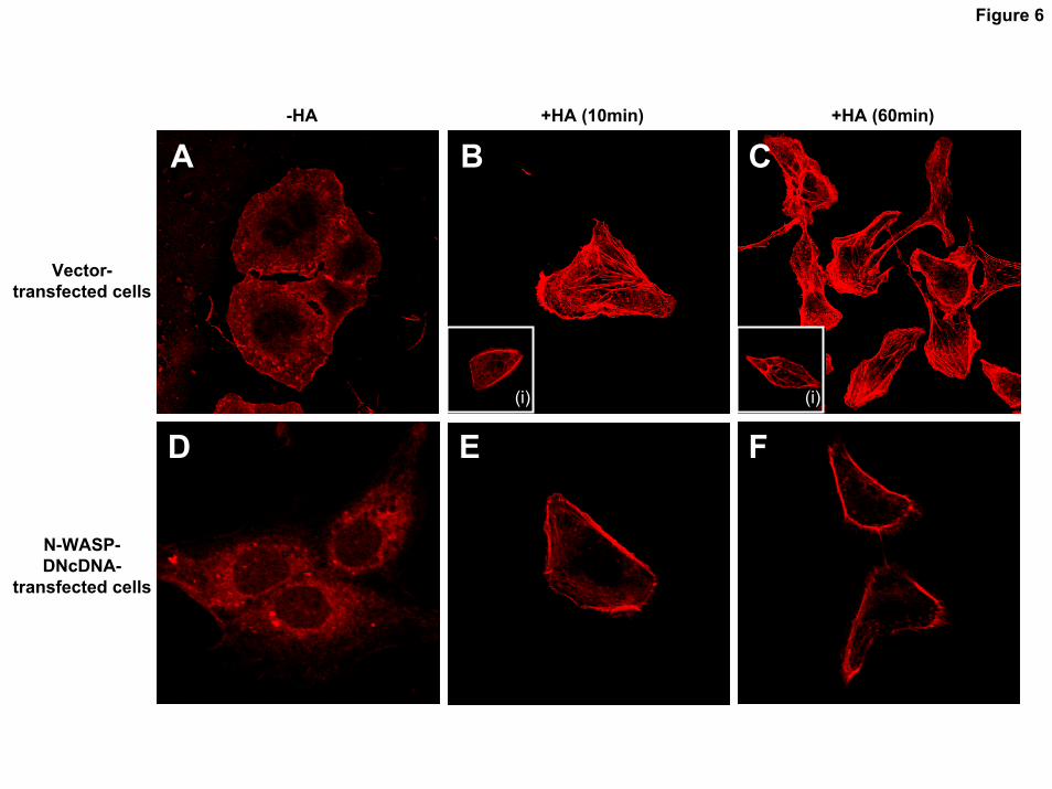
Figure 6
-HA +HA (10min) +HA (60min)
(i) (i)
A B C
D E F
Vector-transfected cells
N-WASP-DNcDNA-
transfected cells
by guest on October 12, 2018 http://www.jbc.org/ Downloaded from

Figure 7AA
1 2 3 4Immunoblot
a: Anti-N-WASP
b: Anti-ErbB2
c: Anti-CD44
IP: AntiIP: Anti--CD44CD44
BBImmunoblot
1 2 3 4
CC Cytosolic FractionCytosolic Fraction
a: Anti-p-ErbB2
b: Anti-ErbB2
Cell LysateCell Lysate
1 2 3 4
a: Anti-p-Tyrosine
(I)b: Anti-ß-Catenin
IP: AntiIP: Anti--ßß--CateninCatenin
Nuclear FractionNuclear Fraction
b: Anti- ß-Catenin
a: Anti-p-Tyrosine
1 2 3
IP: AntiIP: Anti--ßß--CateninCatenin
4
(II)
by guest on October 12, 2018
http://ww
w.jbc.org/
Dow
nloaded from

Figure 8
ßß--CateninCatenin ToproTopro--33 OverlayOverlay
a
a
b
b
c
c(ii) (iii)
(i)
(ii) (iii)
(i)
(iii)
(i)
(ii)
a b
AA
a b c
BB
CC
c
DD
by guest on October 12, 2018
http://ww
w.jbc.org/
Dow
nloaded from
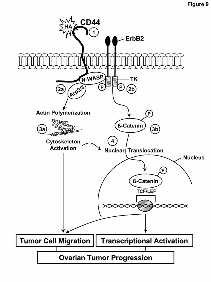
Figure 9
Transcriptional ActivationTranscriptional Activation
CD44CD44
2bN-WASP
Actin Polymerization
TCF/LEF
PP
NucleusNuclear Translocation
Arp2/3
HA
P
ß-Catenin
1ErbB2ErbB2
TK
2a
P
ß-Catenin3a 3b
4Cytoskeleton Activation
Tumor Cell MigrationTumor Cell Migration
Ovarian Tumor ProgressionOvarian Tumor Progression
by guest on October 12, 2018
http://ww
w.jbc.org/
Dow
nloaded from

Lilly Y.W. Bourguignon, Karine Peyrollier, Eli Gilad and Amy Brightmantumor cells
nuclear translocation, transcriptional upregulation and cell migration in ovarian -cateninβ2 activation leading to β(N-WASP) promotes actin polymerization and Erb
Hyaluronan-CD44 interaction with Neural Wiskott-Aldrich Syndrome protein
published online November 8, 2006J. Biol. Chem.
10.1074/jbc.M604672200Access the most updated version of this article at doi:
Alerts:
When a correction for this article is posted•
When this article is cited•
to choose from all of JBC's e-mail alertsClick here
by guest on October 12, 2018
http://ww
w.jbc.org/
Dow
nloaded from