hunter, sally m. (2009) identification of novel modifiers
TRANSCRIPT

This file is part of the following reference:
Hunter, Sally M. (2009) Identification of novel modifiers of chromosome inheritance: using a
genetically sensitised Drosophila model. PhD thesis, James Cook University.
Access to this file is available from:
http://eprints.jcu.edu.au/10926

CHAPTER 1: INTRODUCTION
2
1.1 THE PERPETUATION OF THE BLUEPRINT FOR LIFE
The perpetuation of life depends on the ability of living cells to both sustain and
replicate themselves. Cell division is a complex and highly regulated process whose
purpose is to distribute exactly half of the duplicated genome into each daughter cell,
and also to distribute the organelles and other cellular components evenly. This
process can and, not infrequently, does go awry, resulting in an abnormal
chromosome complement (aneuploidy) in the daughter cells. In humans, the effects
of an abnormal number of chromosomes vary from a relatively mild disturbance of
cellular function to lethality, with the vast majority of chromosomal variations not
being compatible with life. At the level of the whole organism the most widely
recognised chromosomal abnormality is Trisomy 21 or Down Syndrome, which
results from an extra copy of chromosome 21. Exactly why Down Syndrome in
particular is so common in humans remains elusive, as do the predominant cellular
and molecular causes behind chromosome missegregation. This chapter introduces
the human health consequences of aneuploidy and the current state of research
knowledge of the underlying causes and cellular processes involved in cell division.
This is followed by an in-depth look at key molecules underlying these processes,
and finally a look at a model system to allow investigation of the molecular causes
of chromosome missegregation.
1.2 HUMAN ANEUPLOIDY SYNDROMES
The phenomenon of chromosome missegregation is of considerable medical
significance in terms of infertility and chromosomal abnormality disorders.
Aneuploidy, an abnormal chromosome complement, arises through translocation of
parts of chromosomes or missegregation of whole chromosomes during mitosis or
meiosis. Aneuploidy arising in the gametes can lead to an aneuploid conceptus. In
humans, the majority of aneuploid foetuses die in utero or, if full term is reached,
suffer significant development defects and mental retardation. Despite the rigorous
regulation of DNA replication and chromosome segregation at the cellular level,
aneuploidy is a surprisingly common occurrence. An estimated 15-20% of all
human oocytes are chromosomally abnormal, with 5% of all clinically recognised

3
pregnancies displaying aneuploidy (Hassold et al., 2007). An estimated 57% of all
spontaneous abortions in women under 35 years of age during the first trimester are
due to chromosomal abnormalities, while this figure increases to greater than 80% in
women over the age of 35 (Hogge et al., 2003; Hassold et al., 1980; Hassold et al.,
1978). The vast majority of monosomies are lethal, with only monosomy of the X
chromosome being compatible with life. Trisomies of some of the smaller
chromosomes (eg 13, 15, 21, 22) can be viable, however, trisomic individuals suffer
varying developmental issues.
The most common human trisomy leading to a live birth, trisomy 21 (T21 or Down
Syndrome), is the leading cause of mental retardation in children, occurring in
approximately 0.45% of human conceptions and between 1/319 and 1/1000 live
births (Wiseman et al., 2009). Down Syndrome (T21), originally termed
"mongolism" or "mongolian imbecility", was first described by John Langdon
Haydon Down in 1866. As early as 1909 a link between birth order and Down
Syndrome had been observed, which led to the suggestion that children born later to
an older mother were more likely to suffer Down Syndrome (Shuttleworth, 1909).
From the 1930s several theories of a maternal age effect based on surveys and
statistical analysis were suggested (Penrose, 1934; Smith and Record, 1955). In
1959 Lejeune and Jacobs et al. determined the karyotypic abnormality underlying
Down Syndrome, most commonly an extra copy of the small acrocentric
chromosome 21.
For half a century links between maternal age, meiotic recombination, meiotic
missegregation events and aneuploidy have been recognised, but until recently,
surprisingly little progress had been made towards determining the molecular
mechanisms for meiotic aneuploidy. What is known is that two distinct mechanisms
exist: non-disjunction (ND), where chromosomes or chromatids fail to separate and
precocious division (PD), where chromosomes or chromatids separate prior to
anaphase (Angell, 1991). In meiosis I (MI) ND results in the loss or gain of a whole
extra chromosome, while PD results in gain or loss of a single chromatid
(Rosenbusch, 2006; Pellestor et al., 2002). The majority of evidence gathered
through DNA polymorphism and cytogenetic studies looking at the origin,
recombination rates and sites of recombination along the extra chromosome in T21

4
concluded that the majority of missegregation occurs during MI from non-
disjunction of bivalents (Valle et al., 2006; Perroni et al., 1990; Stewart et al., 1988;
Hamers et al., 1987; Warren et al., 1987). In contrast, studies using direct karyotype
analysis of unfertilised oocytes and their polar bodies indicate that PD has been
underestimated as a source of aneuploidy, most likely because earlier studies
assumed ND as the sole mechanism and scored single chromatids as whole
chromosomes (Pellestor et al., 2002; Angell, 1997). Kuliev et al. (2004) found that
single chromatids represented the majority of MI abnormalities, indicating a higher
rate of PD over ND. This study also indicated that MI errors are likely to occur only
slightly more frequently than MII errors, however, randomly segregating extra
chromatids appear to be directed to the polar bodies and therefore MII errors are
more frequently nullified and under-represented in conceptions.
Recurrence of T21 in subsequent pregnancies is higher in younger mothers, but the
incidence of T21 generally is higher in older mothers, with recent evidence
suggestive of different pathways for reaching T21 (Hulten et al., 2008; Hassold and
Hunt, 2008; Oliver et al., 2008). The exact circumstances that lead to cells failing to
segregate all their chromosomes correctly remain unclear. However, many genetic,
cellular and environmental factors are likely to contribute, as discussed below.
1.2.1 Current theories on the origins of female meiotic aneuploidy
1.2.1.1 Gender- and species-specific susceptibility
Compelling evidence from trisomy and polar body chromosome analyses suggests
that, when compared to human spermatogenesis, or oogenesis in other mamamalian
species, human oogenesis is acutely susceptible to chromosome segregation errors
(Pacchierotti et al., 2007; Fragouli et al., 2006). Studies mapping the origin of the
extra chromosomes indicate that ~90% of T21 cases are of maternal origin, while
~10% are of paternal origin (Hassold and Hunt, 2001). Although a maternal age
effect has also been demonstrated in mouse models, the effect is an order of
magnitude lower than that observed in human females. Such a dramatic difference
in error rates between spermatogenesis and oogenesis is less surprising as these are
quite distinct developmental processes and with significant divergence of the

5
regulatory mechanisms and pathways through evolution. Oogenesis in mammals, on
the other hand, is mechanistically very similar between species in terms of the
development of oocytes during embryogenesis, the presence of a prolonged meiotic
arrest, as well as conservation of key cell cycle checkpoints and regulatory
pathways. Why the error rate in humans is atypical, among mammals, is presently
unknown. It may be indicative of unique environmental influences specific to
humans, or of unidentified environmental, genetic or structural factors in some
human chromosomes that can influence their heritability and continuance in the
human genome pool.
1.2.1.2 Old oocytes
Spermatogenesis and oogenesis have quite distinct spindle requirements as
spermatogenesis involves two equal divisions resulting in four haploid gametes,
whereas oogenesis involves two asymmetrical cytokinesis events to produce a single
gamete and two polar bodies that contain the discarded chromosomes. In male
humans, meiosis commences when sperm production is required at the onset of
puberty and sperm are produced continuously throughout the lifetime of a man,
albeit with some decline in quality over time. In stark contrast, oocytes commence
the journey through meiosis when a female is in utero. Oocytes undergo prolonged
cell cycle arrest and only complete the first meiotic division when the oocyte is
released during ovulation sometime after the onset of puberty. This phenomenon of
pausing meiosis for an extended period of time and observations of a severe decline
in oocyte "fitness" after 35 years of age (the maternal age effect) has led many to
hypothesise that as women age, oocyte cell cycle checkpoint control becomes less
stringent and/or cellular repair mechanisms become less efficient, resulting in an
explosive increase in genetically flawed oocytes being ovulated. In women aged 20
years the risk of a T21 birth is 1/1,667, rising to 1/378 at the age of 35 and 1/11 at
the age 49 (ASRM, 2003). This age-dependent risk of aneuploidy also applies to
other chromosomes and the risk of spontaneous abortions increases dramatically
after the age of 35 (ASRM, 2003). It remains a point of debate as to whether errors
arise because the repair and monitoring systems have declined or whether the
remaining oocyte population, when approaching menopause, consists of a higher
proportion of poorer quality oocytes that the body is prepared to risk ovulating as a
response to the evolutionary consequences of not reproducing.

6
1.2.1.3 Altered recombination
Recombination, where homologous chromosomes undergo exchange of genetic
material, is an integral feature of meiosis, with normally at least one recombination
site per chromosome. Chiasmata, the physical sites of crossovers between
recombined chromosomes, are essential for meiotic chromosome cohesion and
accurate chromosome segregation. The number of crossovers per chromosome is
proportional to chromosome length, while the probability of missegregation is
inversely proportional to the number of crossovers and chromosome length. Hence,
small chromosomes (eg. human chromosome 21) suffer a relatively high degree of
missegregation. Investigations into the recombination rates between chromosomes
that have resulted in trisomy 21 have revealed that 45% of these chromosomes were
achiasmate (no recombination sites), compared to 0% of correctly segregated
chromosomes 21 (Oliver et al., 2008; Lamb et al., 1997). The sites of
recombination events have also been found to correlate with risk of T21, with 40%
of investigated T21 chromosomes having a single recombination site at the distal
end of 21q and 29% of investigated T21 chromosomes having a single
recombination site at the proximal end of 21q, compared to normally segregation
chromosomes 21 which had 56% of single crossovers at a median 21q position
(Oliver et al., 2008; Lamb et al., 1997).
Robles et al. (2009) have demonstrated that the number and positioning of
recombination sites are quite different between spermatocytes and oocytes, possibly
linking recombination with the sex-specific difference in T21 origin. It is not
unusual in humans for small chromosomes to have only a single recombination
event; however, the positioning of the crossover can be crucial (Robles et al., 2009).
When a homologous chromosome pair carry a single crossover positioned close to
the telomeres, the chiasma may not be sufficient to physically resist the tension of
the mitotic spindle causing premature bivalent separation. This telomeric crossover
characteristic is more commonly associated with T21 cases occurring with young
mothers. Conversely, chiasmata located in the pericentromeric region can also
induce missegregation events, a characteristic more prevalent in MII-derived T21
cases associated with older women. It is now widely recognised that a subset of MII
missegregation events are associated with pericentromeric recombination events that
arise during MI and only resolve in MII, therefore these cannot be considered to be

7
arising through a unique MII mechanism (Sherman et al., 2006; Hassold et al., 2000;
Hassold and Sherman, 2000). T21 cases associated with achiasmate chromosome
have been identified as equally common between younger and older mothers and no
evidence of a change in exchange rate is observed with increasing age (Oliver et al.,
2008; Lamb et al., 2005; Sherman et al., 2006). In younger women susceptible
recombination configurations (i.e. pericentromeric or telomeric positioning of
chiasmata) appear to be a leading risk factor for chromosome 21 MI missegregation,
while in older women MI missegregation frequently occurs with chromosomes that
have non-susceptible recombination configurations (Sherman et al., 2006). This is
suggestive of a general decline in the meiosis machinery fidelity with age.
The original studies into the origin of the extra chromosome in T21 developed the
theory that if the two copies of a maternal (or paternal) chromosome 21 were
heterozygous at the centromeric region then non-disjunction must have occurred
during MI (Figure 1.1). By this logic, chromosomes with homozygous centromeric
regions must have non-disjoined during MII. These analyses worked on the
assumption that the MI bivalents undergo non-disjunction, resulting in an extra
bivalent at MII and that both products of the MII division will each carry an extra
chromosome. In contrast, studies using MII arrested oocytes from assisted
reproduction programs have identified single extra chromosomes at MII as well as
the predicted extra bivalent chromosome pair (Fragouli et al., 2006; Mahmood et al.,
2000; Angell, 1997; Dailey et al., 1996; Angell et al., 1994; Angell et al., 1991).
This clearly demonstrates an alternative mechanism for aneuploidy to occur and
indicates that non-disjunction may not in fact be the main mechanism for trisomy,
but that premature separation of the kinetochores could drive chromatids to act as
univalents with the inheritance pattern being determined by the placement of
crossovers. It is also predicted that the more distal a crossover, the more likely that
chromatids rather than homologues will separate at the first meiotic division if
bivalents have undergone PD (Figure 1.1) (Wolstenholme and Angell, 2000; Angell,
1997). While this situation may resolve into a normal chromosome complement,
issues arise if the chromatids segregate randomly in MI and raises questions about
theories arising from studies that assumed MI or MII origin based on centromeric
homology.

8
Figure 1.1: Possible meiosis I chromosome configurations. (A) Normal meiosis I: homologous chromosomes synapse and segregate away from each other. (B) Non-disjunction of homologues: the bivalents fail to resolve into univalents and consequently missegregate to the same pole. (C) Pre-division of bivalents: cohesion is lost between homologues and chromatids; the remaining distal crossover directs the chromatids to segregate rather than the homologues.
Prometaphase I Metaphase I Anaphase I
(A) Normal meiosis I
(B) Non‐disjunction of bivalents
(C) Pre‐division of bivalents

9
1.2.1.4 Meiotic drive
Maternal age currently remains the major correlated risk factor for aneuploid
conceptions. This phenomenon is mathematically predicted by the Meioitic Drive
(MD) theory (Day and Taylor, 1998; Malik and Bayes, 2006), which assumes that
homologous chromosomes are driven to divide towards the ovum and away from the
polar body. Based on the asymmetric nature of female meiosis, the theory predicts
that females will be affected during MI, as this is when the greatest selective
pressure is applied as one homologue in a pair segregates to the ovum while the
other segregates to the polar body with no hope of contributing to the next
generation. The mathematical models predict the exponential increase in trisomic
conceptions that is observed in human females over the age of 35 years. An
explanation for this phenomenon is that as menopause, the finalé for chromosomal
inheritance from females, is approaching, meiotic drive exceeds the control
measures in place to prevent aneuploidy (Day and Taylor, 2006). The MD theory
only predicts the outcome of selective pressure on chromosome segregation and
does not indicate any molecular mechanism by which this may come to pass.
However, various examples exist of chromosomes that contain selfish DNA
elements that enable preferential inheritance, such as increased microtubule binding
sites at the centromere (Presgraves et al., 2009; Palestis et al., 2004; Henikoff and
Malik, 2002).
1.2.1.5 Grandmaternal effect
Recent research has implicated grandmaternal age as a significant contributor to risk
of T21 conceptions in daughters (Malini and Ramachandra, 2006). In contrast to
western studies that have previously demonstrated strongest correlation with
maternal age, pedigrees in larger Indian families demonstrate an extremely strong
association between grandmaternal age when daughters were conceived and T21
grandchildren. Of the 69 T21 cases investigated, 52 were born of women aged 18-
29 years, whereas in 61 of these cases the grandmothers were aged 30-40 years
when they had conceived the mothers (Malini and Ramachandra, 2006). Although
there is currently little in the way of other studies that support these findings, this
evidence raises interesting questions as to the molecular mechanisms by which
chromosomes mis-segregate to result in T21 conceptions. The most plausible
molecular explanation for the grandmaternal effect involves epigenetic mechanisms

10
and may indicate that reduced recombination rates are not maternal-age dependent,
but arise from much earlier events during development when grandmaternal
epigenetic influences alter recombination events in utero.
1.2.1.6 Ovarian mosaicism
In contrast to the historic wisdom that the majority of trisomies result from primary
non-disjunction of homologues during MI, there is evidence to support a theory that
many human females are actually trisomy 21 ovarian mosaics (Hulten et al., 2008;
Sachs et al., 2004; Tseng et al., 1994). In the Hulten study every ovary analysed
from eight developmentally normal foetuses (aborted for social reasons) was an
ovarian mosaic, however, only 0.2-0.88% of cells analysed were T21. This is
unlikely to explain the high recurrence rates of T21 observed in some women,
however, studies involving women with recurrent T21 conceptions identified T21
cells in the small population of cells from ovarian biopsies and ovulated oocytes,
indicating that in some women the number of T21 cells in the ovaries is likely to be
much higher than 0.2-0.88% (Sachs et al., 2004; Tseng et al., 1994; Nielsen et al.,
1988; Parke et al., 1980). High penetrance of ovarian mosaicism in the human
population potentially provides a new explanation for the observed maternal age
effect, as T21 oocytes have been found to develop more slowly and may be ovulated
much later (Hulten et al., 2008). Hulten et al. (2008) have also suggested that the
altered recombination rates and positions observed in T21 chromosome studies can
be explained by the presence of an extra chromosome 21 and secondary non-
disjunction when the cells divide. Cell division with an extra homologue has been
shown to result in a bivalent and an achiasmatic univalent or a trivalent (Barlow et
al., 2002; Luciani et al., 1976) that are predicted to force recombination events in
proximal or distal positions. Although many aspects of this theory are compelling, it
is currently unclear how this mosaicism would arise.

11
1.2.1.7 Aneugens and environmental effects
Many chemicals and other environmental factors have been implicated in increasing
aneuploidy (Table 1.2). Much of these data, however, comes from unreplicated
small-scale studies. Further evidence of effects on both sexes, replication in other
species and investigation of molecular mechanisms is clearly required. Some
investigations into environmental factors have proved intriguing but inconclusive;
this is most likely due to study design and assumptions employed. Study design is
extremely complex when considering which stage of meiosis may be relevant to
exposure, particularly in females where MI commences in the foetus and completes
at ovulation some 10-50 years later, and the synergistic effects of some chemicals
(Yang et al., 1999). Intracellular environmental factors that influence spindle-
forming ability or cell cycle progression have also been proposed as potential
aneugens. Van Blerkom (1996) demonstrated a link between reduced cellular pH,
impaired spindle formation and aneuploidy in aging oocytes. This situation has been
hypothesised for aging follicles where a hormone imbalance results in compromised
microcirculation, leading to increased intracellular CO2 and decreased pH (Gaulden,
1992).
Table 1.1: Known aneugens
Lifestyle factors Cigarette smoking
Wyrobek et al., 1993; Robbins et al., 1997b, 2003; Shi et al., 2001)
Alcohol (Robbins et al., 1997b) Caffeine (Robbins et al., 1997b)
Radiation Ionising radiation (reviewed in Verger, 1997)
Medications Chemotherapy
(Monteil et al., 1997; Robbins 1997a; Martin et al., 1995, 1997, 1999; De Mas et al., 2001; Frias et al., 2003)
Diazepam (Baumgartner et al., 2001) Oral contraceptive (Yang et al., 1999)
Chemicals Acrylonitrile (Xu et al., 2003) Bisphenol-A (BPA) Hunt et al., 2003; Susiarjo et al., 2007
Pesticides Trichlorfon (Czeizel et al., 1993)
Other (Padungtod et al., 1999; Xia et al., 2004; Recio et al., 2001; Smith et al., 2004)

12
1.2.1.8 Molecular mechanisms
Despite over 100 years of research into the causes of T21 and aneuploidy, much
remains unknown of the pathways, and the exact factors underpinning these
pathways, that lead to chromosome missegregation. The majority of hypotheses for
molecular mechanisms by which aneuploidy arises during meiosis are drawn from
the evidence of sex-specific rates of aneuploidy, the maternal age correlation,
reduced recombination rates and chemical inducers of aneuploidy. A key figure in
the hypothesised molecular mechanisms is the meiotic spindle, as it is central to
accurate chromosome segregation and provides the basis for a molecular checkpoint
in the cell cycle. Failure of the spindle assembly checkpoint (SAC) to halt cells that
have unattached chromosomes would severely disrupt the fidelity of chromosome
segregation and produce aneuploid cells (Vogt et al., 2008). Meiotic recombination
is also a key factor in the development of aneuploidy. As discussed in section
1.2.1.3, recombination rates and the positioning of recombination events both
correlate strongly with risk of chromosome 21 missegregation. Despite the strong
evidence linking recombination rates with aneuploidy, the mechanisms by which
recombination events are altered are still very vague. It is also becoming clear that
aneuploidy arising at different points within meiosis can have quite different
aetiology.

13
1.3 ACCURATE CHROMOSOME SEGREGATION
Missegregation of chromosomes is highly deleterious as it results in an abnormal
chromosome number (aneuploidy), disrupting the delicate balances of gene dosage,
regulatory networks and homeostatic processes that normally exist in a diploid cell.
Aneuploid yeast cells have been demonstrated to have cell cycle progression defects,
increased glucose uptake and increased sensitivity to conditions interfering with
protein synthesis and folding, generally putting them at a proliferative disadvantage
(Torres et al., 2007). The deterimental effects of altered gene dosage have also been
observed in Drosophila and human trisomy 21 fibroblast cell cultures (Lindsley et
al., 1972; Segal and McCoy, 1974). Studies in plants have shown that aneuploidy is
much more deleterious than polyploidy (whole genome duplication) due to the
imbalances generated in the stoichiometry of multi-protein complexes and other
dosage-sensitive relationships (Birchler and Veitia, 2007).
1.3.1 The cell cycle and checkpoints
Integral to the chromosome segregation process are the cell cycle restriction points
and checkpoint pathways. Entry into the cell cycle (Figure 1.2) is restricted in G1 in
response to nutrient availability, cell size, contact inhibition and other external
signals (G1 restriction point). Cell-cycle checkpoints ensure that any DNA damage
is repaired prior to DNA replication or cell division (DNA damage checkpoints G1-
S and G2-M), that chromosomes are fully replicated only once in a single round of
cell division (G2-M checkpoint), and chromosomes are correctly aligned on the
metaphase spindle (spindle assembly checkpoint, SAC) (Figure 1.2). The DNA
replication checkpoint and SAC function at very restricted points in the cell cycle,
halting the S-G2 and metaphase-anaphase transitions, respectively. While the DNA
damage checkpoint is activated in G1, it can halt cell cycle progression in the G1 or
G2 phases and slow down S phase while repair genes are upregulated (Elledge,
1996; Nasmyth, 1996; Vogt et al., 2007; Lukas et al., 2004). Checkpoint-arrested
cells that cannot repair damage, or correct replication or spindle errors, can be
eliminated by apoptosis, a cell death pathway which removes abnormal cells from
the body.

14
The SAC regulates the metaphase-anaphase transition, preventing precocious sister-
chromatid separation. The SAC specifically targets the APC-C through the effector
complex, mitotic checkpoint complex (MCC), made up for MAD2, BUBR1, BUB3
and the APC-C regulatory subunit CDC20 (Sudakin et al., 2001; Shannon et al.,
2002; Poddar et al., 2005). Many other proteins are considered as core components
of the SAC pathway, including BUB1, MAD3, MPS1 and AURORA-B, or are
recognised regulators of the SAC pathway, including ROD, ZW-10, ZWILCH,
PLK1, CDK1-cyclin B (Musacchio and Salmon, 2007; Karess et al., 2005; van Vugt
and Medema, 2005; D'Angiolella et al., 2003; Kallio et al., 2002). The proteins of
the SAC have been demonstrated to accumulate at the kinetochores of chromosomes
that are unattached by spindle microtubules at metaphase. These proteins disperse
once microtubules attach, providing a monitoring system for the orientation of
chromosomes and establishment of tension across the metaphase plate (Chen et al.,
1998; Taylor et al., 1998; Rieder et al., 1995; Rieder et al., 1994). Once the
chromosomes are correctly oriented the SAC becomes inactivated, this allows
activation of the APC-C, which marks Cyclin B and Securin for degradation, driving
on the onset of anaphase (Musacchio and Salmon, 2007; Hagting et al., 2002; Clute
and Pines, 1999). Securin is the stoichiometric inhibitor of Separase, the protease
that proteolytically cleaves the cohesin complex and allows chromosomes to
segregate.
The fidelity of the meiotic spindle has been identified as a pivotal player in the
maternal age effect. The asymmetry of the oogenesis divisions underlies the higher
observed susceptibility female gametogenesis to cytoskeletal abnormalities
(Pacchierotti et al., 2007; Shin and Choi, 2004). A sex-specific weakening of the
SAC has also been suggested as the potential key to the maternal age effect. These
hypotheses are supported by evidence of chemical induction of aneuploidy in MI of
rodents using microtubule damaging agents, including colchicine, griseofulvin,
taxol, vinblastine and benomyl (Swanton et al., 2009; Salmon et al., 1984; Sloboda
et al., 1982; De Brabander et al., 1986; Jordan and Wilson, 1998), which increased
the frequency of aneuploidy in rodent MII oocytes from a baseline of 0.5-1.0% to
23-42% (Mailhes et al., 1990, 1993a, 1993b, 1999; Russo and Pacchierotti, 1988;
Mailhes and Aardema, 1992).

15
Figure 1.2: The cell cycle and checkpoints. A cell that is signalled to replicate passes a restriction point in gap phase 1 (G1), from which point the cell accumulates mass and nutrients and is committed to divide. When sufficient resources have been acquired the cell enters the synthesis phase (S), when the entire genome is replicated once. Following on from replication the cell enters gap phase 2 (G2), when errors or damage that occurred during the replication process are detected and repaired. During the growth phases the organelles and lipid membranes must also be replicated and enlarged in preparation for cell division, which occurs directly following M phase when the replicated genome is divided exactly in half. Specific cyclin-CDK partners are responsible for controlling the entry and exit from each phase of the cell cycle.
CycE‐Cdk2 (CycA‐Cdk2)
CycB‐Cdk1 (CycA‐Cdk1)
G1‐S checkpoint (ATM, ATR, CHK1, CHK2, CDC25)
G2‐M checkpoint (ATM, ATR, CHK1,
CHK2, CDC25, BRCA1)
SAC checkpoint (MAD2, BUBR1, BUB3, CDC20)
CycD‐Cdk4

16
1.3.2 Chromosome cohesion and recombination
Cohesin (discussed in detail in section 1.4) is a protein complex that facilitates
chromatid cohesion and is essential for maintaining the chiasmata that link
homologous chromosomes (Kudo et al., 2009; Kudo et al., 2006; Buonomo et al.,
2000; Van Heemst and Heyting, 2000). Mice lacking SMC1β, a meiosis-specific
cohesin subunit demonstrate an age-dependent increase in aneuploid conceptions
and births closely resembling the age-dependent increase observed in the general
human population (Hodges et al., 2005). This is supported by a Drosophila model
where a reduction in SMC1 levels induces a concomitant increase in recombinant
homologue missegregation as the flies' age (Subramanian and Bickel, 2008). These
models indicate that a lack of cohesin to maintain chiasmata is tolerable in young
females with young oocytes, where other cellular processes are able to compensate.
Cohesin is an essential feature of chiasmata, preventing the crossovers "sliding off"
the end of the chromosome and being lost. A decline in chiasmata frequency with
increasing age was first observed in mouse oocytes by Henderson and Edwards
(1968). More recent studies have shown that, although the original sites of
recombination can be detected in gametes using MLH1 antibodies that localise
specifically to recombination foci, analysis of the physical crossovers frequently
reveals both distal "slippage" of the chiasmata or complete loss of chiasmata in the
absence of SMC1β containing cohesin complexes (Hodges et al., 2005). Once all
chiasmata linking bivalents are lost, nothing remains to hold the chromosome pairs
together and they are able to randomly segregate. Therefore, loss of cohesin from
chromosomes with increasing age is a potential suspect that could explain the
frequent loss of genomic integrity with increasing maternal age. However, if or how
cohesin is lost from meiotic chromosomes is presently unknown.
The observed decrease in chiasmata number with age has led some scientists to
hypothesise that the functionality of the proteins holding chromosomes together,
which are originally loaded in S phase prior to MI arrest, may decline as the proteins
age. There is little in the way of direct evidence to suggest that the same protein
molecules are maintained from foetus to adulthood. Instead, experiments from
several groups have demonstrated that chromosome-bound cohesin complexes are
highly dynamic and undergo frequent exchange during a cell cycle without affecting
chromosome cohesion (McNairn and Gerton, 2009; Gerlich et al., 2006). Other

17
research groups have demonstrated global loading of cohesin in response to DNA
damage following replication, indicating that there are potential mechanisms within
cells by which proteins holding the chromosome together can be replaced.
Therefore, it would seem plausible that, with increasing age, there is a general
decline in the efficiency and stringency of all pathways leading to accurate
chromosome cohesion and segregation.
The close resemblance of the mouse SMC1β and the Drosophila SMC1 models to
the human maternal age effect is very suggestive of the cohesin complex playing a
central role in the maternal age effect. In-depth studies of the cohesin complex and
individual subunits could prove instrumental in making progress on the causes of
aneuploidy.
1.4 THE COHESIN COMPLEX
1.4.1 Identification of the cohesin complex
The mitotic cohesin complex is made up of four protein subunits, SMC1, SMC3,
SCC3 and RAD21 (Figure 1.3), all of which have identifiable homologues in all
other eukaryotes studied to date (Nasmyth et al., 2001) (Table 1.1). Cohesin
components were first identified in budding yeast; Mcd1/Rad21/Scc1 was identified
in genetic screens for genes involved in chromosome structure and later found to be
essential for chromosome condensation and cohesion (Jachymczyk et al., 1977;
Birkenbihl and Subramani., 1995; Guacci et al., 1997; Furuya et al., 1998; Uhlmann
et al., 1999). The Smc1 subunit was initially identified as a protein necessary for
nuclear division (Strunnikov et al., 1993) and later the Smc1, Smc3 and Scc1
subunits were discovered in a genetic screen for high frequency loss of
chromosomes in the absence of Anaphase Promoting Complex (APC) function
(Michaelis et al., 1997). The Scc3 cohesin subunit was first identified by Tόth et al.
(1998), in a screen for genes that permitted nuclear division in the presence of
anaphase inhibitor Pds1/Cut2. The nature of the cohesin complex began to emerge
when it was demonstrated that Scc1 requires Smc1 to associate with chromatin
(Michaelis et al., 1997). This was followed by purification of a multisubunit
complex containing Scc1, Smc1, Smc3 and two other proteins from Xenopus cell

18
extracts (Losada et al., 1998). Cells carrying mutations in components of the
cohesin complex display high frequency chromosome missegregation. In the
presence of a functional SAC, these cells undergo arrest due to an inability to align
chromosome pairs at the metaphase plate and/or segregate the chromosomes
(Michaelis et al. 1997).
Meiotic-specific cohesin isoforms have been identified in vertebrates for the SMC1
(SMC1β), RAD21 (REC8) and SCC3 (STAG3) subunits (Revenkova et al., 2004;
Xu et al., 2004; Watanabe and Nurse, 1999). Cohesin complexes containing these
alternative subunits are only found in meiotic cells, whereas 'mitotic' cohesin
complexes are observed in both mitotic and meiotic cells. The cohesin complex also
has two associated proteins ECO1/CTF7 and PDS5, which are not considered
subunits but are essential for the regulation of cohesin dynamics (Skibbens et al.,
1999; Panizza et al., 2000).
Table 1.2: Cohesin nomenclature in various model organisms.
S. c
erev
isia
e
S. p
ombe
D. m
elan
ogas
ter
X. la
evis
M. m
uscu
lus
H. s
apie
ns
SMC1α Smc1 Psm1 SMC1 SMC1 SMC1α SMC1α SMC1β n.p. n.p. n.p. n.i. SMC1β SMC1β SMC3 Smc3 Psm3 SMC3 SMC3 SMC3 SMC3 SCC1 Mcd1/Scc1 Rad21 dRAD21 xRAD21 mRAD21 hRAD21 REC8 n.p. Rec8 n.p. REC8 REC8 REC8 SCC3 Scc3 Psc3 SA
SA2 STAG1 STAG2
STAG1 STAG2
STAG1 STAG2
STAG3 n.p. Rec11 n.i. STAG3 STAG3 STAG3 ECO1 Eco1 Eso1 DECO
n.i. SAN
n.i. ECO2
n.i.
ESCO1 ESCO2 NAT13
ESCO1 ESCO2 NAT13
PDS5 Pds5 Pds5 PDS5 n.i.
PDS5A PDS5B
PDS5A PDS5B
PDS5A PDS5B
SCC2 Scc2 Mis4 NIPPEDB xSCC2 NIPBL NIPBL SCC4 Scc4 Ssl3 dMAU-2 SCC4 n.i. hMAU-2 n.i. – Not yet identified in this species n.p. – Not present in this species

19
1.4.2 The SMC subunits: SMC1 and SMC3
The structural maintenance of chromosomes (SMC) proteins is a diverse family of
proteins with a conserved SMC structure with globular amino- and carboxy-termini
joined by long α-helical regions. A hinge domain in the centre allows each protein to
fold back on itself to form a long rod-shaped molecule with the globular amino- and
carboxy-termini joining to form an ABC-like ATPase domain. The two cohesin
SMC proteins, SMC1 and SMC3, interact via their hinge regions to form a V-shaped
heterodimer (Figure 1.3) (Anderson et al., 2002) and some lines of investigation
have suggested that the SMC1 and SMC3 ATPase domains are also able to interact
directly and form a closed ring independently of the RAD21 subunit (McIntyre et
al., 2007; Weitzer et al., 2003). ATP hydrolysis at the SMC ATPase head is
stimulated by the carboxy-terminal winged-helix domain of the Scc1 subunit in S.
cerevisiae (Arumugam et al., 2006) and this hydrolysis may facilitate opening of the
SMC1/SMC3 heterodimer hinge domain to allow the ring complex to encircle DNA
strands (Gruber et al., 2006; Aruguman et al., 2003l Weitzer et al., 2003).
In bacteria, a single SMC homodimer complex has been identified that influences
chromosome compaction and segregation and is potentially a key player in super-
coiling (Grauman and Knust, 2009). In contrast, three distinct SMC complexes exist
in metazoans: the condensin complex (SMC2-SMC4 heterodimer), cohesin complex
(SMC1-SMC3 heterodimer), and the SMC5-SMC6 heterodimer. Condensin has
demonstrated chromosome condensation and dosage compensation functions
(Hirano et al., 1997; Jessberger et al., 1998). The cohesin complex (SMC1-SMC3)
is involved in chromosome cohesion and homologous recombination (Michaelis et
al., 1997; Losada et al., 1998), while the SMC5-SMC6 complex has been linked to
DNA damage repair, sister-chromatid recombination (Michaelis et al., 1997;
Jessberger et al., 1998; Losada et al., 1998; Torres-Rosell et al. 2005; Piccoli et al.,
2009) and appears to form an integral Separase-independent cohesin removal
mechanism (Outwin et al., 2009).

20
1.4.3 Kleisins
The subunit referred to as RAD21/Scc1 belongs to a family of proteins referred to as
α-kleisins (Schleiffer et al., 2003; Haering and Nasmyth, 2003) that binds to the
globular ATPase heads of the SMCs via its conserved amino- and carboxy-terminal
regions. The RAD21/Scc1 amino terminus binds to the SMC3 subunit, while the
carboxy-terminus binds the SMC1 subunit in the cohesin ring (Figure 1.3). The
conservation of amino acid sequences at only the amino- and carboxy-terminal
domains in all identified kleisins (α and γ families) (Figure 1.4A) and exclusive
association with SMC proteins indicates that all SMC dimer pairs are likely to have
an associated kleisin subunit and form ring structures (Figure 1.4B) (Schleiffer et al.,
2003). In yeast the cohesin ring is removed from chromosomes by proteolytic
cleavage of the kleisin subunit (Rad21 or Rec8) by the cysteine protease Separase.
Only members of the kleisin-α family are known to be proteolytically cleaved in this
way (Kudo et al., 2006; Buonomo et al., 2000; Yanagida, 2000; Uhlmann, 2001;
Uhlmann et al., 1999).
Figure 1.3: The mitotic cohesin complex. Cohesin complexes consist of four proteins. The two structural maintenance of chromosomes (SMC) subunits, SMC1 and SMC3, form a heterodimer via their hinge regions and ATPase domains. The SCC1/RAD21 subunit binds to the globular terminal domains of SMC1-SMC3 and also interacts with the fourth subunit, SCC3.
SMC ATPase domains
SMC hinge region
SMC3 SMC1
RAD21 SCC3
C
C
N
N
N C

21
The RAD21/Scc1 kleisin subunit was first identified in yeast as a mitotic cohesin
complex factor, however, in both yeast and mammals it has also been demonstrated
to bind to meiotic chromosomes (Xu et al., 2004). REC8 is a meiosis-specific
kleisin with significant amino acid sequence similarity to RAD21 and is essential for
the reductional chromosome segregation of meiosis (Watanabe and Nurse, 1999).
RAD21 has been demonstrated to be necessary for the bipolar positioning of sister-
chromatids at the metaphase plate, where sister kinetochores are attached to opposite
spindle poles. In contrast, REC8 has been demonstrated to be responsible for
monopolar positioning of sister-chromatids in MI, where the sister kinetochores are
attached to the same spindle pole (Figure 1.4C) (Watanabe and Nurse, 1999;
Yokobayashi et al., 2003). These spindle orientations are crucial for the correct
segregation of the chromosomes.
The requirement for both RAD21- and REC8-containg cohesin complexes in meiotic
mammalian cells can be explained in part by their differential chromosomal binding
sites adjacent to the kinetochores (Allshire, 1997; Watanabe and Nurse, 1999; Eijpe
et al., 2003; Kitajima et al., 2003; Xu et al., 2004). Both REC8 and RAD21 are
found at the centromere during MI and MII, where each has unique binding sites and
distinct roles in cohesion and kinetochore orientation. These discrete sites of
localisation indicate that the subunit composition of the cohesin complex is
important for regulation, however, current knowledge cannot sufficiently explain
how this regulation occurs. REC8 has been demonstrated to have key roles in
homologue pairing, synaptonemal complex (SC) formation and meiotic
recombination (Brar et al., 2009), thereby covering many of the additional logistical
requirements that are present in meiosis but not mitosis.
1.4.4 The Scc3 subunit
SCC3, the fourth cohesin subunit, is also highly conserved among multicellular
eukaryotes and is part of the stromal antigen (SA) family of proteins. Three
isoforms (SA1, SA2 and STAG3) have been identified in vertebrates, two isoforms
in Drosophila and only one isoform in both budding and fission yeast (Losada et al.,
2000; Prieto et al., 2001). STAG3 is a meiosis-specific isoform, whereas SA1 and
SA2 are both found specifically in mitotic cohesin complexes (Pezzi et al., 2000).

22
Figure 1.4: Human SMC complexes. (A) The conserved SMC-binding amino- and carboxy-terminal domains of the kleisin protein superfamily (α-helices in blue and β-sheets in red), the central unconserved region is of variable sequence and length between species and family members. (B) Human mitotic cohesin complex with RAD21 α-kleisin (i); human meiotic cohesin with REC8 α-kleisin (ii); human SMC2/SMC4 complex with HCAP-H γ-kleisin (iii); human SMC5/SMC6 complex with a predicted kleisin, currently unidentified (iv). (C) Monopolar orientation of homologous chromosomes in meiosis I directs sister-chromatids to the same spindle pole (i), in contrast in meiosis II and mitosis the sister-chromatids become bipolar oriented and segregate to opposite spindle poles (ii).
Bipolar orientation (meiosis II and mitosis)
Monopolar orientation (meiosis I)
(A)
HEAT
SMC5 SMC6
?
SMC5/SMC6
SMC2 SMC4
HCAP-H
Condensin
HEAT STAG3
Meiotic cohesin
SMC3 SMC1β
REC8
Mitotic cohesin
SMC3 SMC1α
RAD21
SA1/SA2
(B) (i) (ii) (iii) (iv)
(C)

23
The ratio of SA1 and SA2 containing cohesin complexes varies throughout Xenopus
development, with SA1 being the predominant form in embryos (Losada et al.,
2000). SA1 and SA2 also display differential nuclear localisation patterns, with SA1
localising directly to chromatin, while SA2 appears to localise to the nuclear
envelope (Losada et al., 2000). The SCC3/SA subunit binds to the carboxy-terminal
region of RAD21/SCC1 in the yeast cohesin complexes (Figure 1.3). While the
exact role of the SCC3/SA subunit remains to be fully defined, the variations in
isoforms and temporal and spatial expression pattern variation indicate a likely role
for SCC3/SA as a regulatory subunit.
1.4.5 Cohesin forms a ring structure
Although many models have been presented to explain how cohesin modulates sister
chromatid cohesion, the majority of evidence indicates that the cohesin complex
forms a ring-like quaternary structure (Figure 1.5) (Gruber et al., 2003; Ivanov and
Nasmyth, 2005; Ivanov and Nasmyth, 2007; Haering et al., 2008). Cohesin is
believed to either enclose both sister-chromatids following replication of the
chromosomes or to concatenate sister-chromatids through the linking of multiple
cohesin rings. Current evidence indicates cohesin molecules form locked rings that
are sufficiently sizeable to encompass two sister chromatids (Figure 1.5B(i)),
providing the simplest explanation for how cleavage of the RAD21 subunit could
result in release of the sister-chromatid cohesion (Figure 1.5A) (Haering et al.,
2008). An alternative "handcuff" model has been proposed (Figure 1.5B(ii)) (Zhang
et al., 2008), where individual cohesin rings circle single DNA helices. This model
is hypothesised to provide the flexibility cohesin requires to accommodate other
cellular functions such as DNA repair and transcription that occur while cohesin is
bound to chromosomes; a flexibility that is thought to be lacking in the single ring
model. The cellular reality may be that different conformations of cohesin rings are
necessary to achieve the varied roles that cohesin fulfills in chromosome cohesion,
DNA repair and transcription regulation.
Although the finer details of how cohesin acts at the molecular level are still
unresolved, cohesin has been conclusively demonstrated to be capable of linking
DNA molecules and is required to establish and maintain sister-chromatid cohesion.
The cellular cohesin population is highly dynamic and is regulated by many different

24
factors. Cleavage of the RAD21 subunit allows the cohesin ring to open and
dissociate from the chromatids, thereby allowing them to separate to separate and
segregate to their respective spindle poles (Figure 1.5A). This hypothesised
mechanism is supported by experiments where the ring was artificially "opened" by
cleaving the SMC3 subunits using a tobacco etch virus (TEV) protease and TEV
cleavage sites inserted into the coiled-coil of the SMC3 protein (Gruber et al., 2003;
Ivanov and Nasmyth, 2005). Exactly how this opening is achieved is unclear as it
has long been assumed that the cleavage provides an opening at the ATPase heads.
However, it has also been hypothesised that the RAD21 subunit may actually
regulate the opening of the hinge region and that removal of the amino-terminal
portion of RAD21 alters hinge dynamics (Shintomi and Hirano, 2007).
1.4.6 Identified binding sites
In the search for cohesin binding sites, initial indicators pointed towards there being
no cohesin-specific DNA sequence motif. In yeast cohesin complexes seemed to be
mainly found at A-T rich regions located on average every 10-15kb apart. Closer
analysis of the binding sites revealed that cohesin largely associates with
chromosomes at intergenic regions, which in S. cerevisiae happen to be A-T rich
(Tanaka et al. 1999; Laloraya et al. 2000). This is believed to support the concept
that cohesin binding and active transcription must be mutually exclusive, as the
cohesion ring is too small to physically accommodate the transcription machinery
passing through it. However, as de novo cohesin binding has not been demonstrated
to occur between S phase and anaphase, cohesin complexes may simply migrate to
transcriptionally silent loci and intergenic regions to accommodate active
transcription. Evidence from yeast studies provides support for this notion, with
cohesin complexes being translocated from their original loading sites to mainly
congregate in the non-coding regions at the ends of converging transcriptionally
active genes (Bausch et al., 2007; Lengronne et al. 2004); presumably having been
pushed there by the transcription machinery. In contrast to yeast, studies in
Drosophila and human cells have found that the loading factor NippedB (NIBL) and
cohesin colocalise on chromosomes at transcriptionally active sites (Misulovin et al.,
2008; Dorsett et al., 2005). Recruitment of cohesin to these sites appears to be
dependent on the SCC3/SA subunit interacting with the CCTC- binding factor,

25
Figure 1.5: Cohesin and the sister chromatid cohesion model. (A) The cohesin rings encircle the sister chromatids (black) following replication of the genome. This cohesive force is sufficient to oppose the pulling forces of the spindle microtubules (light and dark green) attached to the kinetochore of each chromatid. At the onset of anaphase Separase cleaves the RAD21 subunit of the cohesin rings and the sister chromatids are released. (B) Proposed models for cohesin engagement of chromatids: (i) the widely accepted model where a single ring encompasses both chromatids and (ii) the "hand-cuff" model where single rings encompass single chromatids, with interaction between cohesin subunits/motifs to achieve cohesion. (Adapted from Nasmyth and Schleiffer, 2004).
(A)
(B) (i) (ii)
Spindle microtubules
Kinetochore proteins Cohesin rings
Metaphase‐to‐anaphase transition
RAD21 cleavage
Chromatids

26
CTCF (Rubio et al., 2008; Parelho et al., 2008; Wendt et al., 2008). CTCF has
demonstrated insulator functions, a role in which NippedB and cohesin have now
been implicated (Rubio et al., 2008; Gause et al., 2008). Although there is a strong
association between cohesin localisation and CTCF, cohesin does not exclusively
colocalise with CTCF. It is also currently unclear as to whether CTCF is associated
only with cohesin to facilitate transcription regulation or whether there are additional
roles in chromosome cohesion.
A study performed in human cells found that cohesin complexes appeared to be
recruited to Alu repeats by the chromatin remodelling factor SNF2h (Hakimi et al.
2002). Alu repeats fall into the class of short interspersed DNA elements, with
approximately 500,000 copies per cell of the 300-bp dimeric sequence comprising
~10% of human genomic DNA (Shankar et al., 2009). Interestingly, Alu elements
are G-C rich, which is in complete contrast to the A-T rich cohesin binding regions
identified in yeast (Hakimi et al. 2002). Multiple cohesin loading factors and site
variation may explain the differences observed at identified cohesin binding sites.
The Alu repeats appear to be associated with specific loci and may correlate with the
general transcription activity of the gene. SNF2h and RAD21 are found to both
locate to Alu elements and RAD21 binding is dependent on SNF2h and its ATPase
activity (Hakimi et al. 2002). Histone methylation and acetylation levels appear to
be important factors in SNF2h and Rad21 binding, with experiments using the DNA
methyltransferase inhibitor 5-azacytidine to decrease methylation resulting in
recruitment of RAD21 to Alu sites where SNF2h alone had been localised before
(Hakimi et al. 2002). Similar to the specific methylation observed at centromeres,
methylation of histone H3 lysine four (K4) correlates with SNF2h binding to
chromatin. Additionally, acetylation of H3 and/or H4 underlines the importance of
histone modification in cohesin loading. The absolute necessity for SNF2h ATPase
activity in cohesin binding suggests that chromatin remodeling is an essential step in
cohesin association (Hakimi et al. 2002). Further support for the chromatin-
structure hypothesis is the finding that cohesin shows greatly increased general
affinity to chromatin over naked DNA and that recognised cohesin binding sites in
yeast do not have a conserved sequence, but instead show conservation of
nucleosome structure (Kagansky et al., 2004).

27
1.4.7 Cohesin loading
Cohesin complexes are not capable of spontaneously associating with chromatin.
Instead, cohesin requires other factors to facilitate loading onto chromatin in a cell
cycle-specific manner, and Scc1 and Scc3/SA are not capable of binding to
chromatin in the absence of the SMC subunits. SMC1 and SMC3 bind to DNA via
their C-terminal domains, however, chromatin localisation is notably increased when
all fours subunits of the cohesin complex are present (Akhmedov et al., 1998; Toth
et al,. 1999; Ciosk et al. 2000). Exactly how the cohesin ring functions to open,
encompass the DNA strands, and then close and maintain cohesion has been the
subject of several investigations. Several studies have shed light on this mystery by
demonstrating that the ATPase heads of SMC1 and SMC3 interact directly and
functionally bind ATP (Arumugam et al., 2003; Weitzer et al., 2003). ATP
hydrolysis is believed to facilitate both opening and closing of the cohesin ring to
allow binding to DNA (Arumugam et al., 2003; Weitzer et al., 2003), although
whether this opening occurs at the ATPase heads or at the hinge regions remains to
be determined conclusively (Milutinovich et al., 2007; Gruber et al., 2006; Hirano
and Hirano, 2006).
The proteins Scc2 and Scc4 form a complex that has been identified as a cohesin
loading factor in budding yeast that is present throughout the cell cycle (Ciosk et al.,
2000). Homologues of Scc2 and Scc4 have been identified in fission yeast and some
metazoans (Seitan et al., 2006; Watrin et al., 2006; Bernard et al., 2006; Krantz et
al., 2004; Dorsett, 2004). Scc2 is a member of the Adherin family of proteins,
which have now been suggested to promote the ATP-hydrolysis activity of
SMC1/SMC3 and thus facilitate cohesion ring opening and chromatin loading
(Arumugam et al., 2003; Weitzer et al. 2003). Scc2 promotion of cohesin ATPase
activity and opening of the cohesin ring suggests a direct interaction. Surprisingly,
Scc2 and cohesin components have not been shown to co-immunoprecipitate, and
therefore any direct interaction is likely to be transient. Also, yeast Scc2 and
cohesin fail to co-localise on chromatin (Ciosk et al., 2000) making it difficult to
accurately hypothesise a loading mechanism for cohesin.
As cohesin has been found to preferentially bind at sites of convergent transcription
in genome wide mapping experiments, it is possible that the Scc2/Scc4 interaction

28
with cohesin is very transient with subsequent translocation of cohesin complexes by
the transcription machinery (Glynn et al., 2004; Lengronne et al., 2004). Studies of
the Drosophila Scc2 homologue, NippedB, have also failed to demonstrate co-
immunoprecipitation with cohesin. However, it has been clearly shown that cohesin
and NippedB colocalise on chromosomes, preferentially at sites of active
transcription (Misulovin et al., 2008). It may be that the more open conformation of
actively transcribed DNA is amenable cohesin loading; however, cohesin and
NippedB have also been linked functionally in regulation of transcription. Together
cohesin and NippedB regulate gene expression by as yet unconfirmed mechanisms,
with current evidence indicating a role in modification of long range enhancer
element interactions with target genes (Wendt et al., 2008). This may reflect a
cooperative role in regulation of gene expression which is not present in yeast.
Consistent with this, mutations in the human homologue of Scc2, NIPBL, have been
linked to Cornelia de Lange syndrome, a rare multisystem developmental disorder
that also shows variable somatic karyotype abnormalities (Krantz, et al., 2004;
Tonkin et al., 2004).
Scc2 and Scc4 are both essential for the loading of yeast cohesin on both
chromosome arms and at centromeres, suggestive of a conserved functional role in
these distinct chromatin domains (Ciosk et al., 2000). Other lines of evidence
indicate that the metazoan Scc2/NippedB homologues are not always required for
cohesin loading, in particular at centromeric sites. Scc2-independent loading of
yeast cohesin has been observed when a population of cohesin appears to be
dislodged from the chromatin by the transcription machinery and is then reloaded,
but no longer confers cohesion between sister-chromatids (Bausch et al., 2007).
Scc2-independent loading also occurs when the centromeric region of pre-anaphase
chromosomes undergoes "breathing", where the chromatids transiently separate a
greater distance than can be encompassed by cohesin rings (Ocampo-Hafalla et al.,
2007). The centromeric region appears to provide a unique cohesin binding
environment in metazoans that is differentially regulated compared to chromosomes
arms. In Drosophila at least, NippedB does not appear to be essential for loading of
cohesin at the centromeres of meiotic chromosomes (Gause et al., 2008).
Additionally, other factors involved in heterochromatin and kinetochore formation,
such as histone H3 variant CENP-A (Cse4/Cid) and heterochromatin protein HP1

29
(Swi6), are associated with the concentrated centromeric loading of cohesin (Eckert
et al., 2007; Bernard et al., 2001). Swi6 has been found to co-immunoprecipitate
with Mis4, the fission yeast Scc2 homologue (Fischer et al., 2009), confirming a
direct link between Swi6 activity and cohesin loading.
1.4.8 Establishment of cohesion
The binding of cohesin to chromatin is necessary but not sufficient for the
establishment of cohesion between chromatids. Instead a number of factors,
including Eco1/Ctf7, Wpl1 and Pds5, are also required to “lock” the cohesin ring
around the sister-chromatids, thereby tethering them together. Although cohesin is
observed to associate with chromatin from telophase (in metazoans) or late G1 phase
(in yeast), cohesion is only established with the aid of Eco1/Ctf7 once the
chromosomes are replicated in S phase (Brand and Skibbens, 2005). After cohesion
is established in S phase ECO1 is not required for cohesion maintenance throughout
the G2 and M phases (Toth et al. 1999). Mutations in the human ESCO1 gene are
linked to Roberts Syndrome, a multisystem developmental disorder with patients
displaying chromosomal defects (McDaniel et al. 2005; Vega et al. 2005; Gordillo et
al., 2008). Eco1 encodes an acetyltransferase that has been demonstrated to
acetylate the Mcd1 (Rad21) and Smc3 subunits of yeast cohesin to antagonise the
cohesin displacing activities of Wpl1 (Wapl) (Heidinger-Pauli et al., 2009; Ivanov et
al., 2002). During S phase, when cohesion is established, the Smc3 cohesin subunit
is acetylated by Eco1, causing the Wpl1-Pds5 regulatory complex to temporarily
dissociate from cohesin complexes and thereby allowing cohesin rings to
functionally engage with sister-chromatids (Rowland et al., 2009; Sutani et al.,
2009). Wpl1 normally inhibits cohesion establishment post S phase, however, Eco1
is able to antagonise Wpl1p activity in response to DNA double-stranded breaks
during G2/M through acetylation of Mcd1, resulting in genome-wide loading of
cohesive cohesin (Heidinger-Pauli et al., 2009).
Loading of cohesin and establishment of cohesion have been closely linked to DNA
replication, with further support coming from studies showing that DNA
polymerases are also needed for effective sister-chromatid cohesion (Uhlmann and
Nasmyth, 1998). S. cerervisiae polymerase ε (Pol 2) has an amino-terminal region

30
with essential DNA polymerase activity, while the C-terminal region interacts with
polymerase σ, which acts to significantly increase the polymerase activity of the
polymerase ε holoenzyme. Two redundant genes, Trf4 and Trf5, which are also
nucleotidyl transferases, encode polymerase σ. Trf4 has demonstrated functions in
sister-chromatid cohesion, completion of S phase and DNA repair, and potentially
functions in cohesin loading by competing with the RNA helicase Dbp2 for binding
to Pol 2, where it may affect transient remodelling at the replication fork and thereby
allow interaction with cohesins (Wang et al. 2000). Pol 2 has also been shown to
interact with Smc1 and Eco1/Ctf7, providing a link between progression of the
replication fork and establishment of cohesion (Edwards et al. 2003).
Although the exact mechanisms are still being elucidated, there is a clear link
between activity at the replication fork and cohesin loading and establishment. In S.
cerevisiae, Ctf18 and Elg1 form alternative replication factor C (RFC) and RFC-like
(RLC) protein complexes, respectively, in conjunction with the small RFC subunits
Rfc2-5 (Parnas et al., 2009; Bellaoui et al., 2003). These complexes are present at
the replication fork with Eco1, being required for sister-chromatid cohesion and
DNA repair (Maradeo and Skibbens, 2009; Bellaoui et al., 2003; Mayer et al. 2001).
The RFC and RLC complexes act as clamp loaders for DNA replication clamps,
including PCNA (pol30), and may be involved with polymerase switching at sites of
cohesin binding (Mayer et al. 2001). PCNA is a DNA polymerase processivity
factor that stabilises polymerase association with DNA, and over-expression of
PCNA is found to partially rescue the effects arising from loss of Eco1/Ctf7
(Skibbens et al. 1999). Ctf7 physically interacts with both PCNA and the RFC/RLC
complexes (Moldovan et al., 2006; Majika and Burgers, 2004; Skibbens, 2005;
Kenna and Skibbens, 2003), however, these appear to represent redundant cohesin
loading mechanisms as mutations in PCNA and RFC/RLC components produce
approximately 1/2-1/3 of the cohesion defects observed in cohesin subunit mutants
or Eco1/Ctf7 mutants (Guacci et al., 1997; Michaelis et al., 1997; Skibbens et al.,
1999; Toth et al., 1999; Moldovan et al., 2006; Mayer et al., 2001; Kenna and
Skibbens, 2003; Edwards et al., 2003). Another protein, Ctf4, is present with the
RFC and/or RLC and Eco1 at the replication fork during S phase and is thought to
be a central figure in coordinating DNA replication, checkpoint activation,

31
chromosome cohesion and DNA damage repair (Yoshizawa-Sugata and Masai,
2009). The current hypothesised models involve Eco1-driven partial dismantling of
the replication fork to allow passage through a single cohesin ring. Alternatively,
single cohesin rings may be transiently opened in an Eco1-Pds5-dependent manner,
or potentially Eco1 and Pds5 regulate pre-loaded cohesin ring opening/closing while
driving ring-ring interactions with newly loaded cohesin rings in a catenation
cohesion model (Skibbens et al., 2007).
1.4.9 Cohesin dynamics and cohesion maintenance
Following DNA replication, cohesion is maintained via the interplay of three
cohesin maintenance factors: Wapl (Wpl1), Sororin and Pds5. These proteins are
considered regulatory subunits of the cohesin complex as they coimmunoprecipitate
with cohesin complexes from HeLa cells and are dependent on cohesin for their
association with chromatin (Losada et al., 2005; Sumara et al., 2000; Rankin et al.,
2005; Kueng et al., 2006; Gandhi et al., 2006). In yeast cells Wpl1 and Pds5 have
been found to form a regulatory heterodimer that acts an "anti-establishment" factor
for cohesin mediated cohesion (Sutani et al., 2009). Ctf7 and Pds5 have also been
found to coimmunoprecipitate from yeast cells, with overexpression of either protein
able to suppress the temperature sensitivity of the reciprocal mutant (Noble et al.,
2006). These interactions suggest that Pds5 has a role in cohesion establishment as
well as cohesion maintenance. This role was first recognised for Pds5 in budding
yeast, where loss of Pds5 results in premature chromatid separation and abnormal
chromosome condensation (Hartman et al., 2000; Stead et al., 2003), and has more
recently been shown to result in failure of homologue synapsis during meiosis (Jin et
al., 2009). Pds5 clearly has a complex regulatory role in metazoan cohesin
dynamics, for although Pds5 colocalises with cohesin throughout the cell cycle the
reduction of Pds5 has varying effects on chromosome segregation. In human HeLa
cells reduction of Pds5 results in premature chromosome separation, while mitotic
chromosomes in Xenopus egg extracts lacking Pds5 do not suffer loss of cohesion at
the chromosomes arms, show only a loosening of cohesin at centromeres, and retain
a high level of cohesin on chromosomes into mitosis (Losada et al., 2005). Fission
yeast Pds5 mutants display hypersensitivity to DNA damage and metaphase delay,
as well as some cohesion defects (Wang et al., 2002). Mutations in the human Pds5
homolog, PDS5B, cause development disorders and multisystem defects and are

32
seen in a subset of cases of Cornelia de Lange syndrome that are not associated with
mutations in NIPB-L (Dorsett, 2007; Dorsett, 2004). Differences observed in Pds5
function in different species may reflect the nature of mutant alleles examined or
reflect species-specific variations in cohesin regulation.
At least two populations of cohesin exist in the cell, stably-bound cohesin, which is
predicted to be required for maintaining cohesion, and dynamic cohesin, which
cycles rapidly on and off chromatin with uncertain functional consequences. Wapl
and Sororin have counteractive effects on the residency time of dynamically- and
stably-associated cohesin on chromatin, respectively. Wapl destabilises chromatin
binding by the dynamic cohesin population, thereby reducing residency time,
preventing cohesion establishment in G1 and promoting cohesin removal from
chromatid arms during prophase. Experiments uncoupling the events of cohesin
stabilisation and DNA replication may indicate that cohesin stabilisation is required
for cohesion establishment rather than the other way round (Bernard et al., 2008).
The destabilising activities of Wapl are counteracted in S phase by the
acetyltransferase activity of Eco1, thereby allowing stabilisation of cohesin
association with chromatin and establishment of cohesion (Bernard et al., 2008;
Ben-Shahar et al., 2008; Sutani et al., 2009; Rowland et al., 2009). Overexpression
of Wapl in human cells results in premature sister chromatid separation and
chromosome instability, and upregulation of Wapl has been identified in cells
infected by human papillomavirus (HPV), which are highly susceptible to tumour
progression (Ohbayashi et al., 2007).
Sororin is necessary for maintaining the population of stably bound cohesin and
unlike Wapl, has as yet only been identified in metazoans (Díaz-Martínez et al.,
2007; Rankin et al., 2005). Sororin is required for the maintenance of chromosome
cohesion and DNA damage repair during G2 phase, with Sororin over-expression
resulting in failure of sister-chromatids to resolve and segregate at anaphase.
Sororin-depleted HeLa cells display complete loss of chromatid cohesion in
metaphase and temporary arrest by the spindle checkpoint (Schmitz et al., 2007;
Díaz-Martínez et al., 2007; Rankin et al., 2005).

33
1.4.10 Cohesin dissociation – prophase
In yeast the entire population of chromosome-bound cohesin is cleaved by Separase
at the onset of anaphase to facilitate opening of the cohesin ring and separation of
the sister-chromatids (Uhlmann et al., 1999; Ciosk et al., 1998). In contrast, in
metazoans a prophase dissociation pathway also exists that allows removal of
cohesin molecules from the chromosome arms by a non-proteolytic,
phosphorylation-dependent mechanism (Figure 1.6). At the onset of anaphase, the
small pool of centromeric cohesin and any remaining arm cohesin is removed by
separase (Nasmyth and Schleiffer, 2004; Hauf et al., 2001; Uhlmann et al., 1999;
Ciosk et al., 1998). While arm- and centromere-specific mitotic cohesin complexes
do not differ in their subunit composition, there are clear differences in the
regulation of dissociation of these two populations from chromosomes. The cohesin
prophase dissociation pathway involves POLO (PLK1) kinase phosphorylation of
the Scc3/SA subunit in combination with the cohesin-destabilising activities of Wapl
(Hauf et al., 2005; Sumara et al., 2002). Meanwhile, the centromeric cohesin is
protected and retained until anaphase when the Scc1/RAD21 subunit is cleaved by
Separase. This differential regulation appears to be achieved through protection of
centromeric cohesin from phosphorylation by Shugoshin (SGO1/MEIS332) proteins
and protein phosphatase 2A (PP2A) activity (Kitajima et al., 2006; Riedel et al.,
2006).
MI comprises a particularly complex situation where there are two differentially
regulated segregation events where sister-chromatids are kept together during MI,
while allowing homologous chromosomes pairs to separate (Rabitsch et al. 2004;
McGuinness et al. 2005). Current evidence indicates that the Shugoshin proteins
and PP2A are responsible for protecting the centromeric cohesins of sister-
chromatids from phosphorylation and cleavage during both mitosis and MI
(Kitajima et al., 2006; Riedel et al., 2006; McGuinness et al., 2005). SGO1
localises to centromeres at prophase and then disappears at the onset of anaphase as
would be expected of a metazoan cohesin-protection protein. Loss of SGO1
function results in loss of cohesin at the centromere and consequently loss of sister-
chromatid cohesion causing cells to arrest in a prometaphase-like state.
(McGuinness et al., 2005). SGO1 localisation at the centromere it determined by the
spindle checkpoint protein BUB1, and PP2A and AURORA B kinases.

34
Figure 1.6: Cohesin cell cycle dynamics. Cohesin complexes are regulated through each phase of the cell cycle by a wide variety of proteins. Such tight regulation in loading, maintenance and removal is essential for accurate chromosome segregation.
LOADING SCC2/SCC4
PROPHASE DISSOCIATION PLK1 WAPL SGO1 PP2A BUB1 AURORA B
ESTABLISHMENTECO1 CTF4 RFC RLC MAINTENANCE
PDS5 WAPL SORORIN
METAPHASE‐TO‐ANAPHASE TRANSITION SEPARASE SECURIN CYCLIN B APC‐C
Cohesin complex Centromere Chromatin Nuclear membrane Spindle
G2 G1
S

35
Additionally, BUB1 and AURORA B centromeric localisation is inter-dependent
(Tang et al., 2006; Kawashima et al., 2007; Kitajima et al., 2005; Resnick et al.,
2006) . Loss of BUB1 function results in SGO1 mis-localisation along the entire
length of the chromosome (Bernard et al., 1998; Basu et al., 1999; Kitajima et al.,
2005). It is quite feasible that protein components of the centromere determine the
localisation of BUB1 and AURORA B, which in turn restricts the localisation of
SGO1 to the centromere and thereby effects the differential regulation of
centromeric cohesin that is observed.
Several other factors have been linked with the cohesin prophase dissociation step;
however, less data are currently available for these proteins. Depletion of Hapsin, a
histone H3 kinase, or Prohibitin 2 (PHB2) results in premature sister chromatid
separation in mitosis. Additionally, overexpression of Haspin rescues the cohesion
defect observed in SGO1 depleted cells, while co-depletion of PHB2 and POLO is
able to rescue the cohesion defects of PHB2 mutants (Dai et al., 2006; Takata et al.,
2007).
1.4.11 Cohesin dissociation - anaphase
Although the majority of chromatin bound cohesin is removed from metazoan
chromatid arms during prophase and prometaphase (Figure 1.6), a fraction of
cohesive cohesin remaining on the arms and a significant centromeric pool requires
the activity of Separase for complete removal at anaphase (Figure 1.6) (Nakajima et
al., 2007; Uhlmann et al., 1999; Ciosk et al., 1998). The protease responsible for
cohesin removal, Separase, is held inactive by the regulatory subunit Securin and by
inhibitory phosphorylation of Separase by Cdk1-cyclinB. At the onset of anaphase,
the Anaphase Promoting Complex/Cyclosome (APC-C) marks securin and cyclin B
for degradation by the proteasome via ubiquitylation (Ulhmann, 2001) Once
Separase becomes actived it is free to cleave the RAD21/Scc1 component of cohesin
complexes bound to the chromatin. In vitro studies have also shown cleavage of
cohesin is promoted by POLO phosphorylation of the RAD21 subunit of cohesin
(Hornig et al., 2004; Alexandru et al., 2001). RAD21 cleavage by Separase occurs
at either or both of two conserved sites within the protein sequence (Figure 1.7).
The core consensus cleavage sequence ((DE)XXR/X) is observed in the RAD21 and
REC8 homologues from yeast to humans, with the invariant arginine residue (R)

36
flanking the peptide bond cleaved by Separase (Figure 1.7B). RAD21/Scc1
cleavage leaves the invariant arginine as the carboxy-terminal residue of the amino-
terminal cleavage products, with the amino-terminal residue of the peptide fragment
being a destabilising residue that targets the peptide for degradation by the
ubiquitin/proteosome-dependent N-end rule pathway (Rao et al., 2001).
Degradation of the carboxy-terminal cleavage peptide is essential for chromosome
stability and persistence of this peptide has been found to be lethal in budding yeast
cells (Rao et al., 2001). Studies in human and yeast cells have found that both of the
Separase cleavage sites in RAD21/Scc1 need to be altered in order to inhibit
Separase cleavage and chromosome segregation. This inhibition of chromosome
segregation resulted in a substantial increase in anaphase bridges and aneuploidy
(Toyoda et al., 2002; Hauf et al., 2001; Tomonaga et al., 2000).

37
Figure 1.7: Cleavage-dependent cohesin removal. At the metaphase-anaphase transition the APC-C initiates degradation of securin and cyclin B, releasing their inhibition of separase activity. (A) Separase (red scissors) proceeds to cleave the RAD21 subunit of all cohesin complexes remaining on the chromosomes at this time. (B) RAD21 contains two separase cleavage sites, which have a consensus sequence that is evolutionary conserved from yeast to humans (Hs, Homo sapiens; Sc, Saccharomyces cerevisiae; Sp, Shizosaccharomyces pombe; Dm, Drosophila melanogaster) in RAD21 and REC8 homologues. (C) Cleavage of the RAD21 Separase sequences occurs immediately after the conserved arginine residue (R).
N C
RAD21
RAD21 Hs 167 DREIMRE 445 IEEPSRL
Sc 175 LVEVGRR 263 SVEQGRR
Sp 174 SIEAGRN 226 SIEVGRD
Dm 170 TPEIIRC 469 APEVLRA
(B)
(C)
(A)
175aa 474aXXEXXR/X XXEXXR/X
715aa
SMC3 SMC1
RAD21 SCC3
C
C
N
N
SEPARASE
N C

38
1.5 DROSOPHILA MELANOGASTER AS A MODEL TO STUDY
ANEUPLOIDY
1.5.1 Aneuploidy studies in Drosophila
Drosophila melanogaster has a long history of being utilised as a model organism
for human aneuploidy research (Birchler et al., 2001; Morton et al., 1990; Grell and
Valencia, 1964; Dallapiccola et al., 1978). Drosophila achiasmate chromosomes
mimic the altered recombination and non-disjunction that are believed to underlie
some meiotic aneuploidies (Koehler and Hassold, 1998) and have been used
extensively to identify genes that influence chromosome inheritance. In-depth
studies in Drosophila to determine the minimum functional centromere resulted in
the development of minichromosomes, a collection of reduced chromosomes with
compromised inheritance rates that have also been a key tool in identifying genetic
factors that influence chromosome inheritance (Dobie et al., 2001; Sun et al., 1997,
Cook et al., 1997). More recently a Drosophila model system for studying the
maternal age effect in older oocytes has been developed (Jeffreys et al., 2003).
Many genes that normally function to prevent aneuploidy have been discovered and
functionally analysed in Drosophila, including the kinetochore protein Mitch
(Williams et al., 2007), the SAC proteins Rod, Zw-10 , BubR1 and Bub3 (Karess
and Glover, 1989; Williams et al., 1992; Lopes et al., 2005; Logarinho et al., 2004)
the checkpoint protein Chk2 (Takada et al., 2003), the APC-C subunit ida (Bentley
et al., 2002), the cell cycle regulator Myb (Manak et al., 2002) and the meiotic
recombination protein Ord (Bickel et al., 1996).
Induction of aneuploidy has also been studied in Drosophila using various chemicals
that humans are commonly exposed to or that are informative about cellular
mechanisms (Osgood and Cyr, 1998; Ferguson et al., 1996; Liang and Brinkley,
1985; Held, 1982; Zimmering, 1982; Traut, 1981; Traut, 1978).
1.5.2 The development of the Drosophila GMR>Rad21NC mutant
A study was undertaken by Keall (2005), to characterise Drosophila Rad21 with the
view of utilising the Drosophila cohesin complex as an instrument for investigating
chromosome segregation and aneuploidy. Studies with human and yeast non-
cleavable RAD21/Scc1 had demonstrated inhibition of the normal segregation of

39
chromosomes resulting in a substantial increase in anaphase bridges and aneuploidy
(Toyoda et al., 2002; Hauf et al., 2001; Tomonaga et al., 2000). From these data it
was hypothesised that ectopic expression of non-cleavable RAD21 (RAD21NC) in
Drosophila should produce similar cellular effects and an observable phenotype.
The work of Keall (2005) identified two Separase cleavage sites in the Drosophila
melanogaster RAD21 sequence based on their similarity to known RAD21/Scc1
cleavage sites in humans and yeast (Figure 1.7). Mutation of these Separase sites
and ectopic expression of the RAD21 cleavage variant transgenes in the developing
Drosophila eye (Figure 1.8) was indeed observed to result in disturbance of normal
tissue development (Keall, 2005).
The Separase cleavage sequence is highly conserved between species, with an
invariant arginine residue denoting the point of cleavage, which occurs at the peptide
bond between the arginine and the following residue (Figure 1.9). Site-directed
mutagenesis of the invariant arginine residues to alanine at the two identified
Drosophila RAD21 cleavage sites (ie R175 and R474) was the tactic used to inhibit
Separase cleavage (Figure 1.9). Ectopic expression in the developing eye of the
RAD21N (R175A) protein with the single amino-terminal mutation produced no
observable phenotypic effects, in contrast to ectopic expression of the RAD21C
(RA474AG) protein with the single carboxy-terminal mutation or RAD21NC
(R175A/RA474AG) protein with both sites mutated, which both produced a reduced
and disorganised eye phenotype (Figure 1.9). Both the overall size of the eye and
the organisation of the ommatidia were severely reduced in the RAD21NC cleavage-
mutant transgenics (Keall, 2005). The ability of the RAD21C cleavage variant to
produce a mutant phenotype was in contrast to the previous studies in yeast and
human cells, as these studies had found mutation of both Separase cleavage sites
necessary to produce inhibition of chromosome segregation (Toyoda et al., 2002;
Hauf et al., 2001; Tomonaga et al., 2000). The phenotype produced by RAD21C
and the lack of phenotype produced by RAD21N led Keall (2005) to speculate that
the carboxy-terminal Separase cleavage site was the dominant site of RAD21
cleavage in Drosophila.

40
Figure 1.8: Drosophila eye development. The compound eye is a particularly useful and powerful organ for performing genetic screens because of its highly organised nature and being unnecessary for viability and fertility. In the developing eye cells proliferate asynchronously until the morphogenetic furrow passes from the posterior to the anterior of the eye-antennal imaginal disc, inducing a G1 arrest ahead that synchronises the cells followed by a final round of division of a subset of cells termed the 2nd mitotic wave. Cells posterior to the MF undergo differentiation as they mature and are recruited to ommatidia. The compound eye contains approximately 750 ommatidia, hexagonal clusters of 11 very specifically arranged cells that are formed into very exact diagonal rows. This array is highly sensitive to perturbations in eye development, such as alterations in cell numbers, ommatidial rotation, and cell recruitment to ommatidia. Transgenes can be ectopically expressed in the eye using a tissue-specific drive and the yeast Gal4-UAS system. The eye-specific driver Glass Multimer Reporter (GMR) uses the eye-specific transcription factor glass (gl) to drive expression of Gal4 in cells posterior to the morphogenetic furrow. Gal4 binds specifically to the Upstream Activated Sequences (UAS) associated with the transgene, thereby allowing expression of the transgene in developing eye cells (green circles). (Adapted from Freeman, 1997).
Anterior
Posterior
Morphogenetic furrow (MF)
MF movementG1 arrest
Proliferation
Transgene expressing cells
2nd mitotic wave

41
Figure 1.9: Drosophila Rad21 cleavage variants. (A) Separase cleavage of the RAD21 protein occurs after the invariant arginine residue (red). The Drosophila Rad21 cDNA was altered, by site-directed mutagenesis, at one or both of the amino- and carboxy-terminal Separase cleavage sites. At the amino-terminal cleavage site the arginine reside (R) was mutated to alanine (A), while at the carboxy-terminal cleavage site the arginine residue and consecutive alanine (RA) residue were mutated to alanine and glycine (AG). (B) Expression of the double-mutant Rad21NC under the control of the GMR>Gal4 driver resulted in a change in eye phenotype, with significant and reproducible reduction in the size and organisation of the eye (B) compared to a wildtype eye (A). This GMR>Rad21NC mutant eye phenotype was also found to be modifiable in a heterozygous mutant background; with modifiers showing both suppression (GMR>Rad21NC/Egfrk05115) (C) and enhancement (GMR>Rad21NC/dlUY2278) (D).
175aa 474aa
TPEIIRC APEVLRA
715aa
TPEIIAC APEVLAG
RAD21WT
RAD21NC
TPEIIAC APEVLRA RAD21N
TPEIIRC APEVLAG RAD21C
(A)
(B)
(A) (B) (C) (D)

42
From the above observations it was hypothesised that cleavage-resistant RAD21 was
being incorporated into functional cohesin rings that were inhibiting the segregation
of replicated chromosomes. By using markers of mitosis and apoptosis, Keall
(2005) showed that there was an increased mitotic index and increased level of
apoptosis in the eye-antennal imaginal discs of the Rad21NC transgenics, with an
eye-specific driver, compared to wildtype. These results are consistent with the cells
being unable to segregate their replicated chromosomes and consequently arresting
in mitosis and eventually apoptosing due to an inability to resolve the segregating
chromatin masses.
One of the strengths of the Drosophila system is its utility in performing genome-
wide genetic screening. The Drosophila GMR>Rad21NC model was judged to be a
suitable and effective tool to screen for modifiers of chromosome inheritance. This
was based on initial observable changes in the GMR>Rad21NC rough-eye phenotype
when the transgene was expressed in heterozygous mutant backgrounds for genes
known to regulate cohesin and chromosome segregation. This preliminary testing of
the screening tool was performed by Keall (2005) and included: the RAD21
cleaving protease subunits, three-rows and pimples (Separase homologue); the
cohesin loading factor, NippedB (Scc2 homologue); the mitotic cell cycle regulator,
Cyclin B; and the centromeric cohesin maintenance factors, polo kinase and mei-
S332. These genes were all found to modify the GMR>Rad21NC in a manner
consistent with and predictable from their known functions.
A genome-wide genetic screen was carried out by Keall (2005), observing whether
large chromosomal deletions, when hemizygous, were capable of altering the rough-
eye phenotype produced by GMR>Rad21NC. In this screen, Keall (2005) used the
'Deficiency Kit' of overlapping deletions to screen more than 90% of the Drosophila
melanogaster genome. With this approach, 57 interacting regions that either
suppressed or enhanced the GMR>Rad21NC phenotype (Figure 1.10) were identified.
Keall also identified 11 interactors to the gene level in 8 of these regions.

43
1.6 THE SCOPE OF THIS PROJECT
All disease manifestations are a combination of an individual's environment,
underlying genetics and stochastic events (Figure 1.11). Dissecting the relative
contribution of these factors is a complex task that is compounded by the complexity
of mammalian genomics and cell biology, and the relative ubiquity of many
environmental influences that may contribute to the outcome. Aneuploidy is another
instance where many factors influence the outcome. Genetic risk factors have long
been recognised for many human diseases; however, knowledge of the molecular
mechanisms that contribute to aneuploidy is still relatively scarce. Additionally,
despite many decades of research, the vast majority of evidence for the origins of
meiotic aneuploidy remains at the macroscopic level of maternal age effect,
recombination rates and the incidence of meiotic aneuploidy in human populations.
Much additional information is to be garnered regarding the molecular mechanisms
behind mitotic and meiotic aneuploidy, and studies in model organisms have and
will continue to play a key part of this process.
Figure 1.10: Cytological positions of chromosomal deletion regions that modified the GMR>Rad21NC rough-eye phenotype. Enhancing deletions are indicated by the red brackets and suppressing deletions are indicated by the green brackets. The size of the bracket is indicative of the size of the deletion.

44
Following on from the work of Keall (2005) several key issues remained outstanding
in the investigation of chromosome cohesion and segregation modifiers using the
GMR>Rad21NC model. Firstly, it remained to be definitively demonstrated that
mutation of the Drosophila Rad21 Separase cleavage sites resulted in an observable
change in RAD21 cleavage patterns and that inhibition of RAD21 cleavage resulted
in an observable chromosome segregation phenotype at the cellular level. Secondly,
further characterisation of the GMR>Rad21NC phenotype was necessary to
understand the full extent of the cellular effects of inhibiting chromosome
segregation. And finally, the identity of the modifier loci, within the chromosomal
regions identified in the original Deficiency Kit screen, remained to be determined at
the molecular level.
This project aimed to identify novel modifiers of chromosome segregation and
aneuploidy-survival genes using Drosophila melanogaster as a model organism.
Drosophila models offer the benefits of a multicellular system that yeast models
lack, without the complexities and costs encountered when working with
mammalian models. Homologues for the mitotic cohesin subunits SMC1, SMC3,
RAD21 and SA/SCC3 have all been identified in Drosophila, along with key
cohesin regulators including SCC2, DECO/ECO1, WAPL, POLO and Separase
(Williams et al., 2003; Verni et al., 2000; Warren et al., 2000; Jones and Sgouros,
Figure 1.11: Disease manifestation. Disease manifestation is dependent on an individual's age, underlying genetics and their environment.
Environment(age)
Stochastic events
Genetics

45
2001; Sunkel and Glover, 1988). The lack of an identifiable Rec8 homologue in
Drosophila is suggestive of conservation of cohesin regulation between somatic
cells and gametes, and opens up the possibility of using the Drosophila RAD21
cohesin in a somatic screen for regulators of chromosome segregation. Additionally
RAD21-containing cohesin complexes are involved in meiosis in other species.
Drawing on the initial studies undertaken by Keall (2005), this metazoan model of
chromosome missegregation has been utilised to identify novel modifiers of
chromosome cohesion and segregation. By using a system of excessive cohesion it
was hoped that genes able to reduce cohesion would be identified and that these
genes would form a plausible shortlist of genes that may act as risk factors for loss
of cohesion and aneuploidy in humans. Defective chromosome cohesion is a risk
factor for chromosome missegregation and aneuploidy in MI, MII and mitosis. The
following chapters describe characterisation of the GMR>Rad21NC mutant, a
genome wide modifier screen to identify metazoan-specific chromosome cohesion
regulators, the identification of 133 modifying loci at the molecular level and
refinement of this modifier list using secondary assays.

46
CHAPTER 2:
MATERIALS AND METHODS

47
2.1 GENERAL LABORATORY CONSUMABLES
2.1.1 General laboratory chemicals
All chemicals used were of analytical grade and were obtained from the following
companies: Sigma, Sigma-Aldrich, ProSciTech, MP Biomedicals, Spectrum,
Univar, Calbiochem, Amresco, Fluka and Lancaster. Distilled water was obtained
using a Barnstead Nanopure Type I ultrapure water system.
2.1.2 Commonly used solutions
• Tris-Acetate EDTA (TAE): 40mM Tris, 20mM acetate, 2mM EDTA, pH 8.1
• Tris-EDTA (TE): 10mM Tris-HCl, pH7.5, 1mM EDTA
• 1X PBS: 140mM NaCl, 3mM KCl, 2mM KH2PO4,
10mM Na2HPO4
• 1X PBT: 1X PBS, 0.1% Triton X-100
• Luria-Bertani broth (LB): 1% tryptone, 0.5% yeast extract, 1% NaCl, pH 7
• LB with ampicillin (LBA): LB, 75µg/mL ampicillin
• LB plates: LB plus 1.5% agar
• Resuspension Solution: 50mM Tris-HCl, 10mM EDTA, 100µg/mL
RNase A, pH 7.5
• Lysis Solution: 1% SDS, 0.2M NaOH
• Neutralisation Solution: 1.32M potassium acetate, pH 4.8
• DNA Binding Solution: 1.5g celite [Diatomaceous Earth, Sigma]
suspended in 100mL 7M Guanidine HCl pH 5.5
• Wash Solution: 80mM potassium acetate, 8.3mM Tris-HCl pH 7.5,
40µM EDTA, 50% ethanol.
2.1.3 DNA modifying enzymes
All DNA restriction and modification enzymes were purchased through Promega
Corporation or New England Biolabs. Enzymes were used in accordance with the
manufacturers’ recommendations and/or used in standard procedures described in
Sambrook and Russell (2001).

48
2.2 GENERATION OF PLASMIDS
2.2.1 Minipreparation of plasmid DNA
All minipreparations of plasmid DNA were performed using a modified alkaline
lysis method with laboratory-made solutions and Wizard Spin Columns (Promega).
Overnight cultures in 1.5 or 3mL of LBA medium were pelleted microcentrifuged at
6,500 x g for 3 mins. Bacterial pellets were resuspended in 200µL of Resuspension
Solution. Bacterial cells were lysed by addition of 200µL of Cell Lysis Solution and
within 5 mins this reaction was neutralised by the addition of 200µL of
Neutralisation Solution. The precipitated proteins, chromosomal DNA and SDS
were then pelleted by microcentrifugation at 15,400 x g for 10 minutes. The
supernatant was combined with 1mL of DNA Binding Solution and the solution
pulled through a Wizard Spin column (Promega) using a vacuum manifold.
Columns were then washed with 3mL or more of Wash Solution. Excess ethanol
was removed by centrifuging the columns at 4,400 x g for 60 seconds followed by a
brief air-drying (5 minutes). Plasmid DNA was then eluted from the column by
applying 15-60µL of Tris-EDTA (TE pH7.4) buffer and centrifuging at 6,500 x g for
1 minute.
2.2.2 Large-scale preparation of plasmid DNA
Large-scale plasmid DNA preparations were performed with 25mL or 100mL using
the Qiagen Midiprep Kit according to manufacturer’s recommendations. Plasmid
DNA was eluted using 300µL or 1mL of TE, respectively.
2.2.3 Preparation of Drosophila genomic DNA from single flies
Genomic DNA was prepared from single flies after euthanasing by freezing in a
1.5mL microfuge tube. A 200µl pipette tip containing 30µl of "squishing" buffer
(1mM Tris-Cl pH8.2, 1mM EDTA, 25mM NaCl, 200µg/ml Proteinase K) was then
used to mascerate the fly for 30 seconds without expelling any of the liquid. The
dissociated fly was suspended in the squishing buffer and incubated at room
temperature for 20-30 minutes. The Proteinase K was inactivated by heating the
sample at 95˚C for 2 minutes. Preparations were stored at 4˚C and typically 1µl was
used as template per 10-15µl PCR reaction (Gloor et al., 1993).
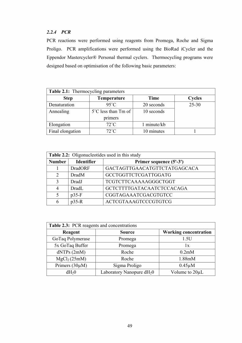
49
2.2.4 PCR
PCR reactions were performed using reagents from Promega, Roche and Sigma
Proligo. PCR amplifications were performed using the BioRad iCycler and the
Eppendor Mastercycler® Personal thermal cyclers. Thermocycling programs were
designed based on optimisation of the following basic parameters:
Table 2.1: Thermocycling parameters Step Temperature Time Cycles
Denaturation 95˚C 20 seconds 25-30 Annealing 5˚C less than Tm of
primers 10 seconds
Elongation 72˚C 1 minute/kb Final elongation 72˚C 10 minutes 1
Table 2.2: Oligonucleotides used in this study Number Identifier Primer sequence (5'-3')
1 DradORF GACTAGTTGAACATGTTCTATGAGCACA 2 DradM GCCTGGTTCTCGATTGGATG 3 DradJ TCGTCTTCAAAAAGGGCTGGT 4 DradL GCTCTTTTGATACAATCTCCACAGA 5 p35-F CGGTAGAAATCGACGTGTCC 6 p35-R ACTCGTAAAGTCCCGTGTCG
Table 2.3: PCR reagents and concentrations Reagent Source Working concentration
GoTaq Polymerase Promega 1.5U 5x GoTaq Buffer Promega 1x
dNTPs (2mM) Roche 0.2mM MgCl2 (25mM) Roche 1.88mM Primers (30µM) Sigma Proligo 0.45µM
dH20 Laboratory Nanopure dH20 Volume to 20µL

50
2.2.5 Agarose gel electrophoresis
Agarose gels were made using 1x Tris-Acetate-EDTA (TAE) buffer (40mM Tris-
acetate, 20mM sodium acetate, 1mM EDTA, pH8.2), 1-2% Molecular Biology
Grade Agarose (Spectrum) and supplemented with 50µg/ml ethidium bromide.
DNA samples were mixed with an appropriate volume of 6x loading dye prior to
loading into wells and run alongside Lamda DNA digested with HindIII/EcoRI
(Promega) as a size marker. Gel electrophoresis was performed in 1xTAE buffer at
100V for 45-60 minutes.
2.2.6 Restriction digest and PCR cleanup
DNA from restriction digests and PCRs intended for use in cloning were purified
using Qiagen's QIAquick Gel Extraction Kit or MinElute PCR Purification Kit.
Both kits were used in accordance with the manufacturer's recommendations.
2.2.7 Ethanol precipitation of DNA
DNA was precipitated where necessary using 1/10 volume of sodium acetate (3M
NaOAc pH5.2) to 1 volume of DNA solution, followed by addition of 2 volumes of
cold 100% ethanol. Following incubation on ice for 30 minutes, samples were
centrifuged at 4˚C for 15 minutes at 15,000 x g. After discarding the supernatant the
DNA pellet was washed with 200µL of 70% ethanol, centrifuged at 4˚C for 5
minutes at 15,000 x g and left to air dry. Finally the pellet was resuspended in 20µL
of Tris-EDTA buffer.
2.2.8 DNA ligation
Ligation reactions were set up using a 3:1 molar ratio of insert DNA:vector DNA
using the following protocol, where X and Y indicate volumes that vary according to
DNA concentration:
T4 DNA ligase (Promega) 1µL 10X Ligase Buffer 1µL Insert DNA XµL Vector DNA YµL ddH20 ZµL (where Z = 8-(X+Y)) TOTAL 10µL
51
2.2.9 Preparation and transformation of competent bacterial cells
Escherichia coli DH12S cells (80dlacZΔM15 mcrA Δ(mrr-hsdRMS-mcrBC)
araD139 Δ(ara, leu)7697 lacX74 galU galK rpsL (StrR) nupG recA1/F’ proAB+
lacIqZΔM15) were made competent using the cold calcium chloride preparation
technique as described in Protocol 25, section 1.116-1.117of Sambrook and Russell
(2001). Transformation of competent bacterial cells was carried out following the
heat shock technique described by Sambrook and Russel (2001).
2.2.10 DNA sequencing
All DNA sequence analysis was performed by Macrogen Inc, Korea, using primers
listed in table 2.2. Template DNA was dried down using ethanol precipitation at
0.5-1µg per reaction and primers were individually dried down at 50pmoles per
reaction prior to shipping to the sequencing facility. Sequencing was performed
using the BigDyeTM terminator cycling conditions and run using Automatic
Sequencer 3730xl.
2.3 GERM-LINE TRANFORMATION OF DROSOPHILA MELANOGASTER
2.3.1 Microinjection of Drosophila embryos
Injection mixes were created by co-precipitating pUAST-derived recombinant
plasmid constructs with pπ25.7wc (Δ2-3 helper transposase) and resuspending in 1x
injection buffer (5mM KCl, 0.1mM PO4 pH7.8) to give a final concentration of
1µg/µL for each plasmid. Particulate matter was removed from the mix by
centrifuging at 15,400 x g for 5 minutes just prior to loading the injection needle.
DNA was delivered to embryos via an Eppendorf Femptojet microinjector with
injection needles pulled from 1mm thin-walled borosilicate glass capillaries was
used for volume-controlled injection of embryos.
W1118 embryos were collected in ½ hourly batches from cages of 1-7 day old adults
maintained on 2% grape-juice agar plates supplemented with live yeast paste. An
Olympus SZ60 dissecting microscope was used to assist manual dechorionation of
300+ w1118 embryos for each injection session. Dechorionated embryos were

52
arranged side-by-side on a glass slide using a strip of low-toxicity double-sided tape
(3M) for adhesion. Embryos were dehydrated for 5-10 minutes before being covered
with Halocarbon 700 oil (Sigma). A small quantity of injection mix was injected
into the posterior cytoplasm of each embryo. Following injection, slides of embryos
were incubated at in an oxygen enriched chamber at room temperature for 24 hours
to promote development. Larvae were removed from the slide to vials of standard
cornmeal treacle medium supplemented with Instant Drosophila media (Sigma) at a
density of 25-35 larvae per 30mL vial.
2.3.2 Identification of transgenic flies and mapping of insertions
All G0 adult flies were individually back crossed to several w1118 flies to allow
identification of germline transformants in the G1 generation on the basis of
inheritance of the w+ eye pigmentation marker carried by the pUAST constructs.
Using G1 flies with pigmented eyes were collected and individually crossed to flies
carrying both second and third chromosome balancer chromosomes (If/CyO;
MKRS/TM6B) with dominant phenotype markers. G2 flies bearing second and third
balancer chromosome markers and pigmented eyes were backcrossed to w1118 stock
and segregation analysis used to assess the chromosomal linkage of insertion site of
the transgene.
2.4 DROSOPHILA MELANOGASTER CULTURING
2.4.1 Drosophila stocks and maintenance
Drosophila stocks used in this study were sourced primarily from Bloomington
Stock Center, Indiana University. Other stocks were sourced from the Vienna
Drosophila RNAi Centre (VDRC) or from other Drosophila laboratories. All stocks
used in this study are listed in Appendices 1-5. All cultures used in this study were
maintained at 18ºC, 25ºC or 29ºC on standard cornmeal-treacle medium. All
crosses, except for the GMR-Gal4 overexpression phenotype crosses, were
performed at 25ºC. All fly work was performed using CO2 to temporarily
anaesthetise flies and an Olympus SZ60 dissecting microscope with an Olympus
LG-PS2 fibre-optic light source was used for viewing.

53
2.4.2 Generation of recombinant stocks
The GMR>UAST-RAD21NC,UAS-p35/CyO stock was generated via meiotic
recombination. Separate stocks carrying GMR>UAST-RAD21NC and UAS-p35
transgenes on the second chromosome were crossed and heterozygous female flies
carrying GMR>UAST-RAD21NC/UAS-p35 were selected and crossed to If/CyO
males. Recombinant progeny were identified by phenotypic suppression of the
RAD21NC rough-eye phenotype by p35 in the presence of curly wings. Diagnostic
PCR was performed using p35 specific primers (Table 2.2) to confirm the presence
of the p35 transgene.
2.5 SDS-PAGE AND WESTERN BLOTTING
2.5.1 Protein expression
In vivo protein expression was driven using the Gal4-UAS system, using either the
eye-specific GMR-Gal4 driver (P{GAL4-ninaE.GMR}) or a ubiquitous heat shock-
Gal4 driver (TM3(hs-Gal4, UAS-EGFP – see Appendix 1). Protein samples were
collected by dissecting eye-antennal imaginal discs from actively wandering third
instar larvae or adult heads after larvae and adults had been washed in series in tap
water, 70% ethanol and distilled water. Imaginal discs were dissected in 1xPBS
(7.5mM Na2PO4, 2.5mM NaH2PO4, 145mM NaCl) before being transferred to cold
protein sample buffer or adult heads were removed using a clean scalpel before
being transferred to cold protein sample buffer. The tissues were disrupted using a
pestle before boiling at 95˚C for 10 minutes, followed by centrifugation at 4˚C at
17,500 x g for 15 minutes. The supernatant was transferred to a fresh chilled 1.5mL
microfuge tube and re-centrifuged at 4˚C at 17,500 x g for 15 minutes. The
supernatant was again transferred to a fresh chilled 1.5mL microfuge tube and either
used immediately or snap frozen in liquid nitrogen and stored at -20˚C for up to 2
weeks.
2.5.2 SDS-PAGE and protein transfer
Proteins were separated on 10% acrylamide gels at 100V for 1.5 hours in 1xRunning
Buffer (25mM Tris, 192mM glycine, 0.1% v/v SDS) at room temperature in a
MiniProtean3 electrophoresis system (BioRad). The same system was used with an

54
ice block to maintain room temperature when samples were transferred to a Trans-
Blot nitrocellulose membrane (BioRad) by wet electroblotting at 100V 0.35A for 1.5
hours in 1xTransfer Buffer (25mM Tris, 192mM glycine, 0.5% v/v SDS, 10% v/v
methanol). The nitrocellulose membrane was prepared by soaking in transfer buffer
for 5 minutes prior to use.
2.5.3 Protein detection
Transfer membranes were blocked, typically overnight, in 8% skim milk powder,
1% BSA and 1% fish skin gelatin (FSG) in 1xTBS. Following a quick rinse in
1xTBS, 0.5% Tween-20 (1xTBS-T), the membrane was incubated overnight with
the primary antibody in antibody solution (0.5% BSA, 0.5% FSG in 1XTBS-T) at
4˚C. Membranes were washed four times for ten minutes each wash in 1xTBS,
0.5% Tween-20, prior to being incubated for a minimum of two hours with the
secondary antibody in antibody solution. After washing four times for ten minutes
in 1xTBS-T, membranes were incubated with ECL reagents for one minute and then
exposed to X-ray film (Kodak BioMax Light) for a variable amount of time. Films
were developed using a Kodak X-OMAT 1000 processor.
Table 2.4: Antibodies used for Western Blots
Antibody Source Working Concentration
Rat anti-Scc1 (1˚) Sunkel Laboratory 1:250
Rat anti-HA (1˚) Roche 200ng/µL
Goat anti-rat-HRP (2˚) Zymed Laboratories 1:5000
2.6 EYE-ANTENNAL IMAGINAL DISC STAINING
2.6.1 Immunofluorescence
Eye-antennal imaginal discs were dissected from 3rd instar larvae in 1xPBS, keeping
the mouthparts attached for ease of manipulation. Dissected tissues were fixed in
fresh 3.7% paraformaldehyde (in PBS) for 45 minutes. The tissues were washed
briefly (5 minutes) in 1xPBT, prior to washing with antibody wash solution (1xPBS,

55
0.2% BSA, 0.1% Triton X-100) for 30 minutes. The tissues were blocked in
antibody block solution (1xPBS, 1% BSA, 0.3% Triton X-100, 10% donkey serum,
1mg/mL RNase A) for at least one hour prior to incubation with the primary
antibody in fresh block solution overnight at 4˚C. Discs were washed with antibody
wash solution and incubated with the secondary antibody for two hours at room
temperature. Finally the discs were washed with antibody wash solution, during
which time the extraneous material was removed from the eye discs by careful
dissection and discarded. Discs were then mounted on glass slides in 80% glycerol
0.5M EDTA and sealed under glass coverslips using clear nail varnish.
Table 2.5: Antibodies used for immunofluorescence
Antibody Source Working
Concentration Rabbit anti-phospho-histone H3 (1˚) Upstate Biotechnology 1:2000
Goat anti-rabbit Alexafluor-488 (2˚) Molecular Probes 1:2000
2.6.2 Acridine orange staining
Eye-antennal imaginal discs were dissected from 3rd instar larvae in 1xPBS, keeping
the mouthparts attached for ease of manipulation. The dissected tissues were then
transferred to a drop of acridine orange stain (Sigma: 1µg/mL in 1xPBS) for 5-10
minutes. The eye discs were then transferred to a drop of 1xPBS to wash for at least
5 minutes, while the mouth parts and other extraneous materials were detached from
the eye discs and discarded. Stained eye discs were mounted on glass slides in
1xPBS, using Cellotape as a support bridge to prevent the coverslips from squashing
the unfixed discs. Discs were immediately viewed using an Olympus BX51
epifluorescence microscope with a GFP filter and images captured using an
Optronics MagnaFire digital camera.

56
2.7 ANALYSIS OF ADULT WINGS
2.7.1 Preservation and mounting
Whole flies were collected and preserved in a solution of 50:50 lactic acid:ethanol
for a minimum of 24 hours (and up to three weeks). Under a dissecting microscope,
the wings were then removed using Dumont No.5 Inox tweezers and mounted flat on
a glass slide in preservation solution. Coverslips were sealed over the wing samples
using clear nail varnish.
2.7.2 Image capture and analysis
Images were captured using brightfield microscopy on an Olympus BX51 and
Optronics MagnaFire digital camera. Once collected, the images were individually
analysed to determine the number of pixels in the wing blade area (red section of
Figure 2.1) using Adobe Photoshop CS2. The wing blade area was analysed as the
hinge region (blue area of Figure 2.1) is not influenced by vg-Gal4 driven
expression. The wing blade area was selected and the hinge region excluded based
on the wing notch landmarks (Figure 2.1).
2.7.3 Wing image analysis
GMR>UAST-Rad21NC modifier alleles were crossed to the vgMQ-Gal4>UAST-
Rad21NC-2HA stock followed by collection of test and control progeny wings for
measurement of wing blade area. Data from each experimental group was
statistically compared to a sibling control group using a Students' Unpaired T-test,
following normalisation of the data. For statistical relevance a minimum cohort (N)
of 12 wings was analysed for each group, this was determined using a nomogram
(Figure 2.2) designed to calculate sample size based on standardised difference and
power. Based on preliminary data, the physically relevant difference between
samples was determined to be 26,500 pixels and the standard deviation was
calculated at 23,000 pixels. Therefore, the standard difference (Δs) = relevant
difference (δ) / estimated standard deviation (s), was calculated as 1.2. To achieve a
power of 80% a total sample size of 24 was required with 12 (N/2) samples for the
experimental and control groups. This, however, was an arbitrary number and being
a biological system test and control groups were not always equal in number, which
required a slight adjustment to the total sample size (N' = 25, N/2 = 12.5) to maintain
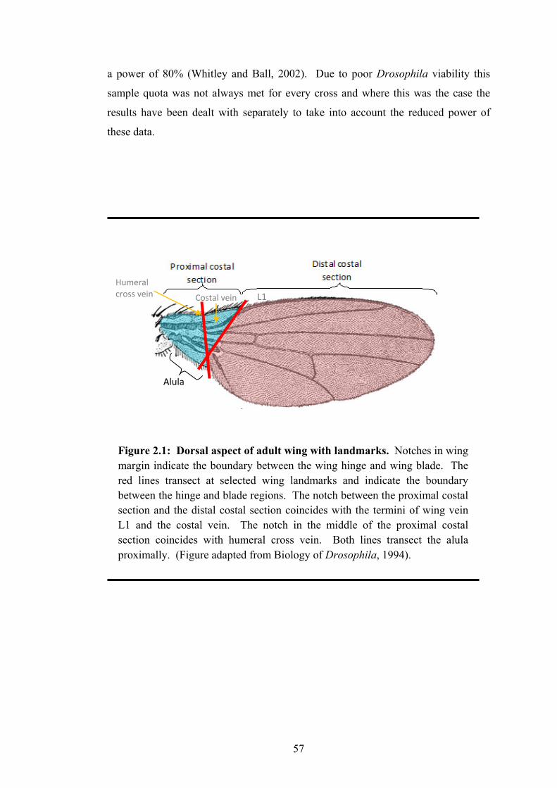
57
a power of 80% (Whitley and Ball, 2002). Due to poor Drosophila viability this
sample quota was not always met for every cross and where this was the case the
results have been dealt with separately to take into account the reduced power of
these data.
Figure 2.1: Dorsal aspect of adult wing with landmarks. Notches in wing margin indicate the boundary between the wing hinge and wing blade. The red lines transect at selected wing landmarks and indicate the boundary between the hinge and blade regions. The notch between the proximal costal section and the distal costal section coincides with the termini of wing vein L1 and the costal vein. The notch in the middle of the proximal costal section coincides with humeral cross vein. Both lines transect the alula proximally. (Figure adapted from Biology of Drosophila, 1994).
Alula
L1 Humeral cross vein Costal vein

58
2.8 MICROSCOPY
2.8.1 Confocal microscopy
All confocal microscopy was carried out using a BIO-RAD Radiance 2000 laser
scanning confocal microscope and images captured using BioRad LaserSharp2000
software.
2.8.2 Scanning electron microscopy (SEM)
Adult flies were prepared for SEMs by anaesthetisation with CO2 before being
transferred to 25% ethanol. The flies were dehydrated by sequentially increasing the
ethanol content to 50%, 75% and 100% and left at each of these stages for a
minimum of 24 hours. The 100% step was repeated three times. When fully
dehydrated, the flies were incubated in a 1:1 mix of hexamethyldisilasane (HMDS)
and ethanol for 10 minutes. This was followed by incubation in 100% HMDS for 10
minutes and repeated three times. Flies were quick dried on Whatman 3MM filter
paper in and then transferred to 4ºC overnight to allow slow drying of the samples.
All steps prior to the drying at 4ºC involving HMDS were performed in the
fumehood for safe ventilation of fumes.
Fly samples were mounted onto stubs with adhesive circles prior to sputter coating
with 5 or 6 layers of either gold or platinum (performed by Dr. Kevin Blake or
Shane Askew using a JEOL sputter coating machine). The coated samples were
then analysed using a JEOL (JSM-541-LV) scanning electron microscope and the
Semaphore digital imaging system.

59
CHAPTER 3: ANALYSIS OF THE DROSOPHILA RAD21 MODEL

60
3.1 INTRODUCTION
In the study of chromosome cohesion and chromosome dynamics that depend on this
cohesion, two obvious strategies for developing a model are decreasing cohesion and
increasing cohesion. In the absence of knowledge of the chromosomal location for
Rad21 and any known mutants that could be used as models of reduced chromosome
cohesion at the time of commencing this study, an alternative option was increasing
cohesion by developing and ectopically expressing a "non-cleavable" cohesin
variant. An additional feature of the non-cleavable cohesin model is that cleavage
occurs at a very specific time in the cell cycle and therefore phenotypes produced
and genetic interactions with these phenotypes can be temporally pinpointed. In
order to truly understand and hypothesise on the genetic interactions uncovered by a
genetic screen, the aetiology of the mutant phenotype needs to be thoroughly
characterised.
This chapter describes the approaches used to generate and characterise a collection
of epitope-tagged Rad21 transgenics. The Rad21NC allele was pursued as a
mechanism for identifying modifiers of chromosome cohesion and in particular,
modifiers that could weaken chromosome cohesion. However, it remained to be
conclusively demonstrated that the proteolytic cleavage pattern of these mutated
RAD21 proteins is altered from that of wildtype RAD21 when expressed in
Drosophila eye cells. It was also unclear whether incorporation of RAD21NC into
functional cohesin complexes was preventing the segregation of the replicated
chromosomes at the cellular level and what the resulting cellular responses to and
consequences of inhibited chromosome segregation in this way may be.
Although Keall (2005) had clearly demonstrated that mitotic arrest and apoptosis
were significant factors underlying the final GMR>Rad21NC eye phenotype
questions remained regarding the extent of the influence of these factors.
Particularly, was the overexpression of RAD21 alone influencing eye development?
Could the transgene expression be influenced through modulation of the driver?
Also, introduction of a single copy of cDNA encoding the anti-apoptotic baculovirus
p35 protein strongly suppressed the GMR>Rad21NC eye phenotype. Therefore,
would increasing the level of p35 further increase the suppression of the phenotype

61
towards wildtype? This chapter describes attempts used to directly and indirectly
determine the consequences of expressing Rad21 and various mutant forms of
Rad21 in the developing eye under the control of GMR-Gal4.
3.2 RESULTS
3.2.1 Generation and expression of Rad21-HA transgenics
3.2.1.1 Generation of Rad21-HA variants
In order to express transgenic RAD21 proteins that could be distinguished from
endogenous RAD21, transgenic Drosophila were generated that could express
Rad21 protein tagged with two tandem copies of the haemagglutinin (HA) epitope
positioned at the carboxy-terminus. The untagged Rad21 variants generated
previously by Keall (2005) had provided inconclusive results when utilised for
Western Blot analyses. Tagging of the Rad21 and Rad21NC constructs was carried
out by Dr. Kylie Gorringe by cutting the Rad21 cDNA sequence using BstBI (Figure
3.2A), followed by ligation with overlapping oligonucleotides containing the HA
coding sequence. Single tagged mutants, Rad21N-2HA and Rad21C-2HA, were later
generated through a "cut and paste" scheme using the Rad21-2HA and Rad21NC-2HA
constructs (Figure 3.2B). The presence of the cleavage-site amino acid substitutions
was confirmed by analysing the constructs for restriction sites introduced by the
original site-directed mutagenesis that altered the Separase cleavage site sequences.
An SphI restriction site introduced at the amino-terminal Separase cleavage site in
the R175A alleles, and an NaeI restriction site introduced in the sequence encoding
the carboxy-teriminal Separase cleavage site RA474AG (Figure 3.2A) aided
molecular analyses. Following subcloning into P-element vector pUAST, NaeI/SphI
double digests of the pUAST-Rad21-2HA constructs produced the expected
restriction fragments, demonstrating the presence of the desired mutations
(R175A/RA474AG) (Figure 3.2, Table 3.1 and Figure 3.3). DNA sequencing
analyses were performed to further confirm the presence of the HA tag sequence and
that the integrity of the cDNA sequence had not been compromised. Independent
transgenic Drosophila lines were generated with the four different Rad21-2HA
constructs (Table 3.2), via microinjection of construct DNA into Drosophila

62
Figure 3.1: The Rad21 plasmid constructs. (A) Schematic of the dRad21 cDNA with amino- and carboxy-terminal sequence changes with introduced restriction sites, primer binding sites and the site of the introduction of the HA tags. (B) Restriction map of the pBluescript KS+-Rad21-2HA construct. The EcoRI, BamHI and XhoI sites were used in the "cut and paste" scheme to generate the Rad21N-2HA and Rad21C-2HA single mutants. The EcoRI and KpnI sites were used to sub-clone Rad21-2HA into the P-element vector pUAST. A second NaeI site (NaeI2) was introduced with the HA tag sequence and the NaeI and SphI sites were used for confirmation digests (Figure 3.2). (Image adapted from Stratagene pBluescript II Phagemid Vectors Instruction Manual).
Rad21 cDNA 2.1kb(A)
NaeI RA474AG DradM DradL
BstBI, site of HA tag
X X
R175A SphI DradORF DradJ
XhoI
EcoRI
BamHI
NaeI1
SphI
NaeI2
X
X
X
BamHI
KpnI
Rad21 cDNA (2145bp)
X
(B)
embryos, as described in section 2.3.1. The transgene insertion sites were mapped
by segregation analysis for all independent transgenic lines, as shown in Table 3.2.

63
Table 3.1: Expected band sizes for SphI/NaeI double digests of the pUAST-Rad21-2HA constructs (based on the Rad21 restriction map in Figure 3.1)
Con
stru
ct
pUA
ST-R
ad21
-2H
A
pUA
ST-R
ad21
N-2
HA
pUA
ST-R
ad21
C-2
HA
pUA
ST-R
ad21
NC-2
HA
NaeI3-NaeI1 10,000bp 10,000bp 10,000bp 10,000bp NaeI1-NaeI3 2600bp SphI-NaeI3 1675bp NaeI1-SphI 925bp NaeI1-NaeI2 1822bp NaeI2-NaeI3 778bp 778bp NaeI1-SphI 925bp SphI-NaeI2 897bp
Figure 3.2: Confirmation digests of pUAST-Rad21-2HA constructs. NaeI/SphI double digests of pUAST-Rad21-2HA (lane 2), pUAST-Rad21N-2HA (lane 3), pUAST-Rad21C-2HA (lane 4) and pUAST-Rad21NC-2HA (lane 5). Lane 1 contains 0.75 µg of λ HindIII/EcoRI DNA ladder (Promega).
897bp 778bp
10000bp
2600bp
1822bp 1675bp
925bp
21226bp
5000bp 4268bp
2027bp 1584bp 1375bp
947bp 831bp
1 2 3 4 5

64
Table 3.2: Rad21-2HA transgenic lines
Construct Independent transgenic
lines
Line identifier
Chromosome mapped to
pUAST-Rad21-2HA (Rad21-2HA)
5
5 2 14.2 3 14.11 X 28.2 2 28.3 3
pUAST-Rad21R175A-2HA (Rad21N-2HA)
2 A 2 R X
pUAST-Rad21RA474AG-2HA (Rad21C-2HA)
9
B 2 C ND E 3 F 3 G 3 H 2 I 3 J 2 K X
pUAST-Rad21R175A/RA474AG-2HA (Rad21NC-2HA)
8
4 3 6 X
14 ND A ND H 2 P ND O 2
XA X

65
Figure 3.3: Ectopic expression of RAD21-2HA variants. The phenotype of the adult eye expressing (A) UAST-Rad21-2HA (line 5), (B) UAST-Rad21N-2HA (line R), (C) UAST-Rad21C-2HA (line H) and (D) UAST-Rad21NC-2HA (line H) under the control of GMR-Gal4. Expression of the RAD21NC-2HA protein produced an eye phenotype that was significantly reduced in overall size and ommatidial organisation (D). No variation was observed for independent transgenic lines of wildtype Rad21 or single mutants (R175A and RA474AG. Only slight variation of the level of eye disorganisation was observed for the different independent Rad21NC transgenic lines tested, presumably due to transgene insert-site effects on expression levels.
(A) (D) (B) (C)
3.2.1.2 Ectopic expression of Rad21-HA variants
The eye-specific driver P{GAL4-ninaE.GMR} (referred to from here as GMR-Gal4
or GMR>) was used to drive expression of the transgenes in the photoreceptor cells
of the developing larval eye imaginal disc. In the adult compound eye the RAD21-
2HA, RAD21N-2HA and RAD21C-2HA proteins did not produce an abnormal
phenotype. In contrast the RAD21NC-2HA protein produced an eye phenotype that
was significantly reduced in overall size and ommatidial organisation compared to
wildtype or eyes expressing the other HA-tagged RAD21 proteins (Table 3.4; Figure
3.3).

66
3.2.2 Generation and expression of Rad21-GFP transgenics
3.2.2.1 Generation of Rad21-GFP variants
It remained to be determined whether a GFP epitope tag did in fact prevent
functional cohesin complexes from forming, as had been previously speculated
following failure of pUAST-Rad21C-GFP transgenics to produce an expected rough-
eye phenotype when expression was driven by GMR-Gal4. This followed on from
data that indicated an untagged pUAST-Rad21C transgene produced a rough-eye
phenotype when driven by GMR-Gal4. Previous studies using wildtype Drosophila
Rad21 tagged with GFP demonstrated that it was localising to the chromosomes in a
pattern consistent with endogenous cohesin, which indicated that the RAD21-GFP
protein was at least localising normally. P-element constructs for Rad21-GFP,
Rad21N-GFP, Rad21C-GFP and Rad21NC-GFP had been generated previously
(Keall, 2005) by cloning wildtype and mutant (R17A and RA474AG) cDNAs
inframe with EGFP coding sequences from pALX190, then subcloning the Rad21-
GFP fragment into pUAST. Transgenic flies existed for the Rad21-GFP and
Rad21C-GFP constructs and so to confirm the hypothesis that GFP was not blocking
cohesin complex formation, the Rad21N-GFP and Rad21NC-GFP transgenics were
also generated (Table 3.3). A total of six independent transgenic lines were
generated for the pUAST-Rad21R175A-GFP construct and two independent trangenic
lines for the pUAST-Rad21R175A/RA474AG-GFP construct, with each of these
insertions being mapped via chromosome segregation analysis (Table 3.3).
Table 3.3: Rad21-GFP transgenic lines
Construct Independent transgenic
lines
Line identifier
Chromosome mapped to
pUAST-Rad21R175A-GFP (Rad21N-GFP)
6
M 2 N 3 O 3 P 3 S 2 T 3
pUAST-Rad21R175A/RA474AG-GFP (Rad21NC-GFP)
2 L 3 Q 3

67
3.2.2.2 Ectopic expression of Rad21-GFP variants
Ectopic expression of RAD21WT-GFP, RAD21N-GFP and RAD21C-GFP under the
control of GMR-Gal4 resulted in normal eye development, whereas ectopic
expression of RAD21NC-GFP produced a severely reduced and disorganised eye
phenotype (Figure 3.4, Table 3.4). The expression of the RAD21-GFP fusion
protein was confirmed by epifluorescence microscopy (data not shown). The ability
of RAD21NC-GFP to produce a phenotype equivalent to that produced by untagged
and HA tagged RAD21NC indicates that GFP-tagging of RAD21 does not prevent
functional incorporation of this subunit into the cohesin complex. This also
indicated that the original analysis deeming the Drosophila carboxy-terminal
Separase cleavage site to be the dominant site of cleavage was inconsistent with
previous data, suggesting that this conclusion may require further investigation (see
section 3.2.5).
Figure 3.4: Ectopic expression of RAD21-GFP variants. (A) GMR>UAST-Rad21WT-GFP (line N2A, Keall 2005), (B) GMR>UAST-Rad21N-GFP (line O), (C) GMR>UAST-Rad21C-GFP (line 19-2, Keall 2005) and (D) GMR>UAST-Rad21NC-GFP (line L). The Rad21WT-GFP, Rad21N-GFP, and Rad21C-GFP mutants (A-C) show no obvious eye abnormalities when expressed under the control of GMR-Gal4, while the Rad21NC double-mutant (D) displays a rough-eye phenotype very similar to that observed with untagged Rad21NC. No variation was observed for independent transgenic lines of wildtype Rad21 or single mutants (R175A and RA474AG. Slight variation of the level of eye disorganisation was observed for independent transgenic lines expressing Rad21NC.
(A) (D) (B) (C)

68
Table 3.4: Rad21 variant phenotypes observed
Construct Tag Eye phenotype
with GMR-Gal4 driver
Reference
pUAST-Rad21 (Rad21)
Untagged Wildtype Keall, 2005
HA-tagged Wildtype This study
GFP-tagged Wildtype This study
pUAST-Rad21R175A (Rad21N)
Untagged Wildtype Keall, 2005
HA-tagged Wildtype This study
GFP-tagged Wildtype This study
pUAST-Rad21RA474AG (Rad21C)
Untagged Reduced size and
ommatidial organisationЭ
Keall, 2005
HA-tagged Wildtype This study
GFP-tagged Wildtype This study
pUAST-Rad21R175A/RA474AG (Rad21NC)
Untagged** Reduced size and
ommatidial organisation
Keall, 2005
HA-tagged Reduced size and
ommatidial organisation
This study
GFP-tagged Reduced size and
ommatidial organisation
This study
ЭThis abnormal phenotype was later found to be incorrect (see section 3.2.5) and therefore no untagged pUAST-Rad21C transgenics exist. **The independent line 24A of untagged Rad21NC transgene recombined with GMR-Gal4 on the 2nd chromosome (Keall, 2005) and balanced over CyO was used for the genome-wide screen and all other Rad21NC experiment unless stated that a HA epitope-tagged Rad21NC was utilised.

69
3.2.3 RAD21 variant cleavage patterns
Although Keall (2005) had characterised the gross phenotypic consequences of
Rad21NC expression, it remained to be demonstrated that mutating the Separase
cleavage sites of RAD21 actually resulted in altered RAD21 protein cleavage
products. Previous efforts using untagged recombinant RAD21 proteins and
Western Blotting to assess cleavage patterns of mutant variants of RAD21 compared
to wildtype RAD21 were inconclusive (Keall, 2005). As a result, experiments
examining proteolysis of HA-tagged and GFP-tagged RAD21 variants were
undertaken to distinguish endogenous and exogenous RAD21 protein signals. HA-
tagged transgenic lines yielded a RAD21-HA protein band on Western Blots of
approximately 120kD (Figure 3.5) and GFP-tagged transgenic lines yielded a
RAD21-GFP protein band 130kD on Western Blots (Figure 3.5), consistent with the
phosphorylated full-length form of RAD21. Full length RAD21 was observed in
protein samples sourced from RAD21WT-2HA (Figure 3.5A), RAD21WT-GFP
(Figure 3.5C), RAD21N-2HA (data not shown), RAD21C-2HA (data not shown),
RAD21NC (Figure 3.5C) and RAD21NC-2HA (Figure 3.5B) transgenics.
Despite extensive efforts and numerous attempts, bands for the wildtype RAD21
cleavage products, expected to be 60kD and 27kD for the amino- and carboxy-
terminal cleavage products, respectively, were not observed using either anti-HA or
anti-Scc1 antibodies. As only a fraction of the total cohesin population in a cell is
loaded onto chromosomes and in metazoans the majority of this is removed during
the prophase dissociation step, only a few percent of the total cellular cohesin
population is proteolytically cleaved by Separase at the onset of anaphase (Hauf et
al., 2001). By expressing these proteins in vivo in the fly eye (driven by GMR-
Gal4), or even ubiquitously (driven by hs>Gal4), the population of cohesin being
cleaved by Separase at any given time is very small due to the asynchronous nature
of the cell divisions

70
Figure 3.5: Detection of epitope-tagged RAD21 protein by Western Blotting. Blot (A): PageRulerTM (Fermentas)(Lane 1), Protein extract from 6 adult Drosophila heads TM3(hs-Gal4),UAS-EGFP/UAST-Rad21WT-2HA, line 28.3)(Lane 2), and Protein extract from 12 adult Drosophila heads TM3(hs-Gal4),UAS-EGFP/UAST-Rad21WT-2HA, line 14.2)(Lane 3); NB – protein samples were collected 10 hours post heat shock treatment. Blot (B): PageRulerTM (Fermentas) (Lane 1); samples of 45 eye-antennal imaginal discs: GMR>UAST-Rad21WT-2HA/CyO (Lane 2), GMR>UAST-Rad21C-2HA/CyO (Lane 3), GMR>UAST-Rad21NC-2HA/CyO (Lane 4). Blot (C): PageRulerTM (Fermentas)(Lane 1), UAST-Rad21WT-GFP/+;TM3(hs-Gal4),UAS-EGFP/+ (line J2A) 5 pupae (Lane 2), TM3(hs-Gal4),UAS-EGFP/UAST-Rad21WT-GFP (line N2A) 3 pupae (lane 3), TM3(hs-Gal4),UAS-EGFP/UAST-Rad21NC 5 pupae (Lane 4), and TM3(hs-Gal4),UAS-EGFP/UAST-Rad21NC 12 pupae (Lane 5); NB – protein samples were collected 16 hours post heat shock treatment. Blots were pr.obed using rat anti-HA (Roche) primary antibody and goat anti-rat-HRP (Zymed Laboratories) secondary antibody.
170 130 100 70 55
40
1 2 3
1 2 3 4 5
170 130 100 70 55
40
170 130 100 70 55
40
1 2 3 4 (A) (B)
(C)

71
3.2.4 Cleavage-resistant RAD21 cellular phenotypes
RAD21NC had been found to produce a demonstrable deleterious effect on the
development of the adult compound eye; however, visualisation of the effects of
RAD21NC at the cellular level remained to be definitively demonstrated. In order to
achieve this, neuroblast squashes were used to allow visualisation of mitotic
chromosomes and analysis of segregation defects. The neuroblast squashes were
performed following a heatshock treatment (8 and 16 hours post-heatshock) to
induce ubiquitous RAD21NC expression in larvae carrying a heatshock-Gal4 driver
and the UAST-Rad21NC transgene. The lethality induced by expressing RAD21NC in
dividing cells results in this phenotype being extremely difficult to capture and
analyse, however, a significant number of neuroblasts from the larval brain had
observable chromosomal defects compared to controls (Table 3.5). The observed
chromosomal defects were tetraploidy, polyploidy, chromosomal fragmentation and
abnormal anaphases, many with lagging chromosomes (Figure 3.6). These
phenotypes were almost entirely absent from hs-Gal4 control brain squashes (Table
3.5).
Table 3.5: hs-Gal4>Rad21NC neuroblast analysis.
Treatment Genotype Diploid Tetra- or
polyploid Fragments Anaphase Total
No
heatshock
hs-Gal4 control
554
(86.4%)
1
(0.2%)
0
(0.0)
86
(13.4)
641
(100.0)
hs-Gal4,
Rad21NC
622
(87.0%)
0
(0.0%)
0
(0.0)
93
(13.0)
715
(100.0)
Heatshock
+8 hours
hs-Gal4 control
505
(81.1%)
0
(0.0%)
0
(0.0)
118
(18.9)
623
(100.0)
hs-Gal4,
Rad21NC
403
(79.0%)
13
(2.5%)
14
(2.7)
80
(15.7)
510
(100.0)
Heatshock
+16 hours
hs-Gal4 control
725
(87.5%)
2
(0.2%)
0
(0.0)
102
(12.3)
829
(100.0)
hs-Gal4,
Rad21NC
678
(66.3%)
199
(19.5%)
64
(6.3)
82
(8.0)
1023
(100.0)
(Data collated by Dr. S. Page)

72
Figure 3.6: Chromosomal segregation abnormalities in neuroblast squashes. Heatshock treated hs-Gal4 (TM3, UAS-EGFP, Ser)/UAST-Rad21NC neuroblast squashes 8 hours post-heatshock: (A) Normal diploid cell with 8 pairs of sister chromatids and (B) normal anaphase. Chromosome segregation abnormalities: (C) tetraploidy, (D) polyploidy and chromosome fragments, and (E and F) anaphases with lagging chromosomes (red arrows). (Images generated by Dr. S. Page)
(C) (D)
(A) (B)
(E) (F)

73
3.2.5 Correction of cleavage site hypothesis As outlined previously in section 3.2.1.2, Keall (2005) proposed the carboxy-
terminal cleavage site (R474) of RAD21 was preferentially used in Drosophila, with
seemingly minimal cleavage occurring at the amino-terminal site (R175). This is in
contrast to RAD21 homologues in other species (Hauf et al., 1999; Uhlmann et al.,
1999; Keall, PhD Thesis), where alteration of both RAD21 cleavage site was
required before chromosome segregation inhibition was observed. The conclusion
that the R474 site was the preferred site of cleavage for Drosophila RAD21 was
based on observations that ectopic expression in the Drosophila eye of the amino-
terminal UAST-Rad21N mutant produced no observable phenotype. In contrast,
ectopic expression of the carboxy-terminal UAST-Rad21C mutant produced a rough-
eye phenotype very similar to the phenotype produced by the “non-cleavable”
mutant, with both the amino- and carboxy-terminal sites mutated (Keall, PhD
thesis). Due to the apparent similarity of the GMR>Rad21C and GMR>Rad21NC
phenotypes, it was concluded that the majority of the RAD21 cleavage must occur at
the carboxy-terminal site because this mutation alone produced a phenotype
equivalent to GMR>Rad21NC. Based on this logic it was also concluded that the
lack of phenotype produced by ectopic expression of UAST-Rad21C-GFP, under
GMR-Gal4 control, was indicative of the GFP epitope blocking functional
incorporation of RAD21-GFP into cohesin rings. Consequently, further work using
the GFP epitope (e.g. UAST-Rad21NC-GFP transgenics) was not pursued at the time.
During the generation of the HA-tagged UAST-Rad21 constructs, it was observed
that DNA sequencing and confirmation restriction digestions indicated that the
expected nucleotide substitutions were present in each construct, eye development
occurred normally when ectopically expressing UAST-Rad21C-2HA using GMR-
Gal4. To rule out the possibility that UAST-Rad21C-2HA simply failed to produce
protein Western Blot analyses were performed and a protein of the correct size
(120kD) was detected with anti-HA antibodies (Figure 3.5). These observations,
suggested an error may have occurred in the earlier analysis of untagged UAST-
Rad21C. Combining this knowledge with previous observations of RAD21-GFP
locating to chromosomes in embryos I hypothesised that GFP was not impeding
functional incorporation of RAD21 and that instead pointed to the need to
characterise the molecular sequences present within the Rad21C transgenics.

74
The simplest explanation for the conflicting results was that a mistake had occurred
during the generation of the transgenic insects. To confirm the molecular nature of
the cDNAs present in the UAST-Rad21C transgenic lines, genomic DNA was
isolated from variant transgenic flies and PCR was performed on genomic DNA
extracted from single flies of each genotype and using primers designed to span
introns and therefore only amplify cDNA (Figure 3.1). Amplification of the amino-
terminal sequence with primers DradORF and DradJ produced a 700bp DNA
product, while amplification of the carboxy-terminal sequence with primers DradM
and DradL produced an 850bp DNA product. The PCR products were then digested
to determine the presence or absence of the NaeI and SphI sites that were introduced
during the site directed mutagenesis procedure to generate the specific R175A and
RA474AG amino acid substitutions (Figure 3.7). Whilst only UAST-Rad21C and
UAST-Rad21NC transgenic mutant lines were found to carry the carboxy-terminal
cleavage site mutation (Figure 3.7), all six of the Rad21 mutant transgenic lines were
found to carry the amino-terminal R175A cleavage site mutation, including lines
labelled UAST-Rad21C. This result was confirmed for available cultures and
independent UAST-Rad21 transgenic lines (data not shown). These data
demonstrated that the UAST-Rad21C transgenics, generated and analysed by Keall
(2005), carried both R175A and RA474AG amino acid substitutions and thus were
in fact UAST-Rad21NC mutants. Consequently, the hypothesis that the RAD21
carboxy-terminus cleavage site is the predominant site at which Separase cleavage
occurs in Drosophila is no longer a justifiable claim.

75
Figure 3.7: Rad21 transgene genotyping by PCR-RFLP analysis. DNA from single-fly preparations was used as template to PCR amplify the Rad21 cDNA present. (A) Rad21 amino-terminal sequence digested with SphI and (B) Rad21 carboxy-terminal sequence digested with NaeI. The gel lanes contain: UAST-Rad21 (lanes 2, template pUAST-Rad21WT plasmid constructЭ), UAST-Rad21N (lanes 3 and 4, lines 4B and D2**), UAST-Rad21C (lanes 5 and 6, lines 1B and P11a**) and UAST-Rad21NC (lanes 7 and 8, lines 11B and N5**). Lanes 1 contains 0.5µg of 100bp Hyperladder (Fermentas). In gel A the presence of a single band of 700bp indicates a lack of an SphI restriction site within the amplicon, while two bands of lenths 460bp and 240bp, respectively indicates the presence of the introduced SphI site associated with the R175A mutation. In gel B the presence of a single band of 850bp indicates a lack of a NaeI restriction site within the amplicon, while the presence of a single 750bp restriction site indicates the presence of the NaeI restriction site a. Э The original pUAST-Rad21WT plasmid DNA was used as control template . ** Transgenic lines generated previously by Keall (2005).
(A)
(B)
1000bp 800bp
600bp
400bp
200bp
1 2 3 4 5 6 7 8
850bp 750bp
1000bp 800bp
600bp
400bp
200bp
1 2 3 4 5 6 7 8
700bp 460bp
240bp

76
3.2.6 Genetic influences on the Rad21NC phenotype
3.2.6.1 Drosophila Rad21 overexpression
RAD21 has many functions aside from chromosome cohesion. It also has roles in
apoptosis, DNA damage repair, gene expression and possibly many other as yet
unrecognised functions. In an attempt to demonstrate that the RAD21NC rough-eye
phenotype was solely due to the introduced mutations and not a result of RAD21
overexpression, multiple copies of wildtype Rad21-2HA transgenes on
chromosomes II and III were simultaneously expressed under the control of the
GMR-Gal4 driver. No abnormal phenotype was observed from ectopic expression
of one, two or three copies of Rad21-2HA (Figure 3.8A-C), indicating that RAD21
overproduction does not produce an observable negative effect on eye development.
Additionally, when Rad21NC and Rad21WT transgenes were co-overexpressed under
the GMR-Gal4 driver strong suppression of the rough-eye phenotype was observed
(Figure 3.8F). The simplest explanation for these observations is the result of
increasing the ratio of cleavable:non-cleavable RAD21 subunit and consequently
decreasing the number of non-cleavable cohesin complexes present on metaphase
chromosomes. Fewer non-cleavable cohesin complexes loaded onto chromosomes
could reduce the strength of the chromosome segregation inhibition and thereby
increase the probability of a euploid chromosome complement. The suppression of
the eye phenotype through concomitant expression of a wildtype Rad21 transgene
further supports the notion that it is the altered cleavage sites and not RAD21
overexpression that is the primary cause of the GMR>Rad21NC rough-eye
phenotype. Interestingly, the introduction of a second copy of the Rad21NC
transgene only slightly enhances the rough-eye phenotype observed compared to
when a single copy is expressed (Figure 3.8E).

77
3.2.6.2 The GMR driver
The eye-specific driver P{GAL4-ninaE.GMR} has been observed to affect
ommatidial development in the absence of a UAS-containing transgene (Figure 3.9)
(Freeman, 1996). The Gal4 system is also temperature sensitive (Duffy, 2002).
Although it is not currently understood how GMR-Gal4 overexpression causes the
ommatidial development effect, an increase in apoptosis has been demonstrated in
the developing eye imaginal discs of both GMR-Gal4 heterozygotes and
homozygotes at 25˚C and 29˚C (Kramer and Staveley, 2003). In the adult
Figure 3.8: Ectopic expression of multiple copies of UAST-Rad21 cDNAs under GMR-Gal4. (A) GMR>1x Rad21WT-2HA (line 28.2), (B) GMR>2x Rad21WT-2HA (line 28.2), and (C) GMR>3x Rad21WT-2HA (lines 28.2 and 14.2) show no variation in eye phenotype. The GMR>Rad21NC phenotype (line 24A, Keall, 2005) (D) is mildly enhanced by introducing a second copy of Rad21NC (line H)(E: GMR>Rad21NC/Rad21NC-2HA) and strongly supressed by introducing a wildtype Rad21 transgene (line 28.2) (F: GMR>Rad21NC/ Rad21WT-2HA).
(A) (B) (C)
(D) (E) (F)

78
compound eye developed at 25˚C homozygous GMR-Gal4 alone produces a distinct
phenotype of gross disorganisation of the ommatidia (Figure 3.9A). Despite
abnormalities being observed by Kramer and Stavely (2003) in the developing eye
discs of heterozygous GMR-Gal4 larvae raised at 25˚C, the adult compound eye of
heterozygous GMR-Gal4 flies has no observable abnormalities (Figure 3.9B)
compared to wildtype eyes. The eyes of heterozygous GMR-Gal4 flies raised at
29˚C demonstrate a relatively mild rough-eye phenotype (Figure 3.9C). An
interesting observation is that introduction of a UAST transgene almost entirely
suppresses the homozygous GMR-Gal4 phenotype (Figure 3.9D). The potential for
genetic modifiers to influence the GMR>Rad21NC eye phenotype through
modification of the driver alone must be kept in mind when undertaking genetic
studies.
Figure 3.9: The effects of the GMR-Gal4 driver. (A) Homozygous GMR-Gal4 25˚C produces a disorganised eye phenotype with little ommatidial organisation remaining, (B) Heterozygous GMR-Gal4 25˚C produces a wildtype phenotype, (C) Heterozygous GMR-Gal4 29˚C produces a milder phenotype with some observable ommatidial orgnisation, (D) Homozygous GMR-Gal4>UAST-Rad21-2HA 25˚C produces a phenotype very close to wildtype in contrast to homozygous GMR-Gal4 alone at 25˚C.
(A) (D) (B) (C)

79
3.2.6.3 Effects on cell survival and cell cycle
P35 is a baculovirus protein that inhibits apoptosis by blocking the activity of the
apoptotic caspases -1, -2, -3, -4, -6, -7, -8 and -10 (Bump et al., 1995; Zhou et al.,
1998). As demonstrated in previous studies (Keall, 2005), introduction of UAS-p35
partially restored the size and organisation defects of GMR>Rad21NC eye phenotype
Figure 3.10). Although co-expression of p35 was observed to significantly suppress
both the size and organisation defects of the GMR>Rad21NC eye phenotype (Figure
3.10), this phenotype still demonstrates significant developmental defects compared
to wildtype. Introduction of a second copy of UAS-p35 has little effect on further
suppressing the eye development defects. The work of Hay et al. (1994)
demonstrated that p35 is capable of inhibiting almost all apoptosis, either naturally
occurring or resulting from radiation induced DNA damage, consistent with the
inhibition of apoptosis observed in acridine orange stained GMR>UAST-
Rad21NC,UAS-p35 (referred to from here as GMR>Rad21NC, p35) eye discs from
third instar larvae (Figure 3.11).
The high level of similarity between the GMR>Rad21NC, p35 rough-eye phenotype
and the rough-eye phenotype produced when Cyclin B is overexpressed and induces
slowing of the cell cycle (Okada et al., 2002), led to exploration of the effects of cell
cycle regulators on the GMR>Rad21NC, p35 phenotype. Increasing the expression
of the Cyclin-dependent kinase Cdc2 was found to suppress the size and
organisation of the GMR>Rad21NC, p35 eye phenotype, while increasing expression
of Cyclin B enhances the disorganisation of the eye phenotype (Figure 3.10C-D).
Cdc2 and Cyclin B are binding partners that are integral in regulating both the entry
to and exit from M phase. Using phospho-histone H3 antibodies to specifically
identify mitotic cells to confirm the results of Keall (2005), it was observed that
there is an increase in the number of mitotic cells posterior to the morphogenetic
furrow (Figure 3.12). The significant increase in mitotic cells suggests that the
cleavage-resistant cohesin is slowing exit from mitosis and inducing mitotic arrest.
These data are suggestive of some of the remaining disorganisation observed in the
GMR>Rad21NC, p35 eye (Figure 3.10) being due to the slowing of the cell cycle that
would occur through inhibition of chromosome segregation and further supported by
modification of the GMR>Rad21NC eye phenotype by alteration of cell cycle
regulator expression levels.

80
Figure 3.11: Acridine orange stained eye-antennal imaginal discs. (A) Wildtype and (B) GMR>UAST-Rad21NC discs and (C) and (D) GMR>UAST-Rad21NC, UAS-p35 discs stained with acridine orange, which is specifically stains apoptosing cells. The intense acridine orange staining posterior to the morphogenetic furrow (position indicated by red arrows) in GMR>UAST-Rad21NC discs is completely absent in the GMR>UAST-Rad21NC, UAS-p35 discs (bottom panels).
(A) (B)
(C) (D)
Figure 3.10: Modification of the GMR>Rad21NC rough-eye phenotype. (A) GMR>UAST-Rad21NC, (B) abolition of apoptosis in the GMR>UAST-Rad21NC, UAS-p35 transgenics results in strong suppression of the size and organisation defects, (C) GMR>UAST-Rad21NC, UAS-Cdc2 eyes show strong suppression of the size and organisation defects, in contrast (D) GMR>UAST-Rad21NC, UAS-CycB eyes show an observable enhancement of the size and organisation defects. .
(A) (D) (C) (B)

81
Figure 3.12: Mitotic indices of wildtype and GMR>Rad21NC eye-antennal imaginal discs. Phospho-histone H3 antibody (magenta) and DNA (blue, To-Pro3) staining of eye discs (A) w1118 control and (B) GMR>UAST-Rad21NC. The morphogenetic furrow is indicated by the red arrows and the width of the bands of mitotic cells is indicated by the yellow bars.
(A) (B)

82
3.3 DISCUSSION
This study, following on from the work of Keall (2005), set out to characterise the
cellular influences underlying the GMR>Rad21NC rough-eye phenotype. To achieve
this aim three main approaches were taken: (i) Generation and ectopic expression of
HA and GFP epitope tagged RAD21 protein variants, (ii) Phenotypic analyses of
Drosophila ectopically expressing these epitope-tagged RAD21 variants, and (iii)
Biochemical analyses. From the data arising through this study and previous
studies, it has been hypothesised that the GMR>Rad21NC rough-eye phenotype
results from increased cell death, reduced proliferation in the 2nd mitotic wave and
perturbation of the cell cycle within the differentiating eye imaginal disc.
Additionally, it was determined that RAD21 overexpression alone does not visibly
contribute to the adult phenotype and that the GMR-Gal4 eye-specific driver has its
own phenotype under certain conditions, which may contribute to potential genetic
interactions with the GMR>Rad21NC eye phenotype.
3.3.1 Altering the cleavage of RAD21
Ectopic expression of Drosophila RAD21 protein with the two putative Separase
cleavage sites altered to prevent proteolytic cleavage by Separase is able to cause a
severe perturbation in normal development (Keall, 2005), supporting the
experimental evidence from previous studies in yeast and human cells demonstrating
inhibition of chromosome segregation and resulting cellular phenotypes due to the
presence of non-cleavable RAD21 (Toyoda et al., 2002; Hauf et al., 2001;
Tomonaga et al., 2000). Attempts to demonstrate an alteration of the Drosophila
RAD21 cleavage variants using Western Blots were largely unsuccessful, most
likely because of the small percentage of cleaved protein present in an asynchronous
cell population at any given time. As yet the only research groups to demonstrate
RAD21 cleavage products, or alteration of cleavage products through mutation, have
been those working with synchronously dividing yeast and human cells (Tomonaga
et al., 2000, Hauf et al., 2001).

83
3.3.2 Characterising the RAD21NC phenotype
Acridine orange staining of GMR>Rad21NC eye discs identified a large increase in
the number of apoptosing cells in the wake of the morphogenetic furrow (Keall,
2005). Ectopic expression of the apoptosis inhibitor p35 under the GMR-Gal4
driver only partially suppresses the rough-eye phenotype, despite all apoptosis in the
eye being inhibited (Figure 3.11). This indicates that cell death is not the only factor
contributing to the reduced and disorganised eye phenotype. The most likely
explanation is that although p35 may allow cells to survive the cellular catastrophe
of inhibited chromosome segregation, these cells are then incapable of replicating to
provide the number of cells required for normal eye development, although other
cellular effects caused by viable karyotypic abnormalities cannot be discounted.
Using phospho-histone H3 staining, Keall (2005) identified an increased number of
mitotic cells in the GMR>Rad21NC eye discs following the 2nd mitotic wave
progression indicating a delay in mitosis. A delay in M phase could well explain the
remaining rough-eye phenotype that is not counteracted by the ectopic P35
expression as similar phenotypes have been observed in studies where a delay in S
phase or M phase is induced (Xin et al., 2002; de Nooij and Hariharan, 1995). It is
thought that this delay uncouples the cell cycle from tissue development progression
and thus when the ommatidia are forming these delayed cells are not ready to be
recruited to specific cell fates as normal, resulting in abnormally sized and shaped
ommatidia (de Nooij and Hariharan, 1995).
The neuroblast data clearly demonstrates that the presence of cleavage resistant
cohesin complexes is impeding chromosome segregation and resulting in aneuploidy
and broken chromosomes (Figure 3.12). A subset of the abnormal anaphases
observed appeared predominantly normal, with only a few lagging chromosomes
rather than complete failure of chromosomes to separate at the onset of anaphase.
The presence of tetra- and polyploid cells indicates that some cell divisions fail and
this is most likely due to excessive chromatin bridges that cannot be resolved,
causing a failure of the abscission required to complete cytokinesis and resulting in
cytokinetic furrow regression (Steigemann et al., 2009). These data demonstrate the
variability of the cellular effects induced by the presence of non-cleavable cohesin
complexes and likely reflects subtle variations in Rad21NC expression. Cells with

84
completely normal chromosome segregation may have replicated their DNA prior to
the induction of Rad21NC expression following the heatshock treatment. Previous
studies of the effects of non-cleavable Rad21/Mcd1 on chromosome segregation
have observed that only 10% of cells segregate chromosomes with a thick bridge of
DNA lingering in the cytokinetic furrow, while the majority of cells demonstrated
very thin DNA bridges linking the chromatin masses (Hauf et al., 2001, Yalon et al.,
2004). The multiple pathways that remove cohesin from chromosomes are likely to
provide significant cellular flexibility in resolving the presence of non-cleavable
cohesin complexes, thereby allowing a subset of dividing cells to correctly segregate
their chromosome complement.
Overexpression of RAD21WT produces no observable phenotype, indicating that the
rough-eye phenotype produced by RAD21NC overexpression results only from
alteration of the Separase cleavage sites and does not result as a consequence of an
excess of RAD21 protein. This is further supported by the consistent rough-eye
phenotype produced by independent transgenic lines expressing both epitope tagged
and untagged RAD21 protein. Additionally, introducing an extra wildtype Rad21
cDNA was found to strongly suppress the GMR>Rad21NC rough-eye phenotype.
This indicates that it is not the level of RAD21 protein present, but the ratio of non-
cleavable cohesin complexes relative to cleavable cohesin complexes that
determines the severity of the eye phenotype.
3.3.3 Altering cell cycle progression
Increased apoptosis alone is not sufficient to explain the GMR>Rad21NC rough-eye
phenotype and it is highly likely that alteration of cell cycle progression is a strong
influencing factor in the final presentation of the GMR>Rad21NC reduced and
disorganised eye. Although it is vital that cell cycle events occur in a very specific
order, the cell cycle is not a rigidly imposed timetable of events; instead the cell can
slow or pause at particular points to allow correction of errors. When chromosome
segregation is inhibited the cell cycle is able to delay cytokinesis, hence the
increased mitotic index observed in GMR>Rad21NC eye discs. This change in cell
cycle progression within the developmental program of the eye can significantly
affect cell differentiation, cell recruitment to specific photoreceptor differentiation

85
pathways and consequently lead to the perturbation of the overall organisation of the
adult compound eye.
An influence on cell cycle progression by GMR>Rad21NC is supported by an
increased mitotic index in developing GMR>Rad21NC eye discs (Figure 3.8), failure
of the anti-apoptotic protein p35 to completely suppress the size and organisation
phenotypes (Figure 3.7) and the modification of the eye phenotype when the cell
cycle regulators Cyclin B and Cdc2 are ectopically expressed. Increased Cyclin B
caused an enhancement of the size and organisation phenotypes, while increased
Cdc2 caused suppression of the size and organisation phenotypes (Figure 3.7).
Cyclin B degradation is required for cells to exit mitosis and so excess Cyclin B
causes cells to linger in mitosis and an increase in mitotic index is observed (Okada
et al., 2002), which would contribute to the increased disturbance of eye
organisation observed. The kinase activity of the Cdc2/Cyclin B complex promotes
entry into mitosis and high levels of endogenous Cyclin B expression is observed
anterior to the morphogenetic furrow (Okada et al., 2002). As a result of the
naturally high endogenous Cyclin B levels, increasing Cdc2 levels is likely to result
in increased kinase activity and prompt cells to precociously entering mitosis
(Marangos and Carroll, 2008). As Cyclin B regulation is more closely linked to exit
of mitosis, while Cdc2 regulations is more closely linked with mitosis entry, this
could explain why the binding partners have opposite effects on the severity of the
eye phenotype.
Cdc2 and Cyclin B also have a critical role in regulating cohesin by inhibiting
Separase activity prior to anaphase (Gorr et al., 2006; Holland and Taylor, 2006).
Increasing Cdc2 or Cyclin B levels could also increase the negative regulation
imposed on Separase and thereby result in reduced Separase activity and
consequently induce enhancement of the rough-eye phenotype by further impeding
chromosome separation. Enhancement is only observed when Cyclin B is
ectopically expressed, while increased Cdc2 has the opposite effect of suppressing
the eye phenotype (Figure 3.9), indicating that other roles for these binding partners
at other times in the cell cycle may have overarching influences on the final
phenotype.

86
3.3.4 The GMR driver
The ability of GMR-Gal4 to influence ommatidial development, independently of
the presence of a UAST transgene, adds a layer of complexity to the issue,
particularly as GAL4 is a yeast protein with no recognisable Drosophila homologue.
The presence of a UAST transgene strongly suppresses the GAL4 overexpression
phenotype (Figure 3.6). Along with a coding sequence of interest, a UAST
transgene carries cis-acting DNA binding sites for Gal4 and frequently an eye
pigmentation marker. The provision of Gal4 binding site may prevent excess Gal4
from perturbing unspecified cellular processes that result in alteration of ommatidial
development. Alternatively, expression of the transgene and pigment protein marker
may reduce Gal4 expression via non-specific competition for limiting transcriptional
or translational factors. Mutant alleles that are able to influence Gal4 expression
levels may subsequently alter GMR-Gal4 induced transgene expression.
3.3.5 The GMR>Rad21NC phenotype as a screening tool
The ability of genes known to be involved in chromosome segregation to influence
the RAD21NC rough-eye phenotype in a predictable manner is a strong indication
that this phenotype is a useful tool for performing genetic screens for modifiers of
chromosome segregation. As is always the case in genetic screens, there is the
potential for other factors to influence outcome of the screen. In the case of this
study we have ascertained that some genes involved in apoptosis and eye
development, and genes capable of influencing transgene expression levels will be
able to modify the GMR>Rad21NC eye phenotype through mechanisms other than
altering chromosome segregation accuracy. These influences were kept in mind
when designing experiments and analysing candidate GMR>Rad21NC modifier loci.
Particular heed was taken of wise words of Hawley and Walker (2003), "The value
of whatever screen you create will be determined by the secondary screens".