human skeleton and locomotion
TRANSCRIPT
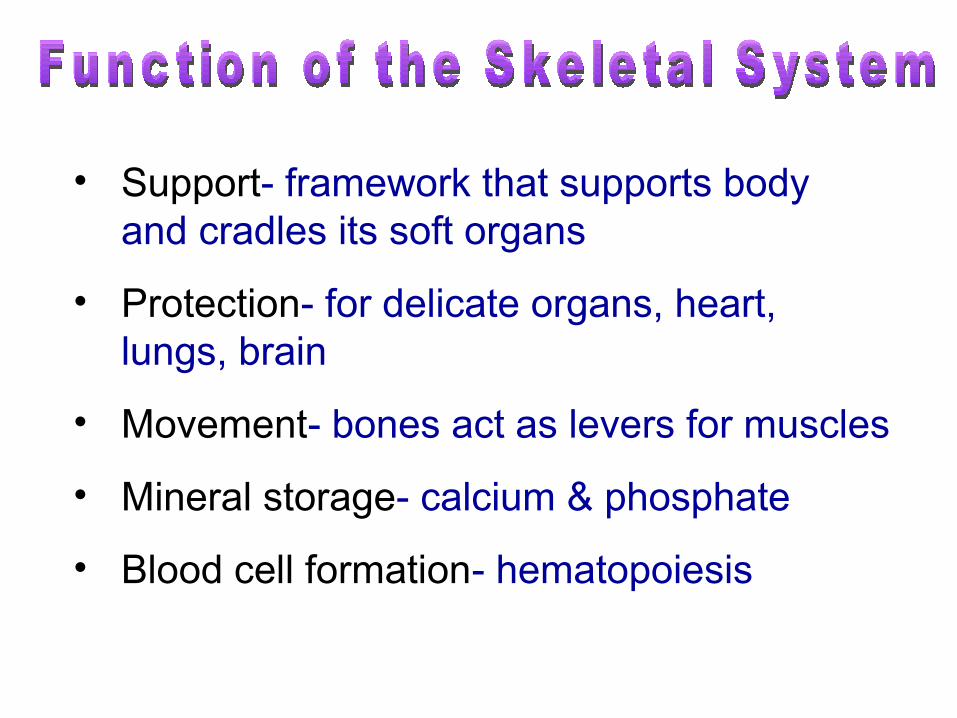
• Support- framework that supports body and cradles its soft organs
• Protection- for delicate organs, heart, lungs, brain
• Movement- bones act as levers for muscles
• Mineral storage- calcium & phosphate
• Blood cell formation- hematopoiesis
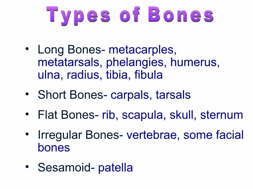
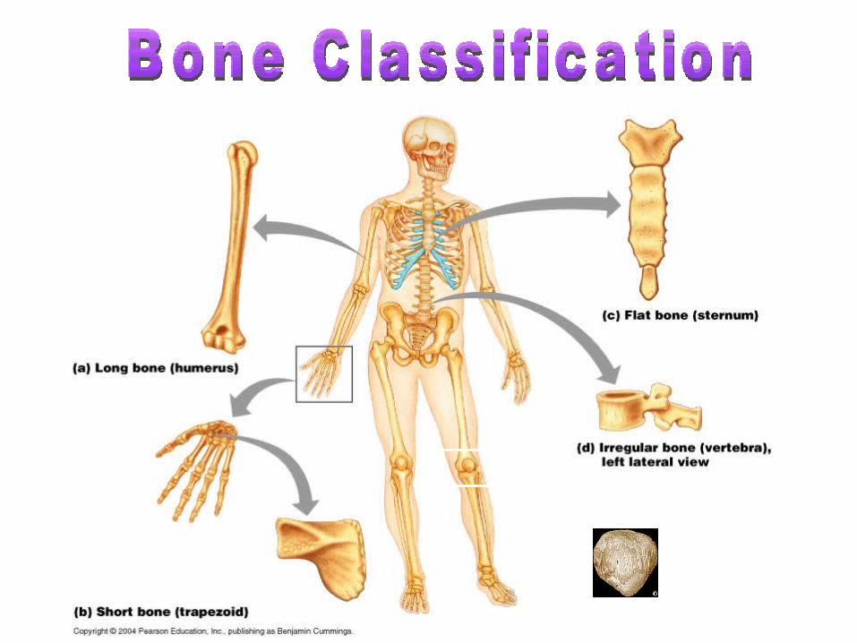
• Long Bones- metacarples, metatarsals, phelangies, humerus, ulna, radius, tibia, fibula
• Short Bones- carpals, tarsals
• Flat Bones- rib, scapula, skull, sternum
• Irregular Bones- vertebrae, some facial bones
• Sesamoid- patella
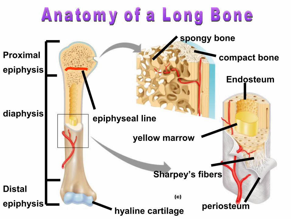
Distal
epiphysis
Proximal
epiphysis
diaphysis
yellow marrow
epiphyseal line
periosteum
compact bone
spongy bone
Endosteum
hyaline cartilage
Sharpey’s fibers
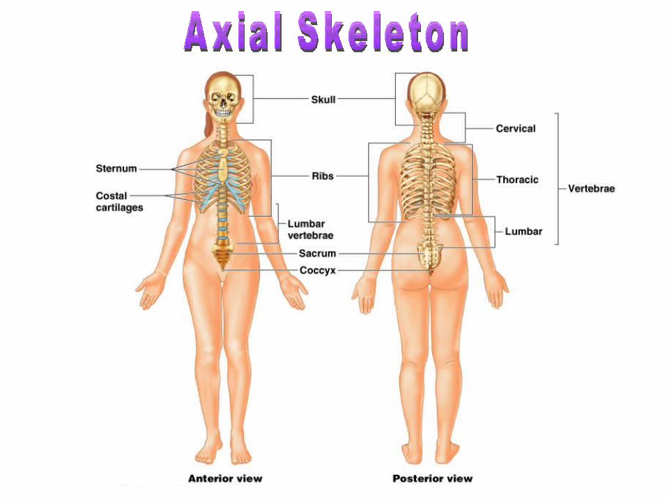
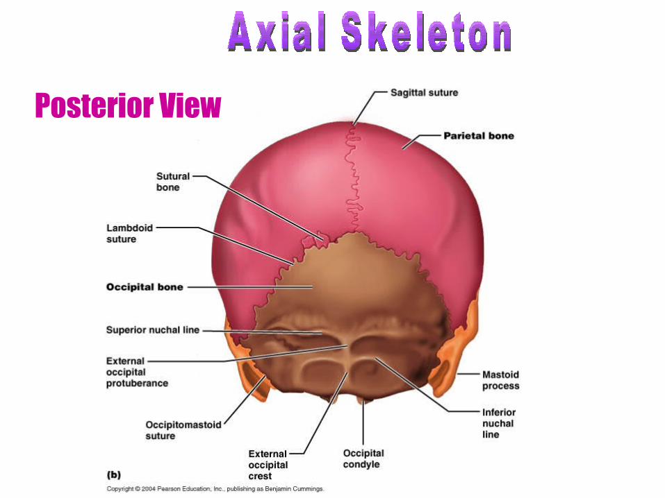
Posterior View
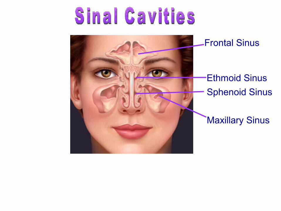
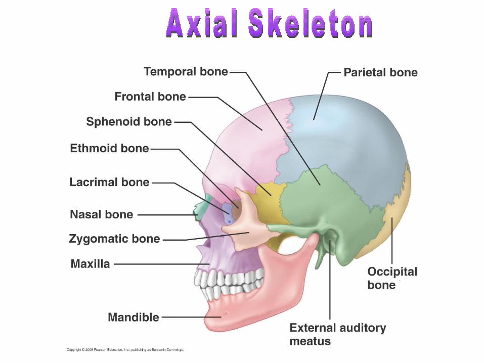
• Warm and moisten air• Lighten the skull• Enhance voice resonance
Frontal Sinus
Ethmoid Sinus
Sphenoid Sinus
Maxillary Sinus
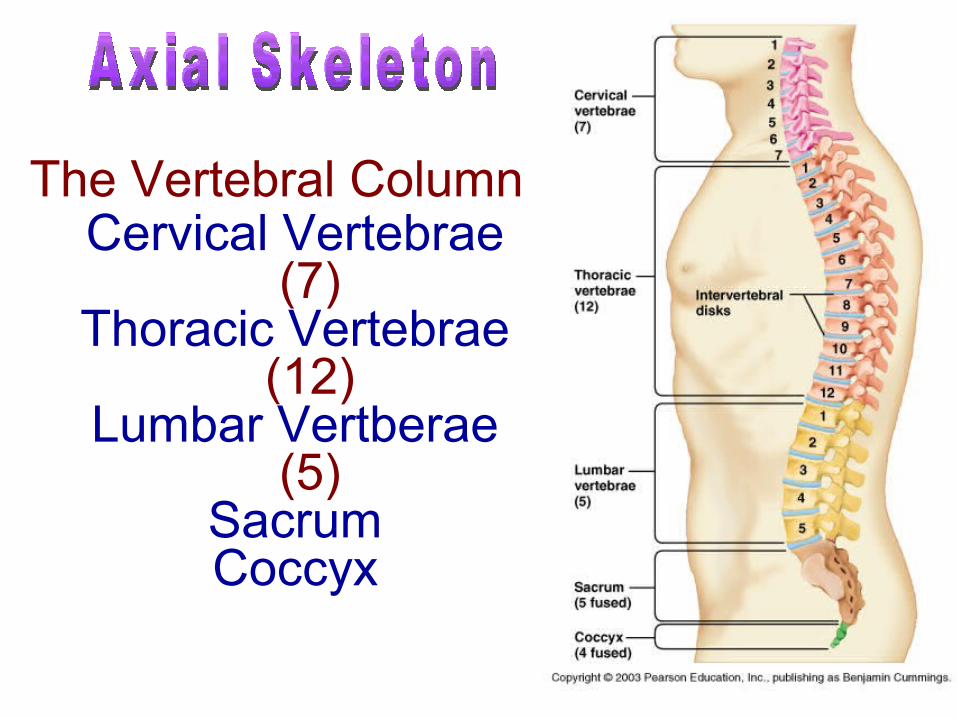
Cervical Vertebrae (7)
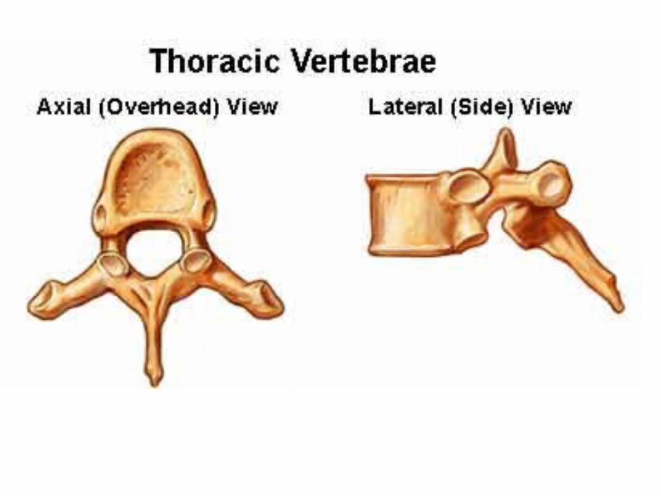
Thoracic Vertebrae (12)
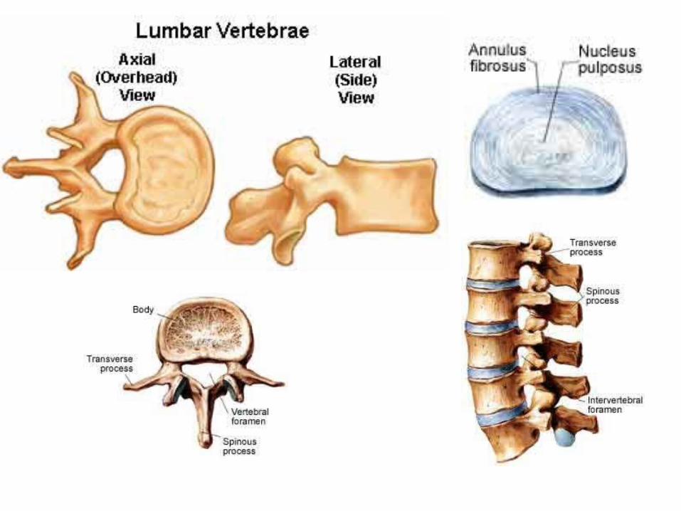
Lumbar Vertberae (5)
SacrumCoccyx
The Vertebral Column
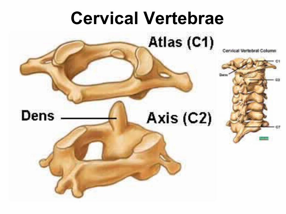
Cervical Vertebrae
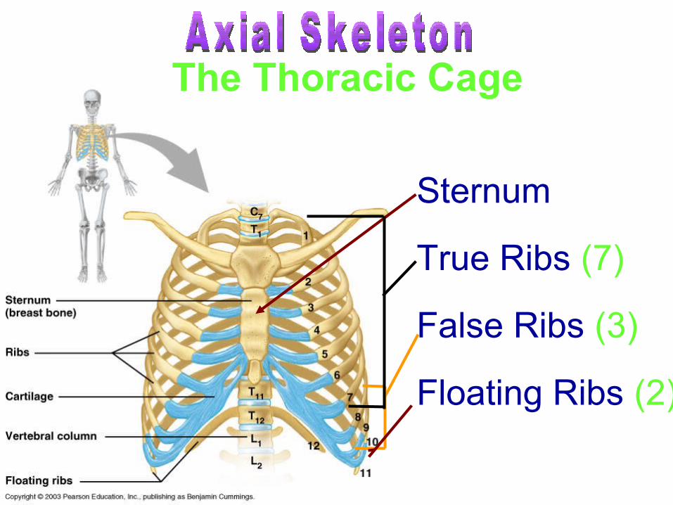
Sternum
True Ribs (7)
False Ribs (3)
Floating Ribs (2)
The Thoracic Cage
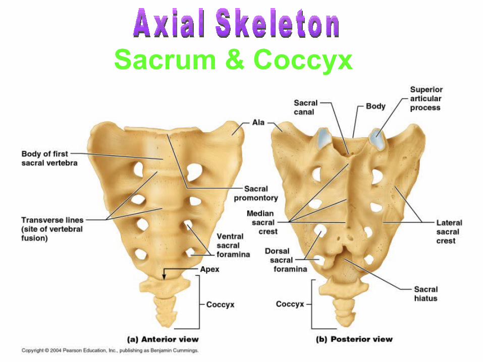
Sacrum & Coccyx
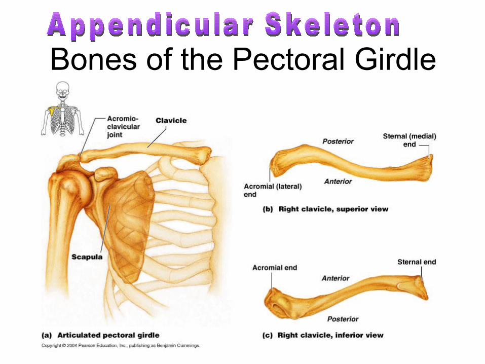
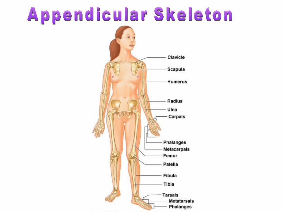
Bones of the Pectoral Girdle
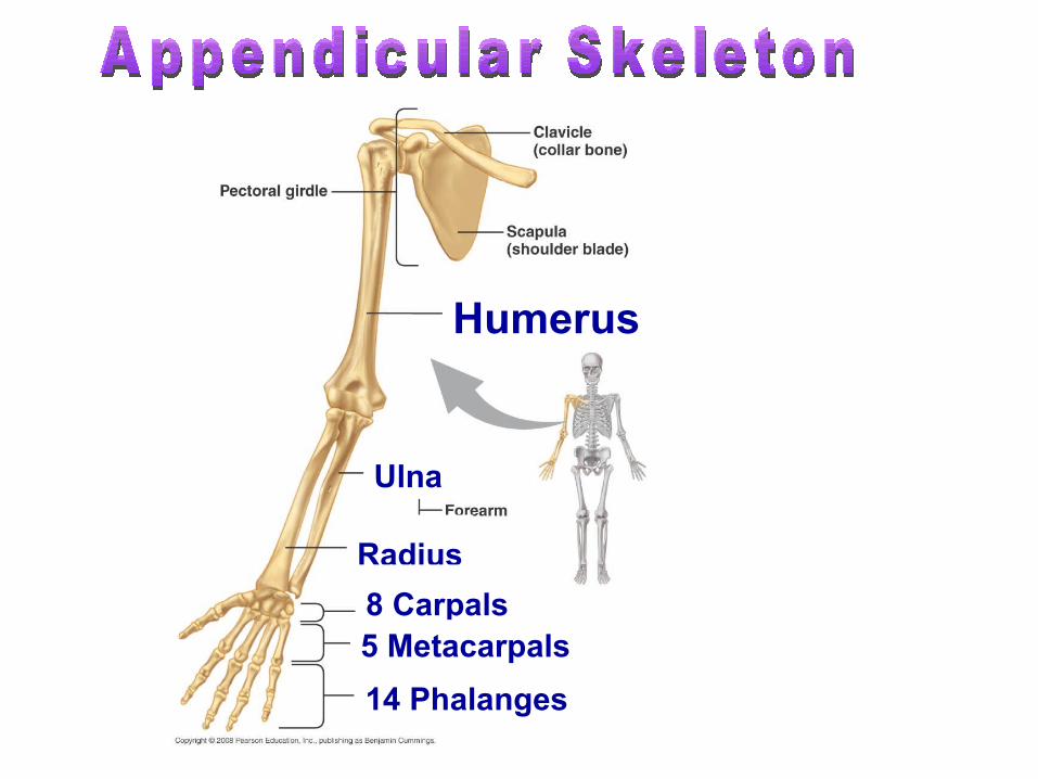
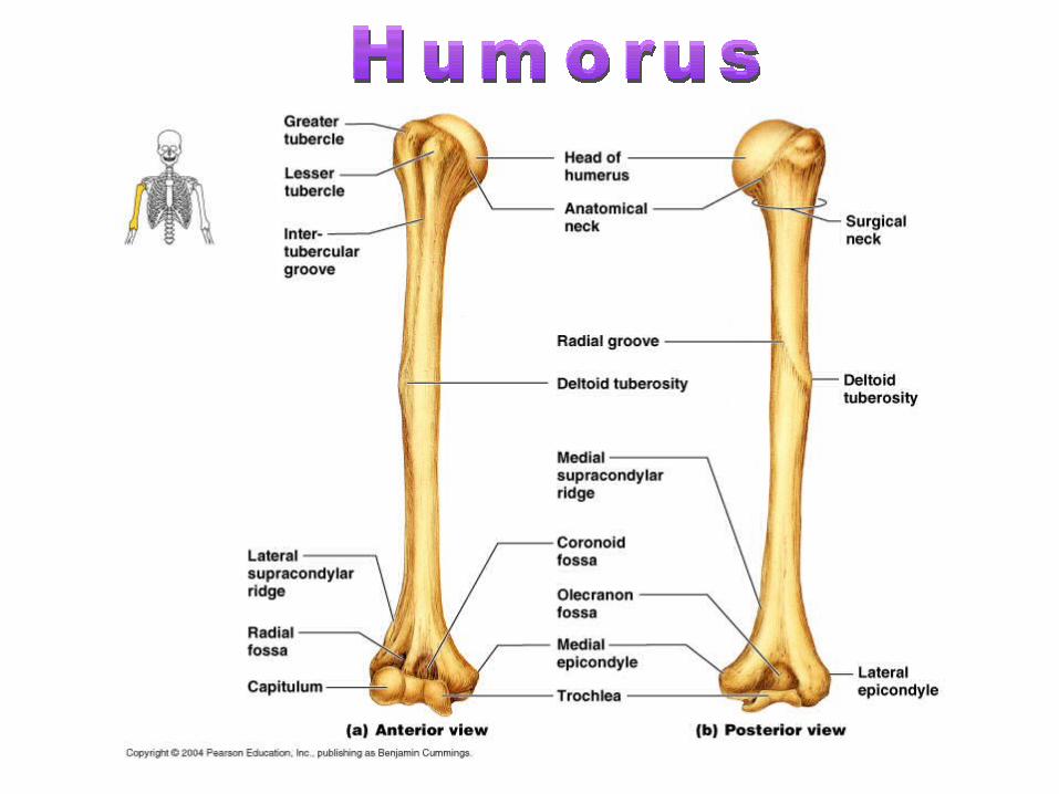
Humerus
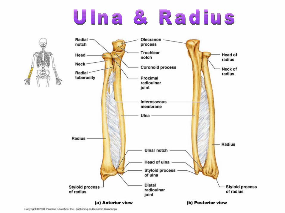
Ulna
Radius
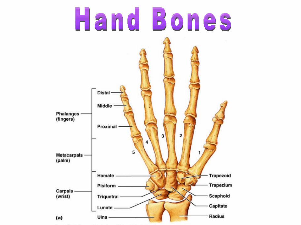
8 Carpals
14 Phalanges
5 Metacarpals
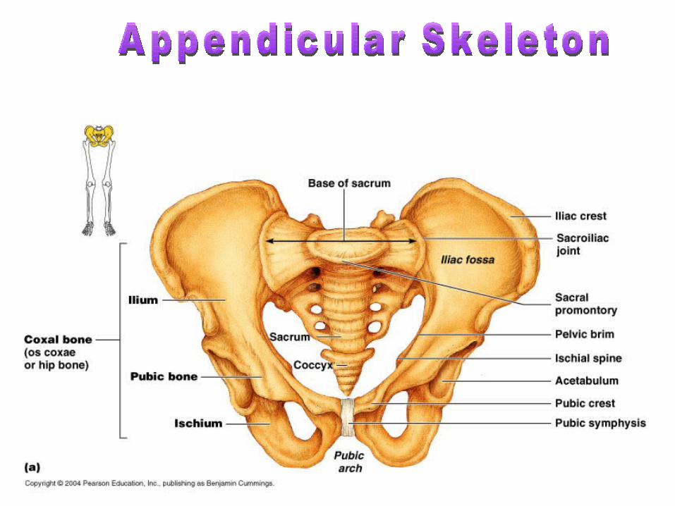
Pelvis
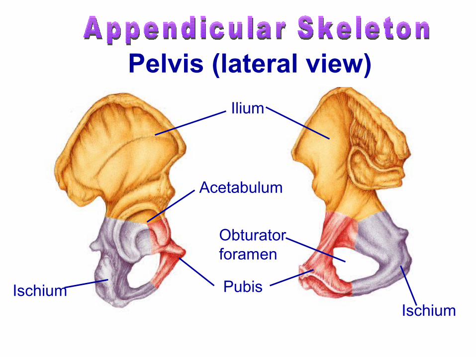
Ischium
Ilium
Acetabulum
Pubis
Ischium
Obturator foramen
Pelvis (lateral view)
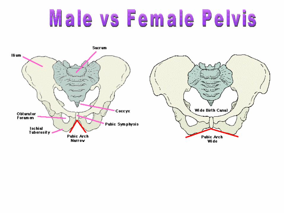
Male Pelvic Girdle
Female Pelvic Girdle
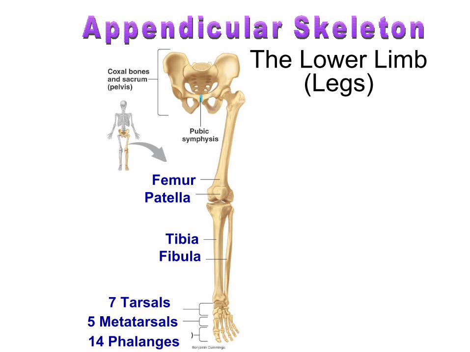
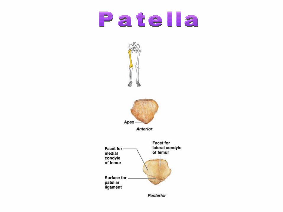
Patella
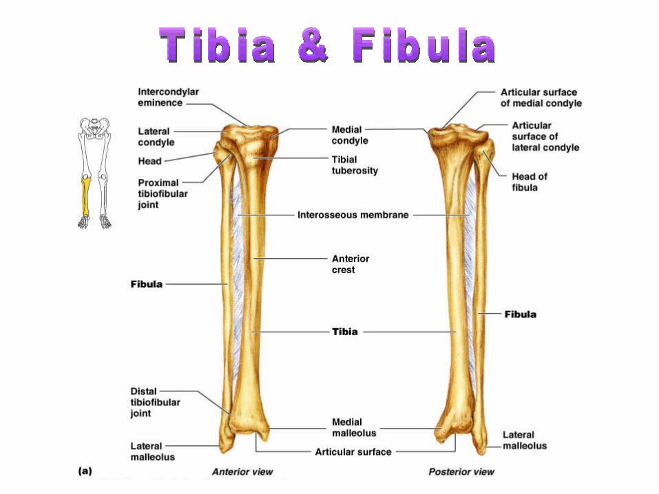
The Lower Limb (Legs)
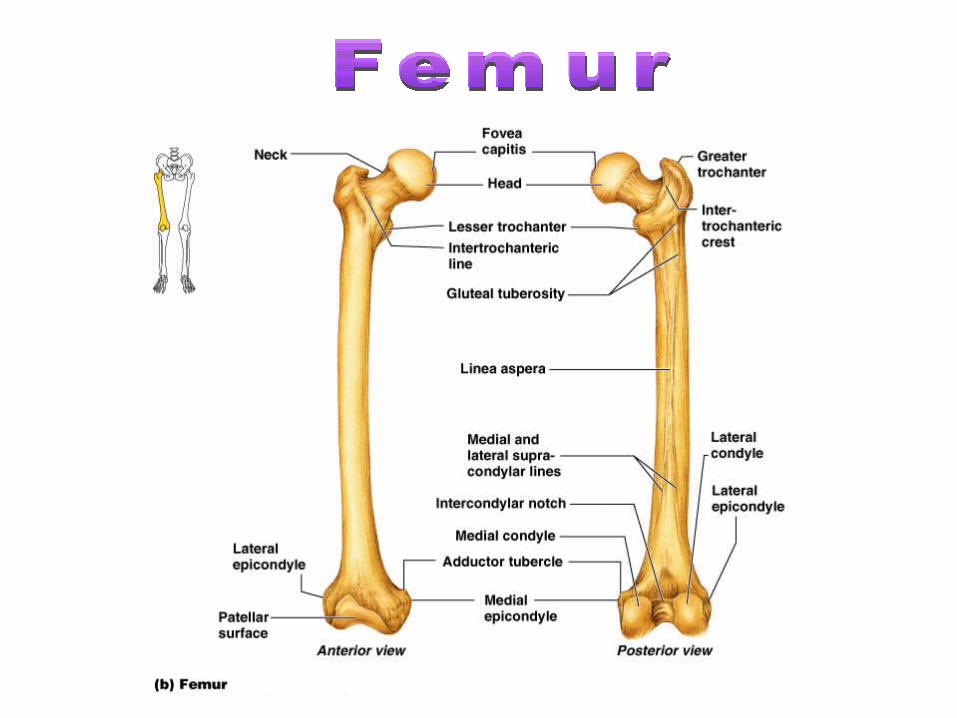
Femur
TibiaFibula
5 Metatarsals14 Phalanges
7 Tarsals
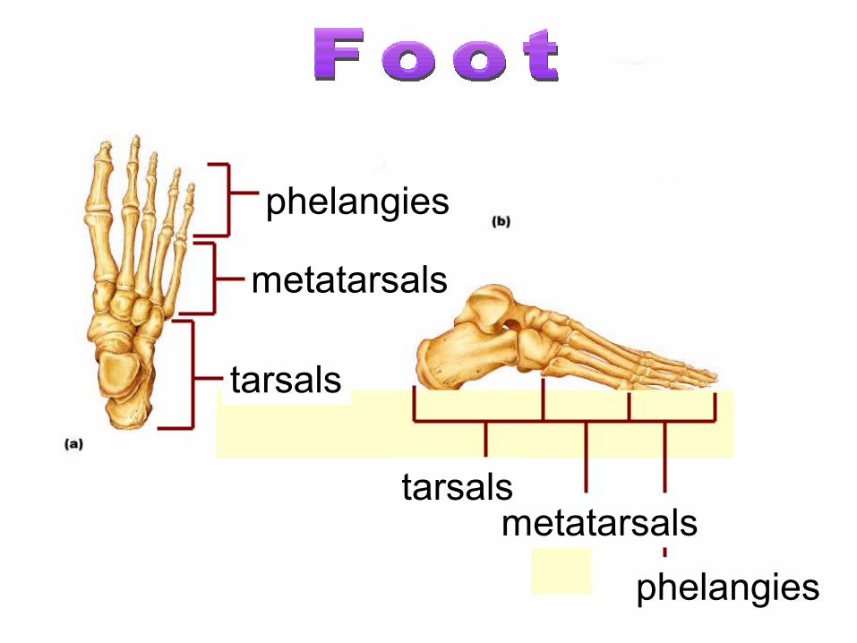
metatarsals
phelangies
tarsals
metatarsals
phelangies
tarsals
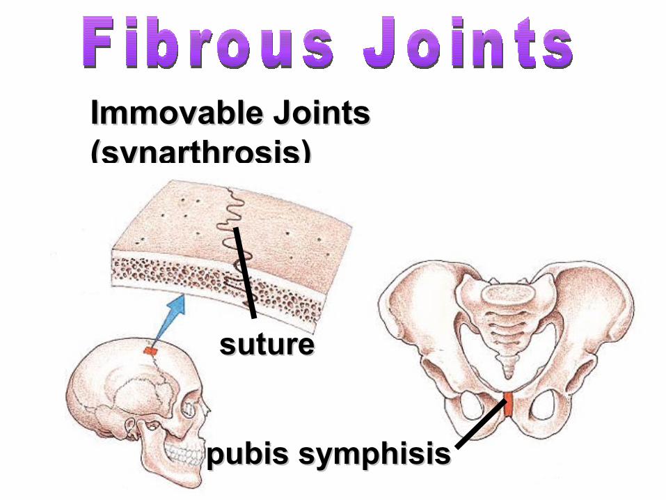
Immovable Joints Immovable Joints (synarthrosis)(synarthrosis)
suturesuture
pubis symphisispubis symphisis
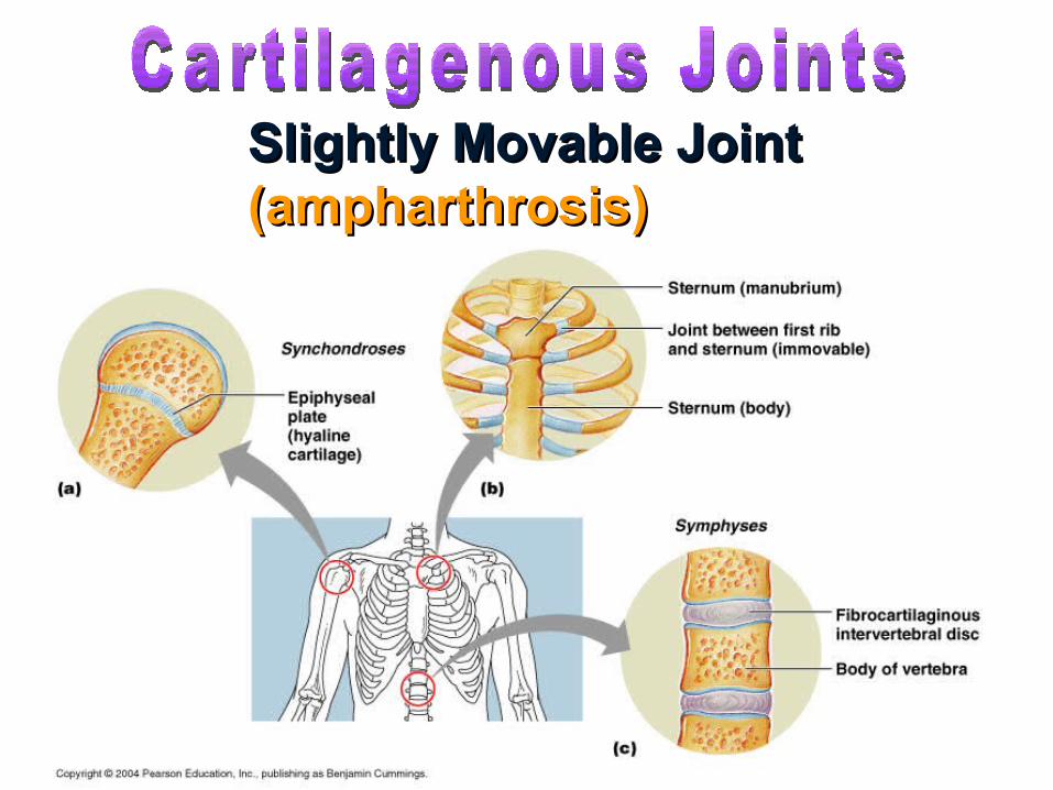
Slightly Movable Joint Slightly Movable Joint (ampharthrosis)(ampharthrosis)
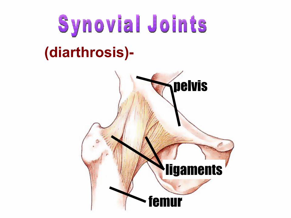
femur
ligaments
pelvis
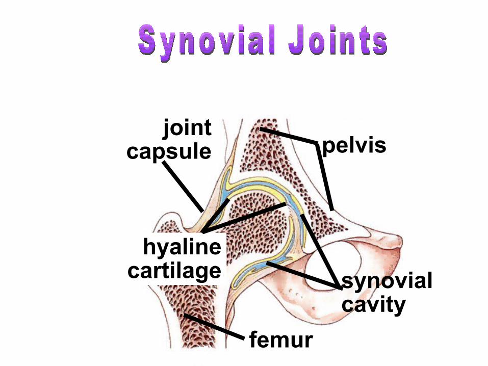
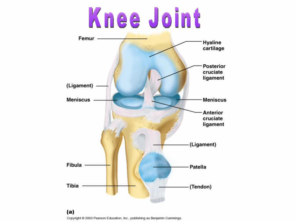
(diarthrosis)- freely moveable
femur
pelvis
hyaline cartilage synovial
cavity
joint capsule
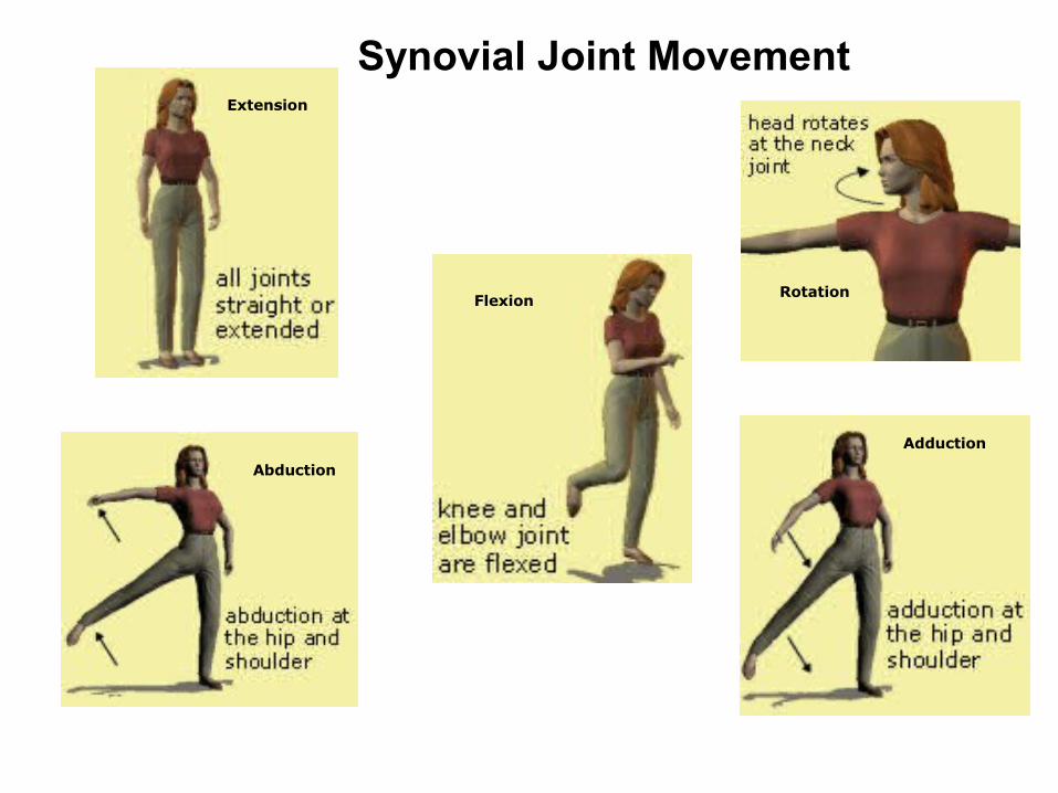
Abduction
Extension
RotationFlexion
Adduction
Synovial Joint Movement
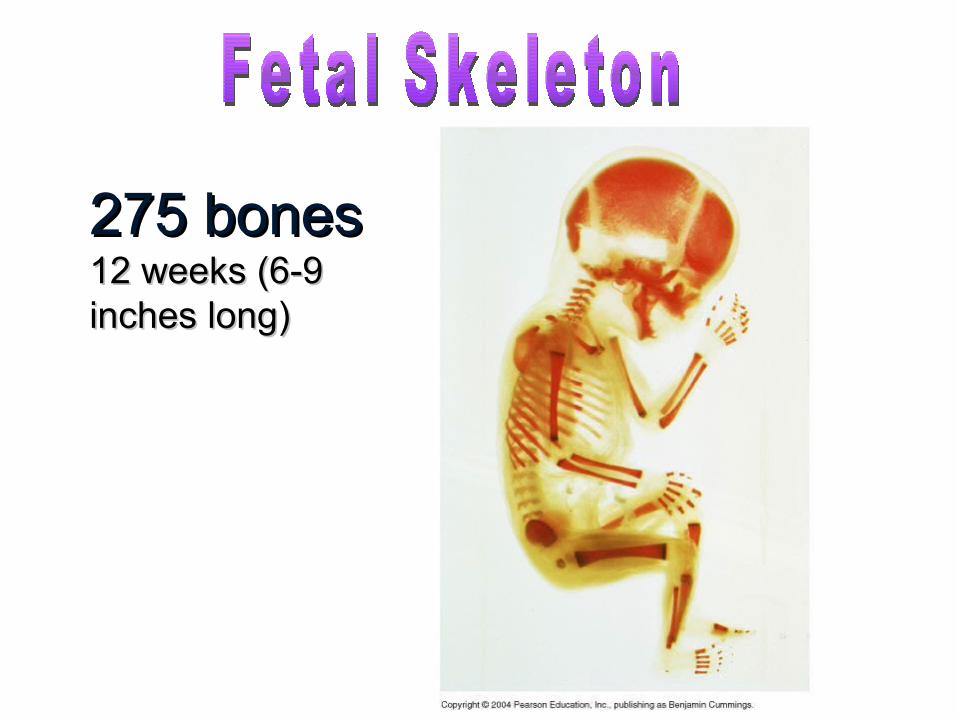
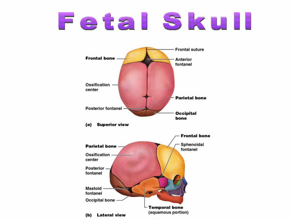
275 bones275 bones12 weeks (6-9 12 weeks (6-9 inches long)inches long)
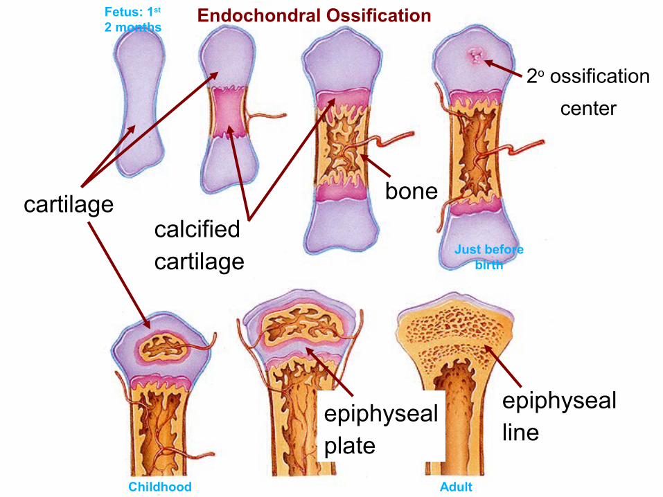
cartilagecalcified cartilage
bone
epiphyseal plate
epiphyseal line
Endochondral Ossification
2o ossification
center
Fetus: 1st 2 months
AdultChildhood
Just before birth
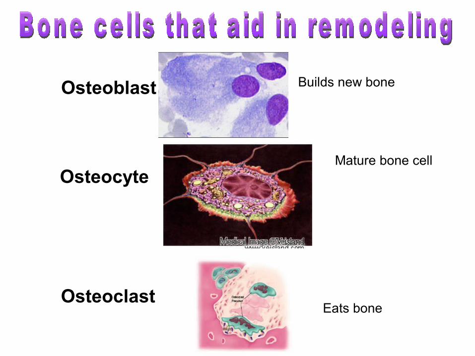
Osteoblast
Osteocyte
OsteoclastEats bone
Builds new bone
Mature bone cell
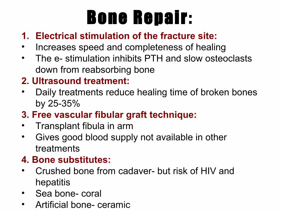
Bone Repair :1. Electrical stimulation of the fracture site:• Increases speed and completeness of healing• The e- stimulation inhibits PTH and slow osteoclasts
down from reabsorbing bone2. Ultrasound treatment:• Daily treatments reduce healing time of broken bones
by 25-35%3. Free vascular fibular graft technique:• Transplant fibula in arm• Gives good blood supply not available in other
treatments4. Bone substitutes:• Crushed bone from cadaver- but risk of HIV and
hepatitis• Sea bone- coral• Artificial bone- ceramic
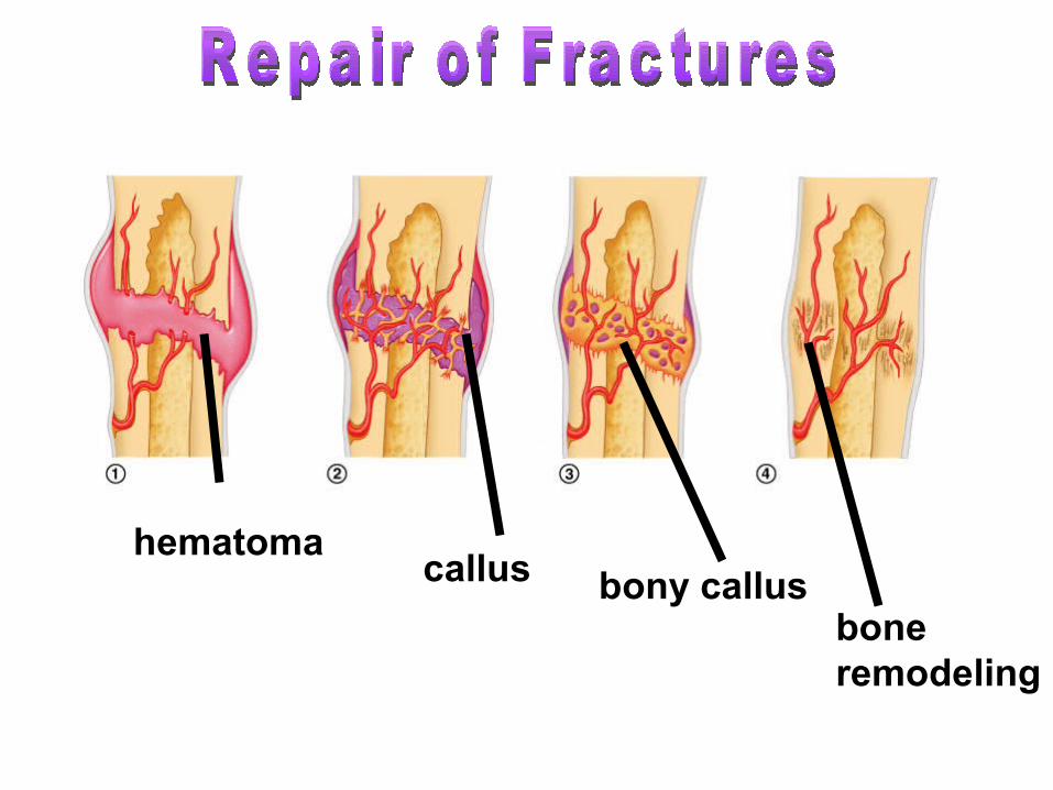
hematomacallus bony callus
bone remodeling
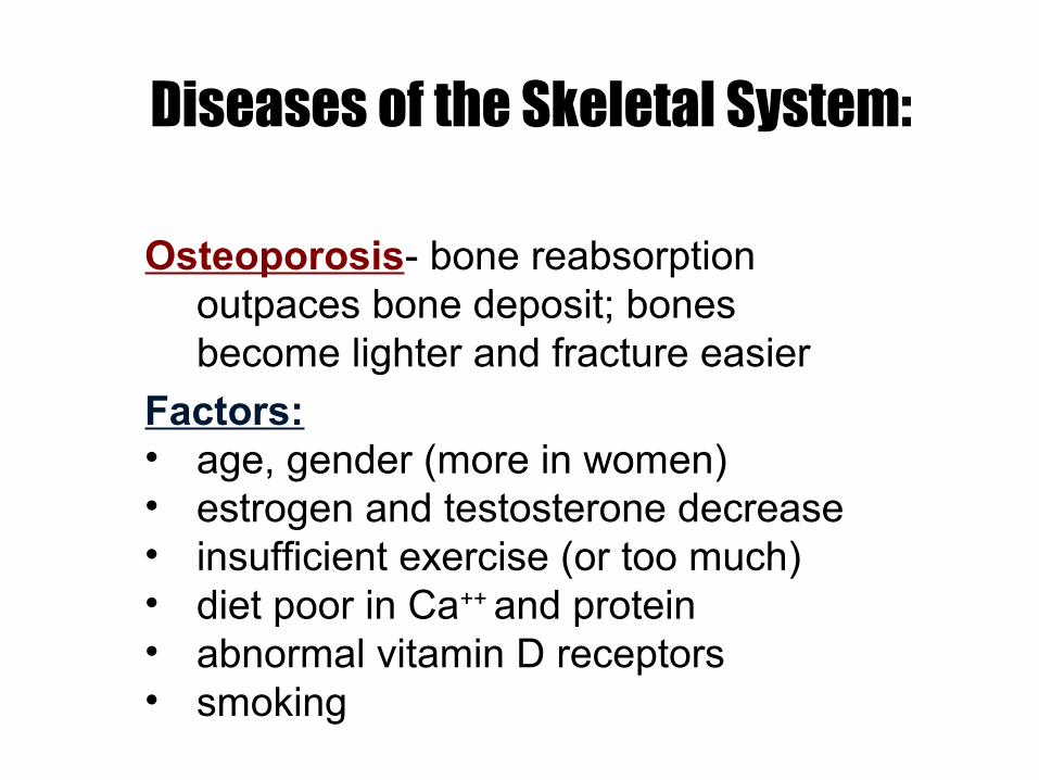
Diseases of the Skeletal System:
Osteoporosis- bone reabsorption outpaces bone deposit; bones become lighter and fracture easier
Factors: • age, gender (more in women)• estrogen and testosterone decrease• insufficient exercise (or too much)• diet poor in Ca++ and protein• abnormal vitamin D receptors• smoking
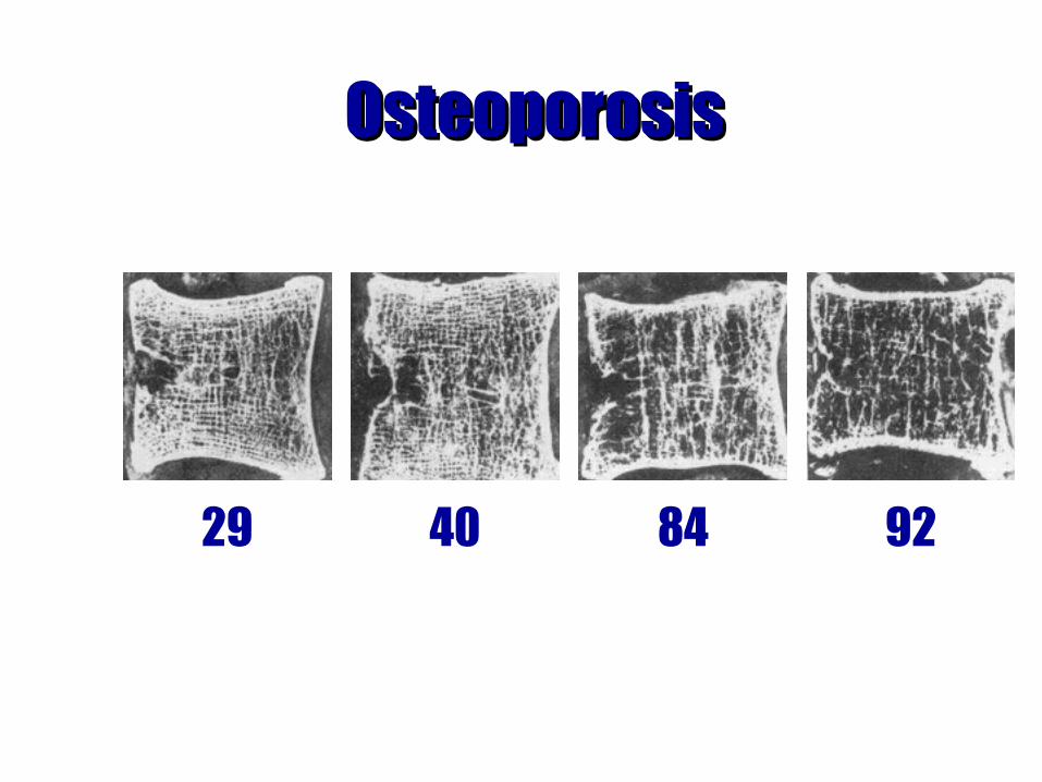
OsteoporosisOsteoporosis
29 40 84 92
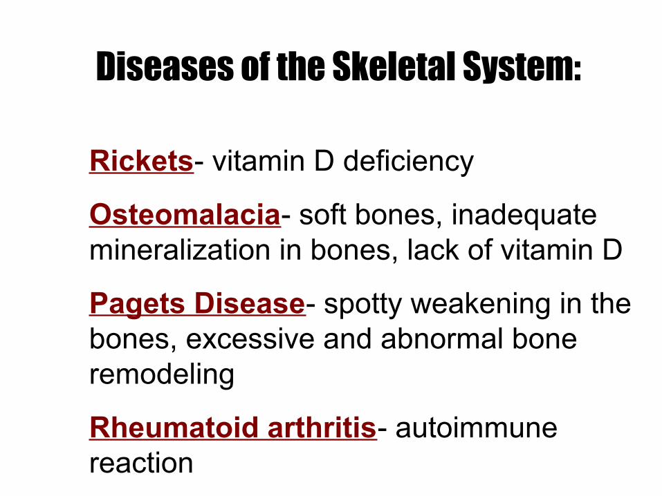
Rickets- vitamin D deficiency
Osteomalacia- soft bones, inadequate mineralization in bones, lack of vitamin D
Pagets Disease- spotty weakening in the bones, excessive and abnormal bone remodeling
Rheumatoid arthritis- autoimmune reaction
Diseases of the Skeletal System: