human nutrition
TRANSCRIPT

WAYNE FERNANDES

Humans have a a complete digestive tract, which begins withthe mouth and ends with the anus.
The major structures of the human digestive tract are the :
Mouth, Pharynx,Oesophagus, Stomach, Small intestine, Large intestine, Rectum and Anus.

Liver
Salivary glands
gall bladder
pancreas


To convert large, often insoluble molecules of food
into smaller soluble molecules capable of being absorbed into the blood or
lymph capillaries associated with the digestive tract and used for various metabolic
processes.

INGESTION
DIGESTION – MECHANICAL AND CHEMICAL
ABSORPTION
ASSIMILATION
EXCRETION


Ingestion: Food is placed into
mouth.
Teeth:
- Incisors cut or bite the food.
- Canines used to tear meat.
- Molars and pre-molars grind the food
fine.
Mechanical digestion


Secreted by the salivary glands (parotid gland, sublingual gland and sub-maxillary gland)
Mix with the food to form a bolus.
Saliva contains the enzyme amylase, that breaks down cooked starch into maltose.
Chemical digestion.

Mixes food with saliva and pushes food between teeth.
Makes swallowing easier.

The bolus is forced down the esophagus when the muscular pharynx contract – swallow process.
Peristalsis (contraction and relaxation of antagonistic circular and longitudinal muscle) of the esophagus pushes the food downwards into the stomach, through the cardiac valve.
No absorption takes place The epiglottis that covers the
trachea prevents food from going
into the trachea when you swallow.


PROCESS OF PERISTALSIS


Food enters the stomach (fundus region) through the cardiac valve.
Remains a half an hour still before the stomach muscles (circular-, longitudinal- and oblique muscles) starts contracting and relaxing (peristalsis).
The food move with circular movements through the stomach (corpus and pyloric regions) and mixes with all the gastric juices.
Mechanical digestion

Gastric juices are secreted after the hormone, gastrin stimulates the parietal cells in the fundusregion of the stomach.
Gastric juices consist of HCl(Acidify stomach and neutralizes the bolus, antiseptic solution, emulsifies fats), digestive enzymes (pepsin, rennin and lipase), mucus (help protect the inner lining of the stomach against enzyme activity) and water.

These gastric juices help with chemical digestion of food and the bolus is now called a chym.
Some absorption occurs in the stomach. Some water, glucose, salt and certain drugs and alcohol pass into the blood capillaries of the stomach wall.


As the chym enters the duodenum (first part of the small intestine) through the pyloric valve it mixes with bile (excreted from the liver or gall bladder) and pancreatic juices.

Secretin is the hormone that stimulates the pancreas to secrete pancreatic juice into the duodenum.
The pancreatic juice contain sodium bicarbonate (neutralizes the chym, antiseptic) and digestive enzymes (Trypsin, amylase and lipase)
Bile is produced in the liver and stored by the gall bladder.
When fatty rich food enters the small intestines, bile is secreted by the gall bladder and transported to the duodenum.


Neutralizes acid from stomachEmulsifies fats (increase surface
area of fats for better digestion)Aid in the absorption of fats.Reduce the fluidity of the chymAntiseptic – prevent decompositionAssist in the absorption of the fat-
soluble vitamins.
The chym then moves through the ileum (second part of small intestines)
It mixes with intestinal juice (succusentericus) that contains the digestive enzymes for the final digestion of food –Peptidase, lipase, lactase, maltase, sucrase
Succus entericus is secreted by 2 glands: Crypt of Lieberkuhn and the glands
of Brunner (both situated in the lining of the small intestine).

The 2 muscle layer of the small intestine helps with the process of peristalsis.
Cross section through the small intestine

The liver produces and secretes bile. It stores glucose in the form of glycogen and is controlled
by the hormone insulin. When the body requires glucose, glycogen is changed back into glucose. The liver also converts glycerol, into glucose.
It can convert excess carbohydrates into fatty acids which combine with glycerol to form fats.
The liver stores blood temporarily, blood is also formed in the liver of embryos.
The liver manufactures plasma proteins e.g. albumen and fibrinogen.
The liver forms heparin which prevents the clotting of blood inside the blood vessels.
The liver detoxifies the body.

• The liver breaks down surplus amino acids through the process of deamination•Change amino acid into ammonia. •Two ammonia molecules combine with one molecule of carbon dioxide to form urea and water. • Deaminated amino acids are converted into glucose (glucose is converted to glycogen and stored in the liver)•Urea is conveyed to the kidneys for excretion.

The hepatic portal vein (transports digested food from the small intestine to the liver) and the hepatic artery (transports oxygen and nutrients to the liver) enter the liver.
Inside the liver the blood of these 2 blood vessels mix and the products transported is exchanged between the blood and the liver cells.
The waste moves out of the liver cells and is transported away from the liver by means of the hepatic vein which joins up with the inferior vena cava.

BLOOD SUPPLY TO THE LIVER CELLS

Food is mainly absorbed from the small intestine, where all final digestive processes take place and the end-products of digestion is formed.

The final products of digestion (glucose, amino acids, fatty acids and glycerol) are formed in the small intestine.
The absorptive surface area is increased by:
The great length of the small intestine (7m)
The circular folds of the mucosa lining of the small intestine.
The millions of villi lining the folds.
The chym is pushed along very slowly through the small intestine, allowing time for absorption to take place.


A villus consist of several capillary arteries, which supply the villus with OXYGEN and capillary veins which carry food away from the villus.
Lacteals (lymph vessels) are found in the centre of each villus used for the absorption of fats.
The vessels are surrounded by connective tissueand a layer of columnar epithelial cells in which goblet cells occur
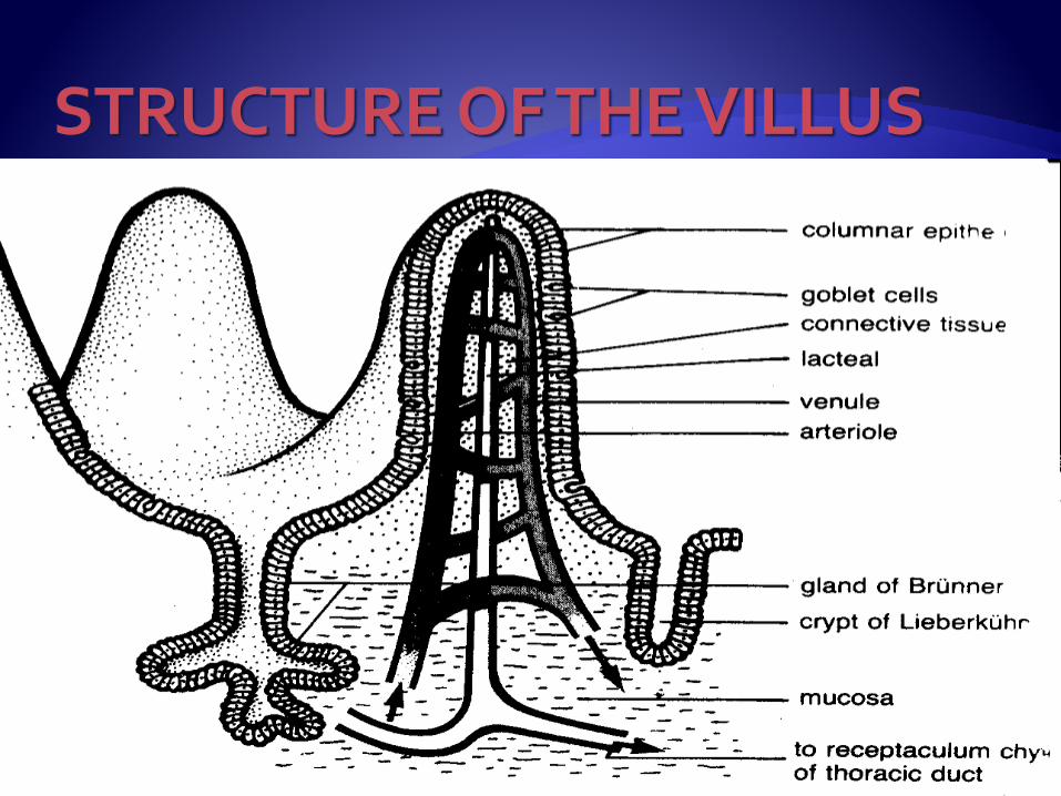

The columnar epithelial cells play an active part in the absorption and are able to allow substances to enter the villi against the concentration gradient.
Amino acids, glucose, mineral salts, water and vitamins are absorbed directly into the blood.

Fatty acids (insoluble) combine with bile salts
To form fatty acid-bile salt complex which is soluble in water.
This complex plus the glycerol component of fat is absorbed by the columnar epithelial cells of the villi.
Inside the villus the fatty acids are freed from the bile salts and recombine with glycerol to form tiny fat globules.
Some of the fat globules are absorbed directly into the blood capillaries.
Most fat globules are absorbed by the lacteals.
The lacteals unite to form small lymph vessels which enter the main lymphatic system.
This fat in the lacteal is now known as chyle, and it reaches the bloodstream in the end.

No digestion takes place in the colon.
Undigested food remains from the small intestine enter the caecum through the ileocaecal valve.
In the colon, water is absorbed so that the chym becomes semi-solid.
Symbiotic bacteria present in the colon act upon the food remains, decomposing them and turning them into faeces.
The bacteria synthesize vitamins of the B group and vitamin K which is essential for the blood clotting process.
Peristalsis in the colon is facilitated by mucus produced by numerous mucous glands.
Mucus assists the passage of faeces and protects the wall of the colon.


By the time the faeces reach the rectum, they consist of approximately 70% water.
Bacteria account for about 30% of the dry mass of faeces
The remainder is made up of food residue, mainly cellulose.


Defication is brought about by contraction of the circular muscles of the rectum, and relaxation of the muscles which make up the anal sphincter.
