human movement - creature and creator
TRANSCRIPT
Picton Session 2 Moving to the Music Brain and Mind
1
Human Movement
What a piece of work is a man,
how noble in reason, how
infinite in faculty, in form and
moving how express and
admirable, in action how like an
angel, in apprehension how like
a god, the beauty of the world,
the paragon of animals.
Shakespeare, 1601, Hamlet, II:2
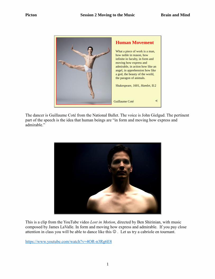
Guillaume Coté
The dancer is Guillaume Coté from the National Ballet. The voice is John Gielgud. The pertinent
part of the speech is the idea that human beings are “in form and moving how express and
admirable.”
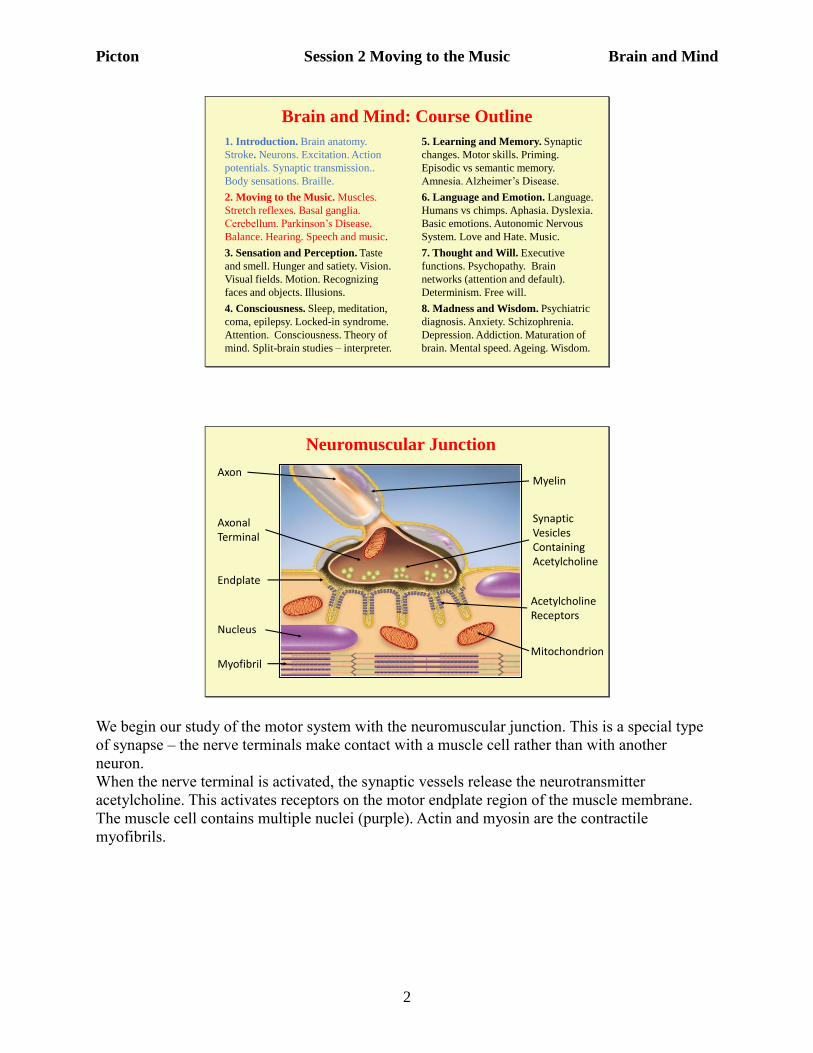
This is a clip from the YouTube video Lost in Motion, directed by Ben Shirinian, with music
composed by James LaValle. In form and moving how express and admirable. If you pay close
attention in class you will be able to dance like this . Let us try a cabriole en tournant.
https://www.youtube.com/watch?v=4OR-n3Rg6E8
Picton Session 2 Moving to the Music Brain and Mind
2
Brain and Mind: Course Outline
1. Introduction. Brain anatomy.
Stroke. Neurons. Excitation. Action
potentials. Synaptic transmission..
Body sensations. Braille.
2. Moving to the Music. Muscles.
Stretch reflexes. Basal ganglia.
Cerebellum. Parkinson’s Disease.
Balance. Hearing. Speech and music.
3. Sensation and Perception. Taste
and smell. Hunger and satiety. Vision.
Visual fields. Motion. Recognizing
faces and objects. Illusions.
4. Consciousness. Sleep, meditation,
coma, epilepsy. Locked-in syndrome.
Attention. Consciousness. Theory of
mind. Split-brain studies – interpreter.
5. Learning and Memory. Synaptic
changes. Motor skills. Priming.
Episodic vs semantic memory.
Amnesia. Alzheimer’s Disease.
6. Language and Emotion. Language.
Humans vs chimps. Aphasia. Dyslexia.
Basic emotions. Autonomic Nervous
System. Love and Hate. Music.
7. Thought and Will. Executive
functions. Psychopathy. Brain
networks (attention and default).
Determinism. Free will.
8. Madness and Wisdom. Psychiatric
diagnosis. Anxiety. Schizophrenia.
Depression. Addiction. Maturation of
brain. Mental speed. Ageing. Wisdom.
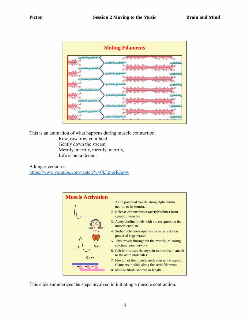
Neuromuscular Junction
MyelinAxon
Axonal Terminal
Synaptic Vesicles ContainingAcetylcholine
Acetylcholine Receptors
Mitochondrion
Nucleus
Myofibril
Endplate
We begin our study of the motor system with the neuromuscular junction. This is a special type
of synapse – the nerve terminals make contact with a muscle cell rather than with another
neuron.
When the nerve terminal is activated, the synaptic vessels release the neurotransmitter
acetylcholine. This activates receptors on the motor endplate region of the muscle membrane.
The muscle cell contains multiple nuclei (purple). Actin and myosin are the contractile
myofibrils.
Picton Session 2 Moving to the Music Brain and Mind
3
Sliding Filaments
This is an animation of what happens during muscle contraction.
Row, row, row your boat
Gently down the stream.
Merrily, merrily, merrily, merrily,
Life is but a dream.
A longer version is
https://www.youtube.com/watch?v=0kFmbrRJq4w
Muscle Activation1. Axon potential travels along alpha motor
neuron to its terminal
2. Release of transmitter (acetylcholine) from
synaptic vesicles
3. Acetylcholine binds with the receptors on the
muscle endplate
4. Sodium channels open and a muscle action
potential is generated
5. This travels throughout the muscle, releasing
calcium from network
6. Calcium causes the myosin molecules to attach
to the actin molecules
7. Flexion of the myosin neck causes the myosin
filaments to slide along the actin filaments
8. Muscle fibrils shorten in length
This slide summarizes the steps involved in initiating a muscle contraction.
Picton Session 2 Moving to the Music Brain and Mind
4
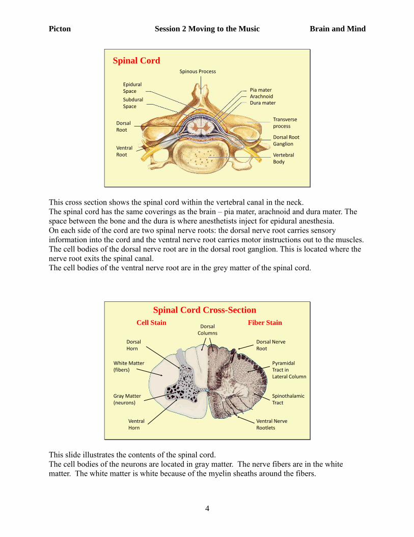
SubduralSpace
Epidural Space
Dorsal Root
Pia materArachnoidDura mater
Transverseprocess
Dorsal RootGanglion
Vertebral Body
Ventral Root
Spinal Cord Spinous Process
This cross section shows the spinal cord within the vertebral canal in the neck.
The spinal cord has the same coverings as the brain – pia mater, arachnoid and dura mater. The
space between the bone and the dura is where anesthetists inject for epidural anesthesia.
On each side of the cord are two spinal nerve roots: the dorsal nerve root carries sensory
information into the cord and the ventral nerve root carries motor instructions out to the muscles.
The cell bodies of the dorsal nerve root are in the dorsal root ganglion. This is located where the
nerve root exits the spinal canal.
The cell bodies of the ventral nerve root are in the grey matter of the spinal cord.
Spinal Cord Cross-Section
White Matter(fibers)
Gray Matter(neurons)
DorsalHorn
VentralHorn
Dorsal Nerve Root
Ventral Nerve Rootlets
Cell Stain Fiber Stain
Pyramidal Tract in Lateral Column
Spinothalamic Tract
DorsalColumns
This slide illustrates the contents of the spinal cord.
The cell bodies of the neurons are located in gray matter. The nerve fibers are in the white
matter. The white matter is white because of the myelin sheaths around the fibers.
Picton Session 2 Moving to the Music Brain and Mind
5
The gray matter is divided into the dorsal horn which has neurons receiving connections from the
incoming sensory fibers, and the ventral horn which contains the motor neurons that innervate
the muscles.
The fibers in the spinal cord go up and down the cord in “columns”
The main connections are the
• dorsal columns which carry fine touch sensation up to the brain.
• pyramidal tracts which carry motor instructions down to the motor neurons in the ventral
horn
• lateral spinothalamic tract which carries pain and temperature sensation up to the brain.
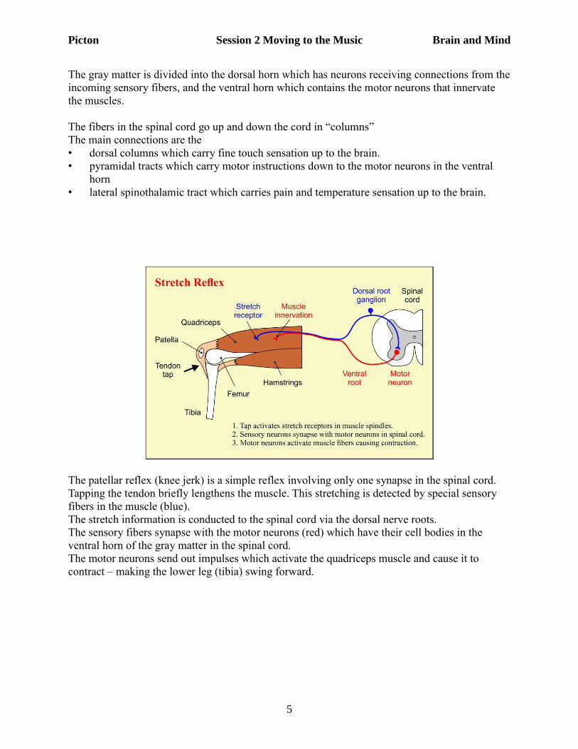
The patellar reflex (knee jerk) is a simple reflex involving only one synapse in the spinal cord.
Tapping the tendon briefly lengthens the muscle. This stretching is detected by special sensory
fibers in the muscle (blue).
The stretch information is conducted to the spinal cord via the dorsal nerve roots.
The sensory fibers synapse with the motor neurons (red) which have their cell bodies in the
ventral horn of the gray matter in the spinal cord.
The motor neurons send out impulses which activate the quadriceps muscle and cause it to
contract – making the lower leg (tibia) swing forward.
Picton Session 2 Moving to the Music Brain and Mind
6
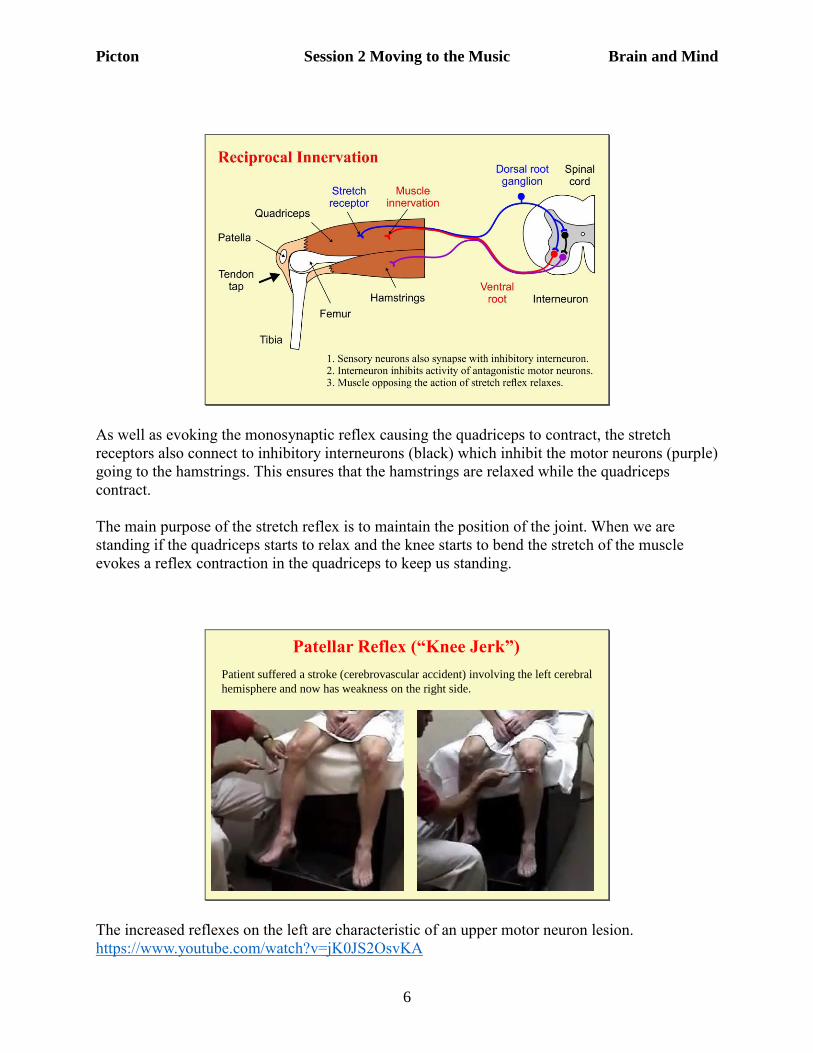
As well as evoking the monosynaptic reflex causing the quadriceps to contract, the stretch
receptors also connect to inhibitory interneurons (black) which inhibit the motor neurons (purple)
going to the hamstrings. This ensures that the hamstrings are relaxed while the quadriceps
contract.
The main purpose of the stretch reflex is to maintain the position of the joint. When we are
standing if the quadriceps starts to relax and the knee starts to bend the stretch of the muscle
evokes a reflex contraction in the quadriceps to keep us standing.
Patellar Reflex (“Knee Jerk”)
Patient suffered a stroke (cerebrovascular accident) involving the left cerebral
hemisphere and now has weakness on the right side.
The increased reflexes on the left are characteristic of an upper motor neuron lesion.
https://www.youtube.com/watch?v=jK0JS2OsvKA
Picton Session 2 Moving to the Music Brain and Mind
7
Frontal Eye Field
Premotor Cortex (Programming)
Primary Somato-sensory Cortex
Primary Motor Cortex
Broca’s Area(Speech)
SupplementaryMotor Area(Initiation)
Movement
Control
Prefrontal Cortices (Planning)
Parietal Cortices (Spatial Control)
Central Sulcus
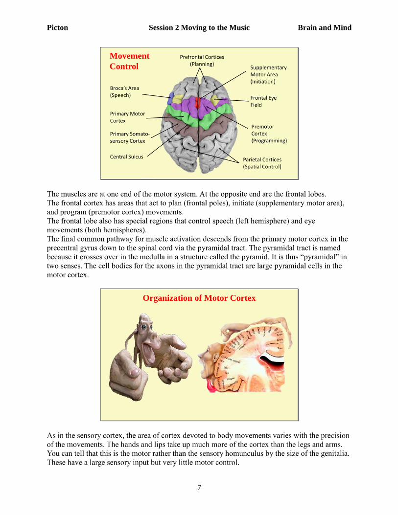
The muscles are at one end of the motor system. At the opposite end are the frontal lobes.
The frontal cortex has areas that act to plan (frontal poles), initiate (supplementary motor area),
and program (premotor cortex) movements.
The frontal lobe also has special regions that control speech (left hemisphere) and eye
movements (both hemispheres).
The final common pathway for muscle activation descends from the primary motor cortex in the
precentral gyrus down to the spinal cord via the pyramidal tract. The pyramidal tract is named
because it crosses over in the medulla in a structure called the pyramid. It is thus “pyramidal” in
two senses. The cell bodies for the axons in the pyramidal tract are large pyramidal cells in the
motor cortex.
Organization of Motor Cortex
As in the sensory cortex, the area of cortex devoted to body movements varies with the precision
of the movements. The hands and lips take up much more of the cortex than the legs and arms.
You can tell that this is the motor rather than the sensory homunculus by the size of the genitalia.
These have a large sensory input but very little motor control.
Picton Session 2 Moving to the Music Brain and Mind
8
Cortical Columns in
Motor Cortex
D – DigitW – WristE – ElbowS – Shoulder
Grasp
Reach
Defense
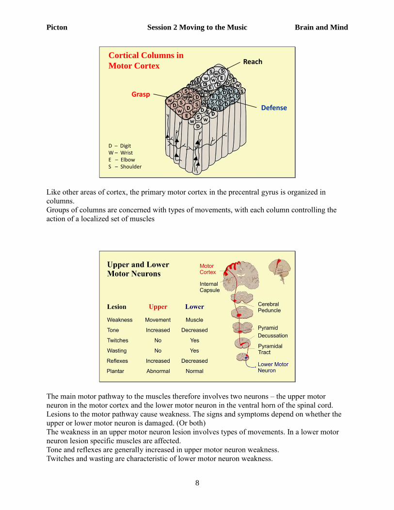
Like other areas of cortex, the primary motor cortex in the precentral gyrus is organized in
columns.
Groups of columns are concerned with types of movements, with each column controlling the
action of a localized set of muscles
The main motor pathway to the muscles therefore involves two neurons – the upper motor
neuron in the motor cortex and the lower motor neuron in the ventral horn of the spinal cord.
Lesions to the motor pathway cause weakness. The signs and symptoms depend on whether the
upper or lower motor neuron is damaged. (Or both)
The weakness in an upper motor neuron lesion involves types of movements. In a lower motor
neuron lesion specific muscles are affected.
Tone and reflexes are generally increased in upper motor neuron weakness.
Twitches and wasting are characteristic of lower motor neuron weakness.
Picton Session 2 Moving to the Music Brain and Mind
9
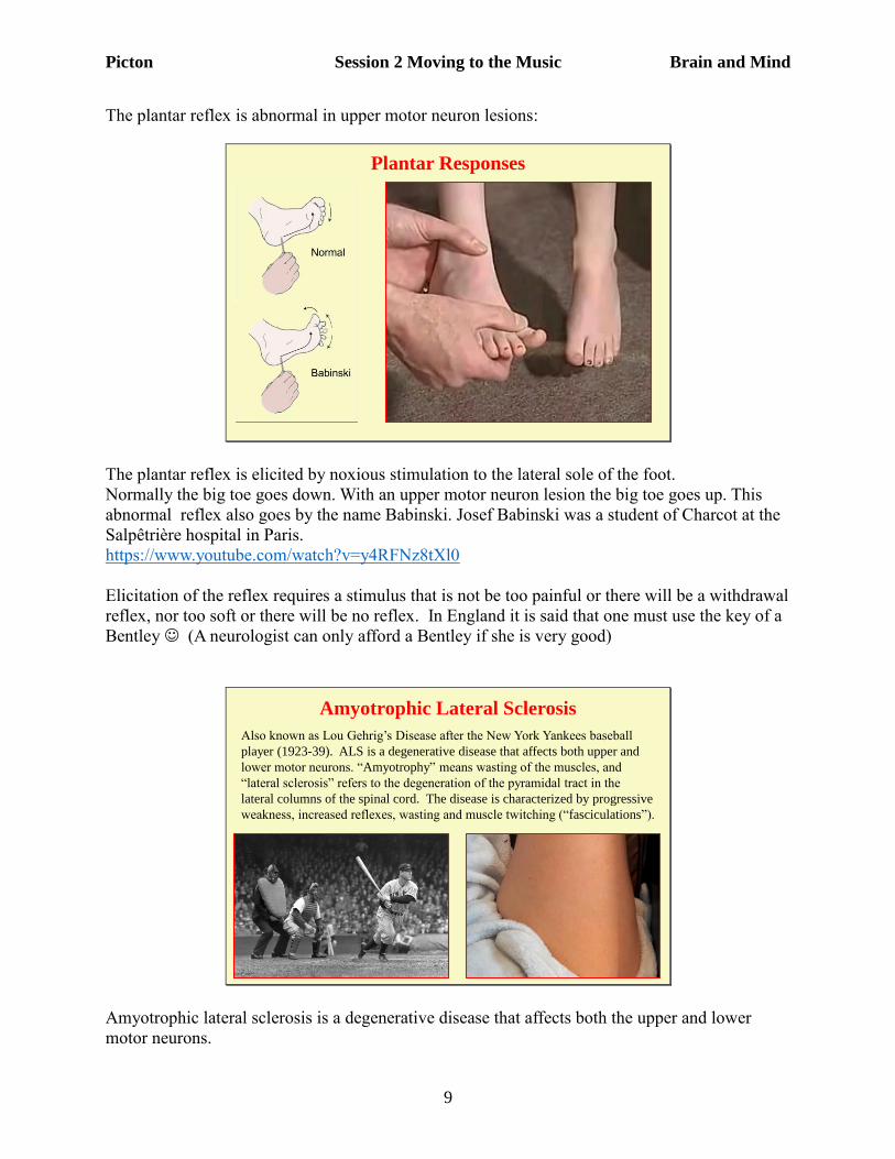
The plantar reflex is abnormal in upper motor neuron lesions:
Plantar Responses
The plantar reflex is elicited by noxious stimulation to the lateral sole of the foot.
Normally the big toe goes down. With an upper motor neuron lesion the big toe goes up. This
abnormal reflex also goes by the name Babinski. Josef Babinski was a student of Charcot at the
Salpêtrière hospital in Paris.
https://www.youtube.com/watch?v=y4RFNz8tXl0
Elicitation of the reflex requires a stimulus that is not be too painful or there will be a withdrawal
reflex, nor too soft or there will be no reflex. In England it is said that one must use the key of a
Bentley (A neurologist can only afford a Bentley if she is very good)
Amyotrophic Lateral Sclerosis
Also known as Lou Gehrig’s Disease after the New York Yankees baseball
player (1923-39). ALS is a degenerative disease that affects both upper and
lower motor neurons. “Amyotrophy” means wasting of the muscles, and
“lateral sclerosis” refers to the degeneration of the pyramidal tract in the
lateral columns of the spinal cord. The disease is characterized by progressive
weakness, increased reflexes, wasting and muscle twitching (“fasciculations”).
Amyotrophic lateral sclerosis is a degenerative disease that affects both the upper and lower
motor neurons.
Picton Session 2 Moving to the Music Brain and Mind
10
Motor ControlMovements are very difficult to control, especially when the mover must
adapt to an unknown environment. Despite the movies, robots cannot yet
perform much better than a human toddler. Robotic failures are similar to
what happens in diseases of the motor system – falls, tremors, inattention,
inability to do two things at once.
Movement requires:
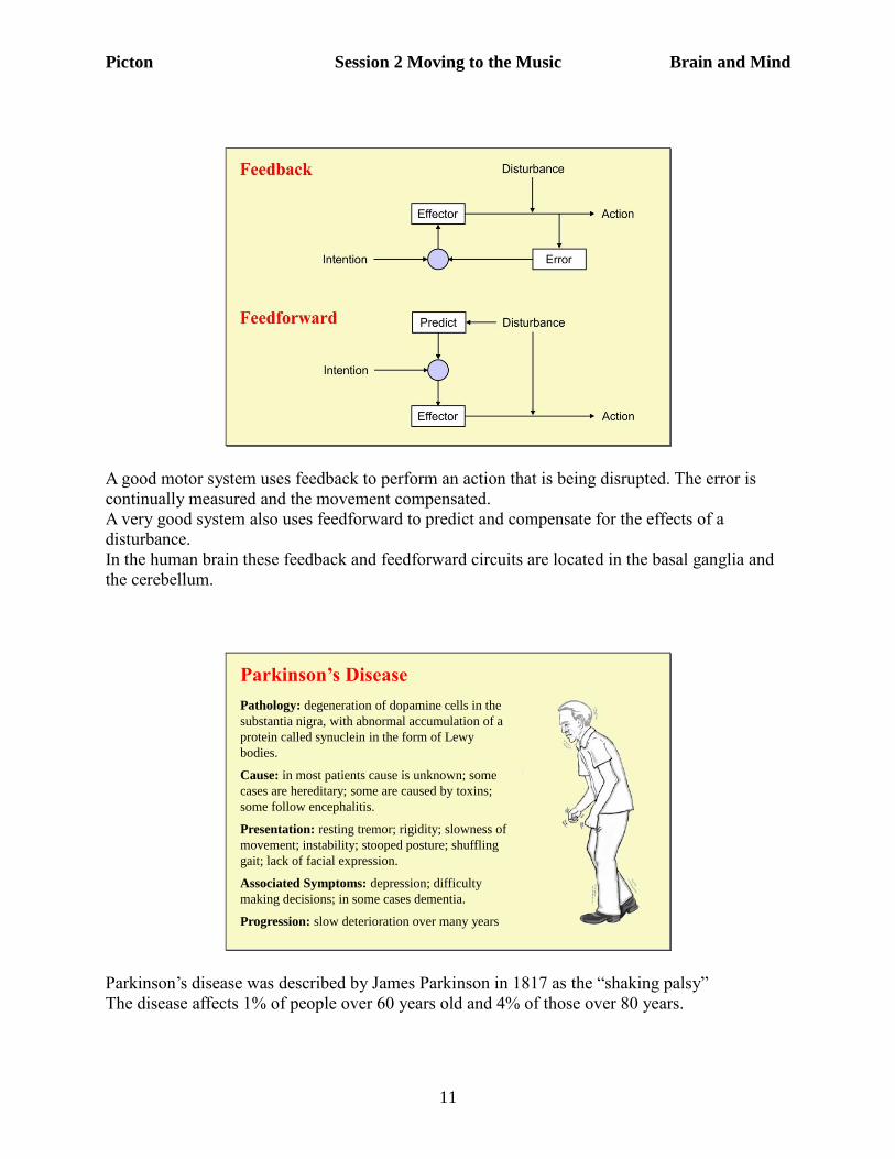
feedback routines to correct for errors.
feedforward programs to anticipate what will happen.
Human movement involves complex interactions between
cerebral cortex (plans, visual control)
basal ganglia (movement in space, learned programs)
cerebellum (error correction, sequencing, learned programs)
vestibular system (body position)
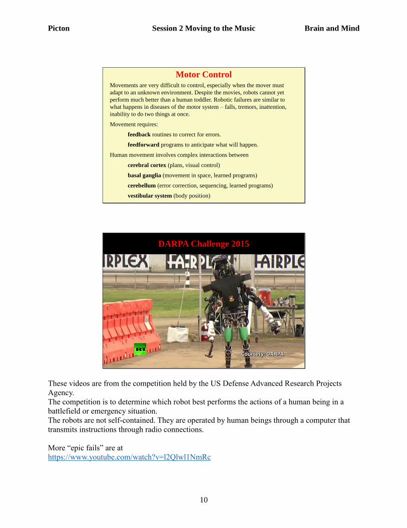
DARPA Challenge 2015
These videos are from the competition held by the US Defense Advanced Research Projects
Agency.
The competition is to determine which robot best performs the actions of a human being in a
battlefield or emergency situation.
The robots are not self-contained. They are operated by human beings through a computer that
transmits instructions through radio connections.
More “epic fails” are at
https://www.youtube.com/watch?v=l2Qlwl1NmRc
Picton Session 2 Moving to the Music Brain and Mind
11
A good motor system uses feedback to perform an action that is being disrupted. The error is
continually measured and the movement compensated.
A very good system also uses feedforward to predict and compensate for the effects of a
disturbance.
In the human brain these feedback and feedforward circuits are located in the basal ganglia and
the cerebellum.
Parkinson’s Disease
Pathology: degeneration of dopamine cells in the
substantia nigra, with abnormal accumulation of a
protein called synuclein in the form of Lewy
bodies.
Cause: in most patients cause is unknown; some
cases are hereditary; some are caused by toxins;
some follow encephalitis.
Presentation: resting tremor; rigidity; slowness of
movement; instability; stooped posture; shuffling
gait; lack of facial expression.
Associated Symptoms: depression; difficulty
making decisions; in some cases dementia.
Progression: slow deterioration over many years
Parkinson’s disease was described by James Parkinson in 1817 as the “shaking palsy”
The disease affects 1% of people over 60 years old and 4% of those over 80 years.
Picton Session 2 Moving to the Music Brain and Mind
12
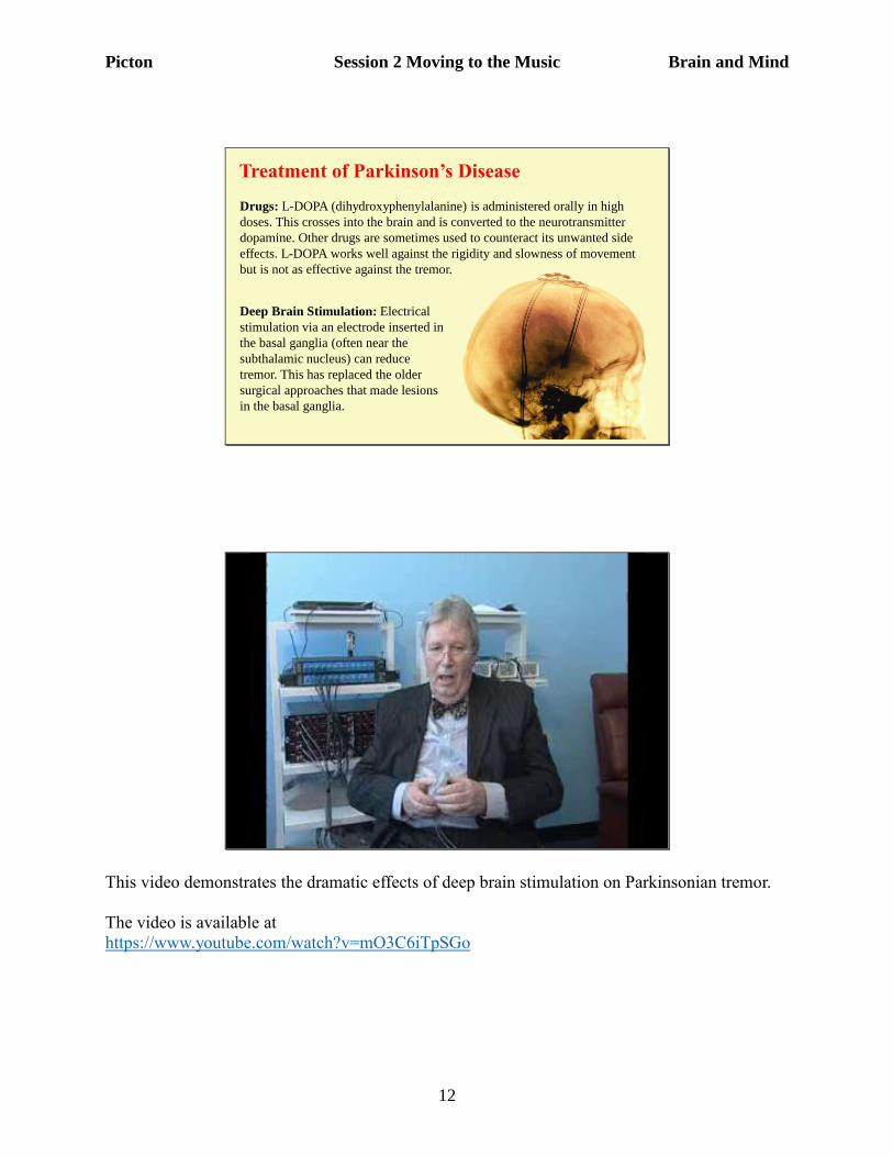
Treatment of Parkinson’s Disease
Drugs: L-DOPA (dihydroxyphenylalanine) is administered orally in high
doses. This crosses into the brain and is converted to the neurotransmitter
dopamine. Other drugs are sometimes used to counteract its unwanted side
effects. L-DOPA works well against the rigidity and slowness of movement
but is not as effective against the tremor.
Deep Brain Stimulation: Electrical
stimulation via an electrode inserted in
the basal ganglia (often near the
subthalamic nucleus) can reduce
tremor. This has replaced the older
surgical approaches that made lesions
in the basal ganglia.
This video demonstrates the dramatic effects of deep brain stimulation on Parkinsonian tremor.
The video is available at
https://www.youtube.com/watch?v=mO3C6iTpSGo
Picton Session 2 Moving to the Music Brain and Mind
13
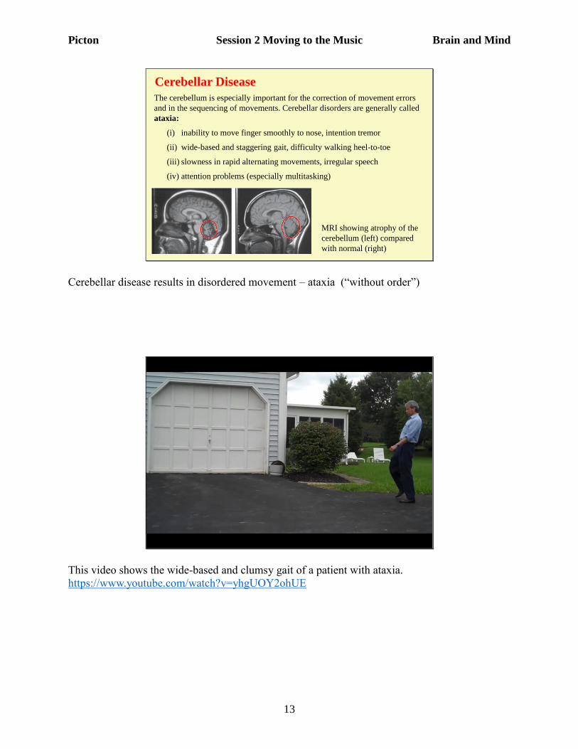
Cerebellar Disease
The cerebellum is especially important for the correction of movement errors
and in the sequencing of movements. Cerebellar disorders are generally called
ataxia:
(i) inability to move finger smoothly to nose, intention tremor
(ii) wide-based and staggering gait, difficulty walking heel-to-toe
(iii) slowness in rapid alternating movements, irregular speech
(iv) attention problems (especially multitasking)
MRI showing atrophy of the
cerebellum (left) compared
with normal (right)
Cerebellar disease results in disordered movement – ataxia (“without order”)
This video shows the wide-based and clumsy gait of a patient with ataxia.
https://www.youtube.com/watch?v=yhgUOY2ohUE
Picton Session 2 Moving to the Music Brain and Mind
14
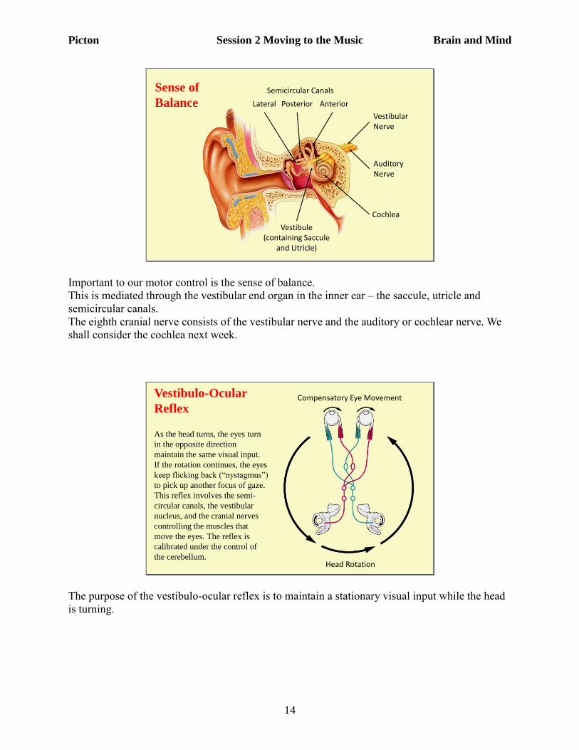
Sense of
BalanceSemicircular Canals
Lateral Posterior Anterior
Vestibule(containing Saccule
and Utricle)
Vestibular Nerve
Auditory Nerve
Cochlea
Important to our motor control is the sense of balance.
This is mediated through the vestibular end organ in the inner ear – the saccule, utricle and
semicircular canals.
The eighth cranial nerve consists of the vestibular nerve and the auditory or cochlear nerve. We
shall consider the cochlea next week.
Vestibulo-Ocular
Reflex Compensatory Eye Movement
Head Rotation
As the head turns, the eyes turn
in the opposite direction
maintain the same visual input.
If the rotation continues, the eyes
keep flicking back (“nystagmus”)
to pick up another focus of gaze.
This reflex involves the semi-
circular canals, the vestibular
nucleus, and the cranial nerves
controlling the muscles that
move the eyes. The reflex is
calibrated under the control of
the cerebellum.
The purpose of the vestibulo-ocular reflex is to maintain a stationary visual input while the head
is turning.
Picton Session 2 Moving to the Music Brain and Mind
15
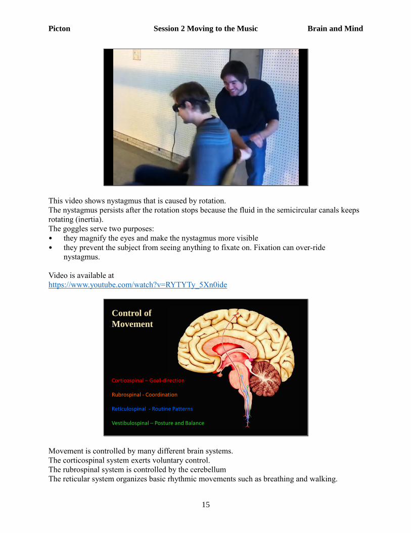
This video shows nystagmus that is caused by rotation.
The nystagmus persists after the rotation stops because the fluid in the semicircular canals keeps
rotating (inertia).
The goggles serve two purposes:
• they magnify the eyes and make the nystagmus more visible
• they prevent the subject from seeing anything to fixate on. Fixation can over-ride
nystagmus.
Video is available at
https://www.youtube.com/watch?v=RYTYTy_5Xn0ide
Control of
Movement
Vestibulospinal – Posture and Balance
Reticulospinal - Routine Patterns
Rubrospinal - Coordination
Corticospinal – Goal-direction
Movement is controlled by many different brain systems.
The corticospinal system exerts voluntary control.
The rubrospinal system is controlled by the cerebellum
The reticular system organizes basic rhythmic movements such as breathing and walking.
Picton Session 2 Moving to the Music Brain and Mind
16
The vestibulospinal system makes the necessary compensations as our body moves (or is moved)
through space.
All of these systems project down to the neurons of the ventral horn of the spinal cord.
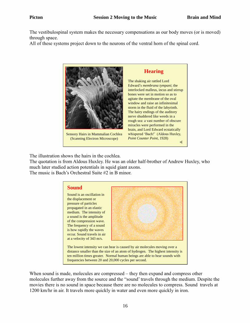
Hearing
Sensory Hairs in Mammalian Cochlea
(Scanning Electron Microscope)
The shaking air rattled Lord
Edward’s membrana tympani; the
interlocked malleus, incus and stirrup
bones were set in motion so as to
agitate the membrane of the oval
window and raise an infinitesimal
storm in the fluid of the labyrinth.
The hairy endings of the auditory
nerve shuddered like weeds in a
rough sea: a vast number of obscure
miracles were performed in the
brain, and Lord Edward ecstatically
whispered ‘Bach!’ (Aldous Huxley,
Point Counter Point, 1928)
The illustration shows the hairs in the cochlea.
The quotation is from Aldous Huxley. He was an older half-brother of Andrew Huxley, who
much later studied action potentials in squid giant axons.
The music is Bach’s Orchestral Suite #2 in B minor.
Sound
Sound is an oscillation in
the displacement or
pressure of particles
propagated in an elastic
medium. The intensity of
a sound is the amplitude
of the compression wave.
The frequency of a sound
is how rapidly the waves
occur. Sound travels in air
at a velocity of 343 m/s.
The lowest intensity we can hear is caused by air molecules moving over a
distance smaller than the size of an atom of hydrogen. The highest intensity is
ten million times greater. Normal human beings are able to hear sounds with
frequencies between 20 and 20,000 cycles per second.
When sound is made, molecules are compressed – they then expand and compress other
molecules further away from the source and the “sound’ travels through the medium. Despite the
movies there is no sound in space because there are no molecules to compress. Sound travels at
1200 km/hr in air. It travels more quickly in water and even more quickly in iron.
Picton Session 2 Moving to the Music Brain and Mind
17
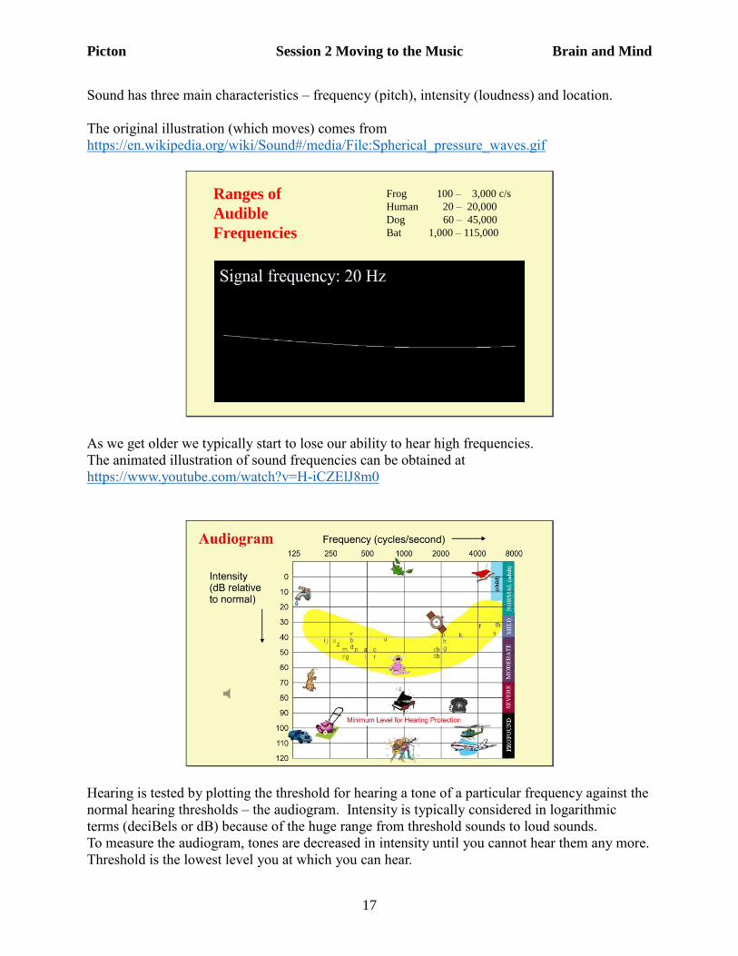
Sound has three main characteristics – frequency (pitch), intensity (loudness) and location.
The original illustration (which moves) comes from
https://en.wikipedia.org/wiki/Sound#/media/File:Spherical_pressure_waves.gif
Ranges of
Audible
Frequencies
Frog 100 – 3,000 c/s
Human 20 – 20,000
Dog 60 – 45,000
Bat 1,000 – 115,000
As we get older we typically start to lose our ability to hear high frequencies.
The animated illustration of sound frequencies can be obtained at
https://www.youtube.com/watch?v=H-iCZElJ8m0
Hearing is tested by plotting the threshold for hearing a tone of a particular frequency against the
normal hearing thresholds – the audiogram. Intensity is typically considered in logarithmic
terms (deciBels or dB) because of the huge range from threshold sounds to loud sounds.
To measure the audiogram, tones are decreased in intensity until you cannot hear them any more.
Threshold is the lowest level you at which you can hear.
Picton Session 2 Moving to the Music Brain and Mind
18
Someone with normal hearing has thresholds near 0 dB on the audiogram.
Someone with a hearing loss would only hear the louder tones and would therefore have
thresholds plotted lower down on the graph.
Normal conversational speech sounds occur in the yellow banana-shaped region of the
audiogram – between 200 and 8000 cycles per second and from 20 to 60 dB above normal
thresholds.
You should protect your ears against sounds over 90 dB above normal thresholds – especially jet
planes and rock bands. Anything that causes a ringing in the ears (tinnitus). Even though there
may be no change in hearing threshold, there is still loss of neurons. As time passes this will lead
to threshold elevation.
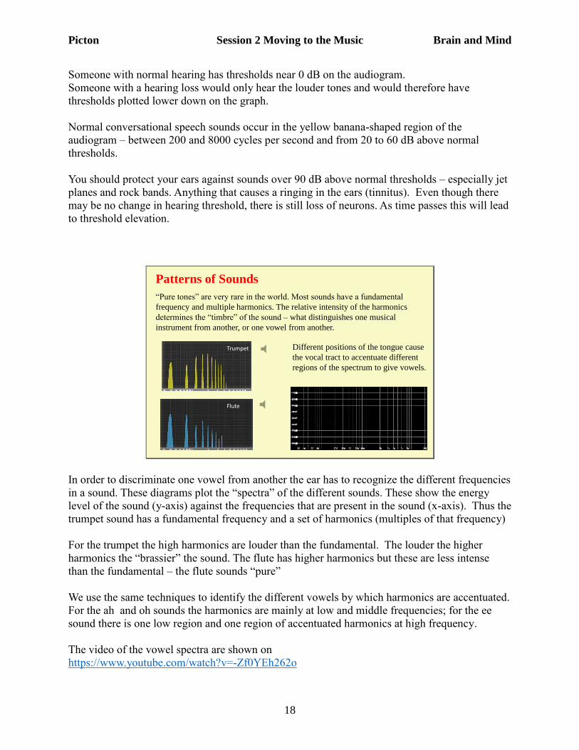
Patterns of Sounds
“Pure tones” are very rare in the world. Most sounds have a fundamental
frequency and multiple harmonics. The relative intensity of the harmonics
determines the “timbre” of the sound – what distinguishes one musical
instrument from another, or one vowel from another.
Trumpet
Flute
Different positions of the tongue cause
the vocal tract to accentuate different
regions of the spectrum to give vowels.
In order to discriminate one vowel from another the ear has to recognize the different frequencies
in a sound. These diagrams plot the “spectra” of the different sounds. These show the energy
level of the sound (y-axis) against the frequencies that are present in the sound (x-axis). Thus the
trumpet sound has a fundamental frequency and a set of harmonics (multiples of that frequency)
For the trumpet the high harmonics are louder than the fundamental. The louder the higher
harmonics the “brassier” the sound. The flute has higher harmonics but these are less intense
than the fundamental – the flute sounds “pure”
We use the same techniques to identify the different vowels by which harmonics are accentuated.
For the ah and oh sounds the harmonics are mainly at low and middle frequencies; for the ee
sound there is one low region and one region of accentuated harmonics at high frequency.
The video of the vowel spectra are shown on
https://www.youtube.com/watch?v=-Zf0YEh262o
Picton Session 2 Moving to the Music Brain and Mind
19
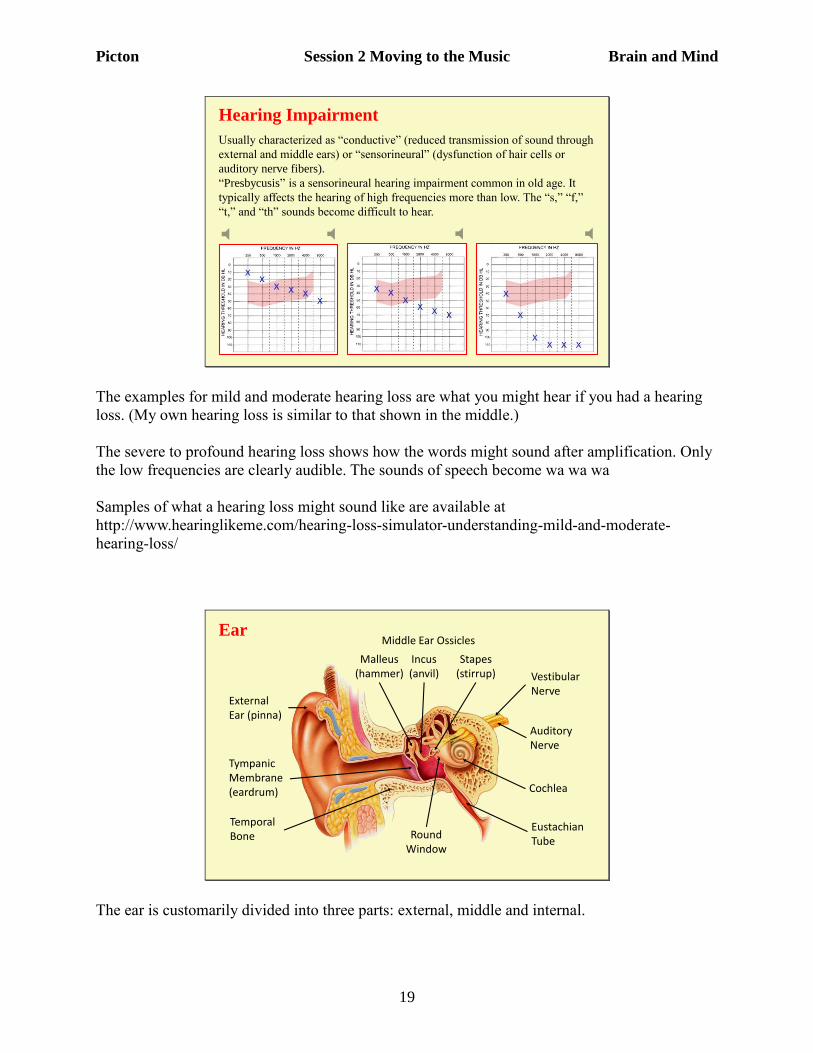
Hearing Impairment
Usually characterized as “conductive” (reduced transmission of sound through
external and middle ears) or “sensorineural” (dysfunction of hair cells or
auditory nerve fibers).
“Presbycusis” is a sensorineural hearing impairment common in old age. It
typically affects the hearing of high frequencies more than low. The “s,” “f,”
“t,” and “th” sounds become difficult to hear.
The examples for mild and moderate hearing loss are what you might hear if you had a hearing
loss. (My own hearing loss is similar to that shown in the middle.)
The severe to profound hearing loss shows how the words might sound after amplification. Only
the low frequencies are clearly audible. The sounds of speech become wa wa wa
Samples of what a hearing loss might sound like are available at
http://www.hearinglikeme.com/hearing-loss-simulator-understanding-mild-and-moderate-
hearing-loss/
EarMiddle Ear Ossicles
Malleus(hammer)
Incus(anvil)
Stapes(stirrup)
Round Window
Vestibular Nerve
Auditory Nerve
Cochlea
Eustachian Tube
TympanicMembrane (eardrum)
Temporal Bone
External Ear (pinna)
The ear is customarily divided into three parts: external, middle and internal.
Picton Session 2 Moving to the Music Brain and Mind
20
The external ear consists of the pinna and the external ear canal. The pinna can help in the
localization of sounds though this is less important in humans than in other mammals such as
dogs and bats.
The middle ear begins at the ear drum. It contains three small bones - hammer, anvil and stirrup.
The eardrum and these ossicles convert the large-amplitude low-force vibrations of the air
molecules to small amplitude high-force vibrations at the oval window. Thus the sound signal is
transferred from gas to fluid with little loss of energy.
The Eustachian tube connects the middle ear to the back of the throat. It equalizes the pressure
across the tympanic membrane. It is normally opened by swallowing. This is what has to be
opened when you descend in an airplane. Holding your nose, blowing and then swallowing
helps.
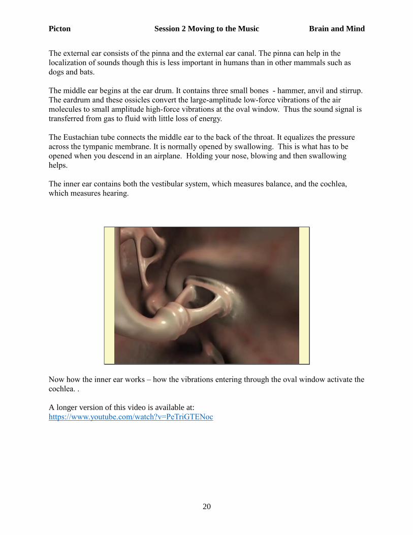
The inner ear contains both the vestibular system, which measures balance, and the cochlea,
which measures hearing.
Now how the inner ear works – how the vibrations entering through the oval window activate the
cochlea. .
A longer version of this video is available at:
https://www.youtube.com/watch?v=PeTriGTENoc
Picton Session 2 Moving to the Music Brain and Mind
21
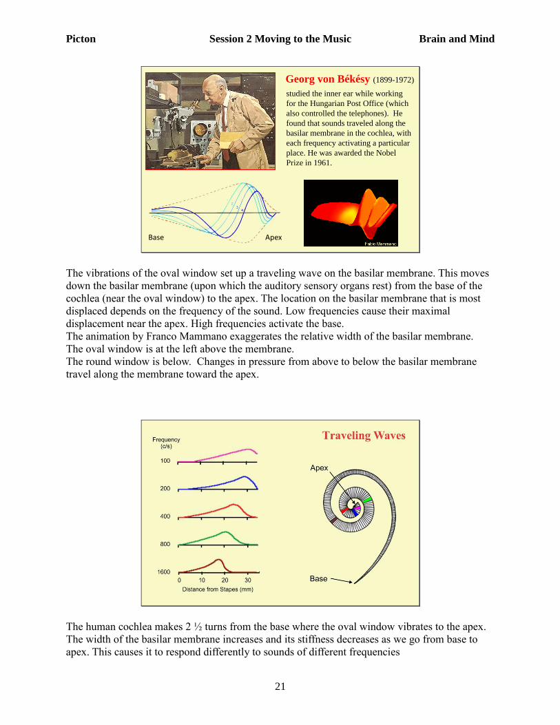
Georg von Békésy (1899-1972)
studied the inner ear while working
for the Hungarian Post Office (which
also controlled the telephones). He
found that sounds traveled along the
basilar membrane in the cochlea, with
each frequency activating a particular
place. He was awarded the Nobel
Prize in 1961.
Base Apex
The vibrations of the oval window set up a traveling wave on the basilar membrane. This moves
down the basilar membrane (upon which the auditory sensory organs rest) from the base of the
cochlea (near the oval window) to the apex. The location on the basilar membrane that is most
displaced depends on the frequency of the sound. Low frequencies cause their maximal
displacement near the apex. High frequencies activate the base.
The animation by Franco Mammano exaggerates the relative width of the basilar membrane.
The oval window is at the left above the membrane.
The round window is below. Changes in pressure from above to below the basilar membrane
travel along the membrane toward the apex.
The human cochlea makes 2 ½ turns from the base where the oval window vibrates to the apex.
The width of the basilar membrane increases and its stiffness decreases as we go from base to
apex. This causes it to respond differently to sounds of different frequencies
Picton Session 2 Moving to the Music Brain and Mind
22
High frequencies activate the base (where the stapes vibrates the oval window) and low
frequencies activate the apex.
This gives a place code for frequency. This basically sets up labelled lines for each frequency.
Neurons activated at the apex code low frequencies; neurons activated at the base code high
frequencies.
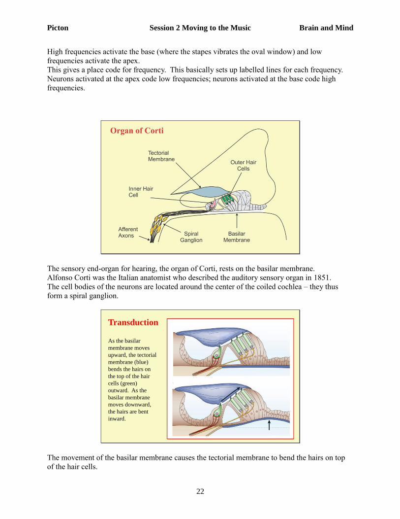
The sensory end-organ for hearing, the organ of Corti, rests on the basilar membrane.
Alfonso Corti was the Italian anatomist who described the auditory sensory organ in 1851.
The cell bodies of the neurons are located around the center of the coiled cochlea – they thus
form a spiral ganglion.
Transduction
As the basilar
membrane moves
upward, the tectorial
membrane (blue)
bends the hairs on
the top of the hair
cells (green)
outward. As the
basilar membrane
moves downward,
the hairs are bent
inward.
The movement of the basilar membrane causes the tectorial membrane to bend the hairs on top
of the hair cells.
Picton Session 2 Moving to the Music Brain and Mind
23
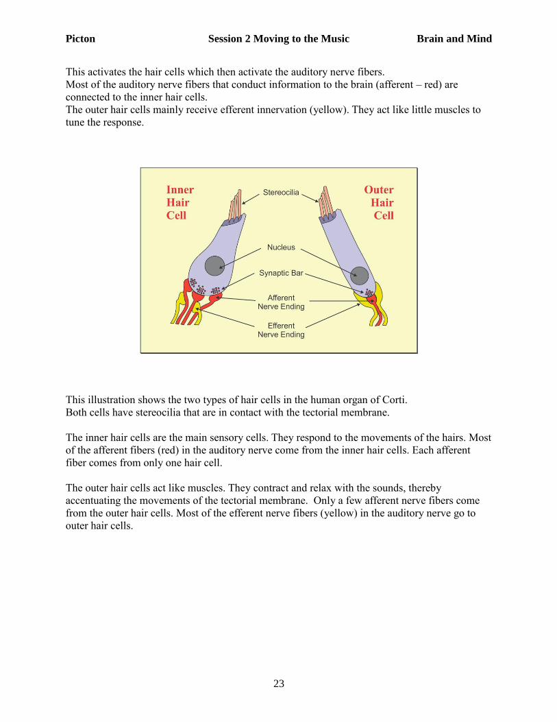
This activates the hair cells which then activate the auditory nerve fibers.
Most of the auditory nerve fibers that conduct information to the brain (afferent – red) are
connected to the inner hair cells.
The outer hair cells mainly receive efferent innervation (yellow). They act like little muscles to
tune the response.
This illustration shows the two types of hair cells in the human organ of Corti.
Both cells have stereocilia that are in contact with the tectorial membrane.
The inner hair cells are the main sensory cells. They respond to the movements of the hairs. Most
of the afferent fibers (red) in the auditory nerve come from the inner hair cells. Each afferent
fiber comes from only one hair cell.
The outer hair cells act like muscles. They contract and relax with the sounds, thereby
accentuating the movements of the tectorial membrane. Only a few afferent nerve fibers come
from the outer hair cells. Most of the efferent nerve fibers (yellow) in the auditory nerve go to
outer hair cells.
Picton Session 2 Moving to the Music Brain and Mind
24
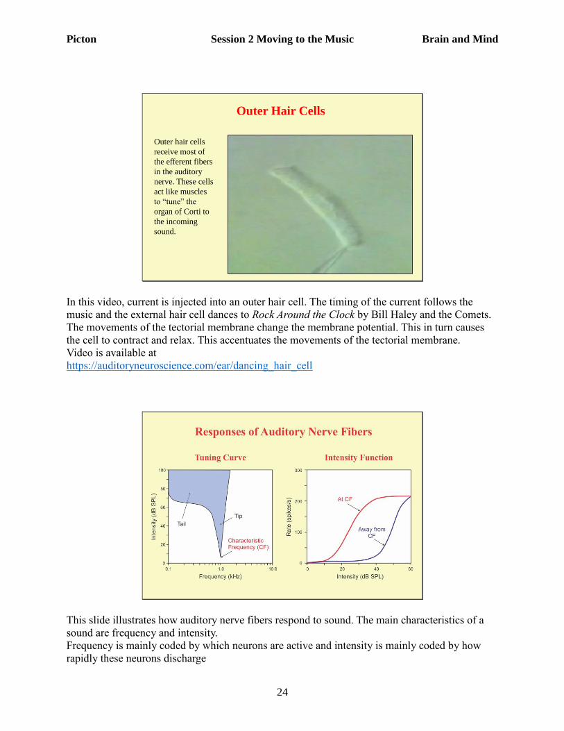
Outer Hair Cells
Outer hair cells
receive most of
the efferent fibers
in the auditory
nerve. These cells
act like muscles
to “tune” the
organ of Corti to
the incoming
sound.
In this video, current is injected into an outer hair cell. The timing of the current follows the
music and the external hair cell dances to Rock Around the Clock by Bill Haley and the Comets.
The movements of the tectorial membrane change the membrane potential. This in turn causes
the cell to contract and relax. This accentuates the movements of the tectorial membrane.
Video is available at
https://auditoryneuroscience.com/ear/dancing_hair_cell
This slide illustrates how auditory nerve fibers respond to sound. The main characteristics of a
sound are frequency and intensity.
Frequency is mainly coded by which neurons are active and intensity is mainly coded by how
rapidly these neurons discharge
Picton Session 2 Moving to the Music Brain and Mind
25
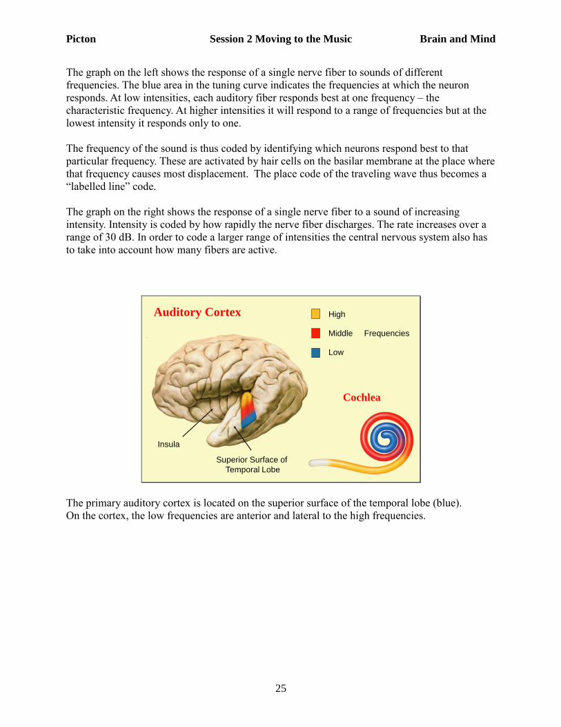
The graph on the left shows the response of a single nerve fiber to sounds of different
frequencies. The blue area in the tuning curve indicates the frequencies at which the neuron
responds. At low intensities, each auditory fiber responds best at one frequency – the
characteristic frequency. At higher intensities it will respond to a range of frequencies but at the
lowest intensity it responds only to one.
The frequency of the sound is thus coded by identifying which neurons respond best to that
particular frequency. These are activated by hair cells on the basilar membrane at the place where
that frequency causes most displacement. The place code of the traveling wave thus becomes a
“labelled line” code.
The graph on the right shows the response of a single nerve fiber to a sound of increasing
intensity. Intensity is coded by how rapidly the nerve fiber discharges. The rate increases over a
range of 30 dB. In order to code a larger range of intensities the central nervous system also has
to take into account how many fibers are active.
Auditory Cortex High
Middle Frequencies
Low
Insula
Superior Surface of
Temporal Lobe
Cochlea
The primary auditory cortex is located on the superior surface of the temporal lobe (blue).
On the cortex, the low frequencies are anterior and lateral to the high frequencies.
Picton Session 2 Moving to the Music Brain and Mind
26
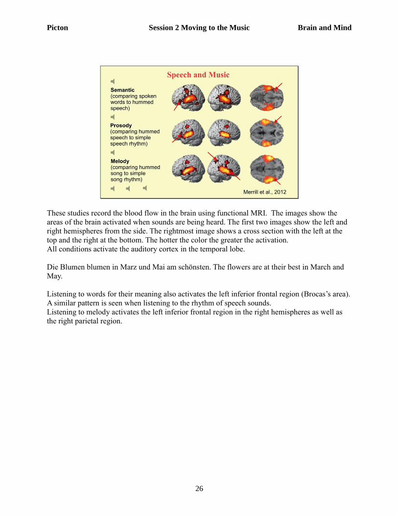
These studies record the blood flow in the brain using functional MRI. The images show the
areas of the brain activated when sounds are being heard. The first two images show the left and
right hemispheres from the side. The rightmost image shows a cross section with the left at the
top and the right at the bottom. The hotter the color the greater the activation.
All conditions activate the auditory cortex in the temporal lobe.
Die Blumen blumen in Marz und Mai am schönsten. The flowers are at their best in March and
May.
Listening to words for their meaning also activates the left inferior frontal region (Brocas’s area).
A similar pattern is seen when listening to the rhythm of speech sounds.
Listening to melody activates the left inferior frontal region in the right hemispheres as well as
the right parietal region.
Picton Session 2 Moving to the Music Brain and Mind
27
Listening to Music
Moon over Bourbon Street, Sting
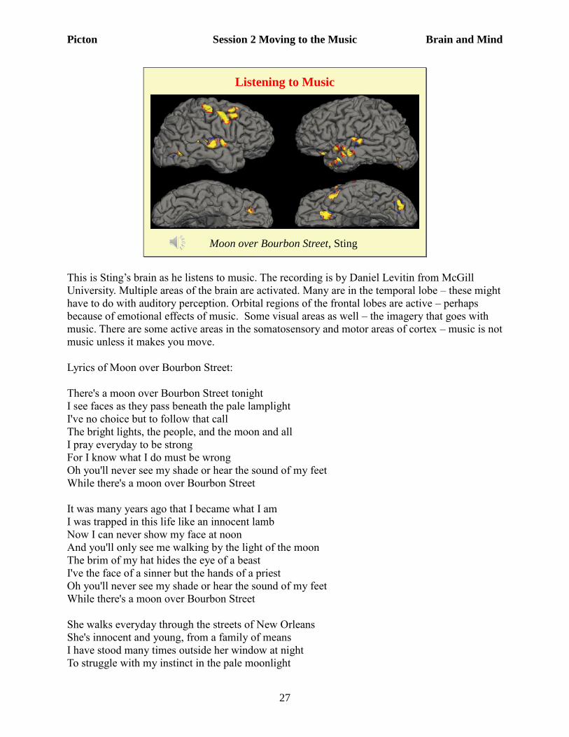
This is Sting’s brain as he listens to music. The recording is by Daniel Levitin from McGill
University. Multiple areas of the brain are activated. Many are in the temporal lobe – these might
have to do with auditory perception. Orbital regions of the frontal lobes are active – perhaps
because of emotional effects of music. Some visual areas as well – the imagery that goes with
music. There are some active areas in the somatosensory and motor areas of cortex – music is not
music unless it makes you move.
Lyrics of Moon over Bourbon Street:
There's a moon over Bourbon Street tonight
I see faces as they pass beneath the pale lamplight
I've no choice but to follow that call
The bright lights, the people, and the moon and all
I pray everyday to be strong
For I know what I do must be wrong
Oh you'll never see my shade or hear the sound of my feet
While there's a moon over Bourbon Street
It was many years ago that I became what I am
I was trapped in this life like an innocent lamb
Now I can never show my face at noon
And you'll only see me walking by the light of the moon
The brim of my hat hides the eye of a beast
I've the face of a sinner but the hands of a priest
Oh you'll never see my shade or hear the sound of my feet
While there's a moon over Bourbon Street
She walks everyday through the streets of New Orleans
She's innocent and young, from a family of means
I have stood many times outside her window at night
To struggle with my instinct in the pale moonlight
Picton Session 2 Moving to the Music Brain and Mind
28
How could I be this way when I pray to God above?
I must love what I destroy and destroy the thing I love
Oh you'll never see my shade or hear the sound of my feet
While there's a moon over Bourbon Street
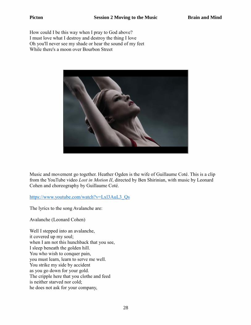
Music and movement go together. Heather Ogden is the wife of Guillaume Coté. This is a clip
from the YouTube video Lost in Motion II, directed by Ben Shirinian, with music by Leonard
Cohen and choreography by Guillaume Coté.
https://www.youtube.com/watch?v=Lxl3AuL3_Qs
The lyrics to the song Avalanche are:
Avalanche (Leonard Cohen)
Well I stepped into an avalanche,
it covered up my soul;
when I am not this hunchback that you see,
I sleep beneath the golden hill.
You who wish to conquer pain,
you must learn, learn to serve me well.
You strike my side by accident
as you go down for your gold.
The cripple here that you clothe and feed
is neither starved nor cold;
he does not ask for your company,
Picton Session 2 Moving to the Music Brain and Mind
29
not at the centre, the centre of the world.
When I am on a pedestal,
you did not raise me there.
Your laws do not compel me
to kneel grotesque and bare.
I myself am the pedestal
for this ugly hump at which you stare.
You who wish to conquer pain,
you must learn what makes me kind;
the crumbs of love that you offer me,
they're the crumbs I've left behind.
Your pain is no credential here,
it's just the shadow, shadow of my wound.
I have begun to long for you,
I who have no greed;
I have begun to ask for you,
I who have no need.
You say you've gone away from me,
but I can feel you when you breathe.
Do not dress in those rags for me,
I know you are not poor;
you don't love me quite so fiercely now
when you know that you are not sure,
it is your turn, beloved,
it is your flesh that I wear.
The meaning of the song is difficult to determine. It may present the Buddhist view of reality as
an avalanche that covers the true soul. However, there are many different interpretations:
http://www.leonardcohenforum.com/viewtopic.php?t=28468