human metapneumovirus and human bocavirus associated with respiratory infection in apulian...
TRANSCRIPT

Virology 417 (2011) 64–70
Contents lists available at ScienceDirect
Virology
j ourna l homepage: www.e lsev ie r.com/ locate /yv i ro
Human metapneumovirus and human bocavirus associated with respiratoryinfection in Apulian population
M. Guido a,⁎, M. Quattrocchi a, A. Campa a, A. Zizza b, P. Grima c, A. Romano d, A. De Donno a
a Laboratory of Hygiene, Department of Biological and Environmental Sciences and Technologies, Faculty of Sciences, University of Salento, Lecce, Italyb Institute of Clinical Physiology, National Research Council, Lecce, Italyc Division of Infectious Diseases, HIV Center, S. Caterina Novella Hospital, Galatina, Lecce, Italyd Laboratory of General Physiology, Department of Biological and Environmental Sciences and Technologies, Faculty of Sciences, University of Salento, Lecce, Italy
⁎ Corresponding author. Fax: +39 832 298626.E-mail address: [email protected] (M. Gu
0042-6822/$ – see front matter © 2011 Elsevier Inc. Aldoi:10.1016/j.virol.2011.04.016
a b s t r a c t
a r t i c l e i n f oArticle history:Received 9 December 2010Returned to author for revision 4 April 2011Accepted 25 April 2011Available online 1 June 2011
Keywords:Human bocavirusHuman metapneumovirusInfluenza virusInfectionSurveillance
We have studied the occurrence of hBoV, hMPV and InfA-B in an Apulian population with respiratory tractinfections. During influenza season 2008–2009, 116 oropharingeal swabs were collected from patientsaffected by Influenza-Like Illness (ILI). The PCR products of hMPVM and HBoVNP-1 genes were sequenced. 78out of 116 samples were positive for at least one respiratory virus; hBoV was detected in 53, hMPV in 22 andInfA-B in 41 out of 116 swabs. A high rate of hBoV infection in adult (18.9%) and elderly (26.4%) subjects wasfound. The co-infection rate was higher for hMPV (18/22 cases, 81.8%) compared to hBoV (26/53 cases, 49.1%),and InfA-B (25/41 cases, 61.0%). Co-infections were common in children. hBoV positive samples shared a highlevel of genetic similarity with the hBoV1 genotype, and hMPV positive samples clustered with A2 subgroup.Our results suggest that hBoV and hMPV play a role in ILI.
ido).
l rights reserved.
© 2011 Elsevier Inc. All rights reserved.
Introduction
Acute respiratory tract infections (ARTI) associate with significantmorbidity worldwide, especially in infants and children. Viruses are aleading cause of ARTI, showing a relevant epidemiological variabilitythat depends on climate and region (Armstrong et al., 1999). Etiologyis still undefined in a significant proportion of ARTI (Monto, 1994;Henrickson et al., 2004).
The impact of ARTI on health and disease is particularly difficult toaddress. Similar symptoms can be generated by a variety of agents[influenza virus (Inf), human adenovirus, respiratory syncytial virus(RSV)]. Moreover, respiratory tract samples from healthy subjects orfrom patients without respiratory symptoms are rarely available.
Viruses, such as Inf, RSV, human adenovirus and coronavirusesNL63 and HKU1 are known to be responsible for the most ofrespiratory tract infections (Kesson, 2007). In a significant proportionof respiratory tract disease, however, no pathogens are identified.
During the past few years, new respiratory viruses have beendiscovered. Today, human metapneumovirus (hMPV), family Para-myxoviridae, is considered one of themost relevant agents of ARTI andit has been reported worldwide (Van den Hoogen et al., 2001;Debiaggi et al., 2006; Kahn, 2006; Sarasini et al., 2006; Boivin et al.,2007).
The hMPV has been associated with ARTI in all age groups, withmore severe disease occurring in young children, elderly individualsand immunocompromised hosts. The virus causes a variety of clinicalsyndromes in children that are typical of the paramyxoviruses,including upper and lower respiratory tract illness. The clinicalcharacteristics of hMPV infections are not distinctive.
It has been reported that the epidemiological and clinicalcharacteristics of hMPV closely resemble those of RSV (Boivin et al.,2002; Van den Hoogen et al., 2004).
Human bocavirus (hBoV) is a novel parvovirus, family Parvovi-ridae, often associated with respiratory tract disease in adult andpediatric patients worldwide. hBoV has been detected in serum andfecal samples, predominantly in children under the age of 2 years withrespiratory infections (Lindner and Modrow, 2008). By means of bothgenetic organization and sequence homology analyses relations tomembers of the two genuses, bovine parvovirus and canine minutevirus, could be established. Consequently, hBoV has been classified asa bocavirus (Allander et al., 2005).
Recently, three viruses closely related to hBoV have been reported,provisionally named human bocavirus 2 (hBoV2), human bocavirus 3(hBoV3), and human bocavirus 4 (hBoV4). It remains unclear whetherhBoV1, hBoV2, hBoV3, and hBoV4 represent unique viral entities ordistant genotypes of a single virus. However, it is a fact that hBoV1infection has been linked with mild to severe lower respiratory tractinfection, but that it has also been detected at low frequency in stoolsamples, while hBoV2, hBoV3 and hBoV4 have been detected in

65M. Guido et al. / Virology 417 (2011) 64–70
gastrointestinal samples (Kapoor et al., 2009; Arthur et al., 2009;Kapoor et al., 2010).
The impact of hMPV and of hBoV on the global epidemiology ofARTI is still unclear. In fact, most reports are retrospective (Peret et al.,2002; Stockton et al., 2002) or predominantly focus on hospitalizedchildren and on children affected by ARTI (Maggi et al., 2003;Wilkesmann et al., 2006; Wolf et al., 2006).
Epidemiological and clinical features of Influenza-like Illness (ILI) ingeneral population need to be highlighted and ongoing investigationsare necessary.We have studied the occurrence of hBoV, hMPV and Inf Aand Inf B (InfA-B) in an Apulian population with respiratory tractinfections.
Results
Prevalence of respiratory viruses
One hundred and sixteen patients (51.3%males, range 0–93 years)with ILI were enrolled in the study.
The oro-pharyngeal swabs were collected from 52 children (aged0–14 years), 22 youths and adults (15–64 years) and 42 elderlysubjects (≥64 years).
Seventy eight out of 116 samples (67.2%) analyzed by themolecular method were positive for one or more respiratory viruses;particularly, InfA-B was detected in 41 (35.3%), hMPV in 22 (19.0%)and hBoV in 53 (45.7%) out of 116 oropharingeal swabs.
Single infection was found in 40.5% (n=47) and double infectionin 20.7% (n=24) of the subjects (Table 2).
The percentage of co-infections was higher for hMPV (18/22 cases,81.8%; Pb0.05 vs. hBoV) compared to hBoV (26/53 cases, 49.1%), and toInfA-B (25/41 cases, 61.0%). HBoV was detected mainly in associationwith InfA-B (72.2%; 13/18). Seven triple infections (6%) were detected.
Age distribution
Viruses were detected only for the period between December andMarchwith a peak on January, with a similar trend for the three viruses.
Table 1Flu A/B, hBoV and hMPV primers and thermal profile used in viral genome amplification.
Detection method Gene Primer sequences 5′-3′
hBoVNested-PCR NP-1 Boca-forward (B1f)
CCAGCAAGTCCTCCAAACTBoca-reverse (B1r)GGAGCTTCAGGATTGGAA
Nested-PCR NP-1 Boca-forward (B2f)GAGCTCTGTAAGTACTATTBoca-reverse (B2r)CTCTGTGTTGACTGAATAC
hMPVNested-PCR M Meta-forward (M1f)
GAGTCCTAYCTRGTAGACAMeta-reverse (M1r)AGTACAGACATDGCWGCA
Nested-PCR M Meta- forward (M2f)CAGCWGCTGTTCAAGTTGMeta-reverse (M2r)YTGTGATGYAGCATACAGA
Flu A/B multiplex real time PCRFluA M GAGTCTTCTAACMGAGGT
GGGCACGGTGAGCGTRAAFAM-TCCTGTCACCTCTGA
FluB M TAGCAGAAGGCCATGAAACGTTCCTAGTTTTACTTGCAJOE-TGTCTCATGGTCATGT
hMPV exhibited a lower level of viral circulation than the other twoviruses (Fig. 1). The distribution of viruses in the age groups showed anincreased presence of infections in children (0–14 age group) followedby elderly. In the 0–14 and 15–64 age groups, infections due to hBoVwere significantly more represented than those due to hMPV (Pb0.05).In the elderly (≥64 years), infections due to hMPV were significantlylower than those due to InfA-B (Pb0.05) and to hBoV (Pb0.01)(Table 2).
Only for hMPV, the distribution was similar in the 15–64 and ≥64age group. All triple infections were found in children (0–14 years),while 54.2% of double infections were among children, 25.0% in the15–64 age group and the remaining 20.8% in elderly.
Phylogenetic analysis
The hMPV M gene and hBoV NP1 gene from 13 samples wereamplified and sequenced. The phylogenetic analysis of the hBoVnucleotide sequences revealed that all hBoV strains clustered with thehBoV1 (NC_007455) genotype (identity range 75.0–100%) (Fig. 2). ThehMPV strains belonged to the A2 subgroup (identity range 74.0–97.0%)(Fig. 3).
Discussion
We have designed a prospective study with the aim of describingthe virological impact of three different respiratory viruses (hMPV,hBoV and InfA-B virus) in subjects with ILI in an Apulian population.
At least one out of the three viruses was identified in 67.2% ofpatients, confirming the widespread distribution in the population(Calvo et al., 2010). hBoV was the most prevalent virus (45.7% ofcases), followed by InfA-B virus (35.3%) and hMPV (19.0%). Thus, ourresults are consistent with data from other groups that attested thepresence of at least one of the potential viral pathogens in a 34% to 95%of cases (Allander et al., 2007; Khetsuriani et al., 2007; Kusel et al.,2007; Gendrel et al., 2007; García-García et al., 2010).
The viruses started spreading during late autumn, with a peak onJanuary; the prevalence of hMPV was lower than the other two
Location Thermal profile
95 °C 5′ (×1)CACCTGC 162–187 95 °C 30″, 63 °C 30″
72 °C 30″ (×35)GCTCTGTG 536–561 72 °C 7′ (×1)
95 °C 5′ (×1)AC 188–208 95 °C 30″, 54 °C 30″
72 °C 30″ (×35)AG 522–542 72 °C 7′ (×1)
95 °C 5′ (×1)CC3 4–24 95 °C 1′, 55 °C 1′
72 °C 1′ (×25)CC 232–251 72 °C 7′ (×1)
95 °C 5′ (×1)44–61 95 °C 1′, 53 °C 1′
72 °C 1′ (×30)G 175–195 72 °C 7′ (×1)
CGAAACGTA 13 FluAv_For207 FluAv_Rev
C-MGB FluAv_TMGCT 317 FluBv_ForTTGAAT 411 FluBv_RevACCT-MGBNFQ FluBv_TM

Table 2Distribution of hBoV, hMPV, and InfA-B of single and co-infections in subjects with ILI (n=116) by age.
InfA/Bn (%)
hMPVn (%)
hBoVn (%)
Totaln (%)
P value
Flu A/B vs hMPV Flu A/B vs hBoV hMPV vs hBoV
Age0–14 (yrs) 21 (51.2) 16 (72.7) 29 (54.7) 52 (44.8) 0.4126 0.1695 0.017515–64 (yrs) 8 (19.5) 3 (13.6) 10 (18.9) 22 (19.0) 0.1623 0.7591 0.0452N64 (yrs) 12 (29.3) 3 (13.6) 14 (26.4) 42 (36.2) 0.0204 0.8134 0.0055Total 41 (35.3) 22 (19.0) 53 (45.7)
Single and co-infectionsTotal 41 (35.3) 22 (19.0) 53 (45.7) – 0.0079 0.1413 0.0001Single infections 16 (39.0) 4 (18.2) 27 (50.9) 47 (40.5) 0.1547 0.3464 0.0104Double infections 18 (43.9) 11 (50.0) 19 (35.8) 24 (20.7) 0.0902 0.2500 0.0104Flu A/B – 5 (45.5) 13 (68.4) – – – –
hMPV 5 (27.8) – 6 (31.6) – – – –
hBoV 13 (72.2) 6 (54.5) – – – – –
Triple infections 7 (17.1) 7 (31.8) 7 (13.2) – – – –
66 M. Guido et al. / Virology 417 (2011) 64–70
viruses. However, the samples with suspected respiratory infectionwere effectively collected by the sentinel physicians only in the periodbetween on October and on March. This short time collection mayhave limited the evaluation of the real time distribution of the viruses.In order to assess the real viral seasonality, oropharyngeal swabsshould have been collected along a 12 month time period for at leastthree consecutive years. It is natural, then, to assess that furtherstudies need to be performed in order to establish whether there is areal epidemiological trend.
The frequency of respiratory viruses was higher in the 0–14 agegroup, where the rate of viral detection reached 75.0%. hBoV wasdetected in 45.7% (53/116 samples) of all the analyzed subjects and in55.8% (29/52) of the children, a rate higher than previously reported(Maggi et al., 2007). However, these results are not fully comparablebecause of the different study design. In fact, none of the previousstudies was prospective; moreover, one of these studies included onlyhospitalized children and showed differences in the rate of hBoVdetection during a period of years (Maggi et al., 2007).
Recent analyses documented the detection of hBoV also inasymptomatic subjects, which suggests that the virus is not a truepathogen but rather a commensal (Blessing et al., 2009; Salmón-Mulanovich et al., 2011). However, although not yet completelydemonstrated, hBoV appears to contribute to the severity of respiratoryillness (Martin et al., 2010). In this study, a limitation is the lack ofcontrol groups without clinical evidences of illness. To assess the realrole of this virus in respiratory infections, it would therefore be useful to
Fig. 1. Number of human bocavirus (hBoV), human metapneumovirus (hMPV) andInfluenza A–B virus per month.
compare a symptomatic group with a control group and an asymptom-atic period with a symptomatic period.
Phylogenetic analysis of the hBoV positive specimen sequencesrevealed that all samples clustered in genotype hBoV1 NP1. Consistentwith other reports, strains of hBoV1 were more frequently identified inrespiratory swabs (Allander et al., 2005; Hishinuma-Igarashi et al.,2009). However, due to the limited number of specimens the presencein the patients of different hBoV genotypes cannot be excluded.
Among the emerging respiratory viruses, hMPV is one of the mostfrequently reported in recent international studies (Van den Hoogenet al., 2001; Boivin et al., 2007). In Italy, hMPV has been variouslyassociated with Upper Respiratory Tract Infections (URTI) or LowerRespiratory Tract Infections (LRTI), affecting from 3% to 13% of childrenwithARTI (Bosis et al., 2005; Gerna et al., 2005; Sarasini et al., 2006). Ourdata do not appear to be in agreement with previous reports, revealinghMPV in 19.0% (22/116) of all samples and in particular in 30.8% (16/52)of children. This virus was detected also in children older than 5 years,which is not in agreement to other serological surveys that showed anearly acquisition of infection (Van den Hoogen et al., 2001; Zappa et al.,2011). Nonetheless, hMPV is an emerging cause of ARTI in ILI patientsand may have a significant clinical impact, especially in children.
With respect to others, a limitation of this study is the search of onlythree respiratory viruses. Further studies should include humanparainfluenza viruses 1–3 (hPIVs), RSV, adenovirus for a more indepth investigation. Furthermore, in this study the respiratoryinfectionswere analyzed for only 1 year and the sampleswere collectedwith limited information on the severity of clinical symptoms.
Phylogenetic analysis of hMPV strains in different countries has beenbased on the comparison of sequences of L, N, F, or P genes (Van denHoogen et al., 2001, 2004a; Boivin et al., 2002, 2004; Bastien et al., 2003;Mackay et al., 2004). In the present study, the RT-PCR was designed forthe M gene to detect hMPV with an equivalent sensitivity for bothgenotypes. Recent studies have validated the use of the M gene (Bellau-Pujol et al., 2005; Chano et al., 2005) and a genetic discrimination hasbeen established (Van den Hoogen et al., 2001; Bastien et al., 2003;Carr et al., 2005; Legrandet al., 2011). In our study, phylogenetic analysisof theM gene showed that the isolated viruses clusteredwith A2 strains.Nevertheless, different hMPV genotypes and their sublineages can co-circulate during the year and one of these can be prevalent (Huck et al.,2006; Caracciolo et al., 2008; Xiao et al., 2010; Legrand et al., 2011).
The strains circulating in Italy were described in several studiespreviously (Sarasini et al., 2006; Gerna et al., 2007; Caracciolo et al.,2008; Larcher et al., 2008; Bosis et al., 2008; Zappa et al., 2011). In allthese studies, co-circulation of different hMPV genotypes wasobserved, but A2 subtype was the most prominent strain. In ourstudy, it has not been possible to assess sub-division of A2 samples in
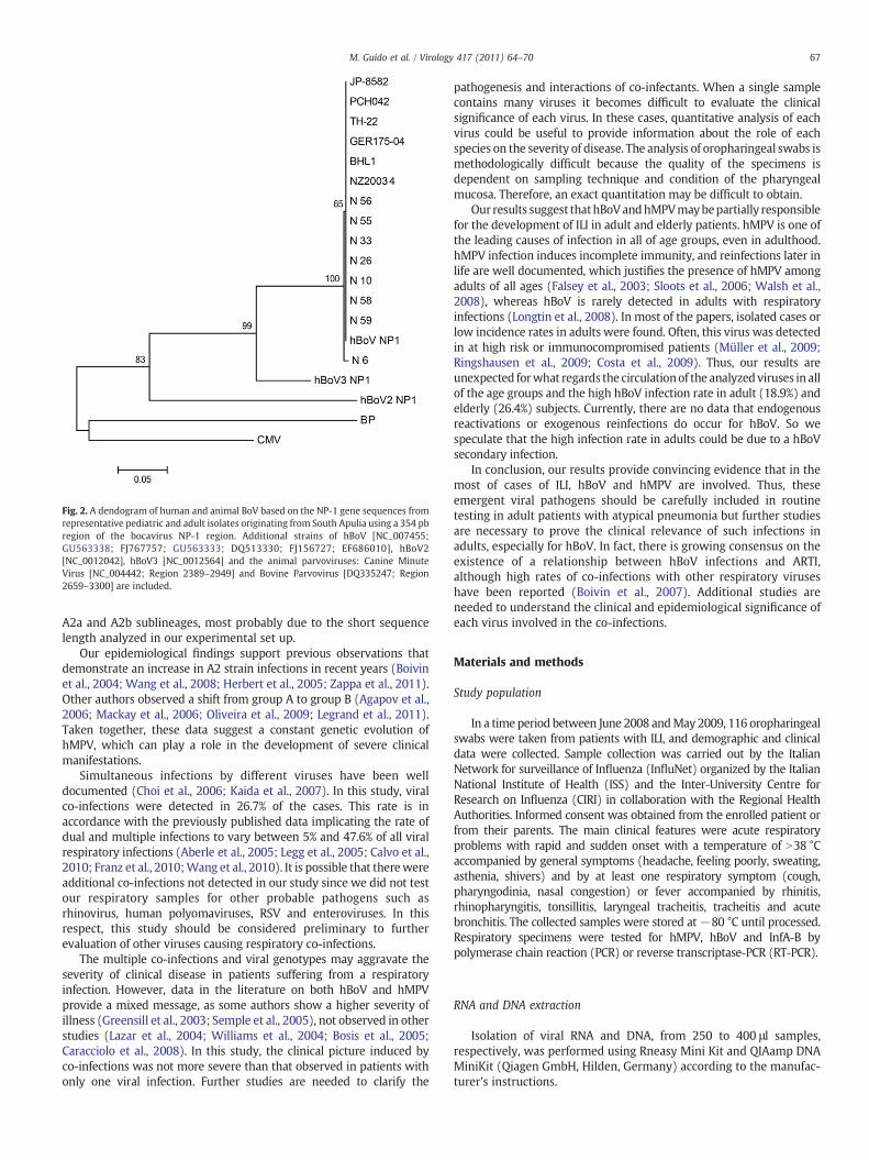
Fig. 2. A dendogram of human and animal BoV based on the NP-1 gene sequences fromrepresentative pediatric and adult isolates originating from South Apulia using a 354 pbregion of the bocavirus NP-1 region. Additional strains of hBoV [NC_007455;GU563338; FJ767757; GU563333; DQ513330; FJ156727; EF686010], hBoV2[NC_0012042], hBoV3 [NC_0012564] and the animal parvoviruses: Canine MinuteVirus [NC_004442; Region 2389–2949] and Bovine Parvovirus [DQ335247; Region2659–3300] are included.
67M. Guido et al. / Virology 417 (2011) 64–70
A2a and A2b sublineages, most probably due to the short sequencelength analyzed in our experimental set up.
Our epidemiological findings support previous observations thatdemonstrate an increase in A2 strain infections in recent years (Boivinet al., 2004; Wang et al., 2008; Herbert et al., 2005; Zappa et al., 2011).Other authors observed a shift from group A to group B (Agapov et al.,2006; Mackay et al., 2006; Oliveira et al., 2009; Legrand et al., 2011).Taken together, these data suggest a constant genetic evolution ofhMPV, which can play a role in the development of severe clinicalmanifestations.
Simultaneous infections by different viruses have been welldocumented (Choi et al., 2006; Kaida et al., 2007). In this study, viralco-infections were detected in 26.7% of the cases. This rate is inaccordance with the previously published data implicating the rate ofdual and multiple infections to vary between 5% and 47.6% of all viralrespiratory infections (Aberle et al., 2005; Legg et al., 2005; Calvo et al.,2010; Franz et al., 2010;Wang et al., 2010). It is possible that therewereadditional co-infections not detected in our study since we did not testour respiratory samples for other probable pathogens such asrhinovirus, human polyomaviruses, RSV and enteroviruses. In thisrespect, this study should be considered preliminary to furtherevaluation of other viruses causing respiratory co-infections.
The multiple co-infections and viral genotypes may aggravate theseverity of clinical disease in patients suffering from a respiratoryinfection. However, data in the literature on both hBoV and hMPVprovide a mixed message, as some authors show a higher severity ofillness (Greensill et al., 2003; Semple et al., 2005), not observed in otherstudies (Lazar et al., 2004; Williams et al., 2004; Bosis et al., 2005;Caracciolo et al., 2008). In this study, the clinical picture induced byco-infections was not more severe than that observed in patients withonly one viral infection. Further studies are needed to clarify the
pathogenesis and interactions of co-infectants. When a single samplecontains many viruses it becomes difficult to evaluate the clinicalsignificance of each virus. In these cases, quantitative analysis of eachvirus could be useful to provide information about the role of eachspecies on the severity of disease. The analysis of oropharingeal swabs ismethodologically difficult because the quality of the specimens isdependent on sampling technique and condition of the pharyngealmucosa. Therefore, an exact quantitation may be difficult to obtain.
Our results suggest thathBoVandhMPVmaybepartially responsiblefor the development of ILI in adult and elderly patients. hMPV is one ofthe leading causes of infection in all of age groups, even in adulthood.hMPV infection induces incomplete immunity, and reinfections later inlife are well documented, which justifies the presence of hMPV amongadults of all ages (Falsey et al., 2003; Sloots et al., 2006; Walsh et al.,2008), whereas hBoV is rarely detected in adults with respiratoryinfections (Longtin et al., 2008). In most of the papers, isolated cases orlow incidence rates in adults were found. Often, this virus was detectedin at high risk or immunocompromised patients (Müller et al., 2009;Ringshausen et al., 2009; Costa et al., 2009). Thus, our results areunexpected forwhat regards the circulationof the analyzedviruses in allof the age groups and the high hBoV infection rate in adult (18.9%) andelderly (26.4%) subjects. Currently, there are no data that endogenousreactivations or exogenous reinfections do occur for hBoV. So wespeculate that the high infection rate in adults could be due to a hBoVsecondary infection.
In conclusion, our results provide convincing evidence that in themost of cases of ILI, hBoV and hMPV are involved. Thus, theseemergent viral pathogens should be carefully included in routinetesting in adult patients with atypical pneumonia but further studiesare necessary to prove the clinical relevance of such infections inadults, especially for hBoV. In fact, there is growing consensus on theexistence of a relationship between hBoV infections and ARTI,although high rates of co-infections with other respiratory viruseshave been reported (Boivin et al., 2007). Additional studies areneeded to understand the clinical and epidemiological significance ofeach virus involved in the co-infections.
Materials and methods
Study population
In a time period between June 2008 andMay 2009, 116 oropharingealswabs were taken from patients with ILI, and demographic and clinicaldata were collected. Sample collection was carried out by the ItalianNetwork for surveillance of Influenza (InfluNet) organized by the ItalianNational Institute of Health (ISS) and the Inter-University Centre forResearch on Influenza (CIRI) in collaboration with the Regional HealthAuthorities. Informed consent was obtained from the enrolled patient orfrom their parents. The main clinical features were acute respiratoryproblems with rapid and sudden onset with a temperature of N38 °Caccompanied by general symptoms (headache, feeling poorly, sweating,asthenia, shivers) and by at least one respiratory symptom (cough,pharyngodinia, nasal congestion) or fever accompanied by rhinitis,rhinopharyngitis, tonsillitis, laryngeal tracheitis, tracheitis and acutebronchitis. The collected samples were stored at−80 °C until processed.Respiratory specimens were tested for hMPV, hBoV and InfA-B bypolymerase chain reaction (PCR) or reverse transcriptase-PCR (RT-PCR).
RNA and DNA extraction
Isolation of viral RNA and DNA, from 250 to 400 μl samples,respectively, was performed using Rneasy Mini Kit and QIAamp DNAMiniKit (Qiagen GmbH, Hilden, Germany) according to the manufac-turer's instructions.

Fig. 3. A dendogram of hMPV based on the M gene sequences from representative pediatric and adult isolates originating from South Apulia using a 151 pb region of the MPVM region.The gene bank access numbers of the referenced strains are: A1 [AF371337, HQ384159, HQ384156, HQ384126, HQ384125, HQ384124, AJ145294], A2 [AJ530095, AJ297749],B1 [HQ384134, HQ384140, HQ384152, HQ384151, HQ384145, HQ384135, HQ384142, HQ384139, AJ525843], and B2 [AJ297748, HQ384157, HQ384146, HQ384150, HQ384154].
68 M. Guido et al. / Virology 417 (2011) 64–70
PCR assays for respiratory viruses
hMPV and InfA-B detection was performed by RT-nested-PCR andhBoV detection by nested-PCR. For reverse transcription, a commercialkit (RevertAidTM First Strand cDNA Synthesis kit, Fermentas LifeSciences) was used. A subsequent nested PCRwas carried out using 5 μlof the first reaction product, which was then added to the reactionmixture to a final volume of 50 μl.
RT-nested-PCRwas designed to amplify the conserved region of thehMPV matrix gene corresponding to matrix protein; nested-PCR wasperformed by using portions of the hBoV NP-1 gene as primers targets.Primer sequences, target genes and thermal profiles are reported inTable 1.
Sequencing and phylogenetic analysis
The PCR products of hMPVM gene (nt 44–61, corresponding to a 151base-pairs fragment) and hBoVNP-1 gene (nt 108–208, corresponding to354 base-pairs fragment) were purified with QIAamp nucleic acidpurification kits (Qiagen, Germany), according to the manufacturer's
protocol. PCR products were sequenced directly or after cloning using theCEQ 8000 (Beckman Coulter) automated sequencer, with the sameforwardprimers (M2f andB2f; see Table 1) used for the PCR. Phylogeneticaffiliation of the obtained sequences was determined by using the BLASTsearch tool at the NCBI web site. Sequence similarity was searched byBLAST (www.ncbi.nlm.nih.gov/blast). Multiple sequence alignment wasconducted with ClustalW2 version 2.1 (www.ebi.ac.uk/clustalw) andphylogenetic trees were constructed by the Neighbor-Joining methodand Kimura 2-Parameter model using Molecular Evolutionary GeneticsAnalysis (MEGA) software version 5.0 (www.xmegasoftware.net).A bootstrap resampling analysis was performed (1000 replicates) totest tree robustness. This analysis was performed on hMPV matrixnucleotide sequences and on a portion of the hBoV NP-1 nucleotidesequences.
Statistical analysis
Statistical analysis was performed with StatView 5.1 Software.Categorical variables between groups were compared with the Fisher'sExact-test. Pb0.05 was considered to be statistically significant.

69M. Guido et al. / Virology 417 (2011) 64–70
Acknowledgments
The study is supported by the Ministry of Education, University andResearch (MIUR), Project of Relevant National Interest (PRIN) 2007:“Prevalence and molecular characterization of new respiratory viruses(human metapneumovirus and human bocavirus) in Apulia” n.2007LPAF42_004 in the framework of “Respiratory infections: molec-ular epidemiology, cross-protection and impact of the preventivestrategies” project.
References
Aberle, J.H., Aberle, S.W., Pracher, E., Hutter, H.P., Kundi,M., Popow-Kraupp, T., 2005. Singleversus dual respiratory virus infections in hospitalized infants: impact on clinicalcourse of disease and interferon-gamma response. Pediatr. Infect. Dis. J. 24 (7),605–610.
Agapov, E., Sumino, K.C., Gaudreault-Keener, M., Storch, G.A., Holtzman, M.J., 2006.Genetic variability of human metapneumovirus infection: evidence of a shift inviral genotype without a change in illness. J. Infect. Dis. 193, 396–403.
Allander, T., Tammi, M.T., Eriksson, M., Bjerkner, A., Tiveljung-Lindell, A., Andersson, B.,2005. Cloning of a human parvovirus by molecular screening of respiratory tractsamples. Proc. Natl. Acad. Sci. USA 102, 12891–12896.
Allander, T., Jartti, T., Gupta, S., Niesters, H.G., Lehtinen, P., Osterback, R., 2007. Humanbocavirus and acute wheezing in children. Clin. Infect. Dis. 44, 904–910.
Armstrong, G.L., Conn, L.A., Pinner, R.W., 1999. Trends in infectious disease mortality inthe United States during the 20th century. JAMA 281, 61–66.
Arthur, J.L., Higgins, G.D., Davidson, G.P., Givney, R.C., Ratclif, R.M., 2009. A novel bocavirusassociatedwith acute gastroenteritis in Australian children. PLos Pathog. 5, e1000391.
Bastien,N.,Normand, S., Taylor, T.,Ward,D., Peret, T.C., Boivin, G., Anderson, L.J., Li, Y., 2003.Sequence analysis of the N, P, M and F genes of Canadian human metapneumovirusstrains. Virus Res. 93, 51–62.
Bellau-Pujol, S., Vabret, A., Legrand, L., Dina, J., Gouarin, S., Petitjean-Lecherbonnier, J.,Pozzetto, B., Ginevra, C., Freymuth, F., 2005. Development of three multiplex RT-PCRassays for the detection of 12 respiratory RNA viruses. J. Virol. Methods 126, 53–63.
Blessing, K., Neske, F., Herre, U., Kreth, H.W., Weissbrich, B., 2009. Prolonged detectionof human bocavirus DNA in nasopharyngeal aspirates of children with respiratorytract disease. Pediatr. Infect. Dis. J. 28 (11), 1018–1019.
Boivin, G., Abed, Y., Pelletier, G., Ruel, L., Moisan, D., Côte, S., Peret, T.C., Erdmann, D.D.,Anderson, L.J., 2002. Virological features and clinical manifestations associated withhuman metapneumovirus: a new paramyxovirus responsible for acute respiratory-tract infections in all age groups. J. Infect. Dis. 186, 1330–1334.
Boivin, G., Mackay, I., Sloots, T.P., Madhi, S., Freymuth, F., Wolf, D., Shemer-Avni, Y.,Ludewick, H., Gray, G.C., LeBlanc, E., 2004. Global genetic diversity of humanmetapneumovirus fusion gene. Emerg. Infect. Dis. 10, 1154–1157.
Boivin, G., De Serres, G., Hamelin, M.E., Côte, S., Argouin,M., Tremblay, G., Maranda-Aubut,R., Sauvageau, C., Ouakki, M., Boulianne, N., Couture, C., 2007. An outbreak of severerespiratory tract infection due to human metapneumovirus in a long-term carefacility. Clin. Infect. Dis. 44, 1152–1158.
Bosis, S., Esposito, S., Niesters, H.G., Crovari, P., Osterhaus, A.D., Principi, N., 2005. Impactof human metapneumovirus in childhood: comparison with respiratory syncytialvirus and influenza viruses. J. Med. Virol. 75, 101–104.
Bosis, S., Esposito, S., Osterhaus, A.D., Tremolati, E., Begliatti, E., Tagliabue, C., Corti, F.,Principi, N., Niesters, H.G., 2008. Association between high nasopharyngeal viral loadand disease severity in childrenwith humanmetapneumovirus infection. J. Clin. Virol.42 (3), 286–290.
Calvo, C., Pozo, F., García-García, M.L., Sanchez, M., Lopez-Valero, M., Pérez-Breña, P.,Casas, I., 2010. Detection of new respiratory viruses in hospitalized infants withbronchiolitis: a three-year prospective study. Acta Paediatr. 99 (6), 883–887.
Caracciolo, S., Minini, C., Colombrita, D., Rossi, D., Miglietti, N., Vettore, E., Caruso, A.,Fiorentini, S., 2008. Human metapneumovirus infection in young children hospital-izedwith acute respiratory tract disease: virologic and clinical features. Pediatr. Infect.Dis. J. 27 (5), 406–412.
Carr, M.J., McCormack, G.P., Crowley, B., 2005. Human metapneumovirus- associatedrespiratory tract infections in the Republic of Ireland during the influenza season of2003–2004. Clin. Microbiol. Infect. 11, 366–371.
Chano, F., Rousseau, C., Laferriere, C., Couillard, M., Charest, H., 2005. Epidemiologicalsurvey of humanmetapneumovirus infection in a large pediatric tertiary care center.J. Clin. Microbiol. 43, 5520–5525.
Choi, E.H., Lee, H.J., Kim, S.J., Eun, B.W., Kim, N.H., Lee, J.A., Lee, J.H., Song, E.K., Kim, S.H.,Park, J.Y., Sung, J.Y., 2006. The association of newly identified respiratory viruseswith lower respiratory tract infections in Korean children, 2000–2005. Clin. Infect.Dis. 43, 585–592.
Costa, C., Bergallo, M., Cavallo, R., 2009. Detection of Human Bocavirus in bronchoalveolarlavage from Italian adult patients. J. Clin. Virol. 45 (1), 81–82.
Debiaggi,M., Canducci, F., Sampaolo,M.,Marinozzi,M.C., Parea,M., Terulla, C., Colombo, A.A.,Alessandrino, E.P., Bragotti, L.Z., Arghittu, M., Goglio, A., Migliavacca, R., Romero, E.,Clementi, M., 2006. Persistent symptomless human metapneumovirus infection inhematopoietic stem cell transplant recipients. J. Infect. Dis. 194, 474–478.
Falsey, A.R., Erdman, D., Anderson, L.J., Walsh, E.E., 2003. Human metapneumovirusinfections in young and elderly adults. J. Infect. Dis. 187 (5), 785–790.
Franz, A., Adams, O., Willems, R., Bonzel, L., Neuhausen, N., Schweizer-Krantz, S.,Ruggeberg, J.U., Willers, R., Henrich, B., Schroten, H., Tenenbaum, T., 2010. Correlation
of viral load of respiratory pathogens and co-infections with disease severity inchildren hospitalized for lower respiratory tract infection. J. Clin. Virol. 48 (4),239–245.
García-García, M.L., Calvo, C., Falcón, A., Pozo, F., Pérez-Breña, P., De Cea, J.M., Casas, I.,2010. Role of emerging respiratory viruses in children with severe acute wheezing.Pediatr. Pulmonol. 45 (6), 585–591.
Gendrel, D., Guedj, R., Pons-Catalano, C., Emerian, A., Raymond, J., Rozenberg, F., Lebon, P.,2007. Human bocavirus in children with acute asthma. Clin. Infect. Dis. 45, 404–405.
Gerna, G., Campanini, G., Rovida, F., Sarasini, A., Lilleri, D., Paolucci, S., Marchi, A., Baldanti,F., Revello, M.G., 2005. Changing circulation rate of human metapneumovirus strainsand types among hospitalized pediatric patients during three consecutive winter-spring seasons. Brief report. Arch. Virol. 150 (11), 2365–2375.
Gerna, G., Sarasini, A., Percivalle, E., Campanini, G., Rovida, F., Marchi, A., Baldanti, F., 2007.Prospective study of human metapneumovirus infection: diagnosis, typing and virusquantification in nasopharyngeal secretions from pediatric patients. J. Clin. Virol. 40(3), 236–240.
Greensill, J., McNamara, P.S., Dove, W., Flanagan, B., Smyth, R.L., Hart, C.A., 2003. Humanmetapneumovirus in severe respiratory syncytial virus bronchiolitis. Emerg. Infect.Dis. 9 (3), 372–375.
Henrickson, K.J., Hoover, S., Kehl, K.S., Hua, W., 2004. National disease burden ofrespiratory viruses detected in children by polymerase chain reaction. Pediatr.Infect. Dis. J. 23, S11–S18.
Herbert, P.L., Abed, Y., van Niekerk, N., Boivin, G., Klugman, K.P., Madhi, S.A., 2005. Humanmetapneumovirus genetic variability, South Africa. Emerg. Infect. Dis. 11, 1074–1078.
Hishinuma-Igarashi, I., Mizuta, K., Saito, Y., Ohuchi, Y., Noda, M., Akiyama, M.,Tsukagoshi, H., Okabe, N., Tashiro, M., Kimura, H., 2009. Phylogenetic analysis ofhuman bocavirus (hBoV) detected from children with acute respiratory infection inJapan. J. Infect. 58 (4), 311–313.
Huck, B., Egger, M., Bertz, H., Peyerl-Hoffman, G., Kern, W.V., Neumann-Haefelin, D.,Falcone, V., 2006. Human metapneumovirus infection in a hematopoietic stem celltransplant recipient with relapsed multiple myeloma and rapidly progressing lungcancer. J. Clin. Microbiol. 44 (6), 2300–2303.
Kahn, J.S., 2006. Epidemiologyofhumanmetapneumovirus. Clin.Microbiol. Rev. 19, 46–57.Kaida, A., Kubo, H., Goto, K., Shiomi, M., Kohndera, U., Iritani, N., 2007. Coinfection of
human metapneumovirus with adenovirus or respiratory syncytial virus amongchildren in Japan. Microbiol. Immunol. 51, 679–683.
Kapoor, A., Slikas, E., Simmonds, P., Chieochansin, T., Naeem, A., Shaukat, S., Alam, M.M.,Sharif, S., Angez, M., Zaidi, S., Delwart, E., 2009. A newly identified bocavirus speciesin human stoll. J. Infect. Dis. 199, 196–200.
Kapoor, A., Simmonds, P., Slikas, E., Li, L., Bodhidatta, L., Sethabutr, O., Triki, H., Bahri, O.,Oderinde, B.S., Baba, M., Bukbuk, D.N., Besser, J., Bartkus, J., Delwart, E., 2010.Human Bocaviruses are highly diverse, dispersed, recombination prone, andprevalent in enteric infections. J. Infect. Dis. 201, 1633–1643.
Kesson, A.M., 2007. Respiratory virus infections. Paediatr. Respir. Rev. 8, 240–248.Khetsuriani, N., Kazerouni, N.N., Erdman, D.D., Lu, X., Redd, S.C., Anderson, L.J., Teague,
W.G., 2007. Prevalence of viral respiratory tract infections in children with asthma.J. Allergy. Clin. Immunol. 119, 314–321.
Kusel, M.M., de Klerk, N.H., Kebadze, T., Vohma, V., Holt, P.G., Johnston, S.L., Sly, P.D.,2007. Early-life respiratory viral infections, atopic sensitization, and risk ofsubsequent development of persistent asthma. J. Allergy Clin. Immunol. 119,1105–1110.
Larcher, C., Pagani, E., Rossi, P., Amato, B., Pescollderungg, L., Campanini, G., Percivalle,E., Huemer, H.P., 2008. Comparison of human metapneumovirus genotypes fromthe province of Bolzano in northern Italy with strains from surrounding regions inItaly and Austria. Jpn. J. Infect. Dis. 61 (2), 154–156.
Lazar, I., Weibel, C., Dziura, J., Ferguson, D., Landry, M.L., Kahn, J.S., 2004. Humanmetapneumovirus and severity of respiratory syncytial virus disease. Emerg. Infect.Dis. 10 (7), 1318–1320.
Legg, J.P., Warner, J.A., Johnston, S.L., Warner, J.O., 2005. Frequency of detection ofpicornaviruses and seven other respiratory pathogens in infants. Pediatr. Infect. Dis.J. 24 (7), 611–616.
Legrand, L., Vabret, A., Dina, J., Petitjean-Lecherbonnier, J., Stéphanie, G., Cuvillon, D.,Tripey, V., Brouard, J., Freymuth, F., 2011. Epidemiological and phylogenic study ofhuman Metapneumovirus infections during three consecutive outbreaks inNormandy. France. J. Med. Virol. 83, 517–524.
Lindner, J., Modrow, S., 2008. Human bocavirus — a novel parvovirus to infect humans.Intervirology 51, 116–122.
Longtin, J., Bastien, M., Gilca, R., Leblanc, E., de Serres, G., Bergeron, M.G., Boivin, G., 2008.Human bocavirus infections in hospitalized children and adults. Emerg. Infect. Dis. 14(2), 217–221.
Mackay, I.M., Bialasiewicz, S., Waliuzzaman, Z., Chidlow, G.R., Fegredo, D.C., Laingam, S.,Adamson, P., Harnett, G.B., Rawlinson, W., Nissen, M.D., Sloots, T.P., 2004. Use of theP gene to genotype human metapneumovirus identifies 4 viral subtypes. J. Infect.Dis. 190, 1913–1918.
Mackay, I.M., Bialasiewicz, S., Jacob, K.C., McQueen, E., Arden, K.E., Nissen, M.D., Sloots,T.P., 2006. Genetic diversity of human metapneumovirus over 4 consecutive yearsin Australia. J. Infect. Dis. 193 (12), 1630–1633.
Maggi, F., Pifferi,M., Vatteroni,M., Fornai, C., Tempestini, E., Anzilotti, S., Lanini, L., Andreoli,E., Ragazzo, V., Pistello, M., Specter, S., Bendinelli, M., 2003. Humanmetapneumovirusassociated with respiratory tract infections in a 3-year study of nasal swabs frominfants in Italy. J. Clin. Microbiol. 41, 2987–2991.
Maggi, F., Andreoli, E., Pifferi, M., Meschi, S., Rocchi, J., Bendinelli, M., 2007. Humanbocavirus in Italian patients with respiratory diseases. J. Clin. Virol. 38, 321–325.
Martin, E.T., Fairchok, M.P., Kuypers, J., Magaret, A., Zerr, D.M., Wald, A., Englund, J.A.,2010. Frequent and prolonged shedding of bocavirus in young children attendingdaycare. J. Infect. Dis. 201 (11), 1625–1632.

70 M. Guido et al. / Virology 417 (2011) 64–70
Monto, A.S., 1994. Studies of the community and family: acute respiratory illness andinfection. Epidemiol. Rev. 16, 351–373.
Müller, A., Klinkenberg, D., Vehreschild, J., Cornely, O., Tillmann, R.L., Franzen, C., Simon,A., Schildgen, O., 2009. Low prevalence of human metapneumovirus and humanbocavirus in adult immunocompromised high risk patients suspected to sufferfrom Pneumocystis pneumonia. J. Infect. 58 (3), 227–231.
Oliveira, D.B., Durigon, E.L., Carvalho, A.C., Leal, A.L., Souza, T.S., Thomazelli, L.M.,Moraes, C.T., Vieira, S.E., Gilio, A.E., Stewien, K.E., 2009. Epidemiology and geneticvariability of human Metapneumovirus during a 4-year-long study in southeasternBrazil. J. Med. Virol. 81, 915–921.
Peret, T.C., Boivin, G., Li, Y., Couillard, M., Humphrey, C., Osterhaus, A.D., Erdman, D.D.,Anderson, L.J., 2002. Characterization of human metapneumovirus isolated frompatients in North America. J. Infect. Dis. 185, 1660–1663.
Ringshausen, F.C., Tan, A.Y., Allander, T., Borg, I., Arinir, U., Kronsbein, J., Hauptmeier,B.M., Schultze-Werninghaus, G., Rohde, G., 2009. Frequency and clinical relevanceof human bocavirus infection in acute exacerbations of chronic obstructivepulmonary disease. Int. J. Chron. Obstruct. Pulmon. Dis. 4, 111–117.
Salmón-Mulanovich, G., Sovero, M., Laguna-Torres, V.A., Kochel, T.J., Lescano, A.G., Chauca,G., Sanchez, J.F., Rodriguez, F., Parrales, E., Ocaña, V., Barrantes, M., Blazes, D.L.,Montgomery, J.M., 2011. Frequency of human bocavirus (HBoV) infection amongchildren with febrile respiratory symptoms in Argentina, Nicaragua and Peru.Influenza Other Respi. Viruses 5 (1), 1–5.
Sarasini, A., Percivalle, E., Rovida, F., Campanini, G., Genini, E., Torsellini, M., Paolucci, S.,Baldanti, F., Marchi, A., Revello, M.G., Gerna, G., 2006. Detection and pathogenicityof human metapneumovirus respiratory infection in pediatric Italian patientsduring winter-spring season. J. Clin. Virol. 35, 59–68.
Semple, M.G., Cowell, A., Dove, W., Greensill, J., McNamara, P.S., Halfhide, C., Shears, P.,Smyth, R.L., Hart, C.A., 2005. Dual infection of infants by human metapneumovirusand human respiratory syncytial virus is strongly associated with severebronchiolitis. J. Infect. Dis. 191 (3), 382–386.
Sloots, T.P.,Mackay, I.M., Bialasiewicz, S., Jacob, K.C.,McQueen, E., Harnett, G.B., Siebert,D.J.,Masters, B.I., Young, P.R., Nissen, M.D., 2006. Human metapneumovirus, Australia,2001–2004. Emerg. Infect. Dis. 12, 1263–1266.
Stockton, J., Stephenson, I., Fleming, D., Zambon, M., 2002. Humanmetapneumovirus as acause of community-acquired respiratory illness. Emerg. Infect. Dis. 8, 897–901.
Van den Hoogen, B.G., De Jong, J.C., Groen, J., Kuiken, T., de Groot, R., Fouchier, R.A.,Osterhaus, A.D., 2001. A newly discovered human pneumovirus isolated from youngchildren with respiratory tract disease. Nat. Med. 7, 719–724.
Van den Hoogen, B.G., Osterhaus, D.M., Fouchier, R.A., 2004. Clinical impact anddiagnosis of human metapneumovirus infections. Pediatr. Infect. Dis. J. 23,S25–S32.
Walsh, E.E., Peterson, D.R., Falsey, A.R., 2008. Human metapneumovirus infections inadults: another piece of the puzzle. Arch. Intern. Med. 168 (22), 2489–2496.
Wang, H.C., Huang, S.W., Wang, S.W., Tsai, H.P., Kiang, D., Wang, S.M., Liu, C.C., Su, I.J.,Wang, J.R., 2008. Co-circulating genetically divergent A2 human metapneumovirusstrains among children in southern Taiwan. Arch. Virol. 153, 2207–2213.
Wang, K., Wang, W., Yan, H., Ren, P., Zhang, J., Shen, J., Deubel, V., 2010. Correlationbetween bocavirus infection and humoral response, and co-infection with otherrespiratory viruses in children with acute respiratory infection. J. Clin. Virol. 47 (2),148–155.
Wilkesmann, A., Schildgen, O., Eis-Hubinger, A.M., Geikowski, T., Glatzel, T., Lentze, M.J.,Bode, U., Simon, A., 2006. Human metapneumovirus infections cause similarsymptoms and clinical severity as respiratory syncytial virus infections. Eur. J.Pediatr. 165, 467–475.
Williams, J.V., Harris, P.A., Tollefson, S.J., Halburnt-Rush, L.L., Pingsterhaus, J.M.,Edwards, K.M., Wright, P.F., Crowe, J.E., 2004. Human metapneumovirus andlower respiratory tract disease in otherwise healthy infants and children. N. Engl. J.Med. 350, 443–450.
Wolf, D.G., Greenberg, D., Kalkstein, D., Shemer-Avni, Y., Givon-Lavi, N., Saleh, N.,Goldberg, M.D., Dagan, R., 2006. Comparison of human metapneumovirus,respiratory syncytial virus and influenza A virus lower respiratory tract infectionsin hospitalized young children. Pediatr. Infect. Dis. J. 25, 320–324.
Xiao, N.G., Xie, Z.P., Zhang, B., Yuan, X.H., Song, J.R., Gao, H.C., Zhang, R.F., Hou, Y.D.,Duan, Z.J., 2010. Prevalence and clinical and molecular characterization of humanmetapneumovirus in children with acute respiratory infection in China. Pediatr.Infect. Dis. J. 29 (2), 131–134.
Zappa, A., Canuti, M., Frati, E., Pariani, E., Perin, S., Ruzza, M.L., Farina, C., Podestà, A.,Zanetti, A., Amendola, A., Tanzi, E., 2011. Co-circulation of genetically distincthuman metapneumovirus and human bocavirus strains in young children withrespiratory tract infections in Italy. J. Med. Virol. 83 (1), 156–164.