human anatomy & physiology - fisiokinesiterapiahuman anatomy & physiology lower extremity...
TRANSCRIPT

Human Anatomy & Human Anatomy & PhysiologyPhysiology
Lower ExtremityLower ExtremityUpper ExtremityUpper ExtremityHead, Neck, Face, & SkullHead, Neck, Face, & SkullAbdomen, Thorax, and Spine Abdomen, Thorax, and Spine
www.fisiokinesiterapia.biz

Types of BonesTypes of Bones
BoneBone-- specialized type of dense connective specialized type of dense connective tissue consisting of bone cells that are fixed tissue consisting of bone cells that are fixed in a matrix. The outer surface is compact in a matrix. The outer surface is compact bone and the inner surface is more porous bone and the inner surface is more porous tissue, tissue, calcellouscalcellous bonebone–– Bone Bone fxnfxn-- body support , organ protection, body support , organ protection,
mvmtmvmt, calcium reservation, and formation of , calcium reservation, and formation of blood cells (blood cells (hematopoesishematopoesis))
FlatFlatIrregularIrregularLong/shortLong/short

Gross StructuresGross Structures
DiaphysisDiaphysis-- the main shaft of the bonethe main shaft of the boneEpihysisEpihysis-- is located at the end of a long is located at the end of a long bone. Bulbous in shape, providing space for bone. Bulbous in shape, providing space for muscle attachment. Composed of muscle attachment. Composed of cancellouscancellousbone giving it a spongy appearancebone giving it a spongy appearancePeriosteumPeriosteum-- a dense, white fibrous a dense, white fibrous membrane, covers the long bones except at membrane, covers the long bones except at the joint surface the joint surface

Tissue PropertiesTissue Properties
Yield Point or Elastic LimitYield Point or Elastic Limit–– When loads exceed the yield When loads exceed the yield piontpiont, the , the
response of the structure is plastic, in response of the structure is plastic, in which when the load is removed, some which when the load is removed, some amount of the deformation will remain. amount of the deformation will remain. LoaadsLoaads exceeding the ultimate failure exceeding the ultimate failure point results in fracture (point results in fracture (fxfx).).

Types of ForcesTypes of Forces
Axial ForceAxial Force-- force acted along the axis of a force acted along the axis of a structurestructureCompression ForceCompression Force-- axial loading that produces a axial loading that produces a squeezing or crushing effectsqueezing or crushing effectTensile ForceTensile Force-- tension or axial loading in the tension or axial loading in the direction opposite that of compressiondirection opposite that of compressionShear ForceShear Force-- Acts parallel or tangent to a plane Acts parallel or tangent to a plane passing thru the objectpassing thru the objectMechanical StressMechanical Stress-- A force divided by the surface A force divided by the surface area over which the force is appliedarea over which the force is applied–– If the force is concentrated over a small area, the If the force is concentrated over a small area, the
mechanical force is relatively high. Itmechanical force is relatively high. It’’s a high magnitude s a high magnitude of stress, rather than a high magnitude of force, that of stress, rather than a high magnitude of force, that tends to result in injury to biological tissuestends to result in injury to biological tissues

StrainStrain-- the amount of deformation an the amount of deformation an object undergoes in response to object undergoes in response to applied force (muscles)applied force (muscles)–– Injury to biological tissues can result from Injury to biological tissues can result from
a single traumatic force of relatively large a single traumatic force of relatively large magnitude, or from repeated force of a magnitude, or from repeated force of a smaller magnitude. Acute trauma is smaller magnitude. Acute trauma is termed termed ““macrotraumamacrotrauma..”” Chronic injury is Chronic injury is termed termed ““microtraumamicrotrauma..””

Grades of StrainsGrades of Strains
Grade IGrade I–– Local pain, increased tension of the muscle, Local pain, increased tension of the muscle,
minor loss of muscle strength, mild swelling, minor loss of muscle strength, mild swelling, echymosisechymosis, localized tenderness, localized tenderness
Grade IIGrade II–– Same as above but moderate Same as above but moderate s/ss/s and impaired and impaired
muscle muscle fxnfxn
Grade IIIGrade III–– Has Has s/ss/s that are severe, loss of muscle that are severe, loss of muscle fxnfxn, and , and
palpated defect in musclepalpated defect in muscle

Soft Tissue TraumaSoft Tissue Trauma–– Contractile tissue are those structures that are Contractile tissue are those structures that are
part of the muscle, its tendon, or its boney part of the muscle, its tendon, or its boney insertioninsertion
–– StrainStrain-- a stretch, rip or tear in the muscle or a stretch, rip or tear in the muscle or adjacent tissue such as the fascia or muscle adjacent tissue such as the fascia or muscle tendontendon
–– SprainSprain-- a stretching or tearing of the fibrous a stretching or tearing of the fibrous connective tissue known as a ligamentconnective tissue known as a ligament
–– HematomaHematoma-- a blood tumor, formed by the a blood tumor, formed by the localization of blood into a clot, which becomes localization of blood into a clot, which becomes encapsulated by connective tissue membranesencapsulated by connective tissue membranes

Skin InjuriesSkin Injuries
A break in the continuity of the soft A break in the continuity of the soft part of the body structures caused by part of the body structures caused by trauma to the tissue.trauma to the tissue.Composed of two layers: Epidermis Composed of two layers: Epidermis and dermisand dermis

Wound ClassificationsWound Classifications
Friction BlisterFriction Blister-- Cont rubbing over the surface of the skin Cont rubbing over the surface of the skin causes a collection of fluid below or within the epidermal layercauses a collection of fluid below or within the epidermal layercalled a blister.called a blister.AbrasionAbrasion-- skin is scraped against a rough surface. The skin is scraped against a rough surface. The epidermis and dermis are worn away, exposing numerous epidermis and dermis are worn away, exposing numerous blood capillariesblood capillariesBruiseBruise-- when a blow compressed or crushes the skin surface when a blow compressed or crushes the skin surface and produces bleeding under the skinand produces bleeding under the skinLacerationLaceration-- a wound in which the skin has been irregularly a wound in which the skin has been irregularly torntornSkin avulsionSkin avulsion-- skin that is torn by the same mechanism as a skin that is torn by the same mechanism as a laceration to the extent that the tissue is ripped off from its laceration to the extent that the tissue is ripped off from its sourcesourceIncisionIncision-- a wound in which the skin has been sharply cuta wound in which the skin has been sharply cutPuncture WoundPuncture Wound-- penetration of the skin by a sharp objectpenetration of the skin by a sharp object

Types of MusclesTypes of Muscles
SmoothSmoothHollow organsHollow organs
StriatedStriatedSkeletal MusclesSkeletal Muscles
CardiacCardiacHeartHeart

Skeletal Muscle Skeletal Muscle PropertiesProperties
Within the cell is a Within the cell is a semifluidsemifluid substance called substance called sarcoplasmsarcoplasmMyofibrils are surrounded by the Myofibrils are surrounded by the endomysiumendomysium, fiber , fiber bundles are surrounded by bundles are surrounded by perimysiumperimysium, and the , and the entire muscle is covered by entire muscle is covered by epimysiumepimysiumThe layers are combined within a fibrous tendonThe layers are combined within a fibrous tendonArteries, veins, lymph vessels, and bundles of nerve Arteries, veins, lymph vessels, and bundles of nerve fibers spread into the fibers spread into the perimysiumperimysium..A complex network of capillaries goes through the A complex network of capillaries goes through the endomysiumendomysium coming into direct contact with muscle coming into direct contact with muscle fibers fibers


Types of Muscle FibersTypes of Muscle Fibers
Slow twitchSlow twitch-- dark fibersdark fibers--RedRed–– Usually resistant to fatigueUsually resistant to fatigue--endurance endurance
fibersfibers
Fast TwitchFast Twitch--light fiberslight fibers--WhiteWhite–– Fatigue quicklyFatigue quickly--anaerobic anaerobic activitesactivites


Muscle ClassificationMuscle Classification
FusiformFusiform–– BicepsBiceps
UnipenateUnipenate–– AbdominalsAbdominals
MultipenateMultipenate–– DeltoidDeltoid

Muscle Cramps & SpasmMuscle Cramps & Spasm
A Cramp is a painful involuntary A Cramp is a painful involuntary contraction of a skeletal muscle or contraction of a skeletal muscle or muscle groupmuscle group–– Cramps have been attributed to the lack Cramps have been attributed to the lack
of water or other electrolytes in relation of water or other electrolytes in relation to muscle fatigueto muscle fatigue
A spasm is a reflex A spasm is a reflex rxnrxn caused by caused by trauma of the MS systemtrauma of the MS system

Overexertion Muscle Overexertion Muscle ProblemsProblems
AcuteAcute--Onset Muscle SorenessOnset Muscle Soreness-- occurs occurs immediately following the physical activity immediately following the physical activity which accompanies fatiguewhich accompanies fatigueDelayedDelayed--Onset Muscle Soreness (DOMS)Onset Muscle Soreness (DOMS)--onset of muscle soreness that appears 24 onset of muscle soreness that appears 24 hrs after activityhrs after activity–– Most intense at 48hrs and gradually subsidesMost intense at 48hrs and gradually subsides–– Leads to increased muscle tension, swelling, Leads to increased muscle tension, swelling,
stiffness, and resistance to stretchingstiffness, and resistance to stretching–– Soreness can be prevented if stretching occurs Soreness can be prevented if stretching occurs
prepre--post activitiespost activities

Muscle stiffnessMuscle stiffness–– Does not produce painDoes not produce pain–– Occurs when a group of muscles have Occurs when a group of muscles have
been worked hard for a long period of been worked hard for a long period of timetime
–– Fluids collect in the muscle during and Fluids collect in the muscle during and after activity and are absorbed at a slow after activity and are absorbed at a slow raterate

Muscle GuardingMuscle Guarding–– Following injury, muscles around an injury act to Following injury, muscles around an injury act to
splint the injured area, thus minimizing painsplint the injured area, thus minimizing pain
Muscle CrampsMuscle Cramps–– Problem related to hard conditioningProblem related to hard conditioning–– Most common cramp in tonic, continuous muscle Most common cramp in tonic, continuous muscle
contractioncontraction–– Caused by the bodies depletion of essential Caused by the bodies depletion of essential
electrolyteselectrolytes
Nerve SupplyNerve Supply
Supplied to ligaments, outer aspect of Supplied to ligaments, outer aspect of synovialsynovial membrane, muscles, and membrane, muscles, and jointsjointsMechanoreceptors are Mechanoreceptors are mylinatedmylinated, , whereas nonwhereas non--mylinatedmylinated fibers are pain fibers are pain receptorsreceptors

Types of JointsTypes of Joints
BallBall--nn--SocketSocket-- allows all possible allows all possible mvmtsmvmts(hip)(hip)HingeHinge-- allows only flex and ext (elbow)allows only flex and ext (elbow)PivotPivot-- only allows rotation around an axisonly allows rotation around an axisEllipsoidalEllipsoidal-- convex head and a concave convex head and a concave socket (wrist)socket (wrist)SaddleSaddle-- allows small amounts of gliding allows small amounts of gliding back and forth or sideways (carpals or back and forth or sideways (carpals or tarsalstarsals))
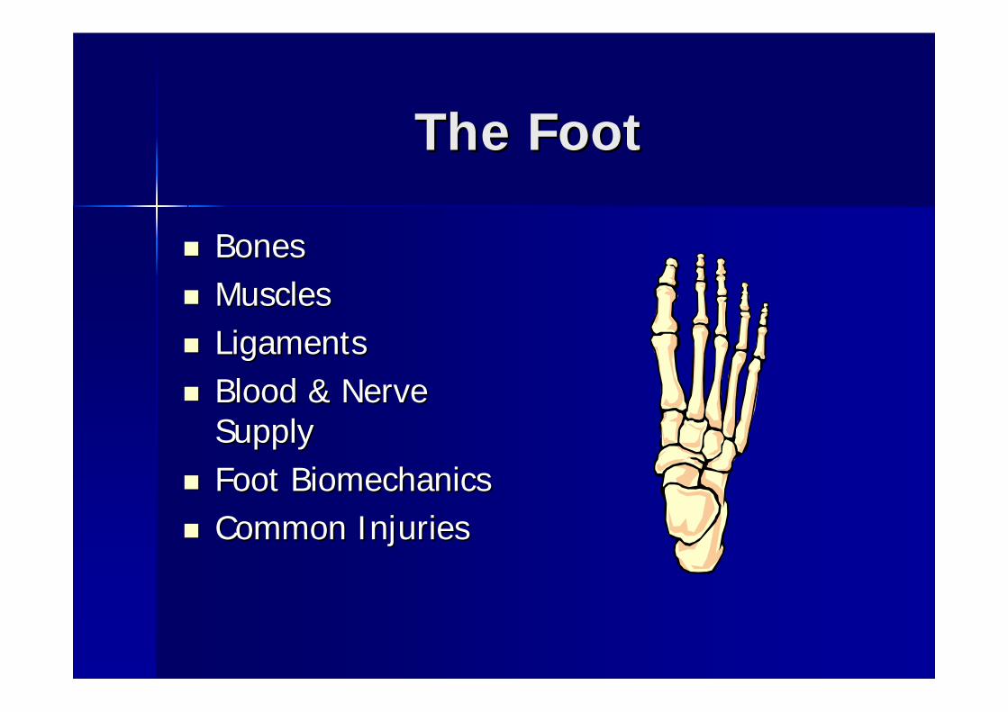
The FootThe Foot
BonesBonesMusclesMusclesLigamentsLigamentsBlood & Nerve Blood & Nerve SupplySupplyFoot BiomechanicsFoot BiomechanicsCommon InjuriesCommon Injuries

The FootThe Foot

The FootThe Foot
26 bones (14 phalanges, 5 26 bones (14 phalanges, 5 metatarsals, 7 metatarsals, 7 tarsalstarsalsTarsalsTarsals: talus, : talus, calcaneuscalcaneus, , navicularnavicular, , cuboidcuboid, 1, 1stst, 2, 2ndnd, & 3, & 3rdrd cuneiformscuneiformsDesigned for strength, flexibility, and Designed for strength, flexibility, and coordinated coordinated mvmtmvmt

Bones of the FootBones of the Foot

Phalanges Phalanges AkaAka ““ToesToes””
Designed to give a Designed to give a wider base of wider base of supportsupport11stst ““toetoe”” halluxhallux: 2 : 2 phalangesphalanges22ndnd thru 5thru 5thth: 3 : 3 phalangesphalanges2 2 SesamoidSesamoid bonesbones–– assist with WBassist with WB–– increase increase mechmech
advantage for flexor advantage for flexor tendonstendons

MetatarsalsMetatarsals
5 bones b/w 5 bones b/w tarsalstarsals and phalangesand phalangesLittle Little mvmtmvmt, ligaments provide , ligaments provide elasticity to foot during WBelasticity to foot during WB QWAQWA11stst metatarsal is largest and strongest metatarsal is largest and strongest and and fxnfxn’’ss as the main body support as the main body support during ambulationduring ambulation

TarsalsTarsals
7 bones: important for support & 7 bones: important for support & locomotionlocomotionCalcaneusCalcaneus-- largestlargestTalusTalus--irregular shaped, most superiorirregular shaped, most superiorNavicularNavicularCuboidCuboid-- lateral aspect of foot, 4lateral aspect of foot, 4thth & 5& 5thth
MetatarsalsMetatarsalsCuneiforms (3)Cuneiforms (3)

ArchesArches
Assist foot in supporting BWAssist foot in supporting BWAbsorbs shockAbsorbs shockGives space for ligaments, blood Gives space for ligaments, blood vessels, and nerves to run on plantar vessels, and nerves to run on plantar surface of the footsurface of the foot

ArchesArches

4 Arches4 Arches
Medial LongitudinalMedial Longitudinal–– main supporting ligament is the plantar main supporting ligament is the plantar
calcanealcalcaneal (spring ligament)(spring ligament)
Stretches and rebounds during AMBStretches and rebounds during AMB

4 Arches4 Arches
Lateral LongitudinalLateral Longitudinal–– Less flexible than the medialLess flexible than the medial–– Runs along the 5Runs along the 5thth metatarsalmetatarsal

4 Arches4 Arches
Anterior Metatarsal ArchAnterior Metatarsal Arch–– Made up of the heads of all 5 metatarsalsMade up of the heads of all 5 metatarsals

4 Arches4 Arches
Transverse ArchTransverse Arch–– Made up of tarsal bonesMade up of tarsal bones–– Gives the shape to the footGives the shape to the foot

DIP, PIP, & IP JointsDIP, PIP, & IP Joints
DIP (distal DIP (distal interphalangealinterphalangeal joint)joint)Found at the distal extremities of the Found at the distal extremities of the proximal and middle phalangesproximal and middle phalangesAllows for Flex/ExtAllows for Flex/ExtCollateral ligaments on medial/lateral Collateral ligaments on medial/lateral
MTP JointMTP Joint
MetatarsalphalangealMetatarsalphalangeal JointJointArticulation b/w metatarsals & Articulation b/w metatarsals & phalangesphalangesAllows for flex/ext, Allows for flex/ext, abdabd/add/addCondyloidCondyloid jointjoint

MusclesMuscles
GasrocnemiusGasrocnemiusSoleusSoleusAnterior Anterior tibialistibialisPeronealPeroneal MusclesMusclesExtensor MusclesExtensor MusclesFlexor MusclesFlexor Muscles
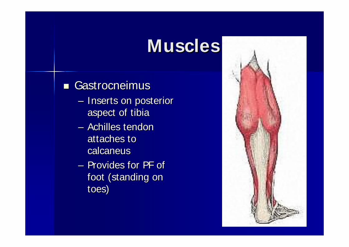
MusclesMuscles
GastrocneimusGastrocneimus–– Inserts on posterior Inserts on posterior
aspect of tibiaaspect of tibia–– Achilles tendon Achilles tendon
attaches to attaches to calcaneuscalcaneus
–– Provides for PF of Provides for PF of foot (standing on foot (standing on toes)toes)

MusclesMuscles
SoleusSoleus–– Runs underneath Runs underneath gastrocsgastrocs–– Aids in PF of footAids in PF of foot


MusclesMuscles
Anterior Anterior TibialisTibialis–– Major DF of foot and Major DF of foot and
great toegreat toe–– Paralysis of this muscle Paralysis of this muscle
results in results in ““drop footdrop foot””–– Can be involved in Ant Can be involved in Ant
Compartment Syndrome Compartment Syndrome which affects the lower which affects the lower legs blood and nerve legs blood and nerve supplysupply
–– Runs the length of the Runs the length of the tibia and also is involved tibia and also is involved in in ““shin splintsshin splints””

MusclesMuscles
PeronealsPeroneals–– Tendons most commonly Tendons most commonly
injured in an inversion spraininjured in an inversion sprain–– Run along the lateral aspect of Run along the lateral aspect of
the lower leg (fibula) around the lower leg (fibula) around the distal portion of the lateral the distal portion of the lateral maleollusmaleollus and insert on the and insert on the lateral aspect of the foot (5lateral aspect of the foot (5thth
MT)MT)–– Tom, Dick, and HarryTom, Dick, and Harry–– Extensive pressure on the Extensive pressure on the
head of the fibula causes head of the fibula causes peronealperoneal palsypalsy……numbness on numbness on the outside of the lower legthe outside of the lower leg
–– Major muscle involved in Major muscle involved in evertingeverting the footthe foot


MusclesMuscles
Extensor MusclesExtensor Muscles–– Run along the dorsal aspect of the foot Run along the dorsal aspect of the foot
and extend the toesand extend the toes–– They insert at the MT, DP and PPThey insert at the MT, DP and PP


MusclesMuscles
Flexor MusclesFlexor Muscles–– Run along the plantar surface of the foot Run along the plantar surface of the foot
and are associated with flexion of the and are associated with flexion of the toestoes
–– Insert on the plantar surface of MT, DP, Insert on the plantar surface of MT, DP, and PPand PP

MusclesMuscles


LigamentsLigaments
Spring LigamentSpring Ligament–– Medial longitudinal archMedial longitudinal arch–– Keeps the shape of the footKeeps the shape of the foot–– Depresses when Depresses when ambamb and reboundsand rebounds–– Maybe outstretched with people who Maybe outstretched with people who
have have ““flat feetflat feet””

LigamentsLigaments
ATF (anterior ATF (anterior talofibulartalofibular ligament)ligament)–– Most commonly injured in an inversion sprainMost commonly injured in an inversion sprain–– AnteriorlyAnteriorly connects the talus and fibula connects the talus and fibula
PTF (posterior PTF (posterior talofibulartalofibular ligament)ligament)–– Another commonly injured ligamentAnother commonly injured ligament–– PosteriorlyPosteriorly connects talus and fibulaconnects talus and fibula
CF (CF (calcaneofibularcalcaneofibular ligament)ligament)–– Commonly injured along with the ATFCommonly injured along with the ATF–– Connects the Connects the calcaneuscalcaneus and the talusand the talus

LigamentsLigaments
Deltoid LigamentDeltoid Ligament–– Fan shapedFan shaped–– Very strong and hard to injureVery strong and hard to injure–– Usually injured with an Usually injured with an eversioneversion sprainsprain–– Runs along the entire medial aspect of Runs along the entire medial aspect of
the medial the medial maleollusmaleollus


Blood & Nerve SupplyBlood & Nerve Supply
Anterior & posterior Anterior & posterior tibialtibial arteriesarteriesTibialTibial NerveNerve--innervates m. of the innervates m. of the back of the leg and back of the leg and plantar aspect of footplantar aspect of footCommon Common PeronealPeronealNerveNerve-- supplies front of supplies front of leg and footleg and foot

Foot BiomechanicsFoot Biomechanics
Phases of WalkingPhases of Walking–– 11stst phase: support phasephase: support phase–– Heel strike to toe offHeel strike to toe off–– 22ndnd phase: swing/recovery phase: swing/recovery –– Immediately after toe off and in position Immediately after toe off and in position
for heel strikefor heel strike

Foot BiomechanicsFoot Biomechanics
Forefoot Forefoot varusvarus–– Excessive Excessive pronationpronation-- Excessive Excessive pronationpronation during support phase during support phase
can lead to stress can lead to stress fxfx, , plantarfacitisplantarfacitis, , Achilles tendonitisAchilles tendonitis

Foot BiomechanicsFoot Biomechanics
Forefoot Forefoot valgusvalgus–– Excessive Excessive supinationsupination–– Excessive Excessive supinationsupination at heel strike can at heel strike can
lead to inversion ankle sprains, ITB lead to inversion ankle sprains, ITB syndrome, syndrome, peronealperoneal tendonitistendonitis

Common InjuriesCommon Injuries
Heel BruiseHeel Bruise–– Extremely painfulExtremely painful–– Etiology: sudden stop/go change from Etiology: sudden stop/go change from
horz/verthorz/vert–– S/S: severe pain, unable to WB, warm & S/S: severe pain, unable to WB, warm &
redness redness –– TX: NWB 24hrs, RICE, NSAIDS, DoughnutTX: NWB 24hrs, RICE, NSAIDS, Doughnut

Image 1 - In the painful heel, the fat pad is compressed and pushed up the side of the foot leaving far less protection for the heel bone.

Common InjuriesCommon Injuries
Bruised Instep (top of foot)Bruised Instep (top of foot)–– Etiology: being stepped on or direct Etiology: being stepped on or direct
contactcontact–– S/S: wearing shoes is painfulS/S: wearing shoes is painful–– TX: ICE, pad the affected areaTX: ICE, pad the affected area

Common InjuriesCommon Injuries
Metatarsal Arch Strain (Metatarsal Arch Strain (PesPes CavusCavus & & PesPes PlanusPlanus))–– Etiology: high arch or flat footEtiology: high arch or flat foot–– S/S: pain or cramping in metatarsal S/S: pain or cramping in metatarsal
regionregion–– TxTx: tape or pad to elevate fallen arch: tape or pad to elevate fallen arch

Common InjuriesCommon Injuries
Turf ToeTurf Toe–– Etiology: results from Etiology: results from
artificial playing surfaces artificial playing surfaces and flexible types of and flexible types of footwearfootwear
S/S: S/S: hyperexthyperext of great of great toe causing toe causing jtjt capsule capsule to be torn from the MT to be torn from the MT headheadTxTx: : –– r/or/o fxfx–– StiffStiff--soled shoessoled shoes–– Tape Tape

Common InjuriesCommon Injuries
Stress Stress fxfx’’ss–– Can occur anywhere Can occur anywhere –– Overuse injury or Overuse injury or
repetitive type repetitive type injuryinjury
–– Starts as a Starts as a weakening of an weakening of an area and can area and can progress into a full progress into a full blown blown fxfx
–– TxTx: Rest, Ice, rehab: Rest, Ice, rehab

Common InjuriesCommon Injuries
Inversion SprainInversion Sprain--““rolling an rolling an ankleankle””–– Etiology: landing on Etiology: landing on
someonesomeone’’s foot, s foot, excessive excessive supinationsupinationand IR of footand IR of foot
–– S/S: severe to mod S/S: severe to mod effusion, effusion, echymosisechymosis, , point tender over ATF, point tender over ATF, CF, & PTF ligamentsCF, & PTF ligaments
–– TX: TX: r/or/o fxfx, , cryokineticscryokinetics, , gentle stretching, gentle stretching, AROM, strengtheningAROM, strengthening


The KneeThe Knee

The KneeThe Knee
BonesBonesMenisciMenisciMusclesMusclesLigamentsLigamentsBlood & Nerve SupplyBlood & Nerve SupplyCommon InjuriesCommon Injuries

The KneeThe Knee
Bones:Bones:–– FemurFemur–– TibiaTibia–– FibulaFibula–– PatellaPatella

The KneeThe Knee
FemurFemur–– Largest & strongest bone in the bodyLargest & strongest bone in the body–– Two Two condylescondyles that articulate with the tibiathat articulate with the tibia


The KneeThe Knee
TibiaTibia–– Medial bone second largest in lower legMedial bone second largest in lower leg–– Two plateaus articulate with femoral Two plateaus articulate with femoral condylescondyles
(concavities where the meniscus sit) (concavities where the meniscus sit) –– Most distal end is known as the medial Most distal end is known as the medial malleolusmalleolus
FibulaFibula–– Smaller bone that runs down the lateral aspect Smaller bone that runs down the lateral aspect
of the lower legof the lower leg–– Most distal portion is known as the lateral Most distal portion is known as the lateral
malleolusmalleolus


The KneeThe Knee
PatellaPatella–– Largest Largest sesamoidsesamoid
bone in the bodybone in the body–– Found in the quad Found in the quad
tendontendon–– Tracking of the Tracking of the
patella depends on patella depends on the pull of the the pull of the quads & patellar quads & patellar tendontendon

The KneeThe Knee
MenisciMenisci–– 2 oval fibro2 oval fibro--cartilages that deepen the cartilages that deepen the
articulation b/w the femur and the tibiaarticulation b/w the femur and the tibia–– Acts as a cushion and shock absorber for the Acts as a cushion and shock absorber for the
kneeknee
Menisci Blood SupplyMenisci Blood Supply–– Each menisci divided into 3 Each menisci divided into 3 circumfrentialcircumfrential zones:zones:–– RedRed--red zone=outer 1/3 good red zone=outer 1/3 good blbl. Supply. Supply–– RedRed--white zone=middle 1/3 minimal white zone=middle 1/3 minimal blbl supplysupply–– WhiteWhite--white zone=inner 1/3 is white zone=inner 1/3 is avascularavascular

The KneeThe Knee
Medial MeniscusMedial Meniscus–– C shapedC shaped–– Attached firmly to the medial Attached firmly to the medial articulararticular
facet of the tibia by the coronary facet of the tibia by the coronary ligamentligament
–– PosteriorlyPosteriorly by the fibers of the by the fibers of the semimembranosoussemimembranosous
The KneeThe Knee
Lateral MeniscusLateral Meniscus–– O shaped O shaped –– Attaches loosely to the lateral Attaches loosely to the lateral articulararticular
capsule and capsule and poplitealpopliteal tendontendon–– Transverse ligament joins the anterior Transverse ligament joins the anterior
portions of the medial and lateral menisci portions of the medial and lateral menisci


MusclesMuscles
FlexorsFlexorsExtensorsExtensorsDynamic StabilizersDynamic Stabilizers


Muscles (flexors)Muscles (flexors)
Biceps Biceps FemorisFemoris

Muscles (flexors)Muscles (flexors)
SemimembranosousSemimembranosous

Muscles (flexors)Muscles (flexors)
SemitendonosousSemitendonosous

Muscles (flexors)Muscles (flexors)
GracilisGracilis–– Abducts, flexes, andAbducts, flexes, and
medially rotates the hipmedially rotates the hip

Muscles (flexors)Muscles (flexors)
SartoriusSartorius–– Allows you to sit cross leggedAllows you to sit cross legged–– Known as the Known as the ““tailor sitstailor sits”” musclemuscle–– ASIS to the ASIS to the tibialtibial headhead

Muscles (extensors)Muscles (extensors)
QuadricepsQuadriceps–– RectusRectus FemorisFemoris–– VMOVMO–– VastisVastis LateralisLateralis–– VastusVastus IntermediusIntermedius

Muscles (dynamic Muscles (dynamic stabilizer)stabilizer)
IT Band (IT Band (iliotibialiliotibial band)band)

LigamentsLigaments
Stabilizing ligaments of the kneeStabilizing ligaments of the kneeACL (anterior ACL (anterior cruciatecruciate ligament)ligament)–– Attaches below and in front of the tibia, Attaches below and in front of the tibia,
runs runs posteriorlyposteriorly to attach laterally to the to attach laterally to the inner surface of the lateral inner surface of the lateral condylecondyle of the of the femurfemur
–– Comprised of 3 twisted bandsComprised of 3 twisted bands–– Prevents anterior translationPrevents anterior translation–– Stabilizes the tibia against excessive IRStabilizes the tibia against excessive IR

LigamentsLigaments
PCL (posterior PCL (posterior cruciatecruciate ligament)ligament)–– Stronger of the twoStronger of the two–– Primary stabilizer of the kneePrimary stabilizer of the knee–– Crosses back of tibia Crosses back of tibia anteriorlyanteriorly to attach to attach
anterior portion of the lateral surface of anterior portion of the lateral surface of the medial the medial condylecondyle of femurof femur
–– Resists IR of tibia Resists IR of tibia –– Helps prevent hyperextension of knee Helps prevent hyperextension of knee
and femur, sliding forward during WBand femur, sliding forward during WB

LigamentsLigaments
MCL (medial collateral ligament)MCL (medial collateral ligament)–– Attaches above the Attaches above the jtjt line on medial line on medial
epicondyleepicondyle of the femur and below on the of the femur and below on the tibia just below the tibia just below the pespes anserineanserine
–– Prevents knee from Prevents knee from valgusvalgus and ER forcesand ER forces–– Approx the width of three fingersApprox the width of three fingers

LigamentsLigaments
LCL (lateral collateral ligament)LCL (lateral collateral ligament)–– Round fibrous cord shaped like a pencilRound fibrous cord shaped like a pencil–– Attaches to lateral Attaches to lateral epicondyleepicondyle of the of the
femur and to the head of the fibulafemur and to the head of the fibula–– Taut during knee ext, relaxed during knee Taut during knee ext, relaxed during knee
flexflex


BursalBursal SacsSacs
BursaBursa–– Reduces friction b/w Reduces friction b/w
anatomical surfacesanatomical surfaces–– SuprapatellarSuprapatellar, ,
prepatellarprepatellar, , infrapatellarinfrapatellar, , pretibialpretibial, and , and gastrocgastroc

Blood & Nerve SupplyBlood & Nerve Supply
Blood supplyBlood supply–– Main supply is Main supply is PoplitealPopliteal artery which stems from artery which stems from
the Femoral arterythe Femoral artery
Nerve SupplyNerve Supply–– TibialTibial nerve supplies hamstrings and nerve supplies hamstrings and gastrocsgastrocs–– Common Common peronealperoneal innervates short head of innervates short head of
biceps biceps femorisfemoris–– Femoral nerve innervates the quads and Femoral nerve innervates the quads and
sartoriussartorius

QQ--AngleAngle
Measured from ASIS to midMeasured from ASIS to mid--patella patella and from and from TibialTibial Tubercle (TT) thru mid Tubercle (TT) thru mid patellapatellaNorms:Norms:–– Men=10 degreesMen=10 degrees–– Women=20 degreesWomen=20 degrees–– 20< leads to pathological conditions 20< leads to pathological conditions
associated with improper patellar trackingassociated with improper patellar tracking

Common InjuriesCommon Injuries
MCL sprainMCL sprain–– Etiology: Etiology: valgusvalgus stress from a direct blow stress from a direct blow
to the lateral side of the kneeto the lateral side of the knee–– Grades I, II, or IIIGrades I, II, or III–– S/S ranges from slight pain and instability S/S ranges from slight pain and instability
to complete tear and loss of to complete tear and loss of fxnfxn, swelling, , swelling, & altered gait& altered gait
–– TxTx: RICE, : RICE, r/or/o ACL, crutches, isometric ACL, crutches, isometric exercises, immobilizer if necessary exercises, immobilizer if necessary

Grade IGrade I–– Outstretching of fibersOutstretching of fibers–– Little or no swellingLittle or no swelling–– Some stiffness and pt tenderSome stiffness and pt tender–– Full AROM &PROMFull AROM &PROM
Grade IIGrade II–– Partial tearing of fibers and tear of the capsular Partial tearing of fibers and tear of the capsular
ligamentligament–– Minimal laxity with slight swelling Minimal laxity with slight swelling –– ModMod--severe severe jtjt stiffness w/ inability to actively ext kneestiffness w/ inability to actively ext knee
Grade IIIGrade III–– Complete tear of the fibersComplete tear of the fibers–– Complete loss of stabilityComplete loss of stability–– Moderate swellingModerate swelling–– Severe pain followed by dull acheSevere pain followed by dull ache–– ++valgusvalgus test w/ full test w/ full jtjt openingopening

Common InjuriesCommon Injuries
LCL SprainLCL Sprain–– Etiology: Etiology: varusvarus stress, less common than stress, less common than
MCLMCL–– S/S: pain and tenderness over LCL, S/S: pain and tenderness over LCL,
swelling, effusion, some swelling, effusion, some jtjt laxity w/ laxity w/ varusvarusstress teststress test
–– TxTx: similar to MCL: similar to MCL

Common InjuriesCommon Injuries
ACLACL–– Etiology: rotary Etiology: rotary
mechanism, tibia ER and mechanism, tibia ER and knee is in knee is in valgusvalguspositionposition
–– S/S: S/S: ““poppop”” usually felt usually felt or they may state that or they may state that their knee feels like it is their knee feels like it is coming apart, rapid coming apart, rapid swelling, can be swelling, can be extremely painful, loss extremely painful, loss of ROM, cannot full ext of ROM, cannot full ext kneeknee
–– TX: RICE immediately, TX: RICE immediately, SxSx

Common InjuriesCommon Injuries
PCLPCL–– Etiology: knee flexed 90 Etiology: knee flexed 90
degrees, falling w/ full wt degrees, falling w/ full wt on anterior aspect of bent on anterior aspect of bent knee, foot in PF or hard knee, foot in PF or hard blow to the front of bent blow to the front of bent knee (MVA)knee (MVA)
–– S/S: S/S: ““poppop”” felt, tenderness felt, tenderness and little swelling in and little swelling in poplitealpopliteal fossafossa, laxity in , laxity in posterior sag test, + posterior sag test, + posterior drawerposterior drawer
–– TxTx: RICE, non: RICE, non--operative operative grades I & II, 6 wks of grades I & II, 6 wks of immobilization, rehab immobilization, rehab

Common InjuriesCommon Injuries
MeniscalMeniscal Lesions Lesions (tears)(tears)–– Etiology: WB with rotary Etiology: WB with rotary
force, medial is injured force, medial is injured more than lateralmore than lateral
–– S/S: deep aching pain S/S: deep aching pain inside the knee, inside the knee, problems kneeling, problems kneeling, walking up/down stairs, walking up/down stairs, squatting, locking up or squatting, locking up or giving way, slow giving way, slow effusion appears after effusion appears after 4848--72 hrs72 hrs
–– TxTx: RICE, MRI, : RICE, MRI, SxSx

Common InjuriesCommon Injuries
BursitisBursitis–– Etiology: cont Etiology: cont
kneeling, trauma, kneeling, trauma, overuseoveruse
–– S/S: localized pain & S/S: localized pain & swelling, redness swelling, redness and increased tempand increased temp
–– TxTx: RICE, : RICE, compression wraps, compression wraps, NSAIDSNSAIDS
Common InjuriesCommon Injuries
Patellar Patellar SubluxSublux/Dislocations/Dislocations–– Etiology: poor tracking of the patella due Etiology: poor tracking of the patella due
toto--wide pelvis, increase in Q angle, wide pelvis, increase in Q angle, shallow femoral grooves, patella shallow femoral grooves, patella altaalta or or bajabaja, , pronatedpronated feetfeet
–– S/S: loss of knee S/S: loss of knee fxnfxn, pain, swelling, , pain, swelling, patella in abnormal position patella in abnormal position
–– TxTx: possible : possible sxsx, isometric ex, rehab & , isometric ex, rehab & strengthening, immobilizationstrengthening, immobilization

Common InjuriesCommon Injuries
ChondromalaciaChondromalacia–– Etiology: abnormal tracking of patellaEtiology: abnormal tracking of patella–– S/S: Stage IS/S: Stage I--swelling & softening of swelling & softening of articulararticular
cartilage, Stage IIcartilage, Stage II-- fissuring of the softened fissuring of the softened cartilage, Stage IIIcartilage, Stage III-- deformation of the surfaces deformation of the surfaces under the patella caused by under the patella caused by defragmentationdefragmentation, , pain pain w/Ambw/Amb, running, stairs, squatting, swelling , running, stairs, squatting, swelling around kneecap, + patellar grindaround kneecap, + patellar grind
–– TxTx: Avoid irritating : Avoid irritating activitesactivites, isometric ex, , isometric ex, NSAIDS, neoprene sleeve, NSAIDS, neoprene sleeve, orthoticsorthotics, smoothing , smoothing of the surface of the patellaof the surface of the patella
Common InjuriesCommon Injuries
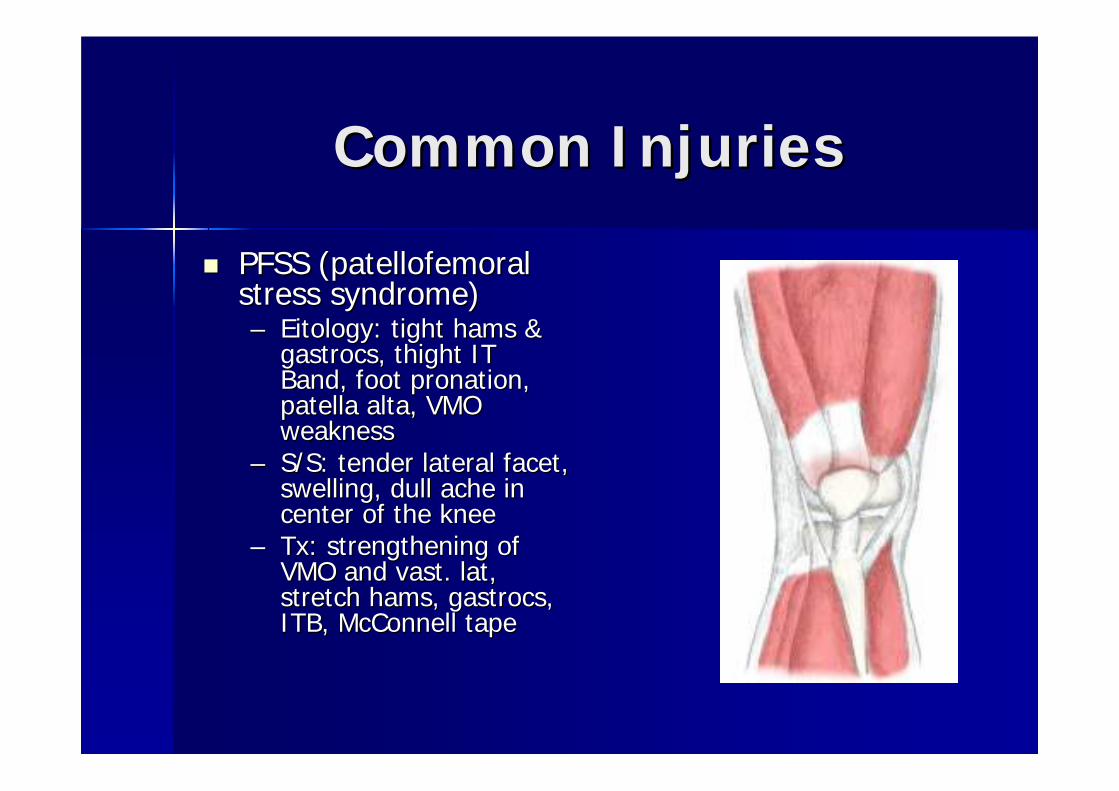
PFSS (PFSS (patellofemoralpatellofemoralstress syndrome)stress syndrome)–– EitologyEitology: tight hams & : tight hams &
gastrocsgastrocs, , thightthight IT IT Band, foot Band, foot pronationpronation, , patella patella altaalta, VMO , VMO weaknessweakness
–– S/S: tender lateral facet, S/S: tender lateral facet, swelling, dull ache in swelling, dull ache in center of the kneecenter of the knee
–– TxTx: strengthening of : strengthening of VMO and vast. lat, VMO and vast. lat, stretch hams, stretch hams, gastrocsgastrocs, , ITB, McConnell tapeITB, McConnell tape

Common InjuriesCommon Injuries
OsgoodOsgood--SchlatterSchlatter’’ssDiseaseDisease–– Etiology: avulsion of the Etiology: avulsion of the
patellar tendon @ the patellar tendon @ the apophysisapophysis of the TTof the TT
–– S/S: repeated irritation, S/S: repeated irritation, swelling, hemorrhage, swelling, hemorrhage, gradual degeneration of gradual degeneration of the the apophysisapophysis
–– TxTx: Decrease stressful : Decrease stressful activities, Ice preactivities, Ice pre--post post activities, isometric ex activities, isometric ex

Hip, Thigh, Groin, PelvisHip, Thigh, Groin, Pelvis

Hip, Thigh, Groin, & PelvisHip, Thigh, Groin, & Pelvis
BonesBonesFemoral TriangleFemoral TriangleMusclesMusclesLigamentsLigamentsBlood & Nerve SupplyBlood & Nerve SupplyCommon InjuriesCommon Injuries

Hip, Thigh, Groin, & Pelvis Hip, Thigh, Groin, & Pelvis
BonesBonesFemurFemur–– Head and neck of the femurHead and neck of the femur–– Greater & lesser Greater & lesser trochanterstrochanters

femurfemur

Hip, Thigh, Groin, & PelvisHip, Thigh, Groin, & Pelvis
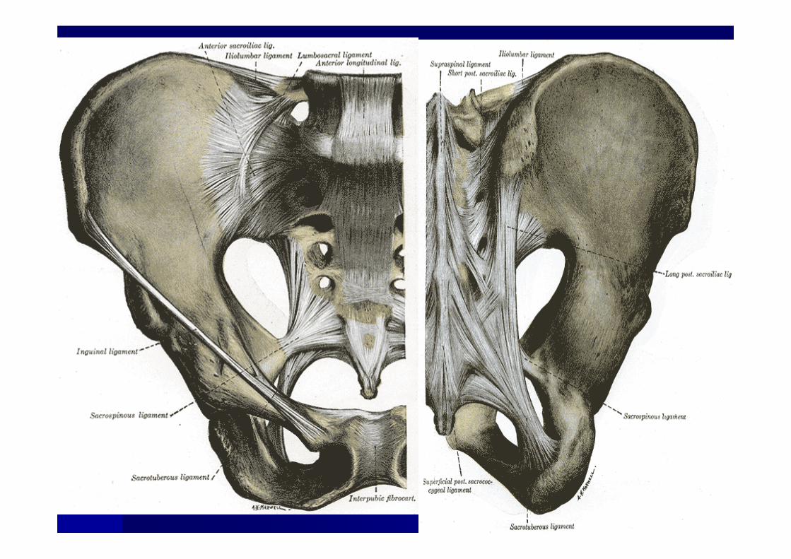
Pelvis (made up of Pelvis (made up of 3 bones)3 bones)IliumIliumIschiumIschiumPubisPubis

Hip, Thigh, Groin, & PelvisHip, Thigh, Groin, & Pelvis
Ball and Socket JointBall and Socket Joint–– Head of femur articulates with the Head of femur articulates with the
acetabulumacetabulum–– One of the most stable joints in the bodyOne of the most stable joints in the body–– If you have stability you give up mobilityIf you have stability you give up mobility–– AcetabularAcetabular ligament deepens the jointligament deepens the joint


Femoral TriangleFemoral Triangle
Inguinal LigamentInguinal LigamentAdductor Adductor LongusLongusSartoriusSartorius
These structures run through it:These structures run through it:--Femoral ArteryFemoral Artery--Femoral NerveFemoral Nerve--Femoral VeinFemoral Vein--Inguinal Lymph NodesInguinal Lymph Nodes

LigamentsLigaments
IliofemoralIliofemoral LigamentLigament–– One of the strongest and largest in bodyOne of the strongest and largest in body–– Also known as the Also known as the ““YY”” ligamentligament


LigamentsLigaments
AcetabularAcetabular LigamentLigament–– Deepens the Deepens the acetabulumacetabulum to create a to create a
deeper socket and making it more stabledeeper socket and making it more stable
Inguinal LigamentInguinal Ligament–– Many times is involved in hernias Many times is involved in hernias –– Part of the femoral trianglePart of the femoral triangle


MusclesMuscles
IliopsoasIliopsoas–– Major hip flexorMajor hip flexor–– Actually is two musclesActually is two muscles

MusclesMuscles
GlutesGlutes–– MaximusMaximus-- Ext & laterally rot hipExt & laterally rot hip–– MediusMedius-- Abducts hipAbducts hip
Anterior fibers: flex & med rot hipAnterior fibers: flex & med rot hipPosterior fibers: ext & lat rot hipPosterior fibers: ext & lat rot hip
–– MinimusMinimus-- abducts & med rot hipabducts & med rot hip

MusclesMuscles
Tensor Fasciae Tensor Fasciae LataeLatae (TFL)(TFL)–– Flexes and Med Rot hipFlexes and Med Rot hip

Blood and Blood and Nerve Nerve SupplySupply
Femoral Artery & Femoral Artery & VeinVeinFemoral NerveFemoral Nerve

Common InjuriesCommon Injuries
Hip bursitisHip bursitisEtiology: repeated Etiology: repeated trauma, overuse injury trauma, overuse injury (from running)(from running)S/S inability to bend , S/S inability to bend , run, pt tender, run, pt tender, swelling, warm, swelling, warm, snapping sensationsnapping sensationTxTx: RICE, ultrasound, : RICE, ultrasound, padding, stretchpadding, stretch
Common InjuriesCommon Injuries
Femoral Stress Fracture Femoral Stress Fracture –– Etiology: Excessive OveruseEtiology: Excessive Overuse–– S/S: Persistent pain in thighS/S: Persistent pain in thigh–– TxTx: NSAIDS, RICE, ROM in pain free range: NSAIDS, RICE, ROM in pain free range
Femoral FractureFemoral Fracture–– Etiology: Excessive blunt traumaEtiology: Excessive blunt trauma–– S/S: deformation and pain over siteS/S: deformation and pain over site–– TxTx: Treat for shock, immobilize, activate EMS: Treat for shock, immobilize, activate EMS

Common InjuriesCommon Injuries
SubluxationSubluxation/dislocation of hip/dislocation of hip–– Etiology: traumatic force along the long Etiology: traumatic force along the long
axis of femur w/ knees bentaxis of femur w/ knees bent–– S/S: flexed, ABD, and IR, head of femur S/S: flexed, ABD, and IR, head of femur
is moved post to is moved post to acetabulumacetabulum. Possible . Possible damage to sciatic nervedamage to sciatic nerve
–– TxTx: Immediate medical attention: Immediate medical attention

Common InjuriesCommon Injuries
LeggLegg--Calve Calve PerthesPerthes DiseaseDisease–– Etiology: disruption of blood circulation to Etiology: disruption of blood circulation to
the head of the femur causing cartilage the head of the femur causing cartilage to become necrotic and flattenedto become necrotic and flattened
–– S/S: pain in groin, limping and referred S/S: pain in groin, limping and referred painpain
–– TxTx: Bed rest and bracing: Bed rest and bracing

Common InjuriesCommon Injuries
Slipped Capita Femoral EpiphysisSlipped Capita Femoral Epiphysis–– Etiology: boys 10Etiology: boys 10--17 who are tall or 17 who are tall or
obese. Hip slips posterior and inferiorobese. Hip slips posterior and inferior–– S/S: hip and knee pain during passive S/S: hip and knee pain during passive
mvmtmvmt as well as limits in ABD, flex, and as well as limits in ABD, flex, and Med RotMed Rot
–– TxTx: Rest & NWB : Rest & NWB

Abdomen, Thorax, & Abdomen, Thorax, & SpineSpine

Abdomen, Thorax, & Abdomen, Thorax, & SpineSpine
BonesBonesMusclesMusclesLigamentsLigamentsBlood and Nerve SupplyBlood and Nerve SupplyCommon InjuriesCommon Injuries

Abdomen & ThoraxAbdomen & Thorax
BonesBones–– Ribs Ribs
11--7 true ribs (7 true ribs (sternalsternal))88--10 are false ribs10 are false ribs1111--12 are floating ribs12 are floating ribs
–– SternumSternumManubriumManubriumBodyBodyXiphoidXiphoid ProcessProcess


Solid OrgansSolid Organs
KidneysKidneysSpleenSpleenLiverLiverPancreasPancreasAdrenal GlandsAdrenal Glands
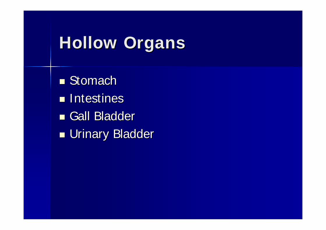
Hollow OrgansHollow Organs
StomachStomachIntestinesIntestinesGall BladderGall BladderUrinary BladderUrinary Bladder

4 Quadrants 4 Quadrants
Right UpperRight Upper–– LiverLiver
Left UpperLeft Upper–– SpleenSpleen–– StomachStomach
Right LowerRight Lower–– Gall BladderGall Bladder–– AppendixAppendix–– KidneyKidney
Left LowerLeft Lower–– KidneyKidney
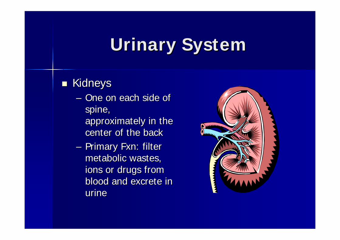
Urinary SystemUrinary System
KidneysKidneys–– One on each side of One on each side of
spine, spine, approximately in the approximately in the center of the backcenter of the back
–– Primary Primary FxnFxn: filter : filter metabolic wastes, metabolic wastes, ions or drugs from ions or drugs from blood and excrete in blood and excrete in urineurine

Urinary SystemUrinary System
Adrenal Glands (Adrenal Glands (akaaka suprarenal suprarenal glands)glands)–– Located on top of each kidney Located on top of each kidney –– Secrete: epinephrine, Secrete: epinephrine, norepinephrinenorepinephrine, ,
cortisolcortisol, estrogen, , estrogen, aldestronealdestrone, and , and androgen androgen

Urinary SystemUrinary System
UretersUreters and Urinary Bladderand Urinary Bladder–– UretersUreters are small tubes that run inferior are small tubes that run inferior
from the bladder. The bladder lies from the bladder. The bladder lies posterior to pubic posterior to pubic symphysissymphysis. In males: . In males: anterior to rectum; in females, anterior to anterior to rectum; in females, anterior to vagina and inferior to uterusvagina and inferior to uterus

Digestive SystemDigestive System
LiverLiver–– Largest internal organ, UR quadLargest internal organ, UR quad–– FxnFxn: absorbs and stores excessive glucose, : absorbs and stores excessive glucose,
process nutrients, detoxifies harmful chemicals, process nutrients, detoxifies harmful chemicals, secretes bilesecretes bile
Bile neutralizes and dilutes stomach acids and aids in Bile neutralizes and dilutes stomach acids and aids in digestion of fat in small intestinedigestion of fat in small intestine
–– HepititisHepititis-- inflammation of the liver caused by inflammation of the liver caused by viral infection or alcohol consumption. If not viral infection or alcohol consumption. If not corrected it can lead to liver cell death and corrected it can lead to liver cell death and cirrhosiscirrhosis
Digestive SystemDigestive System
Gall BladderGall Bladder–– Located inferior to surface of the liverLocated inferior to surface of the liver–– FxnFxn: serves as storage reservoir for bile, : serves as storage reservoir for bile,
secretes those stores into secretes those stores into smsm intestineintestine–– Gall StonesGall Stones-- caused by cholesterol caused by cholesterol
secreted by the liver, block the release of secreted by the liver, block the release of bile, must be removed surgically bile, must be removed surgically

Digestive SystemDigestive System
PancreasPancreas–– Located between the Located between the
small intestine and the small intestine and the spleenspleen
–– FxnFxn: secretes pancreatic : secretes pancreatic juice, which is critical in juice, which is critical in digestion of fats, digestion of fats, carbscarbs, , and proteins. Secretes and proteins. Secretes insulin and insulin and glucagonglucagon, , which control amt of which control amt of glucose and amino acids glucose and amino acids in blood in blood

Digestive SystemDigestive System
StomachStomach–– UL quadUL quad–– Second stage of food break down (first Second stage of food break down (first
stage is in mouth)stage is in mouth)–– Some absorption but mostly in the small Some absorption but mostly in the small
intestineintestine–– Can see stomach ulcers due to an over Can see stomach ulcers due to an over
production of stomach acidsproduction of stomach acids

Lymphatic SystemLymphatic System
SpleenSpleen–– Largest lymph organLargest lymph organ–– FxnFxn: reservoir of red blood cells, : reservoir of red blood cells,
regulates number of RBC, destroys RBC, regulates number of RBC, destroys RBC, produces antibodies, produces produces antibodies, produces lymphocyteslymphocytes
–– Located under diaphragm, left side, Located under diaphragm, left side, behind ribs 9,10, & 11. behind ribs 9,10, & 11.

Digestive SystemDigestive System
Small Intestine (3 Small Intestine (3 parts)parts)–– DuodenumDuodenum–– JejunumJejunum–– IleumIleum
Large intestine (3 Large intestine (3 parts)parts)–– CecumCecum–– ColonColon–– RectumRectum


Common InjuriesCommon Injuries
Evaluation of Abdomen & ThoraxEvaluation of Abdomen & Thorax–– Check chest for asymmetry of breathing, Check chest for asymmetry of breathing,
palpate along ribs, palpate along ribs, intercostalintercostal space, space, costacholandralcostacholandral jxnjxn
–– Check URQ, ULQ, LLQ, LRQ clockwiseCheck URQ, ULQ, LLQ, LRQ clockwise–– Feel and look for muscle guardingFeel and look for muscle guarding–– Distinguish b/w guarding and rigidity Distinguish b/w guarding and rigidity –– Listen and feel for bowel sounds (gurgling Listen and feel for bowel sounds (gurgling
of normal liquid)of normal liquid)


Common InjuriesCommon Injuries
Rib Rib FxFx–– Etiology: direct blow, indirect trauma Etiology: direct blow, indirect trauma
(compression of rib cage), or violent muscle (compression of rib cage), or violent muscle contraction. Direct blow is most dangerous contraction. Direct blow is most dangerous b/cb/cof fragments may displace and cut, tear, of fragments may displace and cut, tear, perforate tissue causing a perforate tissue causing a hemothoraxhemothorax or a or a collapsed lung (collapsed lung (pneumothoraxpneumothorax))
–– S/S: severe pain with inhalation/exhalation, S/S: severe pain with inhalation/exhalation, cough, sneeze, laugh etc.cough, sneeze, laugh etc.
–– TxTx: refer for x: refer for x--rayray

Common InjuriesCommon Injuries
Sternum Sternum FxFx–– Etiology: high impact blow to chest which may Etiology: high impact blow to chest which may
cause a contusion to the underlying cardiac cause a contusion to the underlying cardiac musclemuscle
–– S/S: pt. tenderness, pain exacerbated by deep S/S: pt. tenderness, pain exacerbated by deep inspiration and forceful exhalation, signs of inspiration and forceful exhalation, signs of shock, weak or rapid pulse may indicate a more shock, weak or rapid pulse may indicate a more severe injury.severe injury.
–– TxTx: EMS, X: EMS, X--rays, monitor for heart trauma rays, monitor for heart trauma

Common InjuriesCommon Injuries
HemothoraxHemothorax–– Presence of blood in plural cavityPresence of blood in plural cavity–– Hemorrhage can be Hemorrhage can be negneg of rib of rib fxfx, , –– S/S: Pain on breathing, S/S: Pain on breathing, dyspneddyspned, shock, coughing up frothy , shock, coughing up frothy
blood blood PneumothoraxPneumothorax–– Plural cavity fills with air by an opening in the chest. Air filPlural cavity fills with air by an opening in the chest. Air fills the ls the
cavity and collapses the lung, difficulty breathing and anoxiacavity and collapses the lung, difficulty breathing and anoxiaTension Tension PneumothoraxPneumothorax–– When air filled plural cavity displaces lung and heart towards When air filled plural cavity displaces lung and heart towards
opposite lung compressing itopposite lung compressing it–– S/S: SOB, and pain on injured side, absence of breathing, S/S: SOB, and pain on injured side, absence of breathing,
cyanosis, and distension of neck veins, call EMS immediately. cyanosis, and distension of neck veins, call EMS immediately.
All lung injuries require immediate medical attentionAll lung injuries require immediate medical attention----transporttransport

Common InjuriesCommon Injuries
Traumatic Traumatic AsphyixaAsphyixa–– Results from a violent blow to or Results from a violent blow to or
compression of the rib cage that causes compression of the rib cage that causes cessation of breathingcessation of breathing
–– S/S: purple discoloration of trunk and S/S: purple discoloration of trunk and head, conjunctivas of the eyes display head, conjunctivas of the eyes display bright red colorbright red color
–– TxTx: Rescue Breathing immediately, EMS: Rescue Breathing immediately, EMS
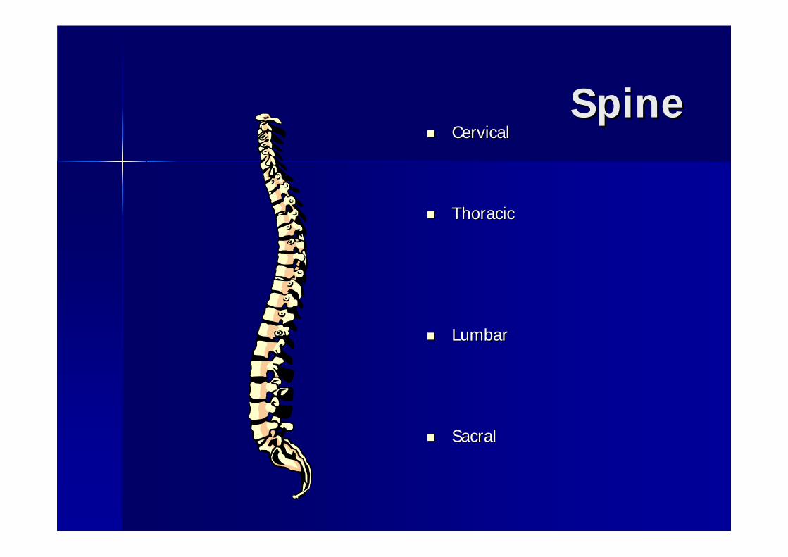
SpineSpineCervical Cervical
ThoracicThoracic
LumbarLumbar
SacralSacral

CC--SpineSpine
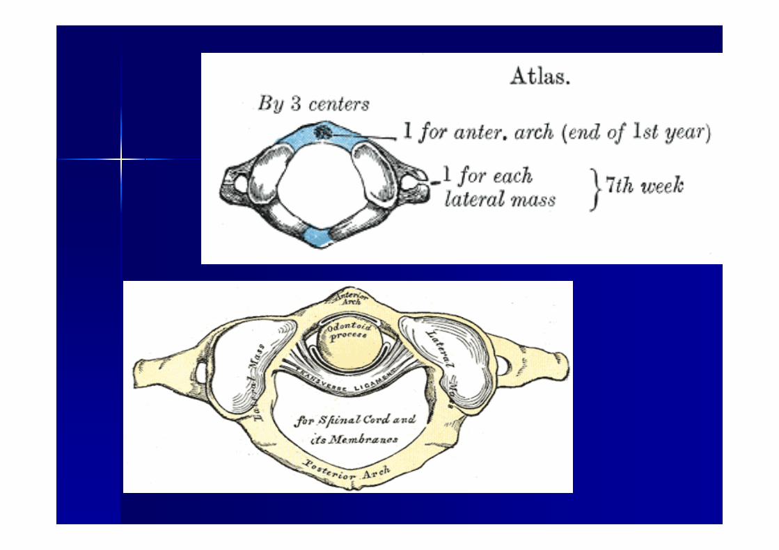
Cervical spine vertebrae 1Cervical spine vertebrae 1--77Atlas (#1)Atlas (#1)–– Upper surface articulates with Upper surface articulates with occiputocciput and allows and allows
for flex and ext. No body or for flex and ext. No body or spinousspinous processprocess
Axis (#2)Axis (#2)–– Allows the skull and atlas to rotateAllows the skull and atlas to rotate–– Tooth like projection from vertebral body that Tooth like projection from vertebral body that
fits into ring of atlas (fits into ring of atlas (odontoidodontoid process)process)
If this If this fxfx, you die, you die……skull slips off severing SCskull slips off severing SC


CC--SpineSpine
Smallest bodiesSmallest bodiesHorizontal Horizontal positioning of the positioning of the spinousspinous processesprocesses
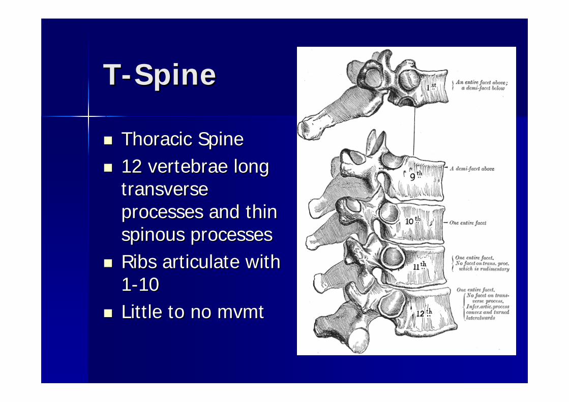
TT--SpineSpine
Thoracic SpineThoracic Spine12 vertebrae long 12 vertebrae long transverse transverse processes and thin processes and thin spinousspinous processesprocessesRibs articulate with Ribs articulate with 11--1010Little to no Little to no mvmtmvmt


LL--spinespine
Lumbar SpineLumbar Spine5 vertebrae5 vertebraeMajor support of the Major support of the lower backlower backLargest and thickest, Largest and thickest, with large with large spinousspinous and and transverse processestransverse processesMuch less flexion than Much less flexion than extension extension

SacrumSacrum
Fusion of 5 vertebraeFusion of 5 vertebraeFuses with the 2 hip Fuses with the 2 hip bones to make pelvisbones to make pelvisRoots of lumbar and Roots of lumbar and sacral nerves pass sacral nerves pass through the 4 foramina through the 4 foramina lateral to the 5 fused lateral to the 5 fused vertebraevertebraeArticulates with Articulates with iliumilium to to make the sacroiliac make the sacroiliac joint (SI): sitting and joint (SI): sitting and standing puts weight at standing puts weight at these jointsthese joints

CoccyxCoccyx
““TailboneTailbone””4 or more fused 4 or more fused vertebraevertebrae

Curves of the SpineCurves of the Spine
CervicalCervical–– ConvexityConvexity
ThoracicThoracic–– ConcavityConcavity
LumbarLumbar–– ConvexityConvexity
SacralSacral–– ConcavityConcavity

LordosisLordosis

IntervertebralIntervertebral DiscsDiscs
Annulus Fibrosis: Annulus Fibrosis: periphery of disc. periphery of disc. Composed of strong, Composed of strong, fibrous tissuefibrous tissueSemifluidSemifluid Nucleus Nucleus PulposusPulposus: in the center : in the center and compressed under and compressed under pressurepressureBoth act as important Both act as important shock absorbers for the shock absorbers for the spinespine

LigamentousLigamentous StructuresStructures
Anterior Longitudinal ligamentAnterior Longitudinal ligament–– Wide, strong band: extends the full length of Wide, strong band: extends the full length of
anterior surface of the vertebral bodiesanterior surface of the vertebral bodies–– Attaches to both vertebral bodies and discs and Attaches to both vertebral bodies and discs and
resists extensionresists extension
Posterior Longitudinal LigamentPosterior Longitudinal Ligament–– Contained within the vertebral canal and extends Contained within the vertebral canal and extends
full length of posterior aspect of bodies and full length of posterior aspect of bodies and limits flexionlimits flexion


Spinal CordSpinal Cord
Contained within the vertebral canal Contained within the vertebral canal and spinal columnand spinal columnFrom the foramen magnum of cranium From the foramen magnum of cranium to the 1to the 1stst or 2or 2ndnd lumbar vertebraelumbar vertebraeLumbar roots and sacral nerves form a Lumbar roots and sacral nerves form a horselikehorselike tail called tail called ““caudacauda equinaequina””

Spinal and Peripheral Spinal and Peripheral NervesNerves
31 pairs of nerves31 pairs of nerves–– 8 cervical, 12 thoracic, 5 lumbar, 5 sacral, and 1 8 cervical, 12 thoracic, 5 lumbar, 5 sacral, and 1 coccygealcoccygeal–– Has a anterior root (motor) and posterior root (sensory)Has a anterior root (motor) and posterior root (sensory)
Each pair has a dermatome (except C1)Each pair has a dermatome (except C1)Specific area of sensation distributionSpecific area of sensation distributionSpinal nerve roots combine to form a network of Spinal nerve roots combine to form a network of nerves known as a nerves known as a ““plexusplexus””–– Cervical (C1Cervical (C1--C4)C4)–– Brachial (C5Brachial (C5--T1)T1)–– Lumbar (L1Lumbar (L1--L4)L4)–– Sacral (L4Sacral (L4--S4)S4)

Cervical Cervical Nerve RootsNerve Roots

Brachial PlexusBrachial Plexus

Lumbar Lumbar PlexusPlexus

Functional AnatomyFunctional Anatomy
Cervical RegionCervical RegionFlexionFlexion–– SCM and scalene groupSCM and scalene group
ExtensionExtension–– Upper trapsUpper traps
Lateral FlexionLateral Flexion–– All muscles on one side contracting unilaterally All muscles on one side contracting unilaterally
RotationRotation–– SCM, SCM, scalenesscalenes, and upper traps contract on , and upper traps contract on
opposite side of rotationopposite side of rotation

Functional AnatomyFunctional Anatomy
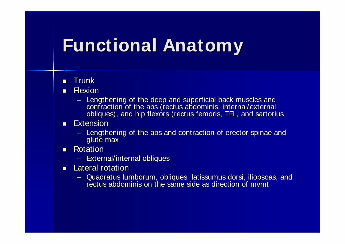
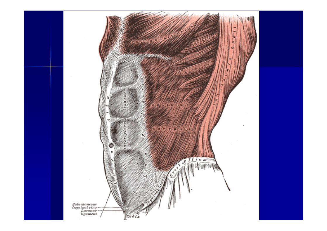
TrunkTrunkFlexionFlexion–– Lengthening of the deep and superficial back muscles and Lengthening of the deep and superficial back muscles and
contraction of the abs (contraction of the abs (rectusrectus abdominisabdominis, internal/external , internal/external obliquesobliques), and hip flexors (), and hip flexors (rectusrectus femorisfemoris, TFL, and , TFL, and sartoriussartorius
ExtensionExtension–– Lengthening of the abs and contraction of erector Lengthening of the abs and contraction of erector spinaespinae and and
gluteglute maxmaxRotationRotation–– External/internal External/internal obliquesobliques
Lateral rotationLateral rotation–– QuadratusQuadratus lumborumlumborum, , obliquesobliques, , latissumuslatissumus dorsidorsi, , iliopsoasiliopsoas, and , and
rectusrectus abdominisabdominis on the same side as direction of on the same side as direction of mvmtmvmt


Common InjuriesCommon Injuries
Cervical Fractures/DislocationsCervical Fractures/Dislocations
EitologyEitology: violent flexion and : violent flexion and rotationofrotationof head (pool head (pool diving), axial loading combined with flexion diving), axial loading combined with flexion (football injury), most common in C4, 5, or 6(football injury), most common in C4, 5, or 6S/S: neck point tenderness and restricted S/S: neck point tenderness and restricted mvmtmvmt, , cervical muscle spasm, pain in neck or chest region, cervical muscle spasm, pain in neck or chest region, numbness in trunk/limbs, weakness or paralysis in numbness in trunk/limbs, weakness or paralysis in limbs/trunk, loss of bladder or bowel controllimbs/trunk, loss of bladder or bowel controlTxTx: EMS immediately, do not move, stabilize head : EMS immediately, do not move, stabilize head and neck and neck

Common InjuriesCommon Injuries
Cervical Sprain (whiplash)Cervical Sprain (whiplash)–– Etiology: head turned suddenly or had Etiology: head turned suddenly or had
forced flexion, extension or rotationforced flexion, extension or rotation–– S/S: tenderness over transverse and S/S: tenderness over transverse and
spinousspinous processes, muscle guarding, processes, muscle guarding, localized pain, and restricted localized pain, and restricted mvmtmvmt
–– TxTx: : r/or/o fxfx, or sc injury, or sc injury

Common InjuriesCommon Injuries
Acute Acute TorticollisTorticollis (wry neck or stiff (wry neck or stiff neck)neck)–– Etiology: pain on one side upon Etiology: pain on one side upon
awakeningawakening

Common InjuriesCommon Injuries
Cervical Cervical StenosisStenosis–– Etiology: narrowing of the spinal canal in Etiology: narrowing of the spinal canal in
the Cthe C--spine region that can impinge the spine region that can impinge the sc. Can occur congenitally or changes in sc. Can occur congenitally or changes in the the vertebaevertebae
–– S/S: pain in limbs, numbness and S/S: pain in limbs, numbness and tingling, associated weakness tingling, associated weakness

Common InjuriesCommon Injuries
Brachial Plexus Brachial Plexus NeruaplaxiaNeruaplaxia (burner), most (burner), most common neurological injurycommon neurological injury–– Etiology: stretching or compression results from Etiology: stretching or compression results from
neck forced laterally to the opposite side or neck neck forced laterally to the opposite side or neck extension, compressed and rotated to the extension, compressed and rotated to the affected sideaffected side
–– S/S: burning sensation, numbness, tingling, and S/S: burning sensation, numbness, tingling, and pain extending from the shoulder down to the pain extending from the shoulder down to the hand, with LOF for several minutes hand, with LOF for several minutes

Common InjuriesCommon Injuries
Lumbar sprains/strainsLumbar sprains/strainsSciaticaSciatica–– Etiology: inflammatory Etiology: inflammatory
response of the sciatic response of the sciatic nerve that has nerve that has associated low back associated low back painpain
–– S/S: shooting pain down S/S: shooting pain down nerve root with possible nerve root with possible numbness and tinglingnumbness and tingling
image 1 - The shaded regions indicate where pain may be felt depending on which part of the
spine is involved.

Forearm, Wrist, Hand, & Forearm, Wrist, Hand, & FingersFingers
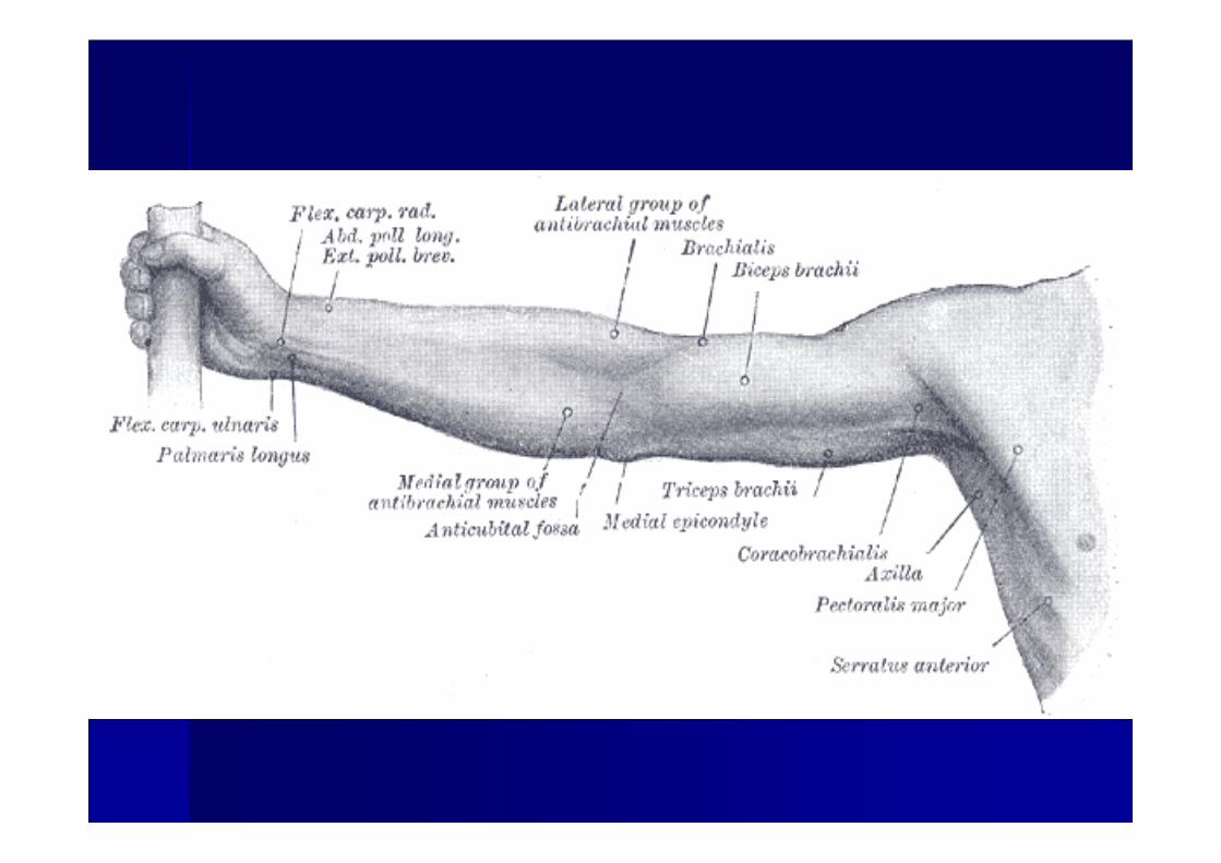

Forearm, Wrist, Hand & Forearm, Wrist, Hand & Fingers Fingers
BonesBonesMusclesMusclesLigamentsLigamentsBlood & Nerve SupplyBlood & Nerve SupplyCommon InjuriesCommon Injuries

ForearmForearm
RadiusRadius–– Thumb side Thumb side
UlnaUlna–– ““pinkypinky”” sideside

Muscles of Muscles of ForearmForearm
Flexors and Flexors and pronatorspronators–– Found Found anterioralyanterioraly
Extensors and Extensors and supinatorssupinators–– Found Found posterioralyposterioraly


Nerve & Nerve & Blood Blood SupplySupply
Most flexors, Most flexors, median nervemedian nerveMost extensors, Most extensors, radial nerveradial nerveBlood: brachial Blood: brachial artery (radial/artery (radial/ulnarulnar))


Wrist, Hand, Wrist, Hand, and Fingersand Fingers
BonesBonesMusclesMusclesLigamentsLigamentsBlood & nerveBlood & nerveCommon injuriesCommon injuries

Wrist, Hand, & Fingers Wrist, Hand, & Fingers
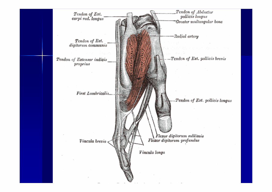
CarpalsCarpals8 carpal bones8 carpal bones–– Proximal row (Proximal row (ulnarulnar to radial): to radial): pisiformpisiform, ,
triquetraltriquetral, , lunatelunate, , scaphoidscaphoid ((navicularnavicular))–– Distal row (Distal row (ulnarulnar to radial): to radial): hamatehamate, ,
capitatecapitate, , trapizoidtrapizoid (lesser (lesser multangularmultangular), ), trapezium (greater trapezium (greater multangularmultangular))
–– Carpals articulate with the metacarpalsCarpals articulate with the metacarpals


Articulations (joints)Articulations (joints)
RadiocarpalRadiocarpal joints (radius and the joints (radius and the carpalscarpals–– CondyloidCondyloid jointjoint–– Allows for flexion, extension, abduction, Allows for flexion, extension, abduction,
circumductioncircumduction
CarpalsCarpals–– ArthrodialArthrodial (gliding) joint(gliding) joint

Articulations (Articulations (concon’’tt))
Metacarpal jointMetacarpal joint–– CondyloidCondyloid jointjoint–– Allows for flexion, extension, add/Allows for flexion, extension, add/abdabd, ,
and and circumductioncircumduction
ThumbThumb–– Saddle jointSaddle joint-- allows rotationallows rotation–– Movements of thumb are different then Movements of thumb are different then
other fingers other fingers

Articulations (Articulations (concon’’tt))
PhalangealPhalangeal jointsjointsHinge jointHinge jointFlexion and extensionFlexion and extension

LigamentsLigaments
WristWrist–– Collateral Collateral ulnarulnar ligament: tip of ligament: tip of styloidstyloid
process of ulna to process of ulna to pisiformpisiform–– Radial collateral: Radial collateral: styloidstyloid of radius to of radius to
scaphoidscaphoid–– Transverse carpal ligament: crosses Transverse carpal ligament: crosses volarvolar
aspect of carpalsaspect of carpalsRoof of the carpal tunnelRoof of the carpal tunnel


LigamentsLigaments
PhalangesPhalangesCollateral ligamentsCollateral ligaments–– MedialMedial–– laterallateral


Muscles, Blood & Nerve Muscles, Blood & Nerve Supply Supply
FlexorsFlexors–– PalmarPalmar side of handside of hand
ExtensorsExtensors–– Back side of handBack side of hand
NervesNerves–– UlnarUlnar–– MedianMedian–– RadialRadial
ArteriesArteries–– Radial and Radial and UlnarUlnar


Common InjuriesCommon Injuries
Wrist sprainWrist sprain–– Etiology: falling on a Etiology: falling on a hyperextendedhyperextended wristwrist–– S/S: pain, swelling, c/o difficulty in S/S: pain, swelling, c/o difficulty in
moving wrist, tenderness, limited ROMmoving wrist, tenderness, limited ROM–– TxTx: RICE, NSAIDS, splinting if necessary, : RICE, NSAIDS, splinting if necessary,
taping taping

Common InjuriesCommon Injuries
Wrist strain (Wrist strain (tenosynovitistenosynovitis) usually the ) usually the extensor extensor carpicarpi radialisradialis longuslongus or or brevisbrevis–– Etiology: repetitive overuse of the wrist Etiology: repetitive overuse of the wrist
tendons and their sheathstendons and their sheaths–– S/S: c/o pain with use or in passive S/S: c/o pain with use or in passive
stretching of tendonsstretching of tendons–– TxTx: Ice massage, NSAIDS, rest, heat after : Ice massage, NSAIDS, rest, heat after
72 hrs, 72 hrs, PREPRE’’ss

Common InjuriesCommon Injuries
TendonitisTendonitisEtiology: overuseEtiology: overuseS/S: c/o pain on active use or passive S/S: c/o pain on active use or passive stretching of tendonstretching of tendonTxTx: Same as : Same as tenosynovitistenosynovitis

Common InjuriesCommon Injuries
Carpal Tunnel Syndrome: Carpal Tunnel Syndrome: inflammtioninflammtionof tendons causing compression of of tendons causing compression of median nervemedian nerve–– Etiology: repeated wrist flexion, direct Etiology: repeated wrist flexion, direct
traumatrauma–– S/S: sensory and motor deficitsS/S: sensory and motor deficits–– TxTx: Rest, immobilization, NSAIDS, ice : Rest, immobilization, NSAIDS, ice
massage, US massage, US


Common InjuriesCommon Injuries
FracturesFractures–– ScaphoidScaphoid--most commonmost common–– BoxerBoxer’’ss-- FxFx of neck of of neck of
55thth MCMC–– BennettBennett’’ss-- FxFx of base of of base of
11stst MCMC–– SmithSmith’’ss-- falling on falling on
hyperextenedhyperextened wrist, wrist, distal radius distal radius fxfx
–– ColleColle’’ss-- falling on falling on outstretched arm, distal outstretched arm, distal radiusradius

The ElbowThe Elbow
BonesBonesMusclesMusclesLigamentsLigamentsBlood & Nerve SupplyBlood & Nerve SupplyCommon InjuriesCommon Injuries


The ElbowThe Elbow
BonesBonesHumerousHumerous, Radius, Ulna, Radius, Ulna–– 2 articulating 2 articulating condylescondyles-- capitulumcapitulum and and
trochleatrochlea–– Flexion/ExtensionFlexion/Extension-- articulation of the articulation of the
trochleatrochlea and and semilunarsemilunar notch (ulna)notch (ulna)–– Pronation/SupinationPronation/Supination--head of radius and head of radius and
capitulumcapitulum
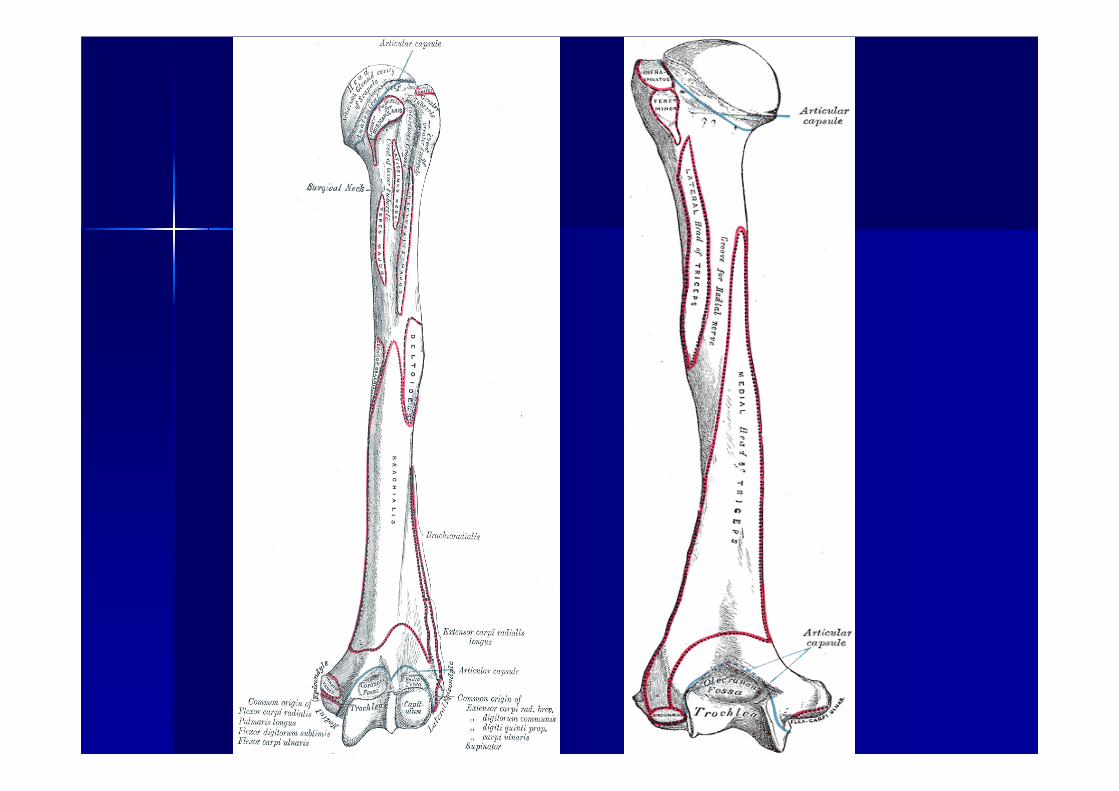


LigamentsLigaments
UlnarUlnar collateral ligament (medial)collateral ligament (medial)Radial collateral ligament (lateral)Radial collateral ligament (lateral)Annular ligamentAnnular ligament–– Stabilizes articulation of radius and radial Stabilizes articulation of radius and radial
notchnotch–– Attached to anterior and posterior radial Attached to anterior and posterior radial
notch and it covers head and neck of notch and it covers head and neck of radiusradius


MusclesMuscles
Flexion: biceps Flexion: biceps brachiibrachii, , brachialisbrachialis, , brachioradialisbrachioradialisExtension: triceps Extension: triceps brachiibrachiiSupinationSupination: biceps : biceps brachiibrachii and and supinatorsupinatorPronationPronation: : pronatorpronator teresteres and and pronatorpronatorquadratusquadratus


MusclesMuscles
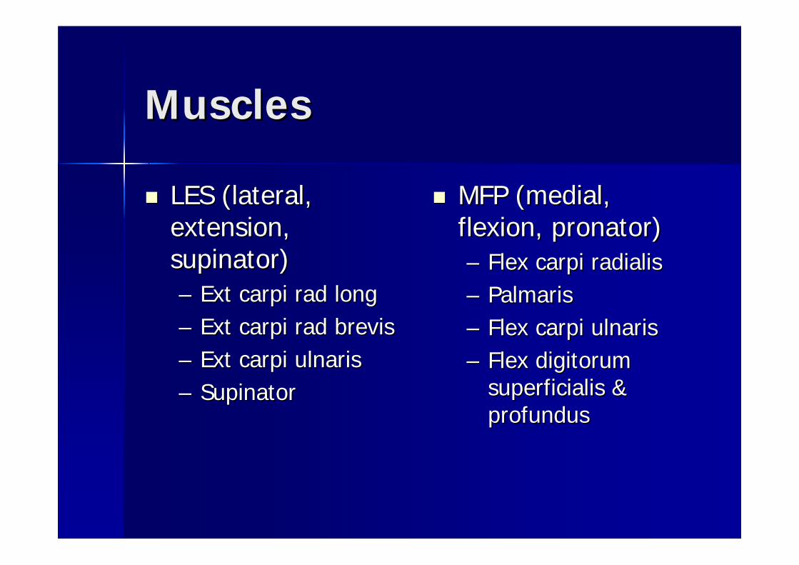
LES (lateral, LES (lateral, extension, extension, supinatorsupinator))–– Ext Ext carpicarpi radrad longlong–– Ext Ext carpicarpi radrad brevisbrevis–– Ext Ext carpicarpi ulnarisulnaris–– SupinatorSupinator
MFP (medial, MFP (medial, flexion, flexion, pronatorpronator))–– Flex Flex carpicarpi radialisradialis–– PalmarisPalmaris–– Flex Flex carpicarpi ulnarisulnaris–– Flex Flex digitorumdigitorum
superficialissuperficialis & & profundusprofundus


Blood & Nerve SupplyBlood & Nerve Supply
BloodBlood–– Brachial and medial arteries: deep within Brachial and medial arteries: deep within
antecubitalantecubital fossafossa–– Veins: front of elbow, Veins: front of elbow, superficalsuperfical and close to and close to
skinskin
NerveNerve–– Roots C5Roots C5--T1T1–– In In cubitalcubital fossafossa, these nerves become the , these nerves become the
musculocutaneousmusculocutaneous, radial and median nerve, radial and median nerve

Common InjuriesCommon Injuries
OlcranonOlcranon bursitis bursitis –– Etiology: blunt trauma to Etiology: blunt trauma to bursalbursal sac sac
superficial to the superficial to the olcranonolcranon processprocess–– S/S: pain and severe spontaneous S/S: pain and severe spontaneous
swelling and point tendernessswelling and point tenderness
Most common injuries due to falling on Most common injuries due to falling on an outstretched arm.an outstretched arm.

Common InjuriesCommon Injuries
Lateral Lateral EpicondylitisEpicondylitis: tennis : tennis elbowelbow–– Etiology: repetitive Etiology: repetitive
trauma to trauma to extersorextersormuscles of the muscles of the lateral lateral epicondyleepicondyle
–– S/S: pain during S/S: pain during and after activity and after activity eventually causing eventually causing weakness in the weakness in the hand and wrist hand and wrist

Common InjuriesCommon Injuries
Medial Medial EpicondylitisEpicondylitis–– Etiology: forceful Etiology: forceful
flexion of wrist and flexion of wrist and extreme extreme valgusvalgustorques of elbowtorques of elbow
–– S/S: pain can be S/S: pain can be centralized or centralized or radiating down arm, radiating down arm, associated associated weakness of wrist weakness of wrist and handand hand

Common InjuriesCommon Injuries
Little League Elbow: 10Little League Elbow: 10--25% of young 25% of young pitchers affectedpitchers affected
–– Etiology: repetitive micro trauma that leads to Etiology: repetitive micro trauma that leads to many disorders of growth in the pitching elbowmany disorders of growth in the pitching elbow
–– S/S: flexion contracture (tightness of anterior S/S: flexion contracture (tightness of anterior joint capsule and weak triceps), locking or joint capsule and weak triceps), locking or catching sensations and decreased ROMcatching sensations and decreased ROM

The Shoulder ComplexThe Shoulder Complex
BonesBonesMusclesMusclesLigamentsLigamentsBlood & Nerve Blood & Nerve SupplySupplyCommon InjuriesCommon Injuries

The Shoulder ComplexThe Shoulder Complex
BonesBones–– ScapulaScapula–– HumerusHumerus–– ClavicleClavicle–– SternumSternum–– RibsRibs

The Shoulder ComplexThe Shoulder Complex
Articulations:Articulations:–– SternoclavicularSternoclavicular (sternum and clavicle) (sternum and clavicle) –– AcromioclavicularAcromioclavicular ((acromionacromion process and process and
clavicle)clavicle)–– GlenohumeralGlenohumeral ((glenoidglenoid fossafossa and humeral and humeral
head)head)–– ScapulothoracicScapulothoracic (scapula and thorax)(scapula and thorax)


LigamentsLigaments
SternoclavciularSternoclavciular-- extremely weak due extremely weak due to boney to boney arrangmentarrangment. Anterior and . Anterior and posterior posterior sternoclavicularsternoclavicular ligament ligament prevents upward translation of the prevents upward translation of the clavicleclavicleAcromioclavicularAcromioclavicular-- Anterior, posterior, Anterior, posterior, inferior, and superior portions join the inferior, and superior portions join the acromionacromion pocesspocess to the clavicleto the clavicle

LigamentsLigaments
GlenohumeralGlenohumeral ligamentsligaments--an anterior an anterior capsule surrounds the joint and is capsule surrounds the joint and is strongly enforced by the superior, strongly enforced by the superior, middle, and inferior GH ligaments.middle, and inferior GH ligaments.


MusclesMuscles
3 Groups3 GroupsGroup 1: Group 1: latissumuslatissumusdorsidorsi and and pecpec majormajorGroup 2: originates Group 2: originates off scapula and off scapula and inserts into the inserts into the humerushumerus; SITTS; SITTS--subscapularissubscapularis, , infraspinatusinfraspinatus, , supraspinatussupraspinatus, and , and teresteres minorminor


MusclesMuscles
Group 3Group 3-- attach axial skeleton attach axial skeleton to the scapulato the scapula……scapular scapular stabilizers: stabilizers: levatorlevator scapula, scapula, trapeziustrapezius, rhomboids, and , rhomboids, and serratusserratus anterior and posterioranterior and posterior
Scapular Rhythm: Scapular Rhythm: mvmtmvmt of the of the scapula relative to the scapula relative to the mvmtmvmt of of the the humerushumerus. First 30 degrees . First 30 degrees is is humerushumerus only, from 30only, from 30--90 90 the scapula moves 1 degree to the scapula moves 1 degree to every 2 degrees of the every 2 degrees of the humerushumerus. At 90 degrees they . At 90 degrees they are =.are =.

Blood & Nerve SupplyBlood & Nerve Supply
Brachial arteryBrachial arteryBrachial PlexusBrachial Plexus


Common InjuriesCommon Injuries
ClavicularClavicular FxFx–– Etiology: fall on outstretched arm, tip of Etiology: fall on outstretched arm, tip of
shoulder or direct impactshoulder or direct impact–– S/S: supports arm of injured side, tilt S/S: supports arm of injured side, tilt
neck towards injured side, deformity, neck towards injured side, deformity, swellingswelling
–– TxTx: immediate referral, X: immediate referral, X--ray, sling and ray, sling and immoblizeimmoblize 66--8 weeks 8 weeks

Common InjuriesCommon Injuries
SternoclavicularSternoclavicular sprainsprain–– Etiology: direct blow Etiology: direct blow
transmitting through transmitting through humerushumerus
–– S/S: grades IS/S: grades I-- little to no little to no pain w/o disability, IIpain w/o disability, II--visual deformity, pain, visual deformity, pain, swelling, point tenderness, swelling, point tenderness, inability to inability to abdabd shoulder or shoulder or bring shoulder across bring shoulder across body, IIIbody, III-- complete complete dislocation with gross dislocation with gross displacement of clavicle at displacement of clavicle at the SC jointthe SC joint
–– TxTx: RICE, refer to MD : RICE, refer to MD

Common InjuriesCommon Injuries
AcromioclavicularAcromioclavicular SprainSprain–– Etiology: direct impact to Etiology: direct impact to
tip of shoulder, upward tip of shoulder, upward force through the long axis force through the long axis of of humerushumerus
–– S/S: grade IS/S: grade I-- PT and PT and discomfort during discomfort during mvmtmvmt, , IIII--displacement of lateral displacement of lateral end of clavicle, and end of clavicle, and inability to completely inability to completely abdabdarm or bring arm across arm or bring arm across chest, IIIchest, III--rupture of AC rupture of AC ligament and CC ligamentligament and CC ligament
–– TxTx: ice and pressure, : ice and pressure, stabilize stabilize jtjt w/ sling, referw/ sling, refer

Common InjuriesCommon Injuries
GH GH SubluxationSubluxation/Dislocation/Dislocation–– Etiology: (Anterior) most common Etiology: (Anterior) most common
is is abdabd, ER, and ext, but also from a , ER, and ext, but also from a direct blow posterior or direct blow posterior or posterolateralposterolateral aspect of shoulder. aspect of shoulder. (Posterior) forced add and IR or fall (Posterior) forced add and IR or fall on extended and IR shoulderon extended and IR shoulder
–– S/S: (Anterior) flattened deltoid, S/S: (Anterior) flattened deltoid, palpation of humeral head at palpation of humeral head at axillaaxilla, , carrying arm in carrying arm in abdabd and ER and ER position. (Posterior) severe pain position. (Posterior) severe pain and disability, arm in add and IR and disability, arm in add and IR with flattened anterior deltoid.with flattened anterior deltoid.
–– TxTx: Immediate immobilization, ice : Immediate immobilization, ice to reduce swelling and to reduce swelling and hemorrhaging, referral to MD, Xhemorrhaging, referral to MD, X--ray to ray to r/or/o associated associated fxfx
Common InjuriesCommon Injuries
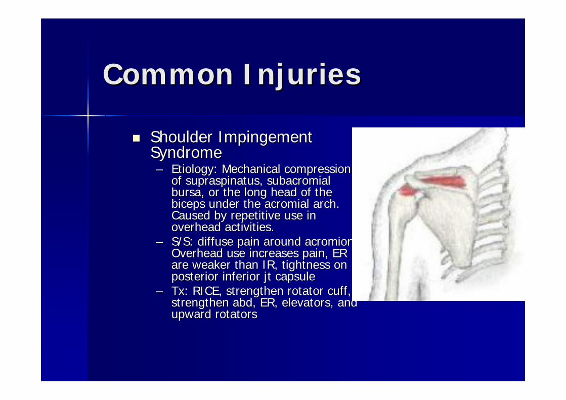
Shoulder Impingement Shoulder Impingement SyndromeSyndrome–– Etiology: Mechanical compression Etiology: Mechanical compression
of of supraspinatussupraspinatus, , subacromialsubacromialbursa, or the long head of the bursa, or the long head of the biceps under the biceps under the acromialacromial arch. arch. Caused by repetitive use in Caused by repetitive use in overhead activities.overhead activities.
–– S/S: diffuse pain around S/S: diffuse pain around acromionacromion. . Overhead use increases pain, ER Overhead use increases pain, ER are weaker than IR, tightness on are weaker than IR, tightness on posterior inferior posterior inferior jtjt capsulecapsule
–– TxTx: RICE, strengthen rotator cuff, : RICE, strengthen rotator cuff, strengthen strengthen abdabd, ER, elevators, and , ER, elevators, and upward rotatorsupward rotators

Throwing DynamicsThrowing Dynamics
Cocking Phase: Cocking Phase: shldshld abdabd, ext, and ER. Can , ext, and ER. Can cause ant cause ant shldshld pain as a result of stretching pain as a result of stretching the the pecpec major, origin of ant deltoid, long major, origin of ant deltoid, long head of biceps, and IRhead of biceps, and IRAcceleration Phase: Acceleration Phase: AbdAbd, IR, and forward , IR, and forward flex. Places tension on post and inferior flex. Places tension on post and inferior capsule, while ant capsule is relaxed.capsule, while ant capsule is relaxed.FollowFollow--Through Phase: Add and IR, the Through Phase: Add and IR, the eccentric load places enormous strain on eccentric load places enormous strain on the ER of the rotator cuff and capsulethe ER of the rotator cuff and capsule

Head, Face, Eyes, Ears, Head, Face, Eyes, Ears, Nose and ThroatNose and Throat
HeadHead–– The skull is composed of 22 bones, the mandible The skull is composed of 22 bones, the mandible
is the only bone not joined by suturesis the only bone not joined by suturesMeningesMeninges–– 3 membranes that protect the brain and SC3 membranes that protect the brain and SC
DuramaterDuramater--outermost dense fibrous inelastic sheath outermost dense fibrous inelastic sheath that covers the brainthat covers the brainArachnoidArachnoid-- extremely delicate, attaches to the SC by extremely delicate, attaches to the SC by silksilk--like strandslike strandsSpace b/w Space b/w arachnoidarachnoid and and piapia mater is called mater is called subarachnoidsubarachnoid space. This space contains cerebrospinal space. This space contains cerebrospinal fluidfluidCerebrospinal fluids main Cerebrospinal fluids main fxnfxn is to act as a is to act as a cusioncusion and and absorb shockabsorb shockPiaPia Mater which is thin, delicate, and highly vascularMater which is thin, delicate, and highly vascular
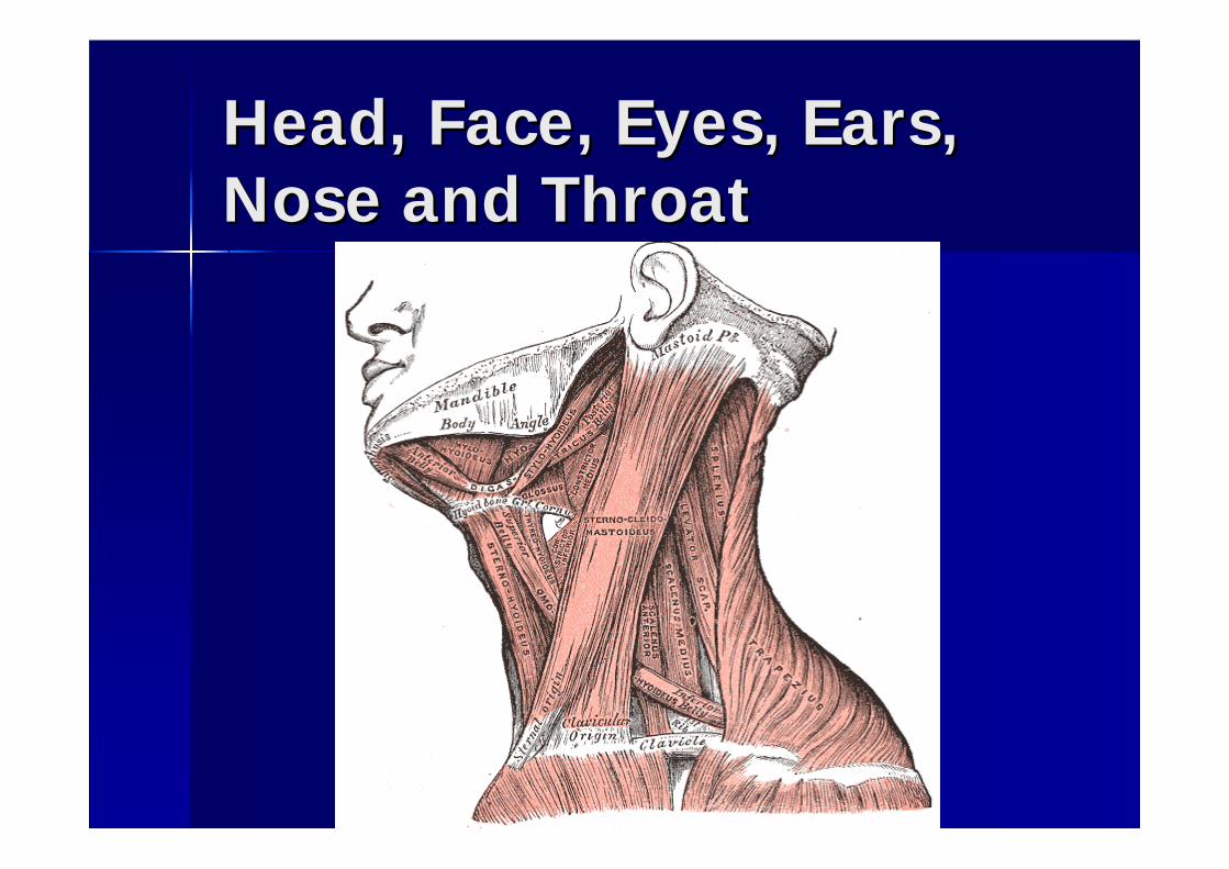
Head, Face, Eyes, Ears, Head, Face, Eyes, Ears, Nose and ThroatNose and Throat

Head, Face, Eyes, Ears, Head, Face, Eyes, Ears, Nose and ThroatNose and Throat

Common InjuriesCommon Injuries
Skull Skull FxFx–– Etiology: blunt traumaEtiology: blunt trauma–– S/S: c/o severe headaches and nausea, possible S/S: c/o severe headaches and nausea, possible
deformity, blood in middle ear or ear canal, deformity, blood in middle ear or ear canal, bleeding from nose, bleeding from nose, ecchymosisecchymosis around eyes around eyes (raccoon eyes), (raccoon eyes), ecchymosisecchymosis behind ears (battle behind ears (battle signs), cerebral spinal fluid may also appear in signs), cerebral spinal fluid may also appear in ear and noseear and nose
–– TxTx: immediate hospitalization: immediate hospitalization

Common InjuriesCommon Injuries
Cerebral Concussions (mild)Cerebral Concussions (mild)–– Etiology: either a direct blow to the head by an object or Etiology: either a direct blow to the head by an object or
moving head meets a fixed objectmoving head meets a fixed object–– S/S: headache, tinnitus, nausea, irritability, confusion, S/S: headache, tinnitus, nausea, irritability, confusion,
dizziness, LOC, dizziness, LOC, anterogradeanterograde/retrograde amnesia, inability /retrograde amnesia, inability to concentrate, blurred vision, photophobia, and sleep to concentrate, blurred vision, photophobia, and sleep disturbancesdisturbances
–– TxTx: If there is any LOC the athlete MUST be removed from : If there is any LOC the athlete MUST be removed from competition. If ATC finds an unconscious athlete, they competition. If ATC finds an unconscious athlete, they must assume a neck/SC injury.must assume a neck/SC injury.
Athlete shouldnAthlete shouldn’’t be permitted to return to play t be permitted to return to play until ALL until ALL postconcussionpostconcussion s/ss/s are resolved are resolved

Common InjuriesCommon Injuries
PostPost--Concussion SyndromeConcussion Syndrome–– Etiology: Etiology: s/ss/s of a concussion that lingerof a concussion that linger–– S/S: c/o persistent headache, impaired memory, S/S: c/o persistent headache, impaired memory,
lack of concentration, anxiety, irritability, lack of concentration, anxiety, irritability, giddiness, fatigue, depression, and visual giddiness, fatigue, depression, and visual disturbances. Symptoms may last for weeks or disturbances. Symptoms may last for weeks or even months before resolving.even months before resolving.
–– TxTx: not to allow athlete to return until all : not to allow athlete to return until all s/ss/s are are no longer present no longer present

Common InjuriesCommon Injuries
Second Impact Syndrome (ages >20)Second Impact Syndrome (ages >20)–– Etiology: occurs Etiology: occurs b/cb/c of rapid swelling and of rapid swelling and
herniationherniation of brain after a 2of brain after a 2ndnd head injury, before head injury, before the 1the 1stst injury had time to healinjury had time to heal
–– S/S: within 15 sec to several min the condition S/S: within 15 sec to several min the condition worsens rapidly. Pupils dilate, loss of eye worsens rapidly. Pupils dilate, loss of eye mvmtmvmt, , LOC leading to coma, and respiratory failureLOC leading to coma, and respiratory failure
22ndnd impact syndrome is a life threatening impact syndrome is a life threatening condition with a mortality rate of 50%condition with a mortality rate of 50%–– TxTx: must be addressed within 5 min by dramatic : must be addressed within 5 min by dramatic
life saving measures life saving measures

Common InjuriesCommon Injuries
Epidural Epidural HematomaHematoma–– Etiology: a blow to the head or skull Etiology: a blow to the head or skull fxfx
can cause tearing of the can cause tearing of the meningealmeningealartery. Blood accumulation and a artery. Blood accumulation and a hematomahematoma occur occur extremlyextremly fastfast
–– S/S: most cases LOC, signs gradually S/S: most cases LOC, signs gradually worsen, worsen, dialationdialation of one pupilof one pupil
–– TxTx: medical emergency: medical emergency

Common InjuriesCommon Injuries
SubduralSubdural HematomaHematoma–– Etiology: results from acceleration/deceleration Etiology: results from acceleration/deceleration
forces that tear vessels that lie b/w the forces that tear vessels that lie b/w the duramaterduramater and brain. Symptoms appear slowly and brain. Symptoms appear slowly b/cb/c the venous structures are affectedthe venous structures are affected
2 types:2 types:Simple Simple subduralsubdural-- collection of blood w/o injury to cerebrumcollection of blood w/o injury to cerebrumComplicated Complicated subduralsubdural-- both blood pooling and injury to the both blood pooling and injury to the cortex increase cortex increase intercerebralintercerebral pressure (50% mortality rate)pressure (50% mortality rate)
–– S/S: simple no LOC, complicated almost always S/S: simple no LOC, complicated almost always LOC and dilation of one pupil, associated LOC and dilation of one pupil, associated headache, dizziness, nausea, or sleepinessheadache, dizziness, nausea, or sleepiness
–– TxTx: immediate medical attention: immediate medical attention

Common InjuriesCommon Injuries
ZygomaticZygomatic Complex Complex FxFx (cheekbone)(cheekbone)–– Etiology: direct blow, 3Etiology: direct blow, 3rdrd most commonmost common–– S/S: deformity, usually nosebleed S/S: deformity, usually nosebleed
((epistaxisepistaxis), seeing double (), seeing double (diplopiadiplopia), ), cheek numbnesscheek numbness

Common InjuriesCommon Injuries
Mandible Mandible FxFx–– Etiology: 2Etiology: 2ndnd most common, direct blowmost common, direct blow–– S/S: possible deformity, loss of normal S/S: possible deformity, loss of normal
occlusion, pain occlusion, pain bittingbitting downdown
DislocationDislocation–– Etiology: direct blow with mouth openEtiology: direct blow with mouth open–– S/S: locked open mouth, jaw S/S: locked open mouth, jaw mvmtmvmt
impossibleimpossible

Common InjuriesCommon Injuries
Nasal Nasal FxFx–– Etiology: most common facial Etiology: most common facial fxfx, frontal , frontal
or lateral blow to noseor lateral blow to nose–– S/S: profuse hemorrhaging, immediate S/S: profuse hemorrhaging, immediate
swelling, deformity, and swelling, deformity, and crepituscrepitus upon upon palpationpalpation
–– TxTx: refer to MD : refer to MD

Common InjuriesCommon Injuries
Orbital Orbital FxFx–– Etiology: direct blow to the eyeball Etiology: direct blow to the eyeball
forcing it forcing it posteriorlyposteriorly–– S/S: S/S: diplopiadiplopia, restricted , restricted mvmtmvmt of eye, of eye,
downward displacement of eye, pain downward displacement of eye, pain swelling, and hemorrhageswelling, and hemorrhage
–– TxTx: refer to MD: refer to MD

Common InjuriesCommon Injuries
Corneal AbrasionCorneal Abrasion–– Etiology: athlete with object in eye will Etiology: athlete with object in eye will
rub eye, in doing so, they will create an rub eye, in doing so, they will create an abrasionabrasion
–– S/S: severe pain, watering of eye, S/S: severe pain, watering of eye, photophobia, spasm of eye lidphotophobia, spasm of eye lid
–– TxTx: sterile rinse, cover, and refer: sterile rinse, cover, and refer