hsp90-cdc37 chaperone complex regulates ulk1- … chaperone complex regulates ulk1- and...
TRANSCRIPT

Molecular Cell
Article
Hsp90-Cdc37 Chaperone Complex RegulatesUlk1- and Atg13-Mediated MitophagyJoung Hyuck Joo,1,7 Frank C. Dorsey,3,7,8 Aashish Joshi,1,7 Kristin M. Hennessy-Walters,1,7 Kristie L. Rose,3
Kelly McCastlain,1 Ji Zhang,2 Rekha Iyengar,1 Chang Hwa Jung,4 Der-Fen Suen,5 Meredith A. Steeves,3
Chia-Ying Yang,6,9 Stephanie M. Prater,3 Do-Hyung Kim,4 Craig B. Thompson,6,10 Richard J. Youle,5 Paul A. Ney,2
John L. Cleveland,3 and Mondira Kundu1,*1Department of Pathology2Department of BiochemistrySt. Jude Children’s Research Hospital, Memphis, TN 38105, USA3Department of Cancer Biology, The Scripps Research Institute Florida, Scripps Florida, Jupiter, FL 33458, USA4Department of Biochemistry, Molecular Biology, and Biophysics, University of Minnesota, Minneapolis, MN 55455, USA5National Institute of Neurological Disorders and Stroke, National Institutes of Health, Bethesda, MD 20892, USA6Abramson Family Cancer Research Institute, Department of Cancer Biology, University of Pennsylvania School of Medicine, Philadelphia,PA 19104, USA7These authors contributed equally to this work8Present address: Lilly Research Laboratories, Indianapolis, IN 46037, USA9Present address: Molecular and Cellular Biology Program, Stony Brook University, Stony Brook, New York, NY 11790, USA10Present address: Cancer Biology and Genetics, Memorial Sloan-Kettering Cancer Center, New York, NY 10065, USA*Correspondence: [email protected] 10.1016/j.molcel.2011.06.018
SUMMARY
Autophagy, the primary recycling pathway of cells,plays a critical role in mitochondrial quality controlunder normal growth conditions and in the responseto cellular stress. The Hsp90-Cdc37 chaperonecomplex coordinately regulates the activity of selectkinases to orchestrate many facets of the stressresponse. Although both maintain mitochondrialintegrity, the relationship between Hsp90-Cdc37and autophagy has not been well characterized.Ulk1, one of the mammalian homologs of yeastAtg1, is a serine-threonine kinase required for mi-tophagy. Here we show that the interaction betweenUlk1 and Hsp90-Cdc37 stabilizes and activates Ulk1,which in turn is required for the phosphorylation andrelease of Atg13 from Ulk1, and for the recruitment ofAtg13 to damaged mitochondria. Hsp90-Cdc37,Ulk1, and Atg13 phosphorylation are all required forefficient mitochondrial clearance. These findingsestablish a direct pathway that integrates Ulk1- andAtg13-directed mitophagy with the stress responsecoordinated by Hsp90 and Cdc37.
INTRODUCTION
Hsp90 is an abundant chaperone that directs thematuration andactivation of a restricted group of metastable proteins, typicallykinases and signaling molecules, to orchestrate responses tocellular stress (Li et al., 2009). Most Hsp90 clients adopt theirfinal configuration only once they are posttranslationally acti-vated (e.g., by ligand binding and/or phosphorylation) in
a manner that is facilitated by their interaction with Hsp90. Thehalf-life and thus the activity of most Hsp90 clients relies on theirassociationwith Hsp90 and its cochaperones, as they are rapidlydegraded by the proteasome following release from the chap-erone complex. The expression and activity of heat shockproteins is dramatically induced in response to heat shock andother proteotoxic stressors. This response, coupled with post-translational modifications of client proteins in complex withHsp90, maintains cellular homeostasis by coordinately regu-lating changes in signal transduction pathways and transcrip-tional responses that promote cell survival and proliferation.Maintenance of healthy mitochondria is essential for cellular
homeostasis, as this organelle produces ATP and other essentialmetabolites as well as the building blocks for protein, nucleicacid, and lipid biosynthesis. In addition, mitochondria harborpools of intracellular calcium and are the principal target andrelay center for cell death cascades (de Moura et al., 2010).Hsp90 appears to be involved in mitochondrial homeostasis,specifically by regulating ubiquitin proteasome-mediated turn-over of mitochondrial proteins (Margineantu et al., 2007) andthe maintenance of mitochondrial membrane potential (Kanget al., 2007).Autophagy also has important roles in controlling mitochon-
drial homeostasis (Bhatia-Kissova and Camougrand, 2010). Au-tophagy functions as the primary recycling pathway of the cell,where it directs lysosome-mediated destruction of its cellularcargo, including damaged or dysfunctional mitochondria (Kunduand Thompson, 2008). Flux through the autophagy pathwaymarkedly increases when cells are faced with metabolic or pro-teotoxic stress that ensues following exposure to noxious envi-ronmental cues, for example starvation, hypoxia, or heat (Amar-avadi and Thompson, 2007; Liu et al., 2010). Indeed, increasedturnover of mitochondria is manifest under all of these conditions(Gamboa and Andrade, 2010; Kim et al., 2007; Oberley et al.,2008; Zhang et al., 2008), and dysregulation of this process is
572 Molecular Cell 43, 572–585, August 19, 2011 ª2011 Elsevier Inc.
linked to disease, including diabetes, neurodegeneration, andcancer (de Moura et al., 2010; Gottlieb and Carreira, 2010).Despite the importance of Hsp90 and autophagy in maintainingmitochondrial integrity and cellular homeostasis, the interplay ofthe Hsp90 chaperone complex and autophagy in mitochondrialclearance has not been explored.In yeast, the serine-threonine kinase Atg1 directs the autoph-
agy machinery to appropriate cargo in response to changes inthe availability of carbon and nitrogen (Mizushima, 2010). Ulk1,one of the mammalian homologs of Atg1, is required for starva-tion-induced autophagy (Chan et al., 2007) and for clearance ofmitochondria in terminally differentiating erythroid cells (Kunduet al., 2008). Here, we report that Ulk1 function requires its phys-ical interaction with Hsp90 and the kinase-specific cochaperoneCdc37. This interaction promotes Ulk1 stability and activation,and is necessary for Ulk1-directed phosphorylation of its inter-acting partner Atg13 at serine 318. Further, Atg13 phosphoryla-tion promotes its release from Ulk1 and its localization todamaged mitochondria. Accordingly, Hsp90, Cdc37, Ulk1kinase activity, and Atg13 phosphorylation are all required forefficient mitochondrial clearance. These findings define anUlk1- and Atg13-dependent pathway that integrates autophagyinto the Hsp90-coordinated stress response to govern mito-chondrial homeostasis.
RESULTS
Ulk1 Interacts with the Hsp90-Cdc37 ChaperoneComplexUlk1 plays a critical role in the autophagy-mediated clearance ofmitochondria during erythroidmaturation (Kundu et al., 2008). Togain insight into Ulk1 regulation, we used an unbiased proteo-mics approach to identify Ulk1-interacting proteins. Hsp90and Cdc37 were identified as Ulk1-interacting partners byLC/MS and immunoblot analyses following affinity purificationof Flag-tagged Ulk1 from K562 cells (Figure 1A) and NTAP-tagged Ulk1 from 293T cells (data not shown). Cdc37 was alsolinked to Ulk1 in a large-scale proteomics-based screen ofautophagy networks (Behrends et al., 2010). The observedinteractions between Ulk1 and Hsp90-Cdc37 were not anartifact of Ulk1 overexpression, as endogenous anti-Ulk1 immu-noprecipitates from wild-type mouse embryonic fibroblasts(MEFs) contained endogenous Hsp90 and Cdc37 (Figure 1B).Similarly, endogenous Ulk1 was detected in endogenous anti-Cdc37 immunoprecipitates fromwild-typeMEFs (see Figure S1Aavailable online).Activation of specific kinase clients by the Hsp90-Cdc37
chaperone complex involves the assembly of a salt-stableheterocomplex of Hsp90, Cdc37, and the kinase client (Hartsonet al., 2000). The formation of these metastable complexes isblocked by Hsp90 antagonists such as 17-allylamino-17-deme-thoxygeldanamycin (17AAG), a synthetic derivative of geldana-mycin that binds to the N-terminal ATP binding pocket ofHsp90, inhibiting ATP binding and hydrolysis, which are requiredfor chaperone function (Hartson et al., 2000; Pearl and Prodro-mou, 2006). Consistent with the notion that Ulk1 is a client ofthe Hsp90-Cdc37 chaperone complex, treatment with 2.5 mM17AAG for 1 hr disrupted the interaction between Ulk1, Hsp90,
and Cdc37 (Figure 1B, left panels, and Figure S1B). Similarly,silencing cdc37 expression by siRNA disrupted the interactionbetween endogenous Ulk1 and Hsp90 (Figure 1B, right panels).
The Hsp90-Cdc37 Chaperone Complex Regulates Ulk1Kinase ActivityAgents that disrupt the interaction of Hsp90 with its kinaseclients inactivate kinase activity and/or lead to their destructionby the ubiquitin-proteasome pathway (Caplan et al., 2007). Todetermine if the Hsp90-Cdc37 complex regulates Ulk1 kinaseactivity, anti-Flag immunoprecipitates isolated from Flag-Ulk1-expressing K562 cells treated with increasing doses of 17AAGfor 1 hr were incubated with 32P-gATP and the general kinasesubstrate myelin basic protein (MBP). 17AAG treatment inhibitedboth Ulk1 autophosphorylation and phosphorylation of MBPin vitro (Figure 1C). Pulse-chase studies demonstrated thatnewly synthesized Ulk1 migrates faster in an SDS-PAGE geland rapidly shifts up, suggesting that Ulk1 is phosphorylatedshortly after synthesis (Figure 1F, top panel). To test if Hsp90plays a role in these early phosphorylation events, Ulk1 waspurified from transfected 293T cells that were pretreated with17AAG for 5 hr and then pulse labeled with 35S-methionine and35S-cysteine for 15 min. Newly synthesized Ulk1 purified from17AAG-treated cells migrated faster than Ulk1 from vehicle-treated cells, suggesting that disrupting Hsp90 interactionswith newly synthesized Ulk1 inhibits Ulk1 phosphorylation (Fig-ure 1D). Ulk1 isolated from cells under normal growth conditionsis hyperphosphorylated, at least in part due to autophosphoryla-tion (Chan et al., 2009; Dorsey et al., 2009a), and treatment withlambda phosphatase in vitro leads to electrophoretic migrationsimilar to kinase-dead Ulk1 mutants (Dorsey et al., 2009a).Treatment of Ulk1-expressing 293T cells with 17AAG for 5 hrled to the appearance of a faster-migrating form of Ulk1 thatcomigrated with a kinase-dead hypophosphorylated Ulk1mutant (Ulk1-K46A) (Figure S1C), suggesting that Hsp90 inhibi-tion impaired Ulk1 phosphorylation. Finally, silencing cdc37also led to increased Ulk1 electrophoretic mobility, consistentwith a requirement for Cdc37 for Ulk1 activation (Figure 1E).Stable isotope labeling with amino acids in cell culture (SILAC)
followed by high-resolution tandem mass spectrometryconfirmed that the change in Ulk1 phosphorylation followingdisruption of Hsp90 function was due to a decrease in Ulk1kinase activity. To quantify Ulk1 phosphorylation followingHsp90 inhibition, we employed SILAC, where one populationof cells is grown in normal (light) media and another is grown inthe presence of media containing 13C6-labeled lysine and 13C615N4-labeled arginine (heavy) (Amanchy et al., 2005). Ulk1 kinaseactivity is required for phosphorylation of S1047 in the Ulk1 Cterminus (Dorsey et al., 2009a). Indeed, S1047 phosphorylationwas 147-fold more abundant in wild-type Ulk1 versus inkinase-dead Ulk1-K46A (after normalizing to unmodified Ulk1peptides, Figure S1D, top panels). Treatment with 17AAG (5 hr)resulted in a 3-fold decrease (after normalization to unmodifiedpeptides) in S1047 phosphorylation (Figure S1D, bottom panels),consistent with the observation that Hsp90 inhibition impairsphosphorylation of newly synthesized Ulk1 (Figure 1D). Collec-tively, these findings indicate that the interaction of Ulk1 withthe Hsp90-Cdc37 complex is an early event that stabilizes the
Molecular Cell
Hsp90-Cdc37 Regulates Ulk1 and Atg13 Function
Molecular Cell 43, 572–585, August 19, 2011 ª2011 Elsevier Inc. 573

mature phosphorylated conformation of Ulk1, which includesautophosphorylation of S1047.
The Hsp90-Cdc37 Chaperone Complex RegulatesUlk1 StabilityPulse-chase analyses have demonstrated that, under normalgrowth conditions, Ulk1 is a long-lived protein with a half-lifeclose to 24 hr (Dorsey et al., 2009a). We therefore assessedthe effects of disrupting the Hsp90-Cdc37-Ulk1 interaction onUlk1 turnover. Notably, 17AAG treatment triggered rapidturnover of Ulk1 in NTAP-Ulk1-expressing 293T cells comparedto vehicle-treated cells (Figure 1F). The steady-state levels of
endogenous Ulk1 protein were also significantly reduced inMEFs treated with 2.5 mM 17AAG for 5 or 21 hr, without effectson ulk1 mRNA levels (Figure S1E). Similarly, silencing cdc37expression triggered reductions in the steady-state levels ofUlk1 protein, without affecting ulk1 mRNA levels (Figure 1E).The reduction in the steady-state levels of Ulk1 was proportionalto the degree of Cdc37 knockdown, andwasmost apparent withcombined shRNA/siRNA-mediated knockdown of Cdc37 (Fig-ure S1F). In addition, cdc37 knockdown markedly increasedthe sensitivity of Ulk1 to the effects of 17AAG (data not shown),as shown for other bona fide kinase clients of Hsp90 (Smith et al.,2009). Finally, the destabilizing effects of 17AAG on endogenous
A
98624938
28
Flag-G
FP
GFPFlag
-K46
R
Flag-U
lk1
Ulk1HSP90
CDC37
Ulk1
HSP90
CDC37
Flag
IP
MEF:
Ulk1
Hsp90Lysa
tes:BAF++ _____:MG132++ ___ __:17AAG + + + ____
B
NTAP-Ulk1 (+Control)NTAP
NTA
P IP
NTAP-Ulk1 (+17AAG)NTAP
0 2 4 8 11 240 :Time (hrs)
Ulk1 S35
Ulk1 S35
NTA
P IP
Ulk1
Cdc37
Actin
NT cdc37 siRNA:
Lysa
tes
ulk1 MEF
C
F
ulk1 cdc37
1
-1-2-3-4
0
log
(!!
CT)
2
D
E
P Ulk132
P MBP32
Ulk1
Kina
se A
ssay
IP
:17AAG (µM) 0 0.5 2.5
K562+Flag-Ulk1
G
Ulk1 S
NTAPNTAP-Ulk1
35
pre-treatment(5 hrs)
NTA
P IP
:17AAG + +__
293T:
293T:
293T:
Lysa
tes
Ulk
1 IP
NTcd
c37 :siRNA--1 5 180
17AAG (hrs)
ulk1 +/+ -/-MEF:
Ulk1
HSP90
CDC37
HSP90
CDC37
Actin
Ulk1
+/+
+/+
ulk1+/+ ulk1-/-
Figure 1. Ulk1 Kinase Activity and Stability Are Regulated by the Hsp90-Cdc37 Chaperone Complex(A) Hsp90 and Cdc37 were identified as Ulk1-interacting proteins using an unbiased proteomics approach. K562 cells were transfected with the indicated
expression constructs. Ulk1-K46R is aULK1mutant withmildly impaired kinase activity (Dorsey et al., 2009a). The anti-Flag immunoprecipitates were resolved by
SDS-PAGE, and the three bands excised from the silver-stained gel (top panel, marked by arrows) were identified as Ulk1, HSP90b, and CDC37 by liquid
chromatography-mass spectrometry (LC-MS) and Mascot prediction analysis. (Bottom panel) Protein identities were confirmed by immunoblot analysis.
(B) Wild-type MEFs were treated with 17AAG or were transiently transfected with pooled control (nontargeting, NT) or cdc37 siRNA. Endogenous Ulk1 and
interacting proteins were immunoprecipitated using an anti-Ulk1 antibody. Ulk1, Hsp90, and Cdc37 were detected by immunoblot analysis.
(C) Flag-Ulk1 was immunoprecipitated from K562 cells treated with the indicated amount of 17AAG for 1 hr. Ulk1 protein levels were assessed by immunoblot
analysis. Ulk1 kinase activity was determined by in vitro kinase assays. Reactions were analyzed by SDS-PAGE, and 32P-labeled Ulk1 andMBPwere detected by
autoradiography.
(D) 293T cells were transfected with the indicated NTAP vector and treated with either vehicle (!) or 2.5 mM17AAG (+) for 5 hr prior to pulse labeling the cells with35S-Translabel for 15 min. 35S-labeled NTAP-Ulk1 was affinity purified and assessed by autoradiography.
(E) Wild-type (ulk1+/+) MEFs were transfected twice with pooled control (nontargeting) or cdc37 siRNA. (Top panel) Expression of Ulk1, Cdc37, and actin
was assessed by immunoblot analysis 48 hr after the second transfection. (Bottom panel) Expression of mRNA was analyzed by RT-qPCR (TaqMan) analyses
using primer/probe combinations specific for ulk1, cdc37, and 18S RNA. Relative expression (log2) was calculated using the Pffafl comparative Ct method.
Expression of ulk1 and cdc37 in cdc37 siRNA-treated samples was normalized to 18S and calibrated to control samples (i.e., those transfected with nontargeting
siRNA).
(F) Pulse-chase analyses were performed on 293T cells expressing the indicated NTAP vector and treated with either vehicle (control) or 2.5 mM 17AAG.
(G) Wild-type MEFs were pretreated with the H+ATPase inhibitor bafilomycin-A1 (10 nM) or with the proteasome inhibitor MG132 (10 nM) for 1 hr prior to the
addition of 2.5 mM17AAG for 5 hr, as indicated. The levels of Ulk1 andHsp90were determined by immunoblotting. Lysates from ulk1!/!MEFs served as a control.
Molecular Cell
Hsp90-Cdc37 Regulates Ulk1 and Atg13 Function
574 Molecular Cell 43, 572–585, August 19, 2011 ª2011 Elsevier Inc.

Ulk1 were abolished by cotreatment with the proteasomeinhibitor MG132 (Figure 1G), but not the H+ATPase inhibitor, ba-filomycin-A1, which inhibits lysosome-mediated degradation ofautophagic cargo (Yoshimori et al., 1991). Therefore, the interac-tion between Ulk1 and the Hsp90-Cdc37 chaperone complexstabilizes Ulk1, and disrupting this complex triggers Ulk1 degra-dation by the proteasome.The Hsp90-Cdc37 complex regulates the stability and activity
of numerous kinases by direct binding to the catalytic kinasedomain (Caplan et al., 2007). Using Ulk1 deletion constructs,we demonstrated that the N-terminal kinase domain of Ulk1(residues 1–279) was necessary and sufficient for mediatingthe interaction of Ulk1 with Hsp90 and Cdc37 (Figure S2A) andfor regulating its sensitivity to degradation following 17AAGtreatment (Figures S2C and S2D). Notably, although Ulk1 andthe related mammalian Atg1 homolog Ulk2 share significanthomology within their kinase domains, we did not detectCdc37 or Hsp90 in Ulk2 immunoprecipitates (Figure S2B), norwere there decreases in the steady-state levels of Ulk2 followingtreatment with 17AAG (Figure S2C). Furthermore, the stability ofyeast Atg1 was not altered in temperature-sensitive mutants thatdisable Cdc37 andHsp82 (yeast Hsp90) (U. Nair andD. Klionsky,personal communication). Together, these data provideevidence of the specificity of the Ulk1-Hsp90-Cdc37 interactionand suggest an evolutionary divergence in Hsp90 regulation ofUlk1 and other Atg1 homologs.
Hsp90 Regulates Starvation-Induced AutophagyUlk1 has been implicated in starvation-induced autophagy(Chan et al., 2007). Thus, we hypothesized that Hsp90 controlledUlk1-directed, starvation-induced autophagy. Ulk1-deficientMEFs have impaired flux through the autophagy pathwayfollowing amino acid starvation, although given the high rate ofturnover of lipidated LC3 (LC3-II) in these cells it is necessaryto inhibit lysosomal degradation of LC3 using the H+-ATPaseinhibitor Bafilomycin A to appreciate the defect (Jung et al.,2009). To quantify autophagic flux in MEFs, we developeda highly reproducible firefly luciferase-based assay that exploitsthe autophagy-dependent turnover of LC3. LC3 is conjugated tophosphatidylethanolamine (PE) following cleavage of the pro-form at glycine 120, and both cleavage and modification arerequired for fusion of isolation membranes to form autophago-somes. Notably, PE-conjugated LC3 decorates both the innerand outer membranes of autophagosomes and is degradedfollowing fusion of autophagosomes with lysosomes (Dorseyet al., 2009b); thus, rates of LC3 turnover are an accuratemeasure of flux through the autophagy pathway. Firefly lucif-erase was fused in frame with the N terminus of LC3 to generatethe Luc-LC3 reporter. We generated MSCV-based vectors thatexpress either luciferase-LC3 (Luc-LC3) or a fusion point mutant(Luc-LC3G120A) that abolishes PE modification, thus uncou-pling LC3 degradation from the autophagic machinery (Fig-ure S3A). Using this reporter assay, ulk1!/! MEFs exhibitedmarked defects in rates of Luc-LC3 degradation (relative toLuc-LC3G120A) following amino acid starvation (Figure S3B).Indeed, defects in LC3 turnover in ulk1!/! MEFs were compa-rable to those observed inMEFs lacking Atg7, an E1-like enzymerequired for the autophagy pathway (Figure S3B). The defect in
LC3 turnover manifest in ulk1!/! MEFs was rescued followingreconstitution with wild-type Ulk1, but not with the kinase-dead Ulk1-K46A mutant (Figure S3C). Thus, Ulk1 kinase activityis required for starvation-induced autophagy. Finally, this assaywas used to assess the role of Hsp90 in amino acid starvation-induced autophagy. Pretreatment with 17AAG impaired starva-tion-induced autophagy in ulk1+/+ MEFs to an extent similar todefects in LC3 turnover manifest in ulk1!/! cells (Figure S3D).17AAG also impaired accumulation of lipidated LC3 in Bafilomy-cin A-treated, amino acid-starved MEFs (data not shown) andinhibited LC3 punctae formation in a screen for drugs inhibitingstarvation-induced autophagy (Criollo et al., 2010). Collectively,these data indicate that Hsp90 and Ulk1 kinase activity arerequired for starvation-induced autophagy.Autophagy is a cell survival mechanism engaged by metabolic
stress, including acute starvation (Stipanuk, 2009). We thereforeassessed the viability of ulk1+/+ and ulk1!/! MEFs by trypan bluedye exclusion following amino acid deprivation. Notably, starva-tion triggered cell death of ulk1!/! but not ulk1+/+ MEFs (Fig-ure S3E), and this response was mitigated by reconstitutingulk1!/! MEFs with wild-type Ulk1 but not with kinase-deadUlk1-K46A (Figure S3F). Finally, Hsp90 inhibition also aug-mented cell death following amino acid deprivation (Figure S3G),consistent with a role for Hsp90 in promoting survival in responseto metabolic stress.
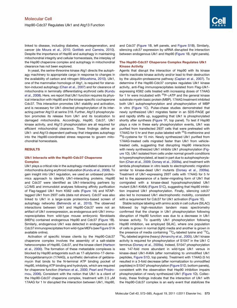
Hsp90 Is Necessary for Autophagy-Mediated Clearanceof Mitochondria during Erythroid DifferentiationMitochondrial clearance occurs during the final steps oferythroid maturation, and this response requires Ulk1 (Kunduet al., 2008). Since the Hsp90-Cdc37 complex was necessaryfor the stability and activity of Ulk1, and 17AAG impairs autoph-agy, we tested the effects of Hsp90 inhibition on mitochondrialclearance during terminal erythroid maturation. Treatment ofdifferentiating erythroid cells with 2.5 mM 17AAG triggeredmarked reductions in Ulk1 protein levels in two independenterythroid cultures (Figure 2A and Figure S4A, left panels) withoutaffecting ulk1 mRNA levels (Figure S4A, right panel). 17AAGtreatment did not impair reticulocyte development (Figure 2B),yet it significantly reduced the number of reticulocytes harboringautophagosomes, especially those containing mitochondria(Figures 2C and 2D) and led to corresponding increases in thelevels of mitochondrial proteins and overall mitochondrial mass(Figures S4A–S4C). Thus, Ulk1 stabilization by the Hsp90-Cdc37 complex is required for efficient autophagy-mediatedclearance of mitochondria during erythroid differentiation.
The Hsp90-Cdc37 Chaperone Complex and Ulk1 AreEssential for Clearance of Depolarized MitochondriaDisease-associated mutations in PARK2 (Abbas et al., 1999;Shimura et al., 2000), the gene encoding the E3 ligase Parkin,impair the elimination of damaged mitochondria (Geisler et al.,2010; Lee et al., 2010; Narendra et al., 2010; Vives-Bauzaet al., 2010). Investigations of Parkin’s role in targetingmitochon-dria for degradation by autophagy have established a highlyreproducible, quantitative, and genetically tractable cell-basedassay for identifying and characterizing genes involved in mito-chondrial clearance (Narendra et al., 2008). The assay involves
Molecular Cell
Hsp90-Cdc37 Regulates Ulk1 and Atg13 Function
Molecular Cell 43, 572–585, August 19, 2011 ª2011 Elsevier Inc. 575
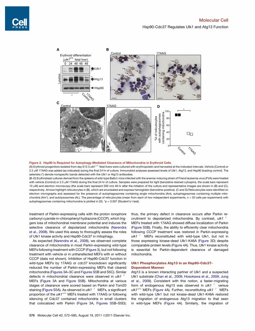
treatment of Parkin-expressing cells with the proton ionophorecarbonyl cyanidem-chlorophenyl hydrazone (CCCP), which trig-gers loss of mitochondrial membrane potential and induces theselective clearance of depolarized mitochondria (Narendraet al., 2008). We used this assay to thoroughly assess the rolesof Ulk1 kinase activity and Hsp90-Cdc37 in mitophagy.
As expected (Narendra et al., 2008), we observed completeclearance of mitochondria in most Parkin-expressing wild-typeMEFs following treatment with CCCP (Figure 3), but not followingtreatment with vehicle or in untransfected MEFs with or withoutCCCP (data not shown). Inhibition of Hsp90-Cdc37 function inwild-type MEFs by 17AAG or cdc37 knockdown significantlyreduced the number of Parkin-expressing MEFs that clearedmitochondria (Figures 3A–3C and Figures S5B and S5C). Similardefects in mitochondrial clearance were observed in ulk1!/!
MEFs (Figure 3A and Figure S5B). Mitochondria at variousstages of clearance were scored based on Parkin and Tom20staining (Figure S5A). As observed in ulk1!/! MEFs, a significantproportion of the ulk1+/+ MEFs treated with 17AAG or followingsilencing of Cdc37 contained mitochondria in small clustersthat colocalized with Parkin (Figure 3A, Figures S5B–S5D);
thus, the primary defect in clearance occurs after Parkin re-cruitment to depolarized mitochondria. By contrast, ulk1!/!
MEFs treated with 17AAG showed diffuse localization of Parkin(Figure S5B). Finally, the ability to efficiently clear mitochondriafollowing CCCP treatment was restored in Parkin-expressingulk1!/! MEFs reconstituted with wild-type Ulk1, but not inthose expressing kinase-dead Ulk1-K46A (Figure 3D) despitecomparable protein levels (Figure 4A). Thus, Ulk1 kinase activityis essential for Parkin-dependent clearance of damagedmitochondria.
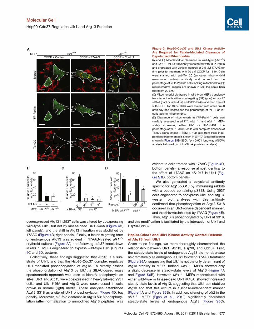
Ulk1 Phosphorylates Atg13 in an Hsp90-Cdc37-Dependent MannerAtg13 is a known interacting partner of Ulk1 and a suspectedUlk1 substrate (Chan et al., 2009; Hosokawa et al., 2009; Junget al., 2009). Consistent with this notion, a faster-migratingform of endogenous Atg13 was observed in ulk1!/! versusulk1+/+ MEFs (Figure 4A). Further, reconstituting ulk1!/! MEFswith wild-type Ulk1 but not kinase-dead Ulk1-K46A restoredthe migration of endogenous Atg13 migration to that seenin wild-type MEFs (Figure 4A). Similarly, the migration of
Control
17AAG
0
20
40
60
80
100
%t o
f ret
icul
ocyt
esw
ith a
utop
hago
som
es c
onta
inin
g m
itoch
ondr
ia
AL
Am*
Am
Control 17AAG
BControl 17AAG
C
0 24 48 48 48
--
Erythroid differentiationLy
sate
s
Control
17AAG
*
Hsp90
Ulk1
Atg13
*
A
D
(ulk1 fetal liver)+/+
*
Figure 2. Hsp90 Is Required for Autophagy-Mediated Clearance of Mitochondria in Erythroid Cells(A) Erythroid progenitors isolated from day E12.5 ulk1+/+ fetal livers were cultured with erythropoietin and harvested at the indicated intervals. Vehicle (Control) or
2.5 mM 17AAG was added (as indicated) during the final 24 hr of culture. Immunoblot analyses assessed levels of Ulk1, Atg13, and Hsp90 (loading control). The
asterisks (*) denote nonspecific bands detected with the Ulk1 or Atg13 antibodies.
(B–D) Erythroblast cultures derived from the spleens of wild-type Balb/c mice infected with the anemia-inducing strain of Friend leukemia virus (FVA) were treated
with vehicle (Control) or 2.5 mM 17AAG during the final 24 hr of culture. Samples were prepared for light (benzidine-stained cytospins, the scale bars represent
10 mM) and electron microscopy (the scale bars represent 500 nm) 48 hr after the initiation of the culture and representative images are shown in (B) and (C),
respectively. Arrows highlight reticulocytes in (B), which are enucleated and express hemoglobin (benzidine-positive). (C and D) Reticulocytes were identified on
electron micrographs and assessed for the presence of autophagosomes containing single mitochondria (Am), autophagosomes containing multiple mito-
chondria (Am*), and autolysosomes (AL). The percentage of reticulocytes (mean from each of two independent experiments, n > 20 cells per experiment) with
autophagosomes containing mitochondria is plotted in (D). *p = 0.007 (Student’s t test).
Molecular Cell
Hsp90-Cdc37 Regulates Ulk1 and Atg13 Function
576 Molecular Cell 43, 572–585, August 19, 2011 ª2011 Elsevier Inc.
overexpressed Atg13 in 293T cells was altered by coexpressingwild-type Ulk1, but not by kinase-dead Ulk1-K46A (Figure 4B,left panels), and the shift in Atg13 migration was abolished by17AAG (Figure 4B, right panels). Finally, a faster-migrating formof endogenous Atg13 was evident in 17AAG-treated ulk1+/+
erythroid cultures (Figure 2A) and following cdc37 knockdownin ulk1!/! MEFs engineered to express wild-type Ulk1 (Figures4C and 5D, bottom).Collectively, these findings suggested that Atg13 is a sub-
strate of Ulk1, and that the Hsp90-Cdc37 complex regulatesUlk1-mediated phosphorylation of Atg13. To directly assessthe phosphorylation of Atg13 by Ulk1, a SILAC-based massspectrometric approach was used to identify phosphorylationsites. Ulk1 and Atg13 were coexpressed in heavy labeled 293Tcells, and Ulk1-K46A and Atg13 were coexpressed in cellsgrown in normal (light) media. These analyses establishedAtg13 S318 as a site of Ulk1 phosphorylation (Figure 4D, toppanels). Moreover, a 3-fold decrease in Atg13 S318 phosphory-lation (after normalization to unmodified Atg13 peptides) was
A
Mer
geYF
P-Pa
rkin
Tom
20
CCCP + Control CCCP + 17AAG CCCP + Control
20
40
60
80
0%Pa
rkin
+ ce
lls w
ithno
mito
chon
dria
+/+ulk1MEF:
17AAG: + __
NT cdc37 siRNA:
20
40
60
80
0%Pa
rkin
+ ce
lls w
ithno
mito
chon
dria
pool #6 #7po
ol
B C D
20
40
60
80
0%Pa
rkin
+ ce
lls w
ithno
mito
chon
dria
-- --Ulk1
K46ARescue:
MEF:
*
*
* * ** *
MEF:
-/-ulk1 +/+
ulk1-/-
ulk1
+/+ulk1
-/-ulk1
Figure 3. Hsp90-Cdc37 and Ulk1 Kinase ActivityAre Required for Parkin-Mediated Clearance ofDepolarized Mitochondria(A and B) Mitochondrial clearance in wild-type (ulk1+/+)
and ulk1!/! MEFs transiently transfected with YFP-Parkin
and pretreated with vehicle (control) or 2.5 mM 17AAG for
5 hr prior to treatment with 20 mM CCCP for 18 hr. Cells
were stained with anti-Tom20 (an outer mitochondrial
membrane protein) antibody and scored for the
percentage of YFP-Parkin+ cells lacking mitochondria (B);
representative images are shown in (A); the scale bars
represent 20 mm.
(C) Mitochondrial clearance in wild-type MEFs transiently
transfected with either nontargeting (NT) (pool) or cdc37
siRNA (pool or individual) and YFP-Parkin and then treated
with CCCP for 18 hr. Cells were stained with anti-Tom20
antibody and scored for the percentage of YFP-Parkin+
cells lacking mitochondria.
(D) Clearance of mitochondria in YFP-Parkin+ cells was
similarly assessed in ulk1+/+, ulk1!/!, and ulk1!/! MEFs
stably expressing either Ulk1 or Ulk1-K46A. The
percentage of YFP-Parkin+ cells with complete absence of
Tom20 signal (mean + SEM, > 100 cells from three inde-
pendent experiments) is shown in (B)–(D) (detailed scoring
shown in Figures S5B–S5D). *p < 0.001 (one-way ANOVA
analysis followed by Holm-Sidak post-hoc analysis).
evident in cells treated with 17AAG (Figure 4D,bottom panels), a response almost identical tothe effect of 17AAG on pS1047 in Ulk1 (Fig-ure S1D, bottom panels).We also generated a polyclonal antibody
specific for Atg13pS318 by immunizing rabbitswith a peptide containing pS318. Using 293Tcells engineered to coexpress Ulk1 and Atg13,western blot analyses with this antibodyconfirmed that phosphorylation of Atg13 S318occurred in an Ulk1-kinase dependent manner,and that this was inhibited by 17AAG (Figure 4E).Thus, Atg13 is phosphorylated by Ulk1 at S318,
and this modification is facilitated by the interaction of Ulk1 andHsp90-Cdc37.
Hsp90-Cdc37 and Ulk1 Kinase Activity Control Releaseof Atg13 from Ulk1Given these findings, we more thoroughly characterized therelationship between UIk1, Atg13, Hsp90, and Cdc37. First,the steady-state levels of endogenous Atg13 did not decreaseas dramatically as endogenous Ulk1 following 17AAG treatment(Figure S6A), suggesting that Ulk1 is not the only determinant ofAtg13 stability in MEFs. Indeed, ulk1!/! MEFs showed onlya slight decrease in steady-state levels of Atg13 (Figure 4Aand Figure S6B). However, ulk1!/! MEFs reconstituted witheither wild-type or kinase-dead Ulk1 (K46A) showed increasedsteady-state levels of Atg13, suggesting that Ulk1 can stabilizeAtg13 and that this occurs in a kinase-independent manner(Figure 4A and Figure S6B). In addition, silencing of ulk2 in theulk1!/! MEFs (Egan et al., 2010) significantly decreasedsteady-state levels of endogenous Atg13 (Figure S6C),
Molecular Cell
Hsp90-Cdc37 Regulates Ulk1 and Atg13 Function
Molecular Cell 43, 572–585, August 19, 2011 ª2011 Elsevier Inc. 577

A C
D
m/z
827 829 831 833 835 837 8390
20
40
60
80
100 834.3339
834.0001
834.6682
835.0029
830.9998831.3331830.6660
827 829 831 833 835 837 8390
20
40
60
80
100 834.3347
834.0010
834.6689
835.0037
831.0002
831.3347
831.6682
504 506 508 510 512 514 5160
20
40
60
80
100 507.3087
512.3129
507.8105
512.8147
508.3123
504 506 508 510 512 514 5160
20
40
60
80
100 512.3134
507.3094
512.8149
507.8112
508.3130
513.3166
830.6661
513.3168
m/z
150X
2.9X
TVQVIVQARUnmodified Atg13 control peptidePhosphorylated Atg13 Ser318 peptide
THCAATPSSSEDTETVSNpSSEGR
38 40 42 44 460
20
40
60
80
1000
20
40
60
80
100
36 38 40 42Time (min)0
20
40
60
80
1000
20
40
60
80
100
Ulk1 K46A+ Atg13(light)
Ulk1 WT+ Atg1317AAG(light)
Ulk1 WT+ Atg13Control(heavy)
Ulk1 WT+ Atg13(heavy)
phosphorylatedS318 peptide
unphosphorylatedS318 peptide
Control17AAG
Ulk1 WTUlk1 K46A
Control17AAG
Ulk1 WTUlk1 K46A
phosphorylatedS318 peptide
unphosphorylatedS318 peptide
% R
elativ
e ab
unda
nce
% R
elativ
e ab
unda
nce
Ulk1
Control 17AAG
Atg13Lysa
tes
:Atg13
:Ulk1:Ulk1 K46A
___
+__
+
_+
+_+
+__
+
_+
+_+
E
:Atg13
:Ulk1:Ulk1 K46A
+--+
:17AAG+
--+
+--+
-- + --
Ulk1
Atg13pS318Atg13
GAPDH
ulk1-/- MEF(+Ulk1)
:siRNA_NT
cdc3
7atg
13
-/-Ulk1
Atg13
Cdc37Lysa
tes
BLy
sate
s
+/+ ulk1-/-
-- --Ulk1
K46A :Rescue
:Genotype
Ulk1
Atg13
Actin
Lysa
tes
Actin
Figure 4. Hsp90 Regulates Ulk1-Directed Phosphorylation of Atg13 at Serine 318(A) Immunoblot analysis of whole-cell lysates of ulk1+/+, ulk1!/!, and ulk1!/! MEFs stably expressing either Ulk1 or Ulk1-K46A.
(B) Immunoblot analysis of whole-cell lysates of 293T cells transiently transfected with the indicated NTAP-tagged expression constructs, which were treated
with vehicle (Control) or 2.5 mM 17AAG for 5 hr.
(C) Immunoblotanalysisofwhole-cell lysatesofulk1!/!orulk1!/!MEFsstablyexpressingUlk1 transfectedwitheitherpoolednontargeting (NT),cdc37, oratg13siRNA.
(D) Quantitative SILAC LC-MS/MS analyses of purified Atg13 from 293T cells expressing either Ulk1 or kinase-dead Ulk1-K46A (top panels); or from Ulk1-
expressing cells treated with vehicle alone (Control) or 2.5 mM 17AAG for 5 hr (bottom panels). (Top left panel) Extracted ion chromatograms showing the relative
abundance of the phosphorylated Atg13 S318 peptide ("40%) from Ulk1-expressing cells, and the predominance of the unmodified Atg13 peptide from Ulk1-
K46A expressing cells. (Top middle panel) Full MS scan of the Atg13 S318 phosphorylated peptide demonstrating a quantitative"150-fold increase (149 ± 26.4)
in S318 phosphorylation of Atg13 when coexpressed with Ulk1 versus Ulk1-K46A (values are the mean ± SD, n = 2). (Bottom left panel) Extracted ion chro-
matogram demonstrating a reduction in the relative abundance of Atg13 S318 phosphorylation from 17AAG-treated cells. (Bottommiddle panel) Full MS scan of
the phosphorylated S318 Atg13 peptide demonstrating an"3-fold reduction (2.9 ± 0.1) in phosphorylation of Atg13 S318 following 17AAG treatment (values are
themean ± SD, n = 2). (Right panels) Full MS scans of one of the several unmodified Atg13 peptides that were used to normalize protein levels, which demonstrate
that purified ‘‘light’’ (left peaks) and ‘‘heavy’’ (right peaks) Atg13 samples were mixed at a ratio very close to 1:1.
(E) 293T cells were transiently transfected with NTAP-Ulk1, NTAP-Ulk1-K46A, and/or Atg13 as indicated, and treated with vehicle (!) or 17AAG (+) for 2 hr. Ulk1,
p318S-Atg13, total Atg13, and GAPDH were detected by immunoblot analyses.
Molecular Cell
Hsp90-Cdc37 Regulates Ulk1 and Atg13 Function
578 Molecular Cell 43, 572–585, August 19, 2011 ª2011 Elsevier Inc.

A DB
E
Ulk1
Cdc37
Atg13
Ulk1
Cdc37
Atg13
Actin
NT cdc37 siRNA:
ulk1-/- MEF(+Ulk1)
Lysa
tes
Ulk
1 IP
Ulk1Atg13Cdc37
Ulk1Atg13Cdc37Actin
0 1 52.5µM 17AAG
Lysa
tes
Ulk
1 IP
:Time (hrs)
NTAP
Ulk1 Ulk1 K46A
NTA
P IP
:17AAG + ___
Ulk1
Atg13Cdc37
C
:NTAP
ulk1-/- MEF(+Ulk1)
Lysa
tes
Ulk
1 IP
+/+
-- --Ulk1
K46ARescue:
MEF: ulk1-/-
Ulk1
Hsp90
Ulk1
Hsp90
Cdc37
Actin
Cdc37
Atg13
Atg13
Ulk1 Ulk1 Ulk1 K46A
HA-Atg13 HA-Atg13 HA-Atg13
Merge Merge Merge
17AAGHA-Atg13+Ulk1
Control HA-Atg13+Ulk1K46A
ControlHA-Atg13+Ulk1
Figure 5. Hsp90-Cdc37 and Ulk1 Kinase Activity Regulate Release of Atg13(A) Immunoblot analysis of anti-Ulk1 immunoprecipitates fromwhole cell lysates of ulk1+/+, ulk1!/!, and ulk1!/!MEFs stably expressing either Ulk1 or Ulk1-K46A.
(B) Immunoblot analysis of NTAP affinity-purified precipitates from 293T cells transfected with the indicated vector and then treated with 2.5 mM 17AAG (+) or
vehicle (!) for 5 hr.
(C) Immunoblot analysis of anti-Ulk1 immunoprecipitates from ulk1!/! MEFs stably expressing Ulk1 and treated with 2.5 mM 17AAG for the indicated times.
(D) Immunoblot analysis of anti-Ulk1 immunoprecipitates from ulk1!/! MEFs stably expressing Ulk1 and transfected twice with pooled nontargeting or cdc37
siRNA.
(E) Representative images of HeLa cells cotransfected with HA-Atg13 and either NTAP-Ulk1 or NTAP-Ulk1-K46A and treated with vehicle (Control) or 2.5 mM
17AAG for 5 hr. Cells were stained with anti-HA and anti-Ulk1 antibodies. Scale bar represents 10 mM.
Molecular Cell
Hsp90-Cdc37 Regulates Ulk1 and Atg13 Function
Molecular Cell 43, 572–585, August 19, 2011 ª2011 Elsevier Inc. 579

indicating that in MEFs both Ulk1 and Ulk2 contribute to stabili-zation of Atg13. By contrast, in erythroid cells, where ulk2mRNAexpression is minimal (Kundu et al., 2008), loss of Ulk1 is suffi-cient to dramatically decrease steady-state levels of endoge-nous Atg13 (Figure S6D). Collectively, these data and thoseshowing that Ulk2 is not a client of Hsp90 (Figure S2) suggestthat Atg13 stability is maintained in MEFs following 17AAGtreatment as a result of its interaction with Ulk2.
Interestingly, immunoprecipitation analyses of ulk1+/+ MEFs,and ulk1!/! MEFs reconstituted with either wild-type orkinase-dead Ulk1-K46A, demonstrated an inverse correlationbetween Ulk1 kinase activity and the relative amount of Atg13present in anti-Ulk1 immunoprecipitates. Although the amountof Hsp90 and Cdc37 immunoprecipitated with Ulk1 wasproportional to the level of Ulk1, higher levels of Atg13 werecoimmunoprecipitated from ulk1!/! MEFs reconstituted withkinase-dead Ulk1-K46A (Figure 5A). Similar findings were ob-served from precipitation of NTAP-Ulk1 or NTAP-Ulk1-K46Aoverexpressed in 293T cells (Figure 5B). Furthermore, treatmentwith 17AAG or knockdown of cdc37, which impaired Ulk1stability and kinase activity (Figure 1), triggered increases inthe amount of Atg13 that coimmunoprecipitated with overex-pressed Ulk1 (Figures 5B–5D). 17AAG treatment also increasedAtg13 immunoprecipitation with endogenous Ulk1 (Figure S6A).Given these findings, we also examined the subcellular distribu-tion of Ulk1 and Atg13 by confocal microscopy. Although Ulk1and Atg13 were diffusely cytoplasmic when coexpressed inHeLa cells, inhibition of Ulk1 kinase activity by treatment with17AAG or by expressing kinase-dead Ulk1-K46A triggered theformation of discrete Ulk1+/Atg13+ foci (Figure 5E) that werenot LC3+ (data not shown). Collectively, these observationssuggest that though the interaction of Ulk1 with Hsp90-Cdc37is necessary for Atg13 phosphorylation, the activation of Ulk1kinase by Hsp90-Cdc37 promotes release of Atg13 from theUlk1-Hsp90-Cdc37 complex.
Atg13 Localizes to Damaged Mitochondriaand Is Necessary for Mitochondrial ClearanceThere are conflicting results regarding the role of orthologousyeast Atg1-Atg13 complex in mitophagy (Kanki and Klionsky,2010; Kanki et al., 2009; Okamoto et al., 2009). Therefore, wetested if Atg13 contributes to mitophagy in mammalian cells.Notably, atg13 knockdown significantly impaired mitochondrialclearance in Parkin-expressing MEFs treated with CCCP(Figures 6A and 6B). Silencing of ATG13 in Parkin-expressingHeLa cells also impaired clearance of depolarized mitochondria(Figure S7B). Strikingly, assessment of the subcellular distribu-tion of Ulk1 and Atg13 in HeLa cells engineered to express Parkinand treated with CCCP demonstrated that Atg13, but not Ulk1,colocalized with Tom20+ mitochondrial clusters in most Parkin-expressing cells (Figure 6C and Figure S7A). Atg13 did notlocalize to mitochondria in the absence of CCCP and Parkin(data not shown). Moreover, overexpressing the kinase-deadUlk1 mutant, which inhibits Atg13 phosphorylation (Figure 4B)and mitochondrial clearance (Figure S7C), prevented Atg13localization to damaged mitochondria (Figure 6C and Fig-ure S7A). Finally, Hsp90 inhibition impaired localization ofAtg13 to damaged mitochondria (Figure 6C and Figure S7A).
Collectively, these findings suggest that mitochondrial damagetriggers Ulk1-dependent phosphorylation and release of Atg13to mitochondria.
Atg13 S318 Phosphorylation Is Requiredfor Parkin-Mediated Mitochondrial ClearanceTo assess if Atg13 phosphorylation on S318 plays roles inParkin-mediated mitophagy, we generated a serine-to-alanine(nonphosphorylatable) substitution mutation of Atg13 (Atg13-S318A). Although Atg13 overexpression had little effect on Par-kin-dependent clearance of depolarized mitochondria, similarlevels of Atg13-S318A exerted a dominant-negative effect onmitophagy (Figures 7A and 7B). Interestingly, although Ulk1-K46A dominantly inhibited both starvation-induced autophagy(data not shown and Chan et al., 2009) and mitochondrialclearance (Figure S7C), overexpression of Atg13-S318A didnot impair starvation-induced autophagy but resulted in in-creased LC3 turnover under normal growth conditions andfollowing nutrient deprivation (Figure S7D). Thus, phosphoryla-tion of Atg13 at S318 is required for mitophagy, but not basalor starvation-induced autophagy, implying that differentialphosphorylation of Atg13 by Ulk1 may influence downstreamfunctions of Atg13.
DISCUSSION
The data presented herein establish that the Hsp90-Cdc37chaperone complex regulates mitophagy by modulating thestability and function of Ulk1 and one of its downstream targets,Atg13. Specifically, the interaction with the Hsp90-Cdc37complex stabilizes Ulk1 by preventing proteasome-mediateddegradation, and this interaction is required for Ulk1 autophos-phorylation and phosphorylation of Atg13 at S318. Strikingly,Ulk1 activation promotes the release of Atg13 from theHsp90-Cdc37-Ulk1 complex and the localization of Atg13 todepolarized mitochondria, where it plays an essential role inParkin-dependent mitophagy. Although Hsp90, Cdc37, andUlk1 regulate the phosphorylation of Atg13 at S318, this modifi-cation alone does not appear to be sufficient for promotingAtg13 release, as increased amounts of Ulk1 were not detectedin immunoprecipitates of Atg13-S318A versus wild-type Atg13,nor did we observe enhanced colocalization of Ulk1 and Atg13-S318A (data not shown). Rather, the release of Atg13 from Ulk1may depend on a kinase-dependent change in conformation ofUlk1 and/or phosphorylation of Atg13 at sites other than S318.Regardless, the finding that the nonphosphorylatable Atg13-S318A mutant dominantly inhibits mitochondrial clearancehighlights the functional significance of Atg13 phosphorylationby Ulk1. Since Hsp90, Cdc37, and Ulk1 together regulate phos-phorylation of Atg13 at S318, and all are required for efficientautophagy-mediated clearance of mitochondria, these findingsdefine a new pathway linking mitochondrial homeostasis withthe cellular stress response coordinated by Hsp90-Cdc37.In contrast to the prevailing view that Ulk1 and Atg13 function
as a complex, our data indicate that Ulk1 kinase activitypromotes the release of Atg13 from Ulk1, and that releasedAtg13 then localizes to ring-like structures around damagedmitochondria, and thereby contributes to their degradation.
Molecular Cell
Hsp90-Cdc37 Regulates Ulk1 and Atg13 Function
580 Molecular Cell 43, 572–585, August 19, 2011 ª2011 Elsevier Inc.

Indeed, silencing of Atg13 or enforced expression of the non-phosphorylatable Atg13-S318A mutant impairs mitochondrialclearance. Curiously, phosphorylation of Atg13 at S318 is notrequired for efficient LC3 conversion and degradation (underbasal conditions or following amino acid deprivation), suggestingS318 phosphorylation of Atg13 may influence cargo selectionduring autophagy.The Hsp90-Cdc37 complex is involved in two distinct
paradigms of autophagy-mediated mitochondrial clearance:Parkin-mediated clearance of depolarized mitochondria, andthe BNIP3L-dependent developmental clearance of mitochon-dria in erythroid cells. Although these pathways differ in the
mechanism by which mitochondria are targeted for degradation(Parkin versus BNIP3L), both rely on Ulk1-mediated activation ofthe autophagy pathway, which is regulated by Hsp90 andCdc37. It is interesting to note that Pink1, a serine-threoninekinase that recruits Parkin to depolarized mitochondria andpromotes clearance (Geisler et al., 2010; Narendra et al., 2010;Vives-Bauza et al., 2010), was identified as a client of theHsp90-Cdc37 chaperone complex (Lin and Kang, 2008; Mori-waki et al., 2008; Weihofen et al., 2008). While the primary defectin mitochondrial clearance occurs after the recruitment of Parkinto mitochondria in ulk1!/! MEFs, and in ulk1+/+ MEFs treatedwith 17AAG or having knockdown of cdc37, we cannot exclude
Actin
Atg13
NT atg13 siRNA:
20
40
60
0
%Pa
rkin
cel
ls w
ithno
mito
chon
dria
MEF+NT siRNA MEF+atg13 siRNA
MEF: ulk1
HA-Ulk1/Tom20
CCCP CCCP HA-Ulk1 HA-Atg13
HA-Atg13/Tom20
CCCP + 17AAGHA-Atg13+Ulk1
HA-Atg13/Tom20
CCCP HA-Atg13+Ulk1K46A
HA-Atg13/Tom20
CCCPHA-Atg13+Ulk1
HA-Atg13/Tom20
0
1K
2K
3K
0
1K
2K
3K Tom20HA-Atg13
0
1K
2K
3K Tom20HA-Atg13
Tom20HA-Atg13
Inte
nsity
(a.u
.)
3 microns 3 microns 3 microns 3 microns0
1K
2K
3K
3 microns0
1K
2K
3K Tom20HA-Ulk1
Tom20HA-Atg13
Mer
ge *
A
C
B
+/+
+NT atg13
Figure 6. Atg13 Is Required for Mitochondrial Clearance and Localizes to DamagedMitochondria in an Ulk1- and Hsp90-Dependent Manner(A and B)Wild-type (ulk1+/+) MEFs were transiently transfected with either pooled nontargeting or atg13 siRNA and YFP-Parkin, and were then treated with 20 mM
CCCP for 18 hr. Cells were then stained with anti-Tom20 antibody and scored for the percentage of YFP-Parkin+ cells lacking mitochondria. Representative
images are shown in (A), and the percentage of YFP-Parkin+ cells with complete absence of Tom20 signal (mean + SEM, >100 cells from three independent
experiments) is shown in (B). *p < 0.001 (Student’s t test). Scale bar represents 20 mM. Atg13 silencing was confirmed by immunoblot analysis (B).
(C) HeLa cells were transfected with YFP-Parkin together with Ulk1, Ulk1-K46A, and/or Atg13 expression constructs (as indicated). Cells were treated with 20 mM
CCCP for 8 hr. As indicated, cells were treated for 5 hr with 2.5 mM17AAG prior to addition of CCCP. Cells were then stained with anti-HA (green) and anti-Tom20
(red) antibodies. Representativemerged images of Parkin+ CCCP-treated cells are shown (top panels). The corresponding single-channel pseudocolored images
are shown in Figure S7A. Line scans (bottom panels) indicate the degree of colocalization between Atg13 (green) and Tom20+ mitochondria (red) in Parkin+ cells
and correlate to the lines drawn in the magnified images (middle panels). Intensity profiles (in arbitrary units) were obtained using NIS elements AR 3.10 software
from Nikon. Scale bar represents 10 mM.
Molecular Cell
Hsp90-Cdc37 Regulates Ulk1 and Atg13 Function
Molecular Cell 43, 572–585, August 19, 2011 ª2011 Elsevier Inc. 581

the possibility that Pink1 function is also affected. In fact, thediffuse localization of Parkin in ulk1!/! MEFs treated with17AAG suggests that another client of Hsp90, perhaps Pink1,recruits Parkin to mitochondria and maintains the minimal levelsof mitochondrial clearance observed the absence of Ulk1.That serine-threonine kinases involved in autophagy (Ulk1)and the mitochondrial targeting pathway (Pink1) are both clientsof the Hsp90-Cdc37 chaperone complex suggests that coordi-nated regulation of mitochondrial turnover is an importanthomeostatic response that must be preserved, even underadverse conditions.It has been suggested that Hsp90 inhibition promotes
autophagy (Qing et al., 2006; Siegelin et al., 2010). Indeed, undernormal growth conditions Hsp90 inhibition triggers an initialUlk1-dependent increase in autophagy-dependent LC3 degra-dation (F.C.D. and M.K., unpublished data), a response consis-tent with the Hsp90-dependent nature of kinases in the PI3K-mTOR pathway (Basso et al., 2002; Gray et al., 2007; Ohjiet al., 2006) that inhibit autophagy (Ravikumar et al., 2004).However, this initial increase in autophagy is self-limiting andis rapidly cancelled by the inactivation of Hsp90-dependentUlk1 activity, the primary autophagy-related target of mTOR(Hosokawa et al., 2009; Jung et al., 2009). In turn, inactivationof Ulk1 cripples the cell’s ability to respond to stimuli that induceautophagy, such as starvation or mitochondrial damage.The exquisite dependence of Ulk1-mediated autophagy and
mitochondrial clearance on Hsp90 function has importantimplications for cancer pathogenesis and treatment. First, ithas been proposed that the destabilization of proteins associ-ated with growth in the hypoxic, nutrient-deprived tumor milieuis compensated by increased expression of Hsp90 and itscochaperones (Whitesell and Lindquist, 2005). Since autophagypromotes survival under these adverse conditions, the stabiliza-tion and activation of Ulk1 by Hsp90 may be important compo-nents of the survival response coordinated by Hsp90 andexploited by certain tumors. Indeed, since many tumors rely onautophagy for survival under stressed conditions (Amaravadiet al., 2007; Livesey et al., 2009; Maclean et al., 2008), theefficacy of Hsp90 inhibitors such as 17AAG may rely on theireffects on Ulk1-mediated autophagy.Collectively, the findings presented herein support a model
(see the Graphical Abstract) whereby interactions of Ulk1 withthe Hsp90-Cdc37 complex stabilize an active form of Ulk1 thatpromotes flux through specific arms of the autophagy pathwayin response to metabolic or proteotoxic cues. Starvation, mito-chondrial depolarization, and other stimuli that trigger autophagyactivate Ulk1, promoting Ulk1-mediated phosphorylation andrelease of its essential interacting partner, Atg13, and increasedflux through the autophagy pathway. Phosphorylation ofAtg13 at specific sites, for example S318, may favor selectivedegradation of mitochondria. As Ulk1 stability and activity relyon the Hsp90-Cdc37 complex, the function of both Ulk1 anddownstream targets, including Atg13, is also subject to regula-tion by environmental and intracellular cues that alter Hsp90
A
B
CCCP CCCP Atg13 Atg13-S318A
%Pa
rkin
ce
lls w
ithno
mito
chon
dria
#
cDNA: _ Atg13 S318A0
10
20
30
40
50
NTAP-Atg13
Tubulin
Atg13S31
8A _
*
+
Atg13
HeLa
Figure 7. Ulk1-Mediated Phosphorylation of Atg13 at S318 IsRequired for Efficient Clearance of Depolarized Mitochondria(A and B) HeLa cells were transiently transfected with YFP-Parkin alone, or
together with either NTAP-tagged Atg13 or Atg13-S318A. The cells were
treated with vehicle (Control) or 20 mMCCCP for 18 hr. Cells were stained with
anti-Tom20 antibody, and YFP-Parkin+ cells were scored based on the pres-
ence or absence of Tom20 staining. Representative images of CCCP-treated
cells are shown in (A); the scale bars represent 20 mm. The percentage of
YFP-Parkin+ cells lacking Tom20 signal (mean + SEM, >100 cells from three
independent experiments) is shown in panel (B). #p < 0.001 (Student’s t test).
Expression of Atg13 and Atg13-S318A was confirmed by immunoblot analysis
(arrows highlight endogenous and NTAP-tagged Atg13 and Atg13-S318A as
indicated); tubulin was used as a loading control. The asterisk (*) denotes
a nonspecific band observed with the anti-Atg13 antibody.
Molecular Cell
Hsp90-Cdc37 Regulates Ulk1 and Atg13 Function
582 Molecular Cell 43, 572–585, August 19, 2011 ª2011 Elsevier Inc.

activity. The Hsp90-Cdc37-to-Ulk1-to-Atg13 pathway thus co-ordinates and integrates autophagy and mitochondrial homeo-stasis with the cellular stress response.
EXPERIMENTAL PROCEDURES
Plasmid ConstructsFlag-tagged Ulk1 and deletion constructs in the pME18S vector (gift of
Dr. Toshifumi Tomoda) (Yan et al., 1998), HA-Atg13 (Jung et al., 2009),
HA-Ulk1 (Jung et al., 2009), and NTAP-Ulk1 (Dorsey et al., 2009a) were previ-
ously described. The YFP-Parkin construct was also described (Narendra
et al., 2008). An insert containing the coding sequences for mCherry-EGFP-
LC3b (from pDEST mCherry-EGFPLC3b vector, a gift of Dr. Terje Johannsen)
was subcloned into the LPC retroviral vector and used to generate stably
expressing MEFs as described (Tresse et al., 2010). Methods used for gener-
ating the following plasmid constructs are described in the Supplemental
Experimental Procedures: Flag- and NTAP- tagged Ulk1 kinase-dead (K46A)
mutants, MSCV-luciferase-LC3B and MSCV-luciferase-G120ALC3B, and
NTAP-Atg13 and NTAP-Atg13-S318A.
ProteomicsUlk1-interacting proteins were visualized by silver staining (Invitrogen)
according to the manufacturer’s protocol. Gel slices containing bands of
interest were digested with trypsin as described (Strader et al., 2006).
Peptides were separated using a 10 cm C18 column; samples were run at
200 nl/min for 45min on a NanoLC (Eksigent). Online nanospray was used
to spray the separated peptides into LTQ (Thermo Electron). Xcalibur was
used to acquire the raw data, and databases including NCBI and Swissprot
were searched using Mascot (Perkins et al., 1999). Criteria used for confident
protein identification were as follows: peptide score R 30; p value of
peptide < 0.05; protein score R 70; number of unique peptides R 2. The
bands shown in Figure 1A were identified as Ulk1 (Mascot score 2274,
85 queries matched, 32 unique peptides), HSP90b (Mascot score 1507,
69 queries matched, 28 unique peptides), and CDC37 (Mascot score 430,
15 queries matched, 8 unique peptides). Details of methods used to quantify
protein phosphorylation by SILAC are described in the Supplemental
Experimental Procedures.
Cell Culture, Transfection, and Drug Treatment; Generation ofMEFs; Immunoprecipitation; Immunoblot Analyses and Antibodies;Pulse-Chase Analyses, Gene Silencing; Microscopy; ErythroidCultures; Flow Cytometry; Luc-LC3 Luciferase Reporter Assay;and Cell Viability AssaysPlease see the Supplemental Experimental Procedures.
In Vitro Kinase AssaysFlag-Ulk1 was immunoprecipitated from K562 cells and eluted with Flag
peptide (Sigma Aldrich) as described above. In vitro kinase reactions were
performed as follows. Eluted Flag-Ulk1 was incubated in kinase buffer
(10mMTris [pH 7.4], 150mMNaCl, 10mMMgCl2, 0.5mMDTT) supplemented
with 1 mg/ml MBP fragments (Sigma Aldrich), 25 mM nonradioactive ATP,
phosphatase inhibitors (Pierce), and 2.5 mCi g-32P-labeled ATP at 37!C for
15 min. Kinase reactions were stopped by adding an equal volume of
23 sample buffer, and samples were immediately boiled and separated on
SDS-PAGE gels, which were then fixed in 30% methanol 10% acetic acid,
stained with Coomassie blue, dried, and exposed to KodakMR resolution film.
Quantitative Real-Time PCRTotal RNA was isolated from cells using TRIzol Reagent (Invitrogen). The
reverse transcription (RT) reaction was carried out using the iScript cDNA
synthesis kit (Bio-Rad) according to the manufacturer’s instructions. TaqMan
Gene Expression Assays containing FAM-labeled primer/probe sets specific
for ulk1, ulk2, akt1, cdc37, and 18S were obtained from Applied Biosystems.
The real-time PCR reactions were performed in a total reaction volume of
25 ml using FastStart TaqMan Probe Master (Roche) reagent and results
were analyzed with the iCycler IQ real-time PCR detection system (Bio-Rad).
Relative expression (log2) was calculated using the Pfaff quantificationmethod
(Pfaffl, 2001) after normalization of cycle thresholds to 18SRNA and calibration
to respective controls.
Statistical AnalysesColocalization of Parkin and mitochondria or mitochondrial clearance was
assessed by visually scoring R100 Parkin-positive cells per condition
from at least three independent experiments and is represented as the
mean ± SEM. Statistical analysis was performed using SigmaPlot; significance
was assessed by two-tailed paired Student’s t test, or one- or two-factor
ANOVA analysis followed by Holm-Sidak post-hoc analysis.
SUPPLEMENTAL INFORMATION
Supplemental Information includes seven figures, Supplemental Experimental
Procedures, and Supplemental References and can be found with this article
online at doi:10.1016/j.molcel.2011.06.018.
ACKNOWLEDGMENTS
We are grateful to Charles J. Sherr (St. Jude Children’s Research Hospital
[SJCRH], Memphis, TN) for providing Cdc37 antibody; Emily Tresse (SJCRH)
for the LPC-mCherry-EGFP-LC3b construct; Toshifumi Tomoda (Beckman
Research Institute of City of Hope, Duarte, CA) for Flag-Ulk1 deletion
constructs; Sharon Tooze (London Research Institute, Cancer Research UK,
London, UK) for providing an Atg13 antibody;Mark Hall (The Scripps Research
Institute [TSRI], Jupiter, Florida) for providing the MSCV-Gateway-IRES-GFP
vector; Reuben Shaw (The Salk Institute for Biological Studies, La Jolla, CA)
for providing ulk1"/" MEFs stably expressing ulk2 shRNA; Jennifer Moore
(SJCRH) for assistance with generation of MEFs; Aaron Poole (SJCRH), Valerie
Cavett (TSRI), Chunying Yang (TSRI), and Laura Alsina (TSRI) for technical
support; the Proteomics Core facilities of Scripps Florida (TSRI) and the
University of Pennsylvania; the Biomedical Imaging Core facilities at SJCRH
(Samuel Connell, Jennifer Peters, and Sharon Frase) and University of Penn-
sylvania (Neelima Shaw and Ray Meade); and Robert Matts (Oklahoma State
University) for helpful discussions. This research was partially supported by
grants from the National Institutes of Health to M.K. (HL084199), F.C.D.
(CA123777), J.L.C. (CA076379), P.A.N. (DK074519), D.-H.K. (GM097057),
and C.B.T. (CA099179); the NINDS intramural program to R.Y; the Burroughs
Welcome Fund to M.K.; the American Society of Hematology to M.K.; and the
American Diabetes Association to D.-H.K. (ADA 7-07-CD-08); by monies from
the State of Florida to J.L.C. at Scripps Florida; and by monies from the Amer-
ican Lebanese Syrian Associated Charities (ALSAC) to P.A.N. and M.K.
Received: November 17, 2010
Revised: April 21, 2011
Accepted: June 24, 2011
Published: August 18, 2011
REFERENCES
Abbas, N., Lucking, C.B., Ricard, S., Durr, A., Bonifati, V., De Michele, G.,
Bouley, S., Vaughan, J.R., Gasser, T., Marconi, R., et al. (1999). A wide variety
ofmutations in the parkin gene are responsible for autosomal recessive parkin-
sonism in Europe. French Parkinson’s Disease Genetics Study Group and the
European Consortium on Genetic Susceptibility in Parkinson’s Disease. Hum.
Mol. Genet. 8, 567–574.
Amanchy, R., Kalume, D.E., and Pandey, A. (2005). Stable isotope labeling
with amino acids in cell culture (SILAC) for studying dynamics of protein abun-
dance and posttranslational modifications. Sci. STKE 2005, pl2. 10.1126/stke.
2672005pl2.
Amaravadi, R.K., and Thompson, C.B. (2007). The roles of therapy-induced
autophagy and necrosis in cancer treatment. Clin. Cancer Res. 13, 7271–7279.
Amaravadi, R.K., Yu, D., Lum, J.J., Bui, T., Christophorou, M.A., Evan, G.I.,
Thomas-Tikhonenko, A., and Thompson, C.B. (2007). Autophagy inhibition
Molecular Cell
Hsp90-Cdc37 Regulates Ulk1 and Atg13 Function
Molecular Cell 43, 572–585, August 19, 2011 ª2011 Elsevier Inc. 583

enhances therapy-induced apoptosis in a Myc-induced model of lymphoma.
J. Clin. Invest. 117, 326–336.
Basso, A.D., Solit, D.B., Chiosis, G., Giri, B., Tsichlis, P., and Rosen, N. (2002).
Akt forms an intracellular complex with heat shock protein 90 (Hsp90) and
Cdc37 and is destabilized by inhibitors of Hsp90 function. J. Biol. Chem.
277, 39858–39866.
Behrends, C., Sowa, M.E., Gygi, S.P., and Harper, J.W. (2010). Network
organization of the human autophagy system. Nature 466, 68–76.
Bhatia-Kissova, I., and Camougrand, N. (2010). Mitophagy in yeast: actors
and physiological roles. FEM. Yeast Res. 10, 1023–1034.
Caplan, A.J., Mandal, A.K., and Theodoraki, M.A. (2007). Molecular chaper-
ones and protein kinase quality control. Trends Cell Biol. 17, 87–92.
Chan, E.Y., Kir, S., and Tooze, S.A. (2007). siRNA screening of the kinome
identifies ULK1 as a multidomain modulator of autophagy. J. Biol. Chem.
282, 25464–25474.
Chan, E.Y., Longatti, A., McKnight, N.C., and Tooze, S.A. (2009). Kinase-inac-
tivated ULK proteins inhibit autophagy via their conserved C-terminal domains
using an Atg13-independent mechanism. Mol. Cell. Biol. 29, 157–171.
Criollo, A., Senovilla, L., Authier, H., Maiuri, M.C., Morselli, E., Vitale, I., Kepp,
O., Tasdemir, E., Galluzzi, L., Shen, S., et al. (2010). The IKK complex contrib-
utes to the induction of autophagy. EMBO J. 29, 619–631.
de Moura, M.B., dos Santos, L.S., and Van Houten, B. (2010). Mitochondrial
dysfunction in neurodegenerative diseases and cancer. Environ. Mol.
Mutagen. 51, 391–405.
Dorsey, F.C., Rose, K.L., Coenen, S., Prater, S.M., Cavett, V., Cleveland, J.L.,
and Caldwell-Busby, J. (2009a). Mapping the phosphorylation sites of Ulk1.
J. Proteome Res. 8, 5253–5263.
Dorsey, F.C., Steeves, M.A., Prater, S.M., Schroter, T., and Cleveland, J.L.
(2009b). Monitoring the autophagy pathway in cancer. Methods Enzymol.
453, 251–271.
Egan, D.F., Shackelford, D.B., Mihaylova, M.M., Gelino, S., Kohnz, R.A., Mair,
W., Vasquez, D.S., Joshi, A., Gwinn, D.M., Taylor, R., et al. (2010).
Phosphorylation of ULK1 (hATG1) by AMP-activated protein kinase connects
energy sensing to mitophagy. Science 331, 456–461.
Gamboa, J.L., and Andrade, F.H. (2010). Mitochondrial content and distribu-
tion changes specific to mouse diaphragm after chronic normobaric hypoxia.
Am. J. Physiol. Regul. Integr. Comp. Physiol. 298, R575–R583.
Geisler, S., Holmstrom, K.M., Skujat, D., Fiesel, F.C., Rothfuss, O.C., Kahle,
P.J., and Springer, W. (2010). PINK1/Parkin-mediated mitophagy is depen-
dent on VDAC1 and p62/SQSTM1. Nat. Cell Biol. 12, 119–131.
Gottlieb, R.A., and Carreira, R.S. (2010). Autophagy in health and disease. 5.
Mitophagy as a way of life. Am. J. Physiol. Cell Physiol. 299, C203–C210.
Gray, P.J., Jr., Stevenson, M.A., and Calderwood, S.K. (2007). Targeting
Cdc37 inhibits multiple signaling pathways and induces growth arrest in
prostate cancer cells. Cancer Res. 67, 11942–11950.
Hartson, S.D., Irwin, A.D., Shao, J., Scroggins, B.T., Volk, L., Huang, W., and
Matts, R.L. (2000). p50(cdc37) is a nonexclusive Hsp90 cohort which partici-
pates intimately in Hsp90-mediated folding of immature kinase molecules.
Biochemistry 39, 7631–7644.
Hosokawa, N., Hara, T., Kaizuka, T., Kishi, C., Takamura, A., Miura, Y., Iemura,
S., Natsume, T., Takehana, K., Yamada, N., et al. (2009). Nutrient-dependent
mTORC1 association with the ULK1-Atg13-FIP200 complex required for
autophagy. Mol. Biol. Cell 20, 1981–1991.
Jung, C.H., Jun, C.B., Ro, S.H., Kim, Y.M., Otto, N.M., Cao, J., Kundu, M., and
Kim, D.H. (2009). ULK-Atg13-FIP200 complexes mediate mTOR signaling to
the autophagy machinery. Mol. Biol. Cell 20, 1992–2003.
Kang, B.H., Plescia, J., Dohi, T., Rosa, J., Doxsey, S.J., and Altieri, D.C. (2007).
Regulation of tumor cell mitochondrial homeostasis by an organelle-specific
Hsp90 chaperone network. Cell 131, 257–270.
Kanki, T., and Klionsky, D.J. (2010). Themolecular mechanism ofmitochondria
autophagy in yeast. Mol. Microbiol. 75, 795–800.
Kanki, T., Wang, K., Baba, M., Bartholomew, C.R., Lynch-Day, M.A., Du, Z.,
Geng, J., Mao, K., Yang, Z., Yen, W.L., and Klionsky, D.J. (2009). A genomic
screen for yeast mutants defective in selective mitochondria autophagy.
Mol. Biol. Cell 20, 4730–4738.
Kim, I., Rodriguez-Enriquez, S., and Lemasters, J.J. (2007). Selective degrada-
tion of mitochondria by mitophagy. Arch. Biochem. Biophys. 462, 245–253.
Kundu, M., and Thompson, C.B. (2008). Autophagy: basic principles and
relevance to disease. Annu. Rev. Pathol. 3, 427–455.
Kundu, M., Lindsten, T., Yang, C.Y., Wu, J., Zhao, F., Zhang, J., Selak, M.A.,
Ney, P.A., and Thompson, C.B. (2008). Ulk1 plays a critical role in the autopha-
gic clearance of mitochondria and ribosomes during reticulocyte maturation.
Blood 112, 1493–1502.
Lee, J.Y., Nagano, Y., Taylor, J.P., Lim, K.L., and Yao, T.P. (2010). Disease-
causing mutations in parkin impair mitochondrial ubiquitination, aggregation,
and HDAC6-dependent mitophagy. J. Cell Biol. 189, 671–679.
Li, Y., Zhang, T., Schwartz, S.J., and Sun, D. (2009). New developments in
Hsp90 inhibitors as anti-cancer therapeutics: mechanisms, clinical perspec-
tive and more potential. Drug Resist. Updat. 12, 17–27.
Lin, W., and Kang, U.J. (2008). Characterization of PINK1 processing, stability,
and subcellular localization. J. Neurochem. 106, 464–474.
Liu, T.T., Hu, C.H., Tsai, C.D., Li, C.W., Lin, Y.F., and Wang, J.Y. (2010).
Heat stroke induces autophagy as a protection mechanism against neurode-
generation in the brain. Shock 34, 643–648.
Livesey, K.M., Tang, D., Zeh, H.J., and Lotze, M.T. (2009). Autophagy inhibition
in combination cancer treatment. Curr. Opin. Investig. Drugs 10, 1269–1279.
Maclean, K.H., Dorsey, F.C., Cleveland, J.L., and Kastan, M.B. (2008).
Targeting lysosomal degradation induces p53-dependent cell death and
prevents cancer in mouse models of lymphomagenesis. J. Clin. Invest. 118,
79–88.
Margineantu, D.H., Emerson, C.B., Diaz, D., and Hockenbery, D.M. (2007).
Hsp90 inhibition decreases mitochondrial protein turnover. PLoS ONE 2,
e1066. 10.1371/journal.pone.0001066.
Mizushima, N. (2010). The role of the Atg1/ULK1 complex in autophagy
regulation. Curr. Opin. Cell Biol. 22, 132–139.
Moriwaki, Y., Kim, Y.J., Ido, Y., Misawa, H., Kawashima, K., Endo, S., and
Takahashi, R. (2008). L347P PINK1 mutant that fails to bind to Hsp90/Cdc37
chaperones is rapidly degraded in a proteasome-dependent manner.
Neurosci. Res. 61, 43–48.
Narendra, D., Tanaka, A., Suen, D.F., and Youle, R.J. (2008). Parkin is recruited
selectively to impaired mitochondria and promotes their autophagy. J. Cell
Biol. 183, 795–803.
Narendra, D.P., Jin, S.M., Tanaka, A., Suen, D.F., Gautier, C.A., Shen, J.,
Cookson, M.R., and Youle, R.J. (2010). PINK1 is selectively stabilized on
impaired mitochondria to activate Parkin. PLoS Biol. 8, e1000298. 10.1371/
journal.pbio.1000298.
Oberley, T.D., Swanlund, J.M., Zhang, H.J., and Kregel, K.C. (2008). Aging
results in increased autophagy of mitochondria and protein nitration in rat
hepatocytes following heat stress. J. Histochem. Cytochem. 56, 615–627.
Ohji, G., Hidayat, S., Nakashima, A., Tokunaga, C., Oshiro, N., Yoshino, K.,
Yokono, K., Kikkawa, U., and Yonezawa, K. (2006). Suppression of
the mTOR-raptor signaling pathway by the inhibitor of heat shock protein
90 geldanamycin. J. Biochem. 139, 129–135.
Okamoto, K., Kondo-Okamoto, N., and Ohsumi, Y. (2009). Mitochondria-
anchored receptor Atg32 mediates degradation of mitochondria via selective
autophagy. Dev. Cell 17, 87–97.
Pearl, L.H., and Prodromou, C. (2006). Structure and mechanism of the Hsp90
molecular chaperone machinery. Annu. Rev. Biochem. 75, 271–294.
Perkins, D.N., Pappin, D.J., Creasy, D.M., and Cottrell, J.S. (1999). Probability-
based protein identification by searching sequence databases using mass
spectrometry data. Electrophoresis 20, 3551–3567.
Pfaffl, M.W. (2001). A new mathematical model for relative quantification in
real-time RT-PCR. Nucleic Acids Res. 29, 2002–2007.
Molecular Cell
Hsp90-Cdc37 Regulates Ulk1 and Atg13 Function
584 Molecular Cell 43, 572–585, August 19, 2011 ª2011 Elsevier Inc.

Qing, G., Yan, P., and Xiao, G. (2006). Hsp90 inhibition results in autophagy-
mediated proteasome-independent degradation of IkappaB kinase (IKK).
Cell Res. 16, 895–901.
Ravikumar, B., Vacher, C., Berger, Z., Davies, J.E., Luo, S., Oroz, L.G.,
Scaravilli, F., Easton, D.F., Duden, R., O’Kane, C.J., and Rubinsztein, D.C.
(2004). Inhibition of mTOR induces autophagy and reduces toxicity of polyglut-
amine expansions in fly and mouse models of Huntington disease. Nat. Genet.
36, 585–595.
Shimura, H., Hattori, N., Kubo, S., Mizuno, Y., Asakawa, S., Minoshima, S.,
Shimizu, N., Iwai, K., Chiba, T., Tanaka, K., and Suzuki, T. (2000). Familial
Parkinson disease gene product, parkin, is a ubiquitin-protein ligase. Nat.
Genet. 25, 302–305.
Siegelin, M.D., Plescia, J., Raskett, C.M., Gilbert, C.A., Ross, A.H., and Altieri,
D.C. (2010). Global targeting of subcellular heat shock protein-90 networks for
therapy of glioblastoma. Mol. Cancer Ther. 9, 1638–1646.
Smith, J.R., Clarke, P.A., de Billy, E., and Workman, P. (2009). Silencing the
cochaperone CDC37 destabilizes kinase clients and sensitizes cancer cells
to HSP90 inhibitors. Oncogene 28, 157–169.
Stipanuk, M.H. (2009). Macroautophagy and its role in nutrient homeostasis.
Nutr. Rev. 67, 677–689.
Strader, M.B., Tabb, D.L., Hervey, W.J., Pan, C., and Hurst, G.B. (2006).
Efficient and specific trypsin digestion of microgram to nanogram quantities
of proteins in organic-aqueous solvent systems. Anal. Chem. 78, 125–134.
Tresse, E., Salomons, F.A., Vesa, J., Bott, L.C., Kimonis, V., Yao, T.P.,
Dantuma, N.P., and Taylor, J.P. (2010). VCP/p97 is essential for maturation
of ubiquitin-containing autophagosomes and this function is impaired by
mutations that cause IBMPFD. Autophagy 6, 217–227.
Vives-Bauza, C., Zhou, C., Huang, Y., Cui, M., de Vries, R.L., Kim, J., May, J.,
Tocilescu, M.A., Liu, W., Ko, H.S., et al. (2010). PINK1-dependent recruitment
of Parkin to mitochondria in mitophagy. Proc. Natl. Acad. Sci. USA 107,
378–383.
Weihofen, A., Ostaszewski, B., Minami, Y., and Selkoe, D.J. (2008). Pink1
Parkinson mutations, the Cdc37/Hsp90 chaperones and Parkin all influence
the maturation or subcellular distribution of Pink1. Hum. Mol. Genet. 17,
602–616.
Whitesell, L., and Lindquist, S.L. (2005). HSP90 and the chaperoning of cancer.
Nat. Rev. Cancer 5, 761–772.
Yan, J., Kuroyanagi, H., Kuroiwa, A., Matsuda, Y., Tokumitsu, H., Tomoda, T.,
Shirasawa, T., and Muramatsu, M. (1998). Identification of mouse ULK1,
a novel protein kinase structurally related to C. elegans UNC-51. Biochem.
Biophys. Res. Commun. 246, 222–227.
Yoshimori, T., Yamamoto, A., Moriyama, Y., Futai, M., and Tashiro, Y. (1991).
Bafilomycin A1, a specific inhibitor of vacuolar-type H(+)-ATPase, inhibits
acidification and protein degradation in lysosomes of cultured cells. J. Biol.
Chem. 266, 17707–17712.
Zhang, H., Bosch-Marce, M., Shimoda, L.A., Tan, Y.S., Baek, J.H., Wesley,
J.B., Gonzalez, F.J., and Semenza, G.L. (2008). Mitochondrial autophagy is
an HIF-1-dependent adaptive metabolic response to hypoxia. J. Biol. Chem.
283, 10892–10903.
Molecular Cell
Hsp90-Cdc37 Regulates Ulk1 and Atg13 Function
Molecular Cell 43, 572–585, August 19, 2011 ª2011 Elsevier Inc. 585