how to make a fast diagnosis
TRANSCRIPT

How to make a fast diagnosis
Cornelia Lass-Flörl Division of Hygiene and Medical Microbiology
Innsbruck Medical University
8th Advances Against Aspergillosis
1 – 3 February 2018
Lisbon, Portugal

Division of Hygiene & Medical Microbiology, Medical University of Innsbruck, Austria
Medical University Hospital, 2400 beds (covers most important medical disciplines)

Hard facts endogenous microflora
fungi can be both colonizers and pathogens organisms from sputum or GI do not necessarily indicate infection
clinical manifestations are non-specific direct examination or cultures from sterile sites are the golden standard
conventional diagnostic tests are insensitive, positive late patients with disseminated infections may have negative blood cultures
vigilance is required in the interpretation of superficial cultures, antigen tests, PCR screening, presence of antibodies and/or metabolites

The patient
Co
nve
nti
on
al t
est
s
An
tige
n t
est
s
Mo
lecu
lar
bas
ed
te
sts
Diagnosis
Imaging Lab parameters
Gliotoxin eNose VOCs
Imaging & siderophores Metabolomics
Ga labelled PETs
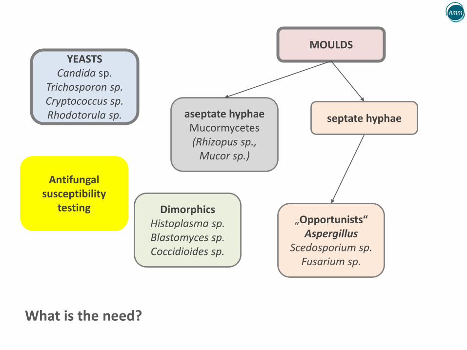
YEASTS Candida sp.
Trichosporon sp. Cryptococcus sp. Rhodotorula sp. aseptate hyphae
Mucormycetes (Rhizopus sp.,
Mucor sp.)
Dimorphics Histoplasma sp. Blastomyces sp. Coccidioides sp.
„Opportunists“ Aspergillus
Scedosporium sp. Fusarium sp.
MOULDS
septate hyphae
What is the need?
Antifungal susceptibility
testing

The human specimen
superficial cultures
sputum, TS, BAL
biopsy
fine needle-aspiration
commensale
flora sterile Infection
blood

Big trocar, big sample

Microscopy & Culture „must have“
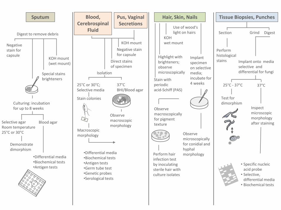
Sputum
Digest to remove debris
Negative stain for capsule
KOH mount (wet mount)
Special stains brighteners
Selective agar Room temperature 25°C or 30°C
Blood agar
Culturing: incubation for up to 8 weeks
Demonstrate dimorphism
•Differential media •Biochemical tests •Antigen tests
Pus, Vaginal Secretions
Tissue Biopsies, Punches
KOH mount
37°C BHI/Blood agar
Blood, Cerebrospinal
Fluid
Negative stain for capsule
Direct stains of specimen
Isolation
25°C or 30°C; Selective media
Stain colonies
Observe macroscopic morphology
Macroscopic morphology
•Differential media •Biochemical tests •Antigen tests •Germ tube test •Genetic probes •Serological tests
Hair, Skin, Nails
Use of wood‘s light on hairs
KOH wet mount
Highlight with brighteners; observe microscopically
Implant specimen on selective media; incubate for 4 weeks
Observe macroscopically for pigment texture
Perform hair infection test by inoculating sterile hair with culture isolates
Observe microscopically for conidial and hyphal morphology
Stain with periodic acid-Schiff (PAS)
Section Grind Digest
Perform histological stains Implant onto media
selective and differential for fungi
25°C - 37°C 37°C
Test for dimorphism
Inspect microscopic morphology after staining
• Specific nucleic acid probe
• Selective, differential media
• Biochemical tests


Microbiology best practice recommendations
Microscopy and stains • Fluids from usually sterile sites and bronchoalveolar lavage (BAL) from patients with
suspected infection should be examined by direct microscopy with suitable methods for fungal detection.
• Adequate tissue for histology (microscopy) and culture should be ensured before processing the rest of the sample.
Optical brighteners are recommended for microscopy on all samples from immunocompromised patients.
Direct fluorescent-antibody staining, PCR, or both is recommended for patients with suspected pneumocystis infection.
India ink staining of cerebrospinal fluid samples from immunocompromised patients is recommended in addition to Gram staining if Cryptococcus capsule antigen (CRAG) testing is not available on site.
BSMM standards, Lancet Infect Dis 2015 15:461-74

Histopathology best practice recommendations
Specialised stains Specialised stains should be done in parallel with standard stains if mycosis or another infection is to be assessed or excluded
• Standard stain: haematoxylin and eosin (H&E) on histopathology slides; Giemsa or Papanicolaou on smears.
• Triple set of stains: Ziehl-Neelsen stain for acid-fast organisms; Gram stain for bacteria, fungi, and others; Grocott silver stain, or periodic acid-Schiff, to highlight fungi.
BSMM standards, Lancet Infect Dis 2015 15:461-74

Histopathology best practice recommendations
Reporting of results Report fungal morphology (yeast or hyphae), including the following:
• Whether a yeast is small, medium, or large.
• Whether a yeast has cross walls or septa (ie, is splitting rather than budding).
• Whether a hyphal form has usual width, or has a dilated, bizarre shape.
• Whether H&E stained fungi are pigmented and brown, or are unpigmented and colorless or pale blue.
Positive results should be telephoned to clinicians immediately.
BSMM standards, Lancet Infect Dis 2015 15:461-74

Microbiology best practice recommendations
Culture and identification • Bronchoscopy fluids should be cultured in suitable media to support fungal growth.
• Yeasts cultured from urine samples should be identified to specie level and reported for all critical care and immunocompromised patients.
All clinical isolates of Aspergillus from patients who will receive antifungal treatment should be identified to species complex level, by referral to a specialist laboratory if necessary.
All fungi (yeasts and moulds) obtained from sterile sites, including blood and continuous ambulatory peritoneal dialysis fluids, and intravenous line tips should be identified to species complex level by referral to a specialist laboratory if necessary.
Bronchoscopy fluid and paranasal sinus material is regarded as sterile in this context for all fungi except Candida spp.
BSMM standards, Lancet Infect Dis 2015 15:461-74

Microbiology best practice recommendations
Antifungal drug-susceptibility testing Isolates of Candida spp from sterile sites, or from patients not responding to
therapy at a minimum should have their susceptibility tested against fluconazole.
• Isolates of Aspergillus fumigatus should have their susceptibility tested against antifungal agents used locally for treatment (eg, itraconazole and voriconazole) if antifungal treatment is given.
Antifungal susceptibility testing of Aspergillus isolates should be performed in patients who are unresponsive to antifungal treatment, for epidemiological purposes or in patients who are clinically suspected suffering from an azole-resistant isolate or in regions with a high prevalence of azole resistance.
BSMM standards, Lancet Infect Dis 2015 15:461-74 ESCMID Aspergillus Guideline, 2018, CMI in press

Serology and/or PCRs are „add on tests“

Copyright 2011 Rainer Poulet

Method Indication Advantages Disadvantages
Galactomannan
(GM)
Early detection of invasive
aspergillosis (IA)
2 serum samples/week,
positive cut-off index > 0.5
1 single samle, positive
cut-of index > 0.7
A screening test to accompany
conventional diagnostic methods in
patients at high risk of IA.
In neutropenic adults
In neutropenic children
Serum value > 1: sign of therapeutic
failure in adults and children
Quantification in BAL (cut-off >1)
and CSF (cut-off > 0.5) (useful in
neutropenic and non-neutropenic
patients)
In non-neutropenic
patients: not the same
diagnostic and prognostic
value
Mold-active antifungal
drug therapy is one of
the factors that may have
an impact on sensitivity
Persistent GM
antigenemia during
therapy is a poor
prognostic sign and
should prompt a
reassessment
Serological and molecular methods in the diagnosis of invasive fungal infections
Lackner & Lass-Flörl, Curr Pharm Res 2013, 19: 3595-614 ESCMID Aspergillus Guideline, CMI in press, 2018
Kullberg et al. N Engl J Med 2015;373:1445

Method Indication Advantages Disadvantages
B-D-glucan
(BG)
Diagnosis of IFI
2 samples/week
(minimum)
Pan-fungal marker in critically ill
patients and in cases of P. jiroveci
pneumonia
Does not cover Mucormycetes and
Cryptococcus neoformans
A frequency of 2 tests per week
seems an appropriate screening
strategy 37% false positive result: 1x 80 pg/mL 23% false positive results: 2x 80 pg/mL
Increases the specificity but
decreases the sensitivity
Site of infection may be important:
patients with tissue infections
failed to show a significant drop in
BG levels despite successful
outcomes
High NPV
False - (+) results (bacteraemia)
Limited experience (less widely used
than GM)
The threshold for positive results
depends on the test that is used:
Fungitell > 80 pg/mL
Wako > 70 pg/mL
Declines slowly in most IA, IC and PCP
patients with appropriate antifungal
therapy;
May persists above the usual
threshold for positivity long after
clinical resolution of the original
infection
Less acurate in hematological patients
Hanson KE et al. PLoS One 2012;7(8):e42282; Koo S et al. Clin Microbiol Infect 2012;18:E122-127; Jaijakul S et al. Clin Infect Dis 2012;55:521-526; ESCMID Aspergillus Guideline, CMI in press, 2018

Method Indication Advantages Disadvantages
Mannan
plus
Anti-mannan
Candidemia Good sensitivity and
specificity when
combined in ICU patients
Early diagnosis prior to
blood culture results
ESCMID Diagnostic &
Management Guideline
for Candida Diseases 2012
recommend this test-
combination, high
negative PV
Limited experience
Non-mycological criterion
The sensitivity and specificity
were 87.5% and 85.5% for
(1→3)-β-D-glucan and 89.3%
and 63.0% for mannan antigen
plus anti-mannan antibody
C. parapsilosis and C.
guilliermondii fungemias were
not detected by the Platelia
Candida Ag Plus assay
Serological and molecular methods in the diagnosis of invasive fungal infections
Mikulska et al, Crit Care Med 2010 14: R222; Held J et al, C Clin Microbiol 2013, 51(4):1158-64; Kullberg et al. N Engl J Med 2015;373:1445

Method Indication Advantages Disadvantages
Molecular methods
(polymerase chain
reaction; PCR)
Most experience of
in-house tests
DNA detection
mainly of Aspergillus
less experience for
Candida
Blood, BAL, …..
Early diagnosis (rapid
techniques), high NPV
High sensitivity (multicopy
genes), capacity for rapid
speciation and ability to
quantitate fungal burden
Low burden of organisms
during bloodstream
infections: <10 CFU/mL
(in 25% <1 CFU/mL) and
intermittent nature of
candidaemia due to
hepatic clearance of fungal
cells and/or periodic
release of cells from deep
organ sites into circulation
Additional techniques
Non-mycological criterion
(they are still in
development)
Limited to reference
laboratories (low
availability)
High costs, improve
technical equipment
Technical difficulties
of efficient fungal DNA
extraction from complex
clinical samples
Serological and molecular methods in the diagnosis of invasive fungal infections
Khot PD and Fredericks DN, Expert Rev Anti Infect Ther 2009; 7:1201-21; Clancy CJ and Nguyen MH, Curr Fungal Infect Rep 2011; 5: 135-140

Assay Methods Fungi Sensitivity (%) Specificity (%) Detection limit Processing time
Specimens
ePlex-BCID-FP GenMark DX „Bedside test“
Ready to use (DNA hybridization and electrochemical detection)
16 fungal targets: C. albicans C. dubliniensis C. famata C. glabrata C. guilliermondii C. kefyr C. krusei C. Lusitaniae C. Parapsilosis C. tropicalis
- - - 1.5 h Positive blood cultures
FilmArray BCID Panel Biomerieux
Ready to use Multiplex PCR
C. albicans C. glabrata C. krusei C. parapsilosis C. tropicalis
100 99.8-100 - 1 h Positive blood cultures
T2 Candida Panel T2 Biosystems
Ready to use (magnetic resonance assay)
C. albicans C. Tropicalis C. parapsilosis C. krusei C. glabrata
91.1 99.4 1 cfu/mL 4.5 h Whole blood
IRIDICA BAC BSI Abbott Diagnostics
PCR & mass spectrometry
Panmicrobial 81 84 8 cfu/mL 6 h Whole blood, sterile fluids, tissue, BAL, endotracheal aspirate
Examples on commercially available DNA-detecting methods for clinical specimens
Tests vary in the target, sen., spec., turnaround time and specimen application!
Lackner M and Lass-Flörl C, Methods Mol Biol. 2017;1508:85

Method Indication Advantages Disadvantages
Laterial Flow Device
Test for IA
Point-of-care test for
invasive aspergillosis
Detects an
extracellular
glycoprotein secreted
during active growth
of Aspergillus via mAB
JF5
Simple, rapid (15 min),
single-use test.
Can be performed in
rudimentary facilities using
BAL or serum specimens
Sensitivities for LFD, GM,
BDG, PCR were between 70
and 88%. Combined GM
(cut off >1.0 OD) with LFD
increased the sensitivity to
94%, while combined GM
(cut off >1.0 OD) with PCR
resulted in 100% sensitivity
(specificity for
probable/proven IPA 95-
98%).
Sensitivity and specificity
of BAL FD tests for
probable IPA were 100%
and 81% (PPV 71%, NPV
100%),
Only few proven patients
BAL Lateral Flow Device Test for IA
ESCMID Aspergillus Guideline, CMI in press, 2018

Microorganism Diagnostic test
Optimal
specimen
type
Sensitivity
(%)
Specificity
(%)
Reasons for false-
positive results
Reasons for false-
negative results Comments
Cryptococcus spp.
Cultures CSF >95 100 Uncommon Uncommon Gold standard, but takes 3–7 days for a positive result.
Histopathology Mostly CSF 75 100 Uncommon Low levels of microorganism
India ink stain often used as a screening test.
Cryptococcal antigen test (LA, EIA, or LFD)
CSF or serum
97 for CSF, 87 for serum
93–100 Trichosporon sp., Capnocytophaga sp., or Stomatococcus sp. invasive infections
Uncommon Most accurate test when performed on CSF. The three methods are comparable, although LA gives more false-positive results. LFD is best for rapid point-of-care diagnosis.
Non-molecular tests used for diagnosis of the most common invasive fungal infectionsa
aBAL, bronchoalveolar lavage; CAGTA, Candida albicans germ tube antibody; CF, complement fixation; CSF, cerebrospinal fluid; EIA, enzyme immunoassay; GM, galactomannan; ID, immunodiffusion; LA, latex agglutination; LFD, lateral-flow device; PPV, positive predictive value; TP, tube precipitin.
Arvanitis M et al. Clin Microbiol Rev. 2014, 27: 490

Microbiology best practice recommendations
Fungal serological and molecular testing • Serum samples from immunocompromised patients with presentations consistent
with cryptococcal meningitis for whom a CSF specimen is not available (eg, cases in which lumbar puncture is contraindicated) should be tested for Cryptococcus spp antigen (CRAG).
Galactomannan screening of serum (two times per week) from patients with haematological malignancies at high risk of invasive aspergillosis should be considered in those not receiving mould-active prophylaxis; optical density (OD) index threshold of 0.5 has a high negative predictive value, enabling invasive aspergillosis to be excluded.
BSMM standards, Lancet Infect Dis 2015 15:461-74

Microbiology best practice recommendations
Fungal serological and molecular testing Galactomannan testing of BAL from patients at high risk of invasive aspergillosis
should be considered, although the current OD index cutoff of 0.5 might change.
β-D-glucan screening of serum from patients at high risk of invasive fungal disease should be considered; a negative result has a high negative predictive value, enabling invasive fungal disease to be excluded.
PCR screening of serum for Aspergillus from patients at high risk of invasive fungal disease should be considered; a negative result has a high negative predictive value, enabling invasive fungal disease to be excluded.
BSMM standards, Lancet Infect Dis 2015 15:461-74

Microbiology best practice recommendations
Therapeutic drug monitoring No indications for therapeutic drug monitoring of amphotericin B or the
echinocandins; measurement of fluconazole concentrations is rarely necessary.
Therapeutic drug monitoring of itraconazole, voriconazole, and posaconazole is usually needed. Specifically, voriconazole monitoring is needed in most patients, and certainly in children, including repeat monitoring after dose changes and shift from intravenous to oral treatment; dose optimisation during long-term therapy needs such monitoring.
Flucytosine monitoring is recommended for all patients receiving treatment.
BSMM standards, Lancet Infect Dis 2015 15:461-74

Fungal diagnosis: limitations
Clinical manifestations are non-specific
Conventional diagnostic tests insensitive, positive late
Fungi can be both, colonizers and pathogens, hence vigilance is required in the interpretation of:
- superficial cultures
- antigen tests, PCR screening, presence of antibodies and/or metabolites
Lass-Flörl 2016, Walsh 2012, Kontoyiannis 2008, Kullberg et al. N Engl J Med 2015;373:1445

Culture Microscopy
Anti-Aspergillus antibodies
Aspergillus antigens
PCR Imaging
Chronic aspergillosis
+ ++ - + Radiography
Invasive aspergillosis
++ - ++ ++ CT scan
Allergic aspergillosis
+/- + - -/+ Radiography
Variable contribution of diagnostic tools according to the disease
ESCMID Aspergillus Guideline, CMI in press, 2018

„Puzzle diagnosis“
Microscopic Examination
Culture
Serology tests
Clinical features
CT

Based on the patient population targeted decide what to do when and how!
Specimens
Patients Tests

Important rules
1. Educate your doctors to give you the „best clinical specimens“
2. Choose tests according your „local epidemiology“ and „patients‘ symptoms & history“
3. Culture and microscopic examinations: must have!
4. Define indirect tests as an „add on“ and have „assay variabilities“ in mind
6. Be aware of the pro & cons
7. No test covers all fungi!

Ausblick 2009


Thank you for your attention!