how the cell divide? - scholar.cu.edu.eg mitotic cell division (mitosis) and meiotic cell division...
TRANSCRIPT

HOW THE CELL DIVIDE?
Cells spend a small part of their life dividing. Cell division is very
tightly controlled, ensuring that everything happens correctly (by
checkpoints) at the right time and in the right order (by regulation).
Cellular division refers to the process by which the living cells divide
through complicated process into 2 or more to transmit its genetic
material for reproduction, tissue renewal (wound healing), growth and
development.
Cell divisions include 2 main events: Cellular and Nuclear divisions.
Cellular divisions (Cytokinesis) refer to the process by which cytoplasm
and cell components are divided. While, nuclear divisions (Karyokinesis)
refer to the process by which a nucleus divides. Two major nuclear
divisions are involved in the genetic continuity of the nucleated cells:
Mitotic cell division (mitosis) and Meiotic cell division (Meiosis).
Mitosis is the process of cell division in which the daughter cells
receive identical copies of DNA of the mother cell. Meiosis is the process
of cell division that results in the formation of cells containing half the
amount of DNA contained in the parent cell, and having different copies
of DNA from one another. The cytoplasm and organelles are usually
shared approximately equally between the daughter cells. So, Mitosis
creates genetically identical species, while Meiosis increases genetic
diversity in a species.
1. CELL CYCLE
The cell cycle occurs from the completion of one division until the
completion of the next division. It involves 3 phases: Interphase (G1, S
and G2), Mitosis (M) followed by Cytokinesis (C). The period between
M and S is called G1 stage and that between S and M is G2 stage (figure
below).

Genetics (B 252) Lecture 2 2017-2018
2
The cell spends 90% of its time in Interphase and only 10% in
Mitosis but, the duration of each phase and stage in eukaryotic cells
depends on the cell type: For a typical rapidly proliferating normal human
somatic cell with a total cycle time of 24 hours (1440 min), the G1 phase
might last about 11 hours, S phase about 8 hours, G2 about 4 hours, and
M about 1 hour. Other types of cells, however, can divide much more
rapidly as budding yeast and embryo cells: Yeast cell has a total cycle
time of 2 hours (120 min), the G1 phase might last about 15 mins, S
phase about 10 mins, G2 about 90 mins, and M about 5 mins.

Genetics (B 252) Lecture 2 2017-2018
3
Other example:
Cell Type Total Time
fly embryo 8 minutes
bacteria 20 minutes
human skin 20 - 24 hours
human liver Once then retain
human nerve never once mature
Chlamydomonous 14 hours
Note: Some cells divide rapidly as beans, for example take 19 hours for
the complete cycle). While other like red blood cells cannot divide at all
as they don‟t contain nucleus. Others, such as nerve cells, lose their
capability to divide once they reach maturity (loss centrosome). Some
cells, such as liver cells, retain but do not normally utilize their capacity
for division. Liver cells will divide if part of the liver is removed. The
division continues until the liver reaches its former size.
Read only: Why can't nerve cells in our brain divide?
However in general, neurons don't divide. There are a few reasons why.
One is structural. The tree shape of a neuron is not really divisible. When
new neurons are created during brain development, they come from
spherical progenitor or neural stem cells, usually in specialized "neuron
factory" regions of the brain. The new neurons travel along cellular
highways and off ramps until they migrate into their final position. From
there, they sprout axons and dendrites and wire themselves into the
surrounding tissue.
Another reason is cranial capacity. Because the size of the skull is fixed,
adding neurons requires killing off and removing neurons someplace else.
Is the brain better off trading old neurons for new ones or using the
neurons it already has?
Genetics (B 252) Lecture 2 2017-2018
4
Which brings us to the third and probably most important reason:
Memories, skills, and the things we have learned throughout life are
represented by the complex tree-shaped structural interconnections within
the fabric of brain tissue. If neurons are taken away, what those neurons
learned is taken away with them. Replacing “old” neurons with “new”
ones would amount to erasing memories.
So generally speaking, the brain is better off with the experienced
neurons that have already been wired up than with new neurons that need
to start from scratch. Besides, the most important part of the brain is not
the neurons, but the connections between them, and those are being
added and removed all the time ("synaptic remodeling") as we acquire
life experience.
STEPS:
Interphase
The time between two successive mitotic divisions is known as
Interphase (Resting or Growth stage). During interphase, the genetic
material in the nucleus is in form of chromatin (uncoiled DNA), which
appears only as dark granules within the nucleus. This appearance may be
because they are uncoiled, long and thin strands. Both nucleolus and
nuclear membranes are present and clearly visible.
In this phase, the cell prepares itself for division through a group of
biological processes for cell growth and accumulating nutrients needed
for mitosis and duplicating its DNA.
The interphase involves 3 stages called G1, S and G2, respectively.
G1 stage (gap1, Pre-DNA synthesis): It lasting in a range of 4-9
hours depending on the type of eukaryotic cells. The cells become
metabolically active (1ry
growth) producing RNA and ribosomes for
protein synthesis; the cell organelles begin to increase in numbers, and

Genetics (B 252) Lecture 2 2017-2018
5
the nucleus and cytoplasm enlarge so, the cell reach their mature size
(small in size from previous division). The genetic materials are 2n in
number (diploid cell), fully extended and single in structure i.e. a
chromatid with a centromere (unduplicated chromosome, Monad).
Cells that have temporarily (reversibly) or permanently stopped dividing
are said to have entered a state of quiescence called G0 phase (Prolonged
G1 phase). This phase refers also as non-dividing phase outside of the
cell cycle (see figure below) in which the cell will readjusted and
stimulated to return to G1 and thereby reenter the cell cycle. If the cells
will never divide again i.e. permanently arrested (never reenter the cell
cycle) it will pass through a process of destruction (apoptosis= suicide).
S stage (DNA synthesis): DNA and histone syntheses lasting in a
range of 6-9 hours depending on the type of eukaryotic cells. DNA and
histone are the main component of chromatids (previously mentioned). At
the end of this stage, monads have been duplicated and became double in
structure i.e. with 2 sister chromatids (duplicated chromosome, dyad)
joined by a centromere (figure below) but still diploid (2n).

Genetics (B 252) Lecture 2 2017-2018
6
Two unduplicated chromosomes Two duplicated chromosomes
Sister chromatids are held together by multi-subunit protein complexes
called cohesin and Shugoshin in interphase and mitosis (figure below).
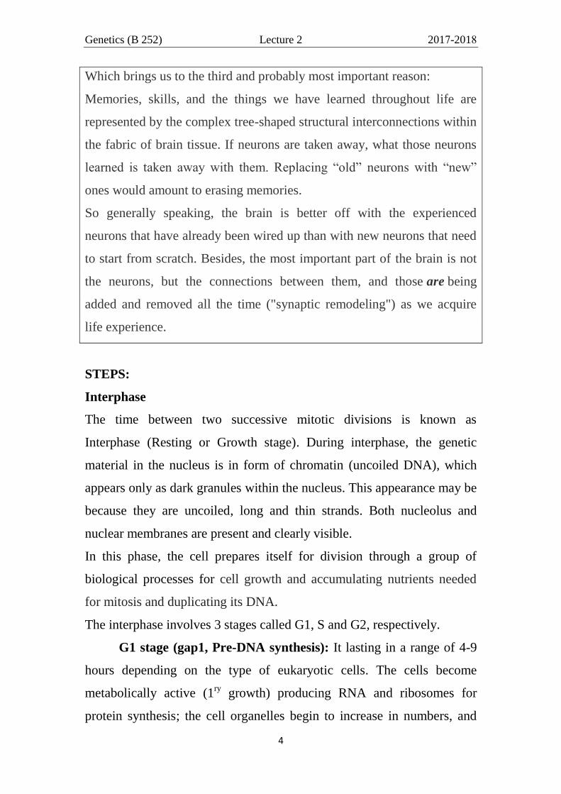
G2 stage (gap2, Post-DNA synthesis): This stage lasting from 2-5
hours in some eukaryotic cells. In which the cell synthesis certain
component required for mitosis (assemble machinery) as microtubules in
plants and microorganisms (centrosomes, centrioles and asters in animal,
figure below), proteins of spindle fiber, enzymes,... and go to the final
preparations of the cell (2nd
growth) before divisions. The chromosomes
are 2n (diploid) double in structure (dyad) but invisible in this form
(uncoil) and the nucleus is filled with chromatin fibers that are formed
when the chromosomes are uncoil.
Duplication
Interphase
Mitosis
Cohesion
Chromatin
network Shugoshin
Genetics (B 252) Lecture 2 2017-2018
7
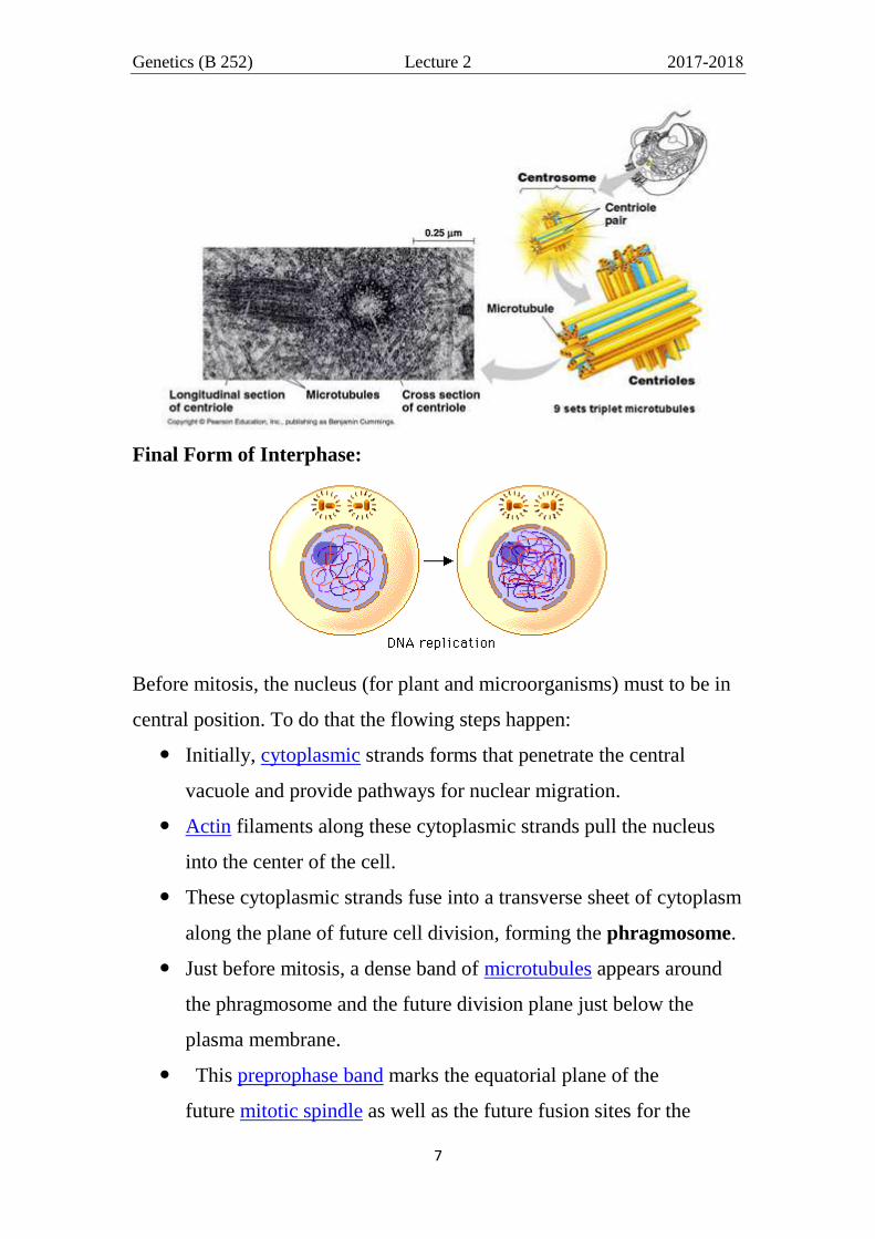
Final Form of Interphase:
Before mitosis, the nucleus (for plant and microorganisms) must to be in
central position. To do that the flowing steps happen:
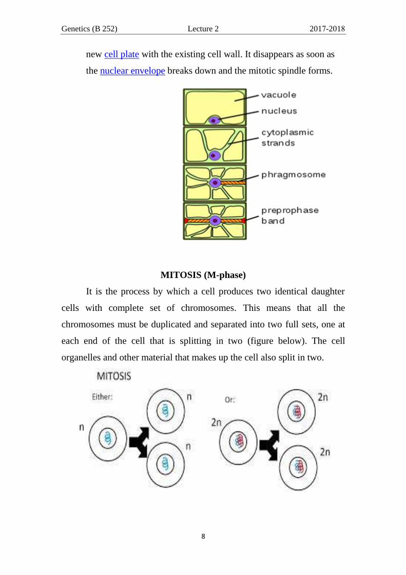
Initially, cytoplasmic strands forms that penetrate the central
vacuole and provide pathways for nuclear migration.
Actin filaments along these cytoplasmic strands pull the nucleus
into the center of the cell.
These cytoplasmic strands fuse into a transverse sheet of cytoplasm
along the plane of future cell division, forming the phragmosome.
Just before mitosis, a dense band of microtubules appears around
the phragmosome and the future division plane just below the
plasma membrane.
This preprophase band marks the equatorial plane of the
future mitotic spindle as well as the future fusion sites for the
Genetics (B 252) Lecture 2 2017-2018
8
new cell plate with the existing cell wall. It disappears as soon as
the nuclear envelope breaks down and the mitotic spindle forms.
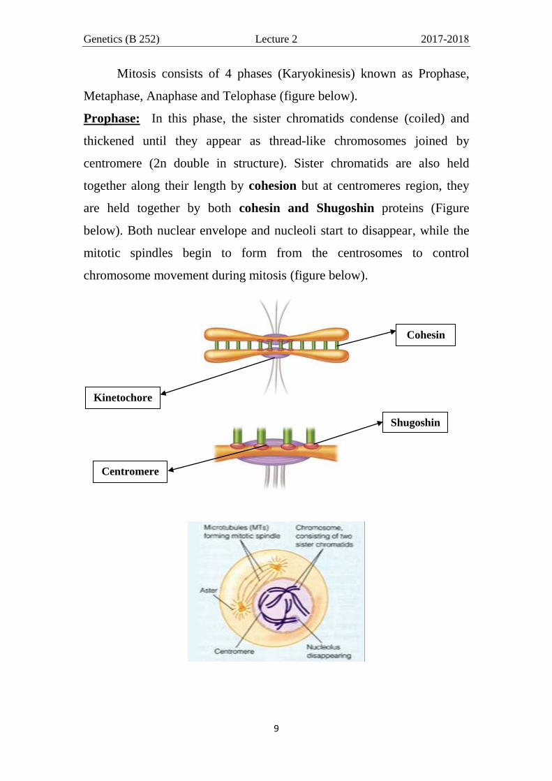
MITOSIS (M-phase)
It is the process by which a cell produces two identical daughter
cells with complete set of chromosomes. This means that all the
chromosomes must be duplicated and separated into two full sets, one at
each end of the cell that is splitting in two (figure below). The cell
organelles and other material that makes up the cell also split in two.
Genetics (B 252) Lecture 2 2017-2018
9
Mitosis consists of 4 phases (Karyokinesis) known as Prophase,
Metaphase, Anaphase and Telophase (figure below).
Prophase: In this phase, the sister chromatids condense (coiled) and
thickened until they appear as thread-like chromosomes joined by
centromere (2n double in structure). Sister chromatids are also held
together along their length by cohesion but at centromeres region, they
are held together by both cohesin and Shugoshin proteins (Figure
below). Both nuclear envelope and nucleoli start to disappear, while the
mitotic spindles begin to form from the centrosomes to control
chromosome movement during mitosis (figure below).
Cohesin
Centromere
Kinetochore
Shugoshin

Genetics (B 252) Lecture 2 2017-2018
10
NOTE: The spindle apparatus (figure below) includes the centrosomes in
animal cell but microtubules in plant cell, the spindle microtubules,
associated proteins and the asters (a radial array of short microtubules in
animal cell). The centrosome replicates in interphase, forming two
centrosomes each with 2 centrioles that migrate to opposite ends of the
cell in prophase. Assembly of spindle microtubules begins in the
centrosome (the microtubule organizing center in plant) and an aster
extends from each centrosome.
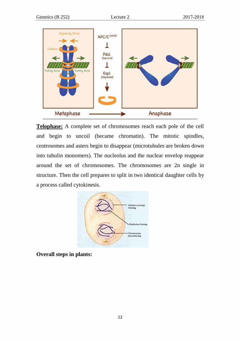
Metaphase: When the mitotic spindle is fully formed, the chromosomes
align themselves along the cell spindle in the middle of the cell (equator,
equatorial plates). This movement is due to: Assembly and disassembly
of microtubules provide force to move chromosomes with the help of the
motor proteins located in kinetochore and poles of cell pull on
microtubules to provide force. The metaphase chromosome (2n double in
structure) appears as two sister chromatids join together by their
centromeres and to the spindles by their kinetochore (figure below). At
this stage, separase enzyme (and others) dissolves the cohesion protein

Genetics (B 252) Lecture 2 2017-2018
11
along the 2 sister chromatids except at centromere were both cohesion
and Shugoshin proteins remains (Figure below).
Anaphase: Both cohesion and shugoshin dissolve by proteiolytic
enzymes so, the sister chromatids (present in equator) split apart at their
centromeres, begin to separate and move to opposite poles of the spindle,
segregating one of the two sister chromatids to each of the opposite ends
of the cell. In this case, each chromatid became a chromosome. The
chromosomes are 2n single in structure (2n monad).
cohesion
Shugoshin
Genetics (B 252) Lecture 2 2017-2018
12
Telophase: A complete set of chromosomes reach each pole of the cell
and begin to uncoil (became chromatin). The mitotic spindles,
centrosomes and asters begin to disappear (microtubules are broken down
into tubulin monomers). The nucleolus and the nuclear envelop reappear
around the set of chromosomes. The chromosomes are 2n single in
structure. Then the cell prepares to split in two identical daughter cells by
a process called cytokinesis.
Overall steps in plants:

Genetics (B 252) Lecture 2 2017-2018
13
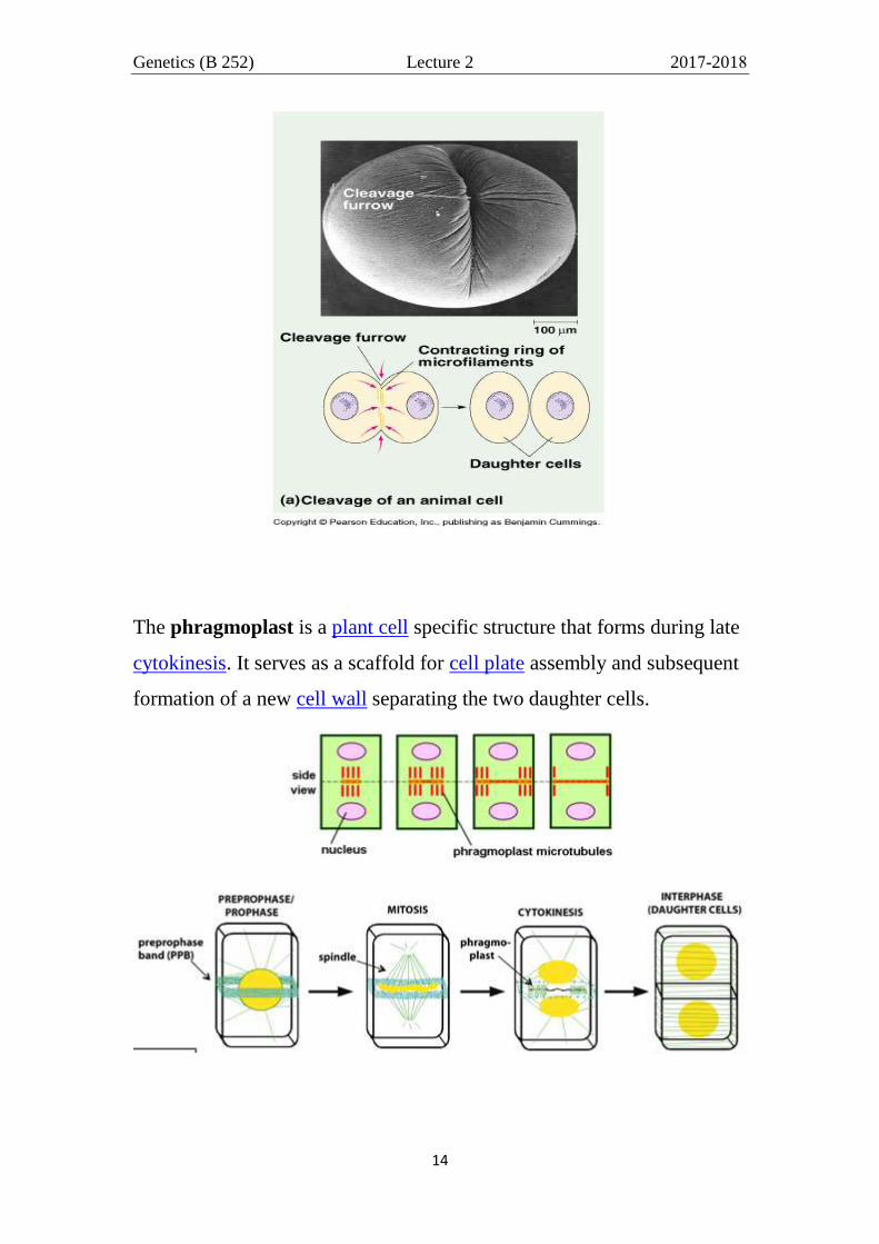
CYTOKINESIS
It usually initiates during the late stages of mitosis (at the end of
telophase), and sometimes meiosis, splitting a cell in two, to ensure
that chromosome number is maintained from one generation to the next
or one cell to another.
In animal, the cell membranes on opposite sides of the cell become
pinched-in (constriction) allowing for the cell to divide. The initial
structure that forms is called a cleavage furrow. The cleavage furrow
continues to pinch in, until the two sides are touching. At this point, there
will be two new cells.
Genetics (B 252) Lecture 2 2017-2018
14
The phragmoplast is a plant cell specific structure that forms during late
cytokinesis. It serves as a scaffold for cell plate assembly and subsequent
formation of a new cell wall separating the two daughter cells.

Genetics (B 252) Lecture 2 2017-2018
15
The phragmoplast is a complex assembly of microtubules (MTs),
microfilaments (MFs), and endoplasmic reticulum (ER) elements, that
assemble in two opposing sets perpendicular to the plane of the
future cell plate during anaphase and telophase. It is initially barrel-
shaped and forms from the mitotic spindle between the two daughter
nuclei while nuclear envelopes reassemble around them. The cell plate
,originates from vesicles of Golgi apparatus, begins to grow and elongate
in the center of the cell (at the region of the metaphase plate), forming a
disc between the two halves of the phragmoplast structure. While new
cell plate material is added to the edges of the growing plate, the
phragmoplast microtubules disappear in the center and regenerate at the
edges of the growing cell plate. The two structures grow outwards until
they reach the outer wall of the dividing cell. If a phragmosome was
present in the cell, the phragmoplast and cell plate will grow outwards
through the space occupied by the phragmosome. They will reach the
parent cell wall exactly at the position formerly occupied by
the preprophase band.

Genetics (B 252) Lecture 2 2017-2018
16
Figure 1 : Cytoskeletal organization in dividing plant cells. Microtubules (green) and actin filaments
(red) are illustrated at successive cell cycle stages in relation to nuclei/chromosomes (yellow) and the
cell plate (black).
Once the cell plate has divided the cell into two cells, it forms the middle
lamella. In the same time the plasma membrane of the maim cell split
and begin to reform in the both daughter cells. Subsequently, the cell will
develop new primary and secondary layers of cell wall (figure below).
The microtubules and actin filaments within the phragmoplast serve to
guide vesicles (from golgi) with cell wall material to the growing cell
plate. Actin filaments are also possibly involved in guiding the
phragmoplast to the site of the former preprophase band location at the
parent cell wall.

Genetics (B 252) Lecture 2 2017-2018
17
While the cell plate is growing, segments of smooth endoplasmic
reticulum are trapped within it, later forming the plasmodesmata (a
narrow thread of cytoplasm that passes through the cell walls of adjacent
plant cells and allows communication between them).
The phragmoplast can only be observed in Embryophytes
(the bryophytes and vascular plants as well as a few advanced green
algae). Some algae use another type of microtubule array, a phycoplast,
during cytokinesis.
NOTE: The cytoplasm and organelles are usually shared approximately
equally between the daughter cells.

Genetics (B 252) Lecture 2 2017-2018
18
CELL CYCLE CHECKPOINTS
Maintenance of genomic stability is needed for cells to survive
many rounds of division throughout their lifetime without disruption. Key
to the proper inheritance of intact genome is the tight temporal and spatial
coordination of cell cycle events to monitor the proper execution of cell
cycle processes to avoid uncontrolled cell division characterizing
malignancy. Those keys are the cell cycle checkpoints.
As we have outlined previously, the cell cycle consists of four
primary stages, G1 (GAP 1, 1ry
growth), S (Synthesis), G2 (GAP 2, 2nry
growth) and M (Mitosis). In order for each of the stages to have good
participation in the cycle, DNA must clear all the checkpoints which it
encounters along the way.
Multiple checkpoints have been identified as G1 checkpoint, DNA
replication checkpoints, G2 checkpoint, antephase checkpoint and Mitotic
spindle checkpoint.
G1 checkpoint (restriction point) is located at the end of the
G1 phase, just before entry into S phase (G1/S) to monitor the size the cell
has achieved since its previous mitosis, nutrition, growth factors and also

Genetics (B 252) Lecture 2 2017-2018
19
to evaluate the condition of the DNA. It is a vital checkpoint making the
key decision of whether the cell should divide, delay division, or enter a
resting stage. If all conditions are “normal”, then the cell is allowed to
proceed from G1 to the S phase of the cycle. If the cell has not reached an
adequate size or if the DNA has been damaged, further progress through
the cycle is arrested until these conditions are “corrected.”
The DNA replication checkpoint is located at the end of the S
phase to ensure the good replication of DNA before entering G2 phase.
The G2 checkpoint is another checkpoint (after completing S and
G2 phases) in which DNA must overcome to complete a successful cycle.
In order for this checkpoint to be passed, the cell has to check a number
of factors, including DNA, to ensure that the cell is ready for advancing
to the M or mitosis phase.
The antephase checkpoint has recently been gaining attention.
The term “antephase” refers to the time in late G2 phase
when energy is being produced and stored for mitosis and signs of
chromosome condensation first become visible until commitment to
mitosis. This checkpoint plays an important role in preventing mitotic
entry (safeguarding) in the presence of various stress conditions by
preventing chromosome condensation and segregation.
The mitotic spindle checkpoint (spindle assembly checkpoint)
occurs at metaphase where all the chromosomes should/have aligned at
the mitotic plate (equator) and be under bipolar tension (tension of both
poles). The tension created by this bipolar attachment is what is sensed,
which initiates the anaphase entry i.e. the anaphase will be blocked if the
chromatids are not properly assembly on mitotic spindle by their
kinetochores. In addition, if this failure to attach correctly to the spindle
passes, it causes an unequal segregation of chromosomes (non-
disjunction), which can lead to cell death or disease.
Genetics (B 252) Lecture 2 2017-2018
20
The DNA damage and spindle assembly checkpoints are
surveillance mechanisms that ensure genomic integrity by delaying cell
cycle progression in the presence of DNA or spindle damages,
respectively until all chromosomes are correctly attached in a bipolar
fashion to the mitotic spindle.
NOTE:
The check for DNA damage in eukaryotic cell division is to
successfully pass accurate DNA strands (mutation free) from parental
genomes to daughter cells as cells mitotically replicates. The passing of
mutation-free DNA will ensure the cycle procedures healthy and
functional cells. However, DNA does not always exist as mutation free
and DNA with mutations (due to either irradiation or chemical
modification) will likely lead to cancer. For the prevention of passing
DNA which could cause replication of cancerous cells, the cell cycle
includes an impressive system of checkpoints that, more or less, scan the
DNA passing through the cycle for mutations (or any damages)
by sensor mechanisms i.e. those checkpoints verify (and assess) whether
the processes (done before or needed) at each phase along the cell
cycle have been accurately completed before progression into the next
phase (Figure below).
Checkpoints along the cycle not only assess the DNA for damage
but can actually act upon it in effort to correct any mutation which is
hindering its advancement in the cycle. Signal Mechanisms within the
checkpoints can delay (or stall) the cycle until mutations are corrected. If
the G1 checkpoint deems the DNA unsuitable for progression it can stop
or delay the process sending it into an optional resting phase known as
G0. A special protein referred to as P53 is essential in the function of the
Genetics (B 252) Lecture 2 2017-2018
21
G1 restriction point as P53 has the ability to detect mutations in the genes
which pass through the checkpoint.
If mutations are irreversible, they can tag a cell for self-
destruction (cell suicide) via apoptosis (effector mechanism) and
thereby block progression through the cell cycle by eliminating the
chance that mutated DNA will be replicated. However, as we all know,
this process is not always flawless, causing the spread of mutation filled,
mutated cells.
Without DNA damage checkpoints throughout the process of cell
division and replication, the transferring of mutated genes would be more
likely. Viable checkpoints and non-damaged P53 are necessary to ensure
that DNA being replicated is mutation free. Mutant cells may spread with
more amplification and at a must quicker rate if it weren‟t for the
detection of checkpoints in the process of cell division.
Regulation of Eukaryotic Cell Cycle
Not all cells proceed through the stages of the cell cycle at the same
rate. Embryonic cells divide very rapidly, while mature cells might divide
rarely, or in response to signals such as wounding or growth factors, or
not at all.
It should seem obvious that the processes that drive a cell through
the cell cycle must be highly regulated and required a number of control
mechanisms to ensure that the resultant daughter cells are viable and each
contains the complement of DNA found in the original parental cell.
They control the timing of events so that each individual process is turned
on and off at the appropriate time, mechanisms to initiate each event in
the correct order and to also ensure that each event is triggered only once
per cell cycle, controls to ensure events occur in a linear, irreversible
Genetics (B 252) Lecture 2 2017-2018
22
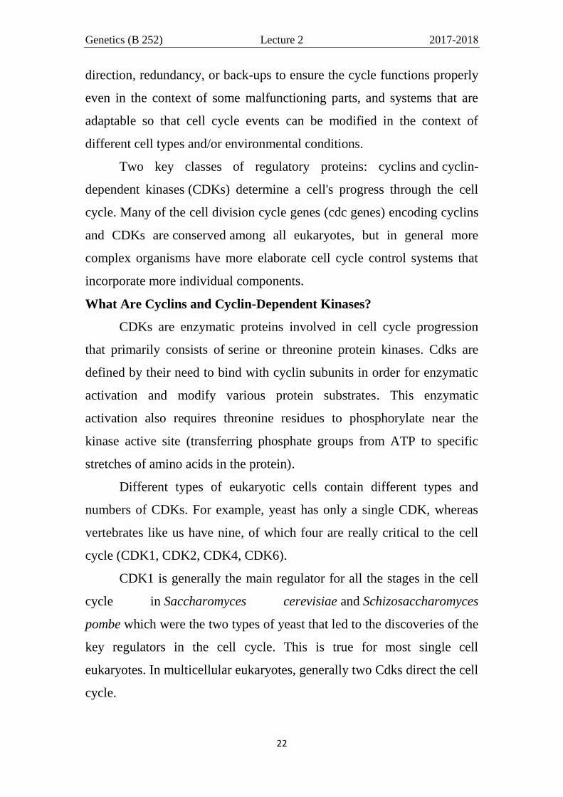
direction, redundancy, or back-ups to ensure the cycle functions properly
even in the context of some malfunctioning parts, and systems that are
adaptable so that cell cycle events can be modified in the context of
different cell types and/or environmental conditions.
Two key classes of regulatory proteins: cyclins and cyclin-
dependent kinases (CDKs) determine a cell's progress through the cell
cycle. Many of the cell division cycle genes (cdc genes) encoding cyclins
and CDKs are conserved among all eukaryotes, but in general more
complex organisms have more elaborate cell cycle control systems that
incorporate more individual components.
What Are Cyclins and Cyclin-Dependent Kinases?
CDKs are enzymatic proteins involved in cell cycle progression
that primarily consists of serine or threonine protein kinases. Cdks are
defined by their need to bind with cyclin subunits in order for enzymatic
activation and modify various protein substrates. This enzymatic
activation also requires threonine residues to phosphorylate near the
kinase active site (transferring phosphate groups from ATP to specific
stretches of amino acids in the protein).
Different types of eukaryotic cells contain different types and
numbers of CDKs. For example, yeast has only a single CDK, whereas
vertebrates like us have nine, of which four are really critical to the cell
cycle (CDK1, CDK2, CDK4, CDK6).
CDK1 is generally the main regulator for all the stages in the cell
cycle in Saccharomyces cerevisiae and Schizosaccharomyces
pombe which were the two types of yeast that led to the discoveries of the
key regulators in the cell cycle. This is true for most single cell
eukaryotes. In multicellular eukaryotes, generally two Cdks direct the cell
cycle.

Genetics (B 252) Lecture 2 2017-2018
23
Cyclins are a family of proteins that form the regulatory subunits,
while CDKs are the catalytic subunits of the activated complex; cyclins
have no catalytic activity and CDKs are inactive in the absence of a
partner cyclin. Each cyclin associates with one or two cyclin-dependent
kinases to be partially activated.
Cyclins CDK Partners
cyclin D
(D1, D2, D3) CDK4, CDK6
cyclin E CDK2
cyclin A CDK2
cyclin B CDK1
CDKs are constitutively expressed in cells whereas cyclins are
synthesized at specific stages of the cell cycle, in response to various
molecular signals. All CDKs exist in similar amounts throughout the
entire cell cycle. In contrast, cyclin manufacture and breakdown varies by

Genetics (B 252) Lecture 2 2017-2018
24
stage with cell cycle progression dependent on the synthesis of new
cyclin molecules.
These proteins are the activators for CDK enzymes. Typically, cyclins are
created or destroyed according to whether they are required which directs
the cell through the various stages of the cell cycle. When cyclins bind
with CDKs, they form a complex where the CDK active site is triggered.
Cyclins are named so because their concentration changes in a cyclical
manner during the cell cycle (figure below).
When cyclin concentrations are low, cyclins detach from CDKs which
in turn inhibit the enzymes activity: cyclins do not have enzymatic
activity alone. After the detachment, CDKs are thought to block their
active site with a protein chain to ensure cyclin concentration increases.
When cyclins are bound to CDKs, they for the promoting factor for
maturation which is primarily the complex formed. This complex is what
stimulates meiosis and the cell cycles in mitosis. It is important to note
that only when this complex is formed, does stimulation occur and not
their subunits alone

Genetics (B 252) Lecture 2 2017-2018
25
How are the CDKs themselves regulated?
The levels of these proteins remain pretty constant throughout the cell
cycle, yet their levels of activity rise and fall cyclically. CDKs need to
hydrolize ATP for energy in order to perform phosphorylation. They
have an ATP binding cleft whose ability to bind ATP is regulated by two
mechanisms. First, CDKs have a „flexible T loop‟ which contains a
threonine (T) residue which normally blocks the ATP binding cleft, but
not when the T is phosphorylated. Second, cyclins bind CDKs and
induce a conformational change that also helps to expose the ATP
binding cleft. Therefore a fully active CDK is one which is both
phosphorylated at the T on the T loop and is bound to a cyclin.
Cyclin/CDK complexes regulate the cell cycle both by promoting
activites for their respective stages, and by inhibiting activites for future
cell cycle stages that must not yet be reached. Therefore cyclins must be
able to be both generated and degraded in order for the cell cycle to
proceed.
All eukaryotes have multiple cyclins, each of which acts during a
specific stage of the cell cycle. All cyclins are named according to the
stage at which they assemble with CDKs. Common classes of cyclins
include G1-phase cyclins, G1/S-phase cyclins, S-phase cyclins, G2-phase
cyclins and M-phase cyclins (table below).
Genetics (B 252) Lecture 2 2017-2018
26
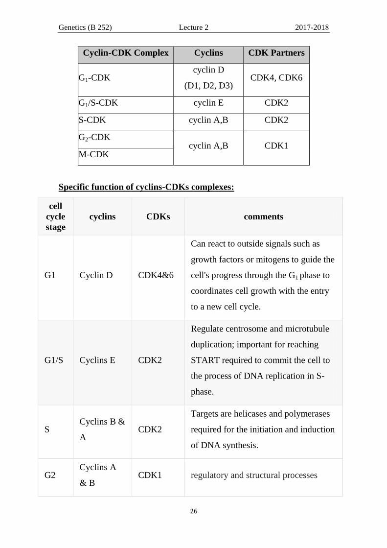
Cyclin-CDK Complex Cyclins CDK Partners
G1-CDK cyclin D
(D1, D2, D3) CDK4, CDK6
G1/S-CDK cyclin E CDK2
S-CDK cyclin A,B CDK2
G2-CDK cyclin A,B CDK1
M-CDK
Specific function of cyclins-CDKs complexes:
cell
cycle
stage
cyclins CDKs comments
G1 Cyclin D CDK4&6
Can react to outside signals such as
growth factors or mitogens to guide the
cell's progress through the G1 phase to
coordinates cell growth with the entry
to a new cell cycle.
G1/S Cyclins E CDK2
Regulate centrosome and microtubule
duplication; important for reaching
START required to commit the cell to
the process of DNA replication in S-
phase.
S Cyclins B &
A CDK2
Targets are helicases and polymerases
required for the initiation and induction
of DNA synthesis.
G2 Cyclins A
& B CDK1 regulatory and structural processes

Genetics (B 252) Lecture 2 2017-2018
27
cell
cycle
stage
cyclins CDKs comments
M
Cyclins
A & B are
synthesized
during S
CDK1
- Regulate G2/M checkpoint.
- drive the cell's entry to promote the
events of mitosis like the assembly of
mitotic spindles and alignment of
sister-chromatids along the spindles.
- Phosphorylate lots of downstream
targets as nuclear envelope and
initiation of prophase, and
subsequently, its deactivation causes
the cell to exit mitosis.
How Do CDKs Control the Cell Cycle?
Interestingly, CDKs require the presence of cyclins to become
partially active. CDKs must also be in a particular phosphorylation state,
with some sites phosphorylated and others dephosphorylated, in order for
activation to occur. When activated by a bound cyclin, CDKs perform a
common biochemical reaction called phosphorylation that activates or
inactivates target proteins to orchestrate coordinated entry into the next
phase of the cell cycle.
Although CDKs are inactive unless bound to a cyclin, there is more
to the activation process than just the interaction of the two parts of the
complex. When cyclins bind to CDKs they alter the conformation of
the CDK resulting in exposure of a spot that is the site of
phosphorylation by another kinase called CDK-activating kinase
(CAK). Following phosphorylation the cyclin-CDK complex is fully
active (figure below).

Genetics (B 252) Lecture 2 2017-2018
28
Cyclin degradation is equally important for progression through the
cell cycle and specific enzymes break down cyclins are present at defined
times in the cell cycle. When cyclin levels decrease, the corresponding
CDKs become inactive. Cell cycle arrest can occur if cyclins fail to
degrade.
CAK phosphorylation is exerted to inhibit CDK activity through
interaction with inhibitory proteins or by inhibitory phosphorylation
events (dephosphorylation). Thus, there is extremely tight control on
the overall activity of each CDK. Proteins that bind to and inhibit
cyclin-CDK complexes are called CDK inhibitory proteins (CKI, for
cyclin-kinase inhibitor), figures below.

Genetics (B 252) Lecture 2 2017-2018
29
Cdk activity can be suppressed both by inhibitory phosphorylation and by inhibitory proteins.
Eukaryotic cell cycle phases with respective cyclin-CDK complexes and inhibitors (CDKs)
Genetics (B 252) Lecture 2 2017-2018
30
IMPORTANCE OF MITOSIS:
Following are the occasions in the lives of organism where mitosis
happens:
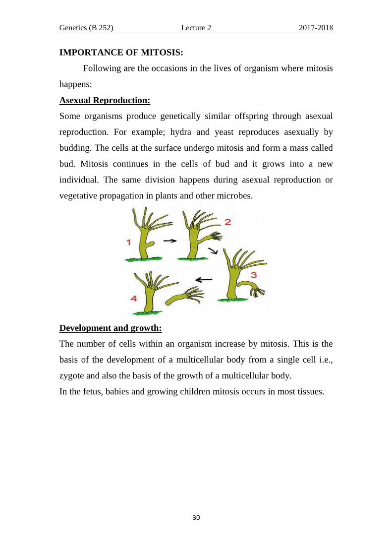
Asexual Reproduction:
Some organisms produce genetically similar offspring through asexual
reproduction. For example; hydra and yeast reproduces asexually by
budding. The cells at the surface undergo mitosis and form a mass called
bud. Mitosis continues in the cells of bud and it grows into a new
individual. The same division happens during asexual reproduction or
vegetative propagation in plants and other microbes.
Development and growth:
The number of cells within an organism increase by mitosis. This is the
basis of the development of a multicellular body from a single cell i.e.,
zygote and also the basis of the growth of a multicellular body.
In the fetus, babies and growing children mitosis occurs in most tissues.

Genetics (B 252) Lecture 2 2017-2018
31
While in adults, however, most tissues do not proliferate but mitosis
occurs regularly at the following sites:
1. Red bone marrow – for production of blood cells (erythropoiesis)
2. Lymphoid tissue - formation of lymphocytes (lymphooiesis)
3. Testes – for spermatogenesis (production of spermatozoa)
4. Epidermis - replacement of superficial skin cells
5. Hair follicles - hair growth
6. Gastro-intestinal tract - renewal of epithelium
Note that most of the neural cells do not perform mitosis so; any damage
in them cannot be repair.
In plants, mitotic cell division mainly takes place in special regions
called meristems. They are either present in Shoot apex or axillary buds
or root tips of the plants for development and growth.

Genetics (B 252) Lecture 2 2017-2018
32
Cell Replacement:
In some parts of body, e.g. skin and digestive tract, cells are constantly
sloughed off and replaced by new ones. New cells are formed by mitosis
and so are exact copies of the cells being replaced. Similarly, RBCs have
short life span (only about 4 months) and new RBCs are formed by
mitosis.

Genetics (B 252) Lecture 2 2017-2018
33
Regeneration:
Some organisms can regenerate (form de novo) their parts of bodies. The
production of new cells is achieved by mitosis. For example; hydra, sea
star and flat worms regenerate their lost part through mitosis.
Flat worm Sea Star
APPLICATIONS:

Genetics (B 252) Lecture 2 2017-2018
34
(1) Clinical
Cancer cells undergo uncontrolled cell proliferation. As such, they are
defects of the control of the cell cycle. Oncogenes )الجينات المسرطنه( are
mutations in the genes that normally control the cell cycle. Chemotherapy
of cancers is aimed towards interrupting the cell cycle and preventing the
cancer cells from proliferating. As a side effect, however, also the normal
sites of cell proliferation are affected resulting in hair loss, intestinal
disorders, anemia and infertility, which return back in normal state after
ending the treatment.
(2)
(3)
Genetics (B 252) Lecture 2 2017-2018
35
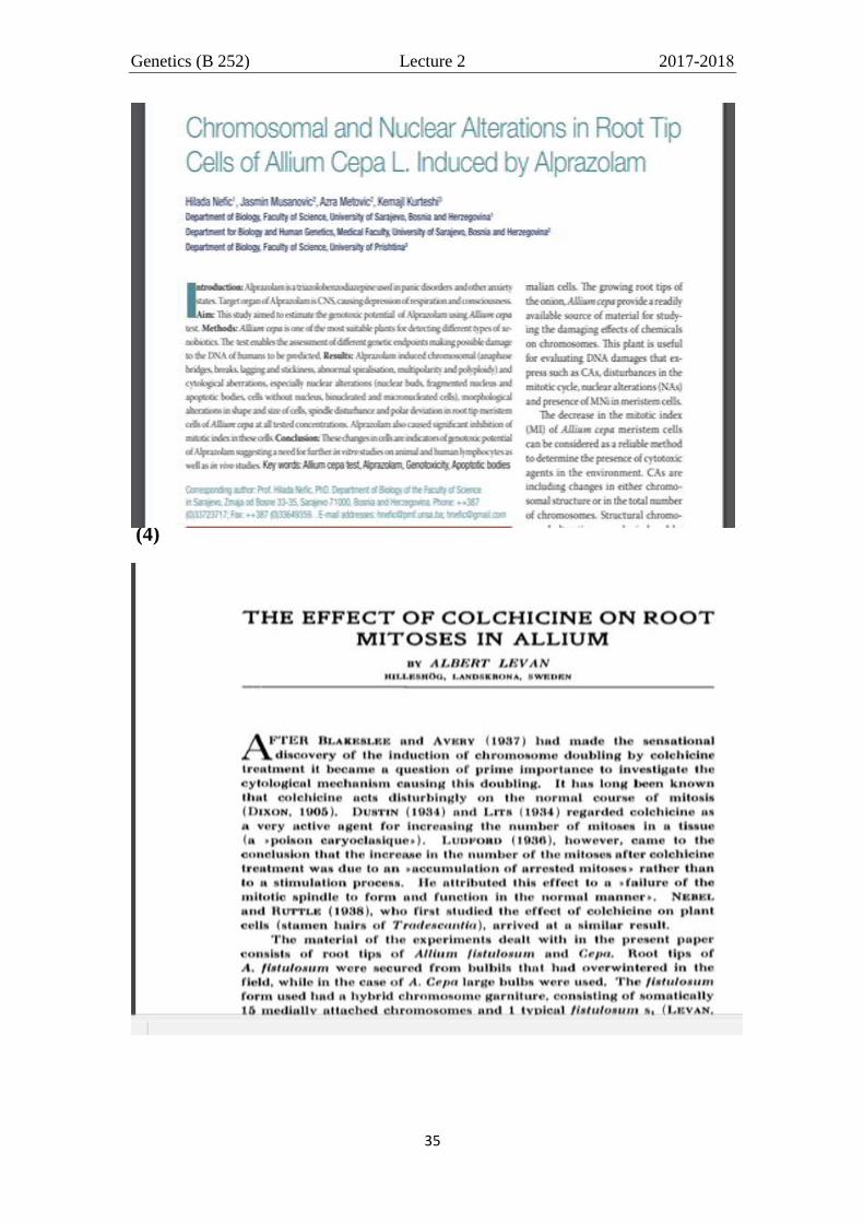
(4)
Genetics (B 252) Lecture 2 2017-2018
36
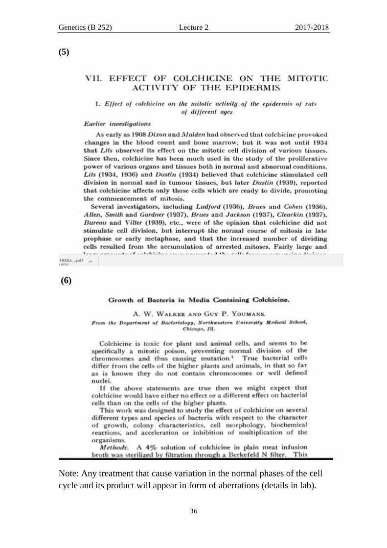
(5)
(6)
Note: Any treatment that cause variation in the normal phases of the cell
cycle and its product will appear in form of aberrations (details in lab).