how cells crawl nhgri-smithsonian collaboration · pdf filedome-shaped tent sitting in the...
TRANSCRIPT

NATIONAL INSTITUTES OF HEALTH • OFFICE OF THE DIRECTOR | VOLUME 21 ISSUE 5 • SEPTEMBER-OCTOBER 2013
How Cells CrawlThe Dynamics of Cell MotilityBY ADAM J. KUSZAK, NIDDK
CONTENTS
FEATURES • |1| How Cells Crawl |1| NHGRI-Smithonian Collaboration |7| Why Do Tumors
Keep on Growing |11| Making Progress Against Rare Adrenal Tumors |14| Camp Fantastic
|16| Town Hall Meeting at NIH with HHS Secretary Kathleen Sebelius
DEPARTMENTS • |2| DDIR: Intramural Research Program |3| News You Can Use: Medical Arts
|4| Training Page |5| NIH in History: Mystery Solved |7| SIG Beat |8| Research Briefs and Other
News |10| Abbreviations |12| Colleagues: Recently Tenured |15| Announcements
CONTINUED ON PAGE 6
CONTINUED ON PAGE 10
In June, the “Genome: Unlocking Life’s Code” exhibi-tion opened at the Smithsonian National Museum of Natural History (Washington, D.C.). The exhibition celebrates the anniversaries of two historic landmarks: the 10th anniver-sary of the Human Genome Project’s completion and the 60th anniversary of James Watson and Francis Crick ’s discovery of DNA’s double-helical structure.
The exhibition is the brain-child of Eric Green, director of the National Human Genome Research Institute (NHGRI), and G. Wayne Clough, secretary of the Smithsonian Institution. It represents the most expansive collaboration to date between the NIH and the Smithson-ian Institution. The high-tech exhibition uses interactive touch screens and high-definition graphics, three-dimensional models, custom animations, and videos of real-life stories to explain the basics of genomics and DNA sequencing technology and to examine both the benefits and the challenges that genomics presents.
One of the exhibition’s displays features the stories of people who have taken part in genomic sequencing studies. Select a medical story, and a video of that individual appears
No, Masur Auditorium hadn’t become a campground. That orange dome-shaped tent sitting in the middle of the stage was a prop for a G. Bur-roughs Mider Lecture given as part of the Wednesday Afternoon Lecture Series. Jennifer Lippincott-Schwartz used it to demonstrate what happens to a cell’s “skeleton,” or more precisely its cytoskel-eton, when it crawls.
Cells crawl for all sorts of reasons: to form new tissue during embryonic devel-opment; to heal wounds; to defend against invading microorganisms; to remodel bone; to regenerate nerves; and more. A cell’s movement is driven by continuous remodel-ing of the cytoskeleton and is mediated by the lamellipodia (tiny filaments composed of a protein called actin) located at the lamella (front edge of the cell). When actin subunits are added to the lamellipodia in a process known as polymerization, a pushing force is generated. After the polymerization has occurred, the molecular motor, myosin II, is added to cause a contractile force. As the actin filament cytoskeleton pushes and contracts, the cell slowly crawls along.
Scientists typically use electron micros-copy to get high-resolution, but static, images of the lamellipodia. Conventional confocal imaging can capture live, but blurry, images of cells crawling. Lippin-cott-Schwartz, a distinguished investigator in the National Institute of Child Health and Human Development (NICHD), has
NHGRI-Smithsonian CollaborationA New Model for NIH OutreachBY KATHERINE WENDELSDORF, NIAID
In June, the “Genome: Unlocking Life’s Code” exhibition opened at the Smith-sonian National Museum of Natural History, representing the most expansive collaboration to date between the NIH and the Smithsonian Institution.
MA
GG
IE BA
RTLE
TT, NH
GR
I

FROM THE DEPUTY DIRECTOR FOR INTRAMURAL RESEARCH
2 THE NIH CATALYST SEPTEMBER-OCTOBER 2013
Most of the r e a ders of this column are aware of the enormous con-tributions to human health that the NIH has made by supporting basic biomedi-cal research. For the NIH intramural research program (IRP) these contribu-tions are ref lected in numerous Nobel prizes to NIH scientists and trainees, other awards, and citations to articles by our highly visible scientists (http://www.irp.nih.gov/about-us/honors).
An equally lasting impact of intramural research has been felt in medicine’s “stan-dard of care” (what is supposed to happen when you enter a doctor’s office for a check-up, diagnosis, or treatment).
The NIH IRP, including the Clinical Center and its talented clinically oriented scientists, has exemplified the importance of evidence-based medicine, dentistry, and even veterinary medicine. Almost every aspect of clinical practice has been profoundly affected by research done at the NIH.
Let’s say you walk into the doctor’s office for a check-up. Blood is drawn to screen for a variety of disorders, such as problems with lipid metabolism that could lead to heart disease, stroke, and kidney disease. The original description of low- and high-density lipoproteins and cholesterol, as well as their association with blood-vessel dis-eases, was worked out at the NIH by Don Fredrickson (NHLBI) and colleagues.
Next, the doctor does a blood count to determine numbers of red cells, white cells, and platelets; to detect malignant disorders of these cells; and to look for infection or bleeding. This procedure used to be a tedious counting process done with
a hemocytometer and a microscope. Now diagnostic labs use a Coulter Counter or similar device. The counter was initially developed by Wallace Coulter, an engi-neer, to count particles in the paint used to protect the surfaces of U.S. Navy vessels. It occurred to him that the principle could be used to measure particles (cells) in blood. The NIH quickly picked up on this concept and developed it for clinical use.
Your doctor is likely to check that your vaccinations are up to date. If you were born in the past 20 years, you would have received in childhood an Haemophilus influenzae vaccine (developed by NICHD scientists Rachel Schneerson and John Robbins to prevent H. influenzae meningitis) and other vaccines—including one for hepati-tis A—developed from NIH work. As an adolescent, you would have been vaccinated against human papillomavirus to prevent cervical cancer (NCI’s John Schiller and Doug Lowy developed that vaccine). Later in life, you will receive a high-dose Herpes zoster vaccination to prevent shingles, thanks to the work of Steve Straus, Phil Brunell, and others in NIAID.
Chest pain is a common complaint. If the pain is severe and acute, your doctor will tell you to take a nitroglycerin tablet (and aspirin) and get to an emergency room, as per a protocol developed at the NIH to reduce the damage from occluded coronary vessels. NHLBI’s Andrew Arai, in collaboration with Suburban Hospital (Bethesda, Md.), is working on a quick way to use functional magnetic resonance imag-ing (fMRI) to determine whether there is restricted blood supply to the heart during
chest pain. We expect that use of coronary fMRI in emergency rooms will eventually provide definitive diagnostic information in minutes instead of hours. Incidentally, much of the software for interpreting both heart and brain MRIs was developed by NHLBI scientists.
The doctor might discover that a con-tributing cause of your chest pain is a severe anemia requiring a blood transfusion to improve oxygen delivery to your heart and other vital organs. Rest assured that the blood you receive will not be contaminated with hepatitis virus, thanks to the pioneer-ing work of the Clinical Center’s Harvey Alter, or human immunodeficiency virus (HIV), thanks to Robert Gallo (NCI) who helped develop the first blood test for HIV. If your anemia is due to bone-marrow failure (aplastic anemia), the standard treatment with immunosuppressive agents is thanks to the work of Neal Young (NHLBI).
The future of medicine will include a large dose of genomic analysis. Need I point out that the genetic code was deciphered by Marshall Nirenberg (NHLBI) and NIH colleagues; that the BLAST (Basic Local Alignment Search Tool) algorithm that we use to find related DNA sequences in existing databases was worked out by Ste-phen Altschul and David Lipman (NCBI, NLM); that many of the genes associated with human genetic diseases were first identified at the NIH and continue to be, through William Gahl’s (NHGRI) Undi-agnosed Diseases Program and other studies of rare diseases; and that use of genomic analysis of bacterial genomes, introduced by Julie Segre (NHGRI), Tara Palmore
The NIH Intramural Research Program: Our Research Changes Medical Practice BY MICHAEL GOTTESMAN, DDIR

http://irp.nih.gov/catalyst 3http://irp.nih.gov/catalyst 3http://irp.nih.gov/catalyst 3
Despite what you may have heard, the Division of Medical Arts is still in business and continuing to provide services to its NIH customers. We have served the NIH community for more than 50 years and distinguished ourselves by winning numerous awards for our work. Our ser-vices are unmatched by outside providers.
But Medical Arts is a fee-for-service organization, and our budget depends on billable work requests from Institutes and Centers (ICs). Recently, there have been fewer requests for our services because ICs have smaller budgets and have had to cut or postpone projects that would have required Medical Arts’ help; advances in technology have allowed ICs to do some things themselves that they used to rely on Medical Arts for; and some ICs are procur-ing similar services from outside vendors.
Medical Arts, therefore, is restructuring and changing the way we do business. We are reducing our space; refining our technol-ogy; and modifying the way our designers and artists work. In essence, Medical Arts is becoming a storefront that will manage and provide the services that support the NIH’s mission, to the extent that revenue continues to cover expenses.
As we transition through this restructuring into FY2014, we will continue to provide:
Express services such as scientific and event posters, programs and brochures, plaques, campus photos, and custom framing
Medical and visual information services:
• Designs for identity graphics, publications, posters, brochures, signage, and exhibits
• Three-dimensional modeling and anima-tion ranging from simple moving diagrams
to photorealistic animations revealing even the most complicated processes
• Digital animations for presentations that can illuminate surgical procedures, edu-cate patients, and communicate complex research by showing a series of steps instead of a single static image
• Illustrations for journal covers and pub-lications; medical and biological drawings; and accurate technical charts, diagrams, and depictions of laboratory equipment
We encourage our customers to contin-ue using our services during the transition. As the budget is developed for FY2014, we will have a better understanding of our ability—and of our customers’ needs—to ensure the future viability of the Medi-cal Arts capabilities. We will keep you posted and will adjust our business plan accordingly.
Medical Arts will be able to complete the services outlined in established Inter/Intra-Agency Agreements. These formal agreements have resulted in vital collabora-tion with other government agencies. You, the Medical Arts customer, can be assured that service levels will not decline.
We also encourage ICs that are procur-ing similar services from outside entities to try using Medical Arts instead during the transition. Our team is ready to do what-ever it takes to get the job done—creatively, efficiently, and expertly. No outside pro-vider can match our unique qualifications, convenience, expertise, and understanding of the NIH community. Medical Arts is dedicated to meeting your needs—each and every time.
For more information, visit medarts.nih.gov
or call 301-496-3221.
http://irp.nih.gov/catalyst 3
NIH Medical Arts We're Much Alive and Changing the Way We Do BusinessBY MEDICAL ARTS STAFF
(CC), and Evan Snitkin (NHGRI), will revolutionize the study of epidemics of multidrug-resistant pathogens.
There are hundreds of rare human dis-eases whose study has been advanced at NIH; important improvements have been made in treating severe psychiatric disorders such as bipolar illness (lithium was first used for this purpose at NIH, and Carlos Zarate in NIMH has introduced ketamine for rapid treatment of depression); fluoride, shown to prevent tooth decay, was first tested by NIDCR; the use of artificial, surgically implanted mitral valves to replace defec-tive heart valves was an NHLBI first; and the development of single- and multiagent chemotherapy to treat cancer was pioneered at the NCI.
And given that the NIH IRP is one of the most credible sources of medical research in the world, it’s not surprising that many treatments, once thought to be effective, failed NIH’s rigorous clini-cal studies (for example, NIDDK tested pancreatic islet-cell transplantation for dia-betes; NCI evaluated bone-marrow trans-plantation after high-dose chemotherapy for breast cancer). I am sure that there are many other examples of how the NIH IRP has influenced health care throughout the world, saving lives and preventing squan-dering of valuable resources.
At a time when everything that the government does is under increased scru-tiny, the NIH IRP seems like a particularly good investment of funds and people.
Sending me any additional examples you have of how the IRP has improved medical care would be much appreciated.
NEWS YOU CAN USE

4 THE NIH CATALYST SEPTEMBER-OCTOBER 2013
THE TRAINING PAGE
The N I H Fel l ow s Edit or i a l Board (FEB) may be one of the best-kept secrets of NIH’s intramural research training program. But it shouldn’t be.
The FEB provides a free, confidential scientific-editing service to all fellows at the NIH and FDA for manuscripts rang-ing from primary data articles to literature reviews to grant proposals. The volunteer editors are fellows and professionals who edit the documents for grammar, form, and clarity and review figures, legends, and other elements. They do not, however, consider the scientific merit of submissions.
Each manuscript is assigned to an asso-ciate editor and three or four primary editors who lead an open discussion with the rest of the board and prepare the final edits and
reports. Each author receives a letter that summarizes the editorial suggestions and a copy of the marked-up manuscript.
The manuscript-editing service benefits more than just the authors. Whether FEB editors are planning to stay at the bench or transition to a career in scientific publish-ing, editing, or writing, they gain valuable experience as they hone their own edito-rial skills. FEB alum Ranjini Prithviraj, a former postdoctoral fellow at the National Institute of Neurological Disorders and Stroke, is now a managing editor at the American Chemical Society (Washington, D.C.). Being an FEB editor trained her to “read, understand, and critique manuscripts” outside of her field and work as part of an editorial team, she said.
FEB was founded in 2002 by the National Cancer Institute’s Center for Cancer Research; in 2005, FEB was expanded NIH-wide. To date, some 500 fellows have served as volunteer editors and have reviewed more than 720 man-uscripts. About 40 FEB members—led by Senior Editor Andrew Broadbent, a Visiting Fellow at the National Institute of Allergy and Infectious Diseases—are active at any given time. In addition, con-sulting editors, many of whom are former FEB members, provide assistance when needed.
For more information, visit FEB at http://ccr.
cancer.gov/careers/feb or e-mail NCIedi-
FROM THE FELLOWS COMMITTEEThe NIH Fellows Editorial Board: A Secret No MoreBY JENNIFER SARGENT, NIAMS
In July, 27 Ph.D. students from 16 institutions took part in the new “Clinical and Translational Research Course for Ph.D. Students,” a two-week course that introduces talented young scientists to “bench-to-bedside” research. They met with intramural Ph.D. investigators who served as translational researcher role models, participated in a mock institu-tional review board, and learned how to file investigational drug applications with the FDA. “This was a diverse group of very bright and engaged students,” said Juan J.L. Lertora, faculty lead for the program. Ph.D. students conducting research at the NIH and U.S. academic institutions are encouraged to apply. “This course has reminded me that my origi-nal goal of improving human health is not absurd or unrealistic,” said Bridget Queenan, a neuroscience graduate student at Georgetown University (Washington, D.C.). “It’s the only thing worth doing.” For more information, visit http://www.cc.nih.gov/training/phdcourse/index.html.
NIH CLINICAL CENTER’S NEW COURSE FOR PH.D. STUDENTSBY JANET HULSTRAND, CC
MA
RIA
NEK
HA
YON
AK
, CC

http://irp.nih.gov/catalyst 5
No, it wasn’t a prototype for a flux capacitor.
The “History Mystery” photo that appeared in the May-June issue of the NIH Catalyst (http://irp.nih.gov/catalyst/v21i3/nih-in-history) elicited 14 responses to our plea for help in identifying the equip-ment used by the late Roderic E. Steele, a researcher in the National Heart, Lung, and Blood Institute (NHLBI) from 1975 to 1988.
Three were whimsical guesses—a flux capacitor (the time-travel machine featured in the Back to the Future trilogy); an early breast pump; and a device to deliver electro-shock therapy. But most respondents pro-vided real clues. We thank everyone who helped identify this object (for individual comments, see http:// irp.nih.gov/catalyst/v21i5/nih-in-history).
So what is it? It turns out that it’s equip-ment for a porous-bottom culture dish (PBCD) that Steele helped to develop for research on epithelial cells.
“This product truly revolutionized the way the epithelial cell community studied cells, particularly for performing transport assays,” said Gregory Germino, Deputy Director of the National Institute of Diabetes and Digestive and Kidney Diseases (NIDDK). “The conductivity meter would have been used to measure resistance, a measure of how ‘tight’ the cell junctions had become, and also how much ionic activity could be induced by various interventions.”
Epithelial cells are tightly packed together and line the body’s major cavities, most organs, blood vessels, and the skin. Molecules—including nutrients, hormones, growth factors, ions, and oxygen as well as carbon dioxide and other waste products—are transported through the cells’ upper and lower membranous surfaces. But such
transport is difficult to measure when the cells are grown in a normal culture dish.
Steele’s PBCD simulated a natural envi-ronment for the cells. The porous bottom allowed for the passage of transported sol-utes and provided a conductive pathway for electrical measurements.
Steele’s journey to create the PBCD began with work he did in the 1970s at Stanford University (Stanford, Calif.). He was studying sodium transport and carbon dioxide production to determine how the chemical energy of cellular metabolism is converted to the electrochemical energy of active transport. Instead of using a conven-tional culture dish, he fashioned a PBCD out of a toad bladder, which he cut in half and suspended like a tiny trampoline across a glass chamber that could simultaneously measure sodium transport and oxygen con-sumption (J Membr Biol 34:289–312, 1977).
By the spring of 1983, Steele was work-ing with T. Andrew Guhl, an inventor and supervisor at Becton Dickinson, on com-mercializing the PBCD. Guhl wrote in a letter to Steele, “I envision this particular product to be the first [to incorporate] a porous membrane for cell-culture experi-ments involving feeding of the cell mono-layer from the basal-lateral surface.”
In 1986, Steele published a paper with Joseph S. Handler (NHLBI) in the American Journal of Physiology that described four porous bottoms they had tried—cel-lulose ester, polycarbonate, collagen, and placental amnion. The first three types were attached to a polycarbonate ring that formed the sides. The amnion bottoms were tucked between two hollow polyethylene stoppers stacked on top of each other in an “embroidery-hoop arrangement” (Am J Physiol 251:C136–C139, 1986, and Methods Enzymol 171:736–744, 1989).
NIH IN HISTORY
NIH scientists helped the Office of NIH History discover that this conductivity meter was part of a system that the late NHLBI scientist Roderic E. Steele started developing in the 1970s: a porous-bottom culture dish that revolution-ized research on epithelial cells.
MIC
HELE LY
ON
S, OFFIC
E OF N
IH H
ISTOR
Y
The group continued their development of the PBCD, publishing an article in 1992 about a more complicated set-up (J Tissue Cult Methods 14:259–264, 1992).
After Steele left NIH in 1988, he con-tinued his association with Handler’s lab. He eventually retired to California, where he died in 2011. His widow did not have any papers or objects to document his work. We were fortunate, however, that a few let-ters and handwritten data sheets mailed between Steele and Guhl were tucked into the box containing the PBCD. Usually, we are not so lucky as to have correspondence between a federal employee inventor and a commercial operation.
The expertise of the scientific commu-nity is an important resource for the NIH Stetten Museum, which covers a broad array of NIH history and research. Without your help, we would never identify everything in our collection. Thank you.
History Mystery Solved Invention that Revolutionized Epithelial Cell ResearchBY MICHELE LYONS, OFFICE OF NIH HISTORY

6 THE NIH CATALYST SEPTEMBER-OCTOBER 2013
played a leading role in advancing micros-copy technologies to allow scientists to see high-resolution images of cells in action.
Her lab helped develop photo-activated localization microscopy (PALM), a tech-nique that uses repeated activation, imaging, and bleaching of photo-switchable fluores-cent probes to obtain resolutions of 10 to 20 nanometers, about 10 times the size of an average protein. PALM and other recent super-resolution imaging techniques have ushered a new era of investigations into cel-lular mechanics.
Lippincott-Schwartz wowed the audience with stunning videos of a live cell crawling. She explained how studies of actin’s continual reorganization at the cell’s lamella have yielded novel insights into the fundamental mechanics of cell motil-ity. She and her NICHD colleagues found that actin arcs of the lamella were the base against which the lamellipodium’s polymer-izing actin filaments protrude (Nature Cell Biol 13:371-382, 2011). Furthermore, the high spatial and temporal resolution analysis
revealed that actin filaments evolve into the actin arcs as they interact with myosin II, demonstrating a heretofore unknown conti-nuity between the lamellipodia and lamella actin networks.
She described how Dylan Burnette, the leading postdoctoral researcher on the proj-ect, studied the spatial relationship between actin and the focal adhesion protein zyxin and found a correlation between the con-tracting actin arcs and focal adhesion points (actin-rich structures that enable cells to adhere to the extracellular matrix). Moving actin arcs slowed down near newly forming focal adhesions, likened to “feet” at the point of contact between the cell and a surface. This interplay between actin arcs and focal adhesions in turn creates an anchored, stiff substrate against which the polymerizing actin filaments extend the plasma mem-brane. The combined dynamics of these systems cause the migrating cells to crawl.
Lippincott-Schwartz and Burnette wondered whether this “contractile appa-ratus” played other roles in a migrating
FEATURE
cell ’s shape. Burnette visualized the arrangement of actin and myosin through-out the cell three-dimensionally. He saw that myosin II was localized to the actin arcs of a cell’s dorsal side, not to the actin stress fibers connecting the dorsal surface to the ventral focal adhesions. His finding suggests that contractile force is localized to the actin arcs.
“What this system really looks like is a tent,” said Lippincott-Schwartz as she walked over to the tent on center stage. The poles represent the actin stress fibers that go from the base to the top of the cell, she explained. As she pushed down on the tent, it flattened just as a crawling cell would.
She hypothesized that such a system underlies the flattening of a cell’s leading edge, allowing the cell to move into the tight spaces of surrounding substrates. Her studies have generated exciting insights into a critical process in developmental biology and cancer metastasis.
Although Lippincott-Schwartz and Burnette have captured astound-ing images of cellular movement using super-resolution microscopy, they are focused on advancing the mechanistic understanding of cell motility rather than repeatedly imaging the same processes with new microscopy techniques.
Lippincott-Schwartz and her trainees are also applying high-resolution imaging techniques to their investigation of the diverse systems of mitochondrial fusion, Golgi organization, glucose uptake, and hepatocyte metabolism. They are poised to make many more great leaps forward in understanding dynamic cellular functions.
To view the video of Lippincott-Schwartz’s
talk, “Navigating the Cellular Landscape with
New Optical Probes, Imaging Strategies, and
Technical Innovations,” visit http://videocast.
nih.gov/launch.asp?17997.
DY
LAN
BU
RN
ETTE
, NIC
HD
How Cells Crawl CONTINUED FROM PAGE 1

http://irp.nih.gov/catalyst 7http://irp.nih.gov/catalyst 7http://irp.nih.gov/catalyst 7
FEATURE
BCIG: A MIMIC-II STORY Monday, September 23, 10:30–11:30 a.m.
Natcher Conf. Center (Building 45), Room B
The NIH Biomedical Computing Interest Group
(BCIG) presents “Knowledge Discovery for
Critical Care: A MIMIC-II Story” by Mengling
Feng (Harvard–MIT), Thomas Brennan (MIT),
and Leo Anthony Celi (Harvard–Beth Israel).
The data generated in the process of medical
care have historically been underused and
wasted. This waste was due in part to the dif-
ficulty of accessing, organizing, and using data
on paper charts. In addition, variability in clini-
cal documentation methods and quality made
the problem even more challenging. Without
a practical way to systematically capture,
analyze, and integrate the information con-
tained in the massive amount of data generated
during patient care, medicine has remained
largely an ad hoc process in which the discon-
nected application of individual experiences
and subjective preferences continues to thwart
continuous improvement and consistent deliv-
ery of best practices to all patients.
The intensive-care unit (ICU) presents an
especially compelling case for clinical data
analysis. The value of many treatments and
interventions in the ICU is unproven, and high-
quality data supporting or challenging specific
practices are embarrassingly sparse. Over the
past decade, the Massachusetts Institute of
Technology, Beth Israel Deaconess Medical
Center (BIDMC), and Philips Healthcare, with
support from the National Institute of Biomedi-
cal Imaging and Bioinformatics, have partnered
to build and maintain the Multi-parameter Intel-
ligent Monitoring in Intensive Care database,
which now holds clinical data from more than
40,000 de-identified stays in BIDMC ICUs and
is shared online with researchers via PhysioNet.
This system uses individual data to benefit the
care of populations and population data to
benefit the care of individuals.
For more information, contact Jim DeLeo at
THE SIG BEAT
NEWS FROM AND ABOUT THE NIH SCIENTIFIC INTEREST GROUPS
http://irp.nih.gov/catalyst 7
Why Do Tumors Keep on Growing? Cancer Therapy Kills Only Part of a TumorBY REBECCA BAKER, NIAID
“I want to get you to change your thinking about how we evaluate cancer,” Antonio “Tito” Fojo told the audience that had gathered in Lipsett Amphi-theater (Building 10) on June 12 for the Great Teachers Tenth Annual John Laws Decker Memorial Lecture. Cancer therapy, he said, kills only part of a tumor—the part that’s sensitive to a can-cer-fighting drug. The rest is resistant and continues to grow.
Fojo, a senior investigator in the Nation-al Cancer Institute, strives to identify the mechanisms of drug resistance so that one day it can be prevented. He conducts basic science and translational research as well as clinical studies in the hopes of finding better therapies. He also excels at teach-ing clinical fellows to rigorously evaluate cancer therapies, work toward improved patient outcomes, and provide empathetic patient care.
In recognition of his skill in training physicians, the NIH Clinical Fellows Committee selected him as the recipi-ent for this year’s Distinguished Clinical Teacher Award.
During his lecture, “Novel Paradigms in Cancer That May Lead to Better Therapies,” Fojo pointed out the blind spots in modern-day cancer treatments: Even after years of research by NIH scientists and others, clini-cians still rely on the same small arsenal of nonspecific cancer-fighting drugs to treat patients. And he bemoaned the proliferation of “me too” drugs that mimic successful therapeutics by targeting, ineffectively, the same signaling pathways.
Fojo challenged the audience to think more critically about cancer treatments and develop better ones. He also highlighted NIH’s innovative research that explores how to get the immune system to attack tumors.
But better research wasn’t all that was on Fojo’s mind—empathetic patient care was, too. “The most important thing NIH clinical fellows can learn is how to take care of patients [who] are enrolled in clinical trials,” he said. Fellows need to learn “how to listen to a patient, empathize with the patient, and be able to attend [to] their needs in diverse ways.”
The Decker Lecture is named in honor of
John Laws Decker, director of the Clinical
Center (CC) 1983–1990, who was lauded by
colleagues for both his bedside manner and
his careful design of clinical trials. He oversaw
major advances at the CC including the devel-
opment of the positron emission tomography
program and the clinical use of magnetic
resonance imaging. To view the June 12, 2013
lecture, visit http://videocast.nih.gov/launch.
asp?17996. For more on Fojo, visit http://
www.irp.nih.gov/pi/antonio-fojo.
At the Great Teachers Tenth Annual John Laws Decker Memorial Lecture, held in June, NCI senior investigator Antonio “Tito” Fojo explained how cancer therapy kills only part of a tumor—the part that’s sensitive to a cancer-fighting drug. The rest is resistant and continues to grow.
ERN
IE BR
AN
SON

8 THE NIH CATALYST SEPTEMBER-OCTOBER 2013
FEATURE
NIAID, CC: INVESTIGATIONAL MALARIA
VACCINE FOUND SAFE AND PROTECTIVE
An investigational malaria vaccine has been
found to be safe, to generate an immune
response, and to offer protection against
malaria infection in healthy adults, accord-
ing to the results of an early-stage clinical
trial conducted at NIH. The vaccine, known
as the PfSPZ vaccine, was developed by sci-
entists at Sanaria Inc. (Rockville, Md.) The
PfSPZ vaccine is composed of live but weak-
ened sporozoites of the species Plasmodium
falciparum, the most deadly of the malaria-
causing parasites.
An important challenge in the continued
development of PfSPZ vaccine is that the
vaccine currently is administered intrave-
nously—a rare delivery route for vaccines.
Previous studies at lower doses showed
that the more common intradermal (into
the skin) and subcutaneous (under the skin)
routes did not yield as strong an immune
response as the intravenous route. Several
follow-up studies are planned, including
research to evaluate the vaccine’s different
dose schedules, possible protection against
other Plasmodium strains, and the durabil-
ity of protection. The researchers may also
evaluate whether higher doses administered
subcutaneously or intradermally provide
the same protection as found in this study.
(NIH authors: R.A. Seder and others, Science
DOI:10.1126/science.1241800)
NCI: HOW CANCER CHROMOSOME ABNOR-
MALITIES FORM IN LIVING CELLS
For the first time, scientists have directly
observed events that lead to the formation of
chromosome abnormalities, called translo-
cations, often found in cancer cells. Despite
many years of research, just exactly how
translocations form in a cell has remained
a mystery. To better understand this pro-
cess, NCI researchers created an experimen-
tal system in which they induced breaks in
the DNA of different chromosomes in living
cells. Using sophisticated imaging technol-
ogy, they were then able to watch as the
broken ends of the chromosomes reattached
correctly or incorrectly inside the cells. The
scientists were able to demonstrate that
translocations can occur within hours of DNA
breaks and that their formation is indepen-
dent of when the breaks happen during the
cell-division cycle. (NCI authors: V. Roukos,
T.C. Voss, C.K. Schmidt, D. Wangsa, and T.
Misteli, Science 341:660–664, 2013)
NIA: DIABETES DRUG IMPROVES HEALTH
AND EXTENDS LIFESPAN IN MICE
Long-term treatment with the type 2 diabetes
drug metformin improves health and longev-
ity of male mice when started at middle age,
reported an international team of scientists
led by NIA researchers. Metformin is known to
enhance insulin sensitivity, prompting the con-
version of glucose to energy and preventing
its build-up in the liver. Metformin also reduce
risk of health problems associated with meta-
bolic syndrome, a condition characterized by an
increased chance for heart disease, stroke, and
2 diabetes. (NIA authors: A. Martin-Montalvo,
E.M. Mercken, S.J. Mitchell, H.H. Palacios, T.M.
Ward, R.K. Minor, M. Bernier, Y. Yu, and R. de
Cabo, Nat Commun 4:2192, 2013)
NIAAA: CHRONIC ALCOHOL USE SHIFTS
BRAIN’S CONTROL OF BEHAVIOR
Chronic alcohol exposure leads to brain adap-
tations that shift behavior control away from
an area of the brain involved in complex
decision-making and toward a region associ-
ated with habit formation, according to a new
study conducted in mice by NIAAA scientists.
The findings provide insight into how exces-
sive drinking affects learning and behavioral
control at the neural level. (NIAAA authors:
L. DePoy, R. Daut, J.L. Brigman, K. MacPher-
son, N. Crowley, O. Gunduz-Cinar, C.L. Pick-
ens, R. Cinar, G. Kunos, D.M. Lonvinger, M.C.
Camp, and A. Holmes, Proc Natl Acad Sci USA
DOI:10.1073/pnas.1308198110)
NIEHS, NCI: 3-D IMAGES SHOW FLAME
RETARDANTS CAN MIMIC ESTROGENS
By determining the 3-D structure of proteins
at the atomic level, NIH researchers have
discovered how some commonly used flame
retardants, called brominated flame retar-
dants, can mimic estrogen hormones and
possibly disrupt the body’s endocrine system.
(NIH authors: R.A. Gosavi, G.A. Knudsen, L.S.
Birnbaum, L.C. Pedersen, Environ Health Per-
spect DOI:10.1289/ehp.1306902)
NINDS: NIH RESEARCHERS DISCOVER HOW
BRAIN CELLS CHANGE THEIR TUNE
Using advanced microscopic techniques,
NINDS researchers recently showed that brief
bursts of chemical energy coming from the
mitochondria may fine-tune brain cell com-
munication. Problems with mitochondrial
energy production and movement throughout
nerve cells have been implicated in Alzheimer
disease, Parkinson disease, amyotrophic lat-
eral sclerosis, and other major neurodegen-
erative disorders. The researchers think these
results will ultimately help scientists under-
stand how mitochondrial problems can lead
to disorders in brain cell communication.
(NINDS authors: T. Sun, H. Quiao, P-Y. Pan, Y.
Chen, Z-H. Sheng, Cell Rep 4:413–419, 2013)
CATALYTIC RESEARCH
Intramural Research Briefs
Read more Research Briefs (and expanded
versions of these) online at http://irp.nih.
gov/catalyst/v21i5/research-briefs.
8 THE NIH CATALYST SEPTEMBER-OCTOBER 2013
The female Anopheles gambiae mosquito can transmit malaria parasites as she takes a blood meal. NIH research-ers recently tested an investigational malaria vaccine and found it to be safe and protective.
T. W
ELLE
MS,
NIA
ID

http://irp.nih.gov/catalyst 9http://irp.nih.gov/catalyst 9
HELA GENOME ACCESS BROKERED BY NIH
NIH announced a privacy agreement with the
family of the late Henrietta Lacks to allow for
the limited use of genomic sequence data from
cells derived from her tumor, commonly known
as HeLa cells. HeLa cells are the most widely
used human cell line in existence today and
have been used in the development of modern
vaccines, cancer treatments, in vitro fertiliza-
tion techniques, and other medical advances.
Lacks died of cervical cancer in 1951, before
scientists were required to get permission to
use a patient’s cells in research.
This spring, German scientists sequenced
the genome of a HeLa cell line and posted the
data in a public database. The data had the
potential to reveal private genetic information
about the Lacks family. NIH Director Francis
Collins and NIH Deputy Director for Science,
Outreach, and Policy Kathy Hudson met with
members of the Lacks family to discuss the
research. They settled on an agreement for
use of HeLa data: Published genomes will be
stored in an NIH-controlled database, accessed
only by researchers who agree to terms of use
defined by a panel that includes Lacks family
members. Collins noted the new policy allows
for public data sharing while showing respect
that NIH and scientists have for the contribu-
tions of the Lacks family. (K.L. Hudson and F.S.
Collins, Nature 500:141–142, 2013)
Rebecca Baker, NIAID
NIH DIRECTOR’S CHALLENGE INNOVATION
AWARDS
The NIH Director’s Challenge Innovation
Awards program is supporting 10 intramural
projects for research on antibacterial drug
resistance. The program, which provides seed
money to stimulate innovative, high-impact
research, gives priority to applications that
involve collaborations among PIs from multiple
NIH Institutes and Centers. This year, $750,000
is supporting the projects, with awards ranging
from $45,000 to $100,000 per year.
PUBLISH AND FLOURISH
It’s difficult to get official numbers, but if you
do a search via PubMed, you will see that NIH
researchers are authoring approximately 500
peer-reviewed journal articles each month.
Some of these articles are featured on flat-
screen monitors at the Clinical Center. Some
are featured in the “Hot Topics” section of
the relatively new Intramural Research Pro-
gram (IRP) Web site at http://irp.nih.gov/
news-and-events/hot-topics.
It’s a fantastic number, considering we
have about 1,000 labs at the NIH. Now the
journal Nature has provided some documen-
tation. According to the Nature Publishing
Index 2012 (http://www.natureasia.com/en/
publishing-index/global/), the NIH ranks #6 in
the number of papers published in the Nature
family of journals. Only #6, you ask? Well,
of course, NIH isn’t competitive in physics,
astronomy, engineering, and many other fields
dominated by the top five on the list: Harvard,
Stanford, Max Planck, Massachusetts Institute
of Technology, and the French National Centre
for Scientific Research (CNRS).
Also, the CNRS and Max Planck organiza-
tions represent a good deal of their nation’s
investment in research; their rankings are
equivalent to counting all NIH-supported
research (extramural and intramural) with a
little NSF and NASA thrown into the mix.
Nevertheless, if you break down the
Nature numbers, the NIH IRP ranked #2 for
Nature Genetics, Nature Neuroscience, Nature
Medicine, and Nature Immunology; #3 for
Nature Structural and Molecular Biology; and
#5 for Nature Chemical Biology.
The bottom line is that we are a productive
bunch, and I hope we can keep up that produc-
tivity and maintain our rankings through the
inevitable downsizing that will occur if budgets
remain flat or additional cuts are mandated.
Michael Gottesman, DDIR
FY2013 and FY2014 Awards:
Anirban Banerjee (NICHD) with researchers
from NIAID: Structural and Chemical Biol-
ogy Approach to Targeting MraY—an Integral
Membrane Enzyme in the Bacterial Peptido-
glycan Biosynthesis Pathway
Robert Danner (CC) with researchers from NCI
and NHGRI: Engineering a Multivalent Bacte-
riophage Targeting KPC+ K. pneumoniae
John Barrett (NHLBI) with researchers from
CC and NIAID: Th17 Cells for Treatment of Car-
bapenem Resistant Klebsiella pneumoniae
Yasmine Belkaid (NIAID) with research-
ers from NCI, NHGRI, and NIAID: Role of the
Microbiota in KPC-producing Klebsiella Pneu-
monia Infection
Karen Frank (CC) with researchers from NCI
and NHGRI: Carbapenemase blaKPC Horizon-
tal Transfer between Enterobacteriacae
Julie Segre (NHGRI) with researchers from CC:
Microbiome Studies of CC Patients at Risk for
Multidrug-Resistant Bacteria
Susan Buchanon (NIDDK) with researchers
from NCI: Klebsiella Capsule as a Target for
Novel Antibiotics: Genetics, Structure/func-
tion and Small Molecule Screens
Adrian Ferre D’Amare (NHLBI) with research-
ers from NCI: High-throughput Discovery of
Antibiotics Against Bacterial Riboswitches
Matthias Machner (NICHD) with researchers
from NCATS: Discovering “Smart” Drugs That
Target Only Pathogenic Bacteria
Roger Woodgate (NICHD) with researchers
from NCATS: Inhibitors of LexA/DinR Cleav-
age Combat Evolution of Antibiotic Resistance
The next competition is scheduled for FY2015.
For more information, visit http://sigs.nih.
gov/challenge/Pages/FundedProjects.aspx.
CATALYTIC RESEARCH
Other Research News

10 THE NIH CATALYST SEPTEMBER-OCTOBER 2013
a year, ranging from school-aged children to retirees, from all over the world. “If I published 100 papers a year, no way would seven million people read them,” Lawrence Brody, chief of NHGRI’s Genome Tech-nology Branch, pointed out.
NHGRI wants to “enhance the pub-lic’s genomic health literacy [and] encour-age understanding of how genomic research is important to individuals, their families and communities,” said ECIB education-outreach specialist Christina Daulton.
In addition to disseminating informa-tion, the exhibition also gathers it. Visitors can voluntarily answer survey questions such as “Is there anything about your genome that you want to remain a mystery?” and “If you could find out one thing about your genome, what would it be?” This Social Genomics Project led by intramural researchers provides the public an opportu-nity to consider the questions and see how other people have responded. It is expected that thousands of people will participate.
The exhibition will be at the National Museum
of Natural History through September
2014. Afterwards, it will tour North America
through 2018. For more information, visit
http://www.mnh.si.edu/exhibits/genome or
http://unlockinglifescode.org.
on a large screen. The people describe their participation in the research and how the results were able to impact their lives.
Visitors are also invited to explore genetic and environmental disease risk with a “wheel of chance.” This interactive activity illustrates that genetic risk factors alone do not deter-mine whether one gets a particular disease. It is important for people to grasp the concept that a combination of genetic, environmental, and random factors influences their chances of developing a particular disease.
The exhibition was developed by museum and NHGRI staff. NHGRI’s Division of Policy, Communications, and Education, and its Education and Commu-nity Involvement Branch (ECIB) directed NHGRI’s involvement in the project. ECIB leads NHGRI’s involvement in helping to build the public understanding of the role of genomics in human health and of the accompanying ethical, legal, and social issues. ECIB provides educational informa-tion on genetics and genomics, distributes information on new genomic technolo-gies, and increases awareness of genomic advances.
NHGRI expects to be able to reach mil-lions of people through the exhibitions. The museum hosts roughly seven million visitors
NHGRI-Smithsonian Collaboration CONTINUED FROM PAGE 1
FEATURE
MA
GG
IE B
AR
TLET
T, N
HG
RI
The high-tech exhibition, developed by the Smithsonian and NHGRI staff, uses interactive touch screens and high-def-inition graphics, three-dimensional models, custom animations, and videos of real-life stories to explain the basics of genomics and DNA sequencing technology and to examine both the benefits and the challenges that genomics presents.
NIH ABBREVIATIONS
CBER: Center for Biologics Evaluation and Research, FDACC: NIH Clinical CenterCCR: Center for Cancer Research, NCICDC: Centers for Disease Control and PreventionCIT: Center for Information TechnologyDCEG: Division of Cancer Epidemiology and Genetics, NCIDOE: Department of EnergyFAES: Foundation for Advanced Education in the SciencesFelCom: Fellows CommitteeFDA: Food and Drug AdministrationFNL: Frederick National LaboratoryIRP: Intramural Research ProgramHHS: U.S. Department of Health and Human ServicesNCATS: National Center for Advancing Translational SciencesNCCAM: National Center for Complementary and Alternative MedicineNCBI: National Center for Biotechnology InformationNCI: National Cancer InstituteNEI: National Eye InstituteNHGRI: National Human Genome Research InstituteNHLBI: National Heart, Lung, and Blood InstituteNIA: National Institute on AgingNIAAA: National Institute on Alcohol Abuse and AlcoholismNIAID: National Institute of Allergy and Infectious DiseasesNIAMS: National Institute of Arthritis and Musculoskeletal and Skin DiseasesNIBIB: National Institute of Biomedical Imaging and BioengineeringNICHD: Eunice Kennedy Shriver National Institute of Child Health and Human DevelopmentNIDA: National Institute on Drug AbuseNIDCD: National Institute on Deafness and Other Communication DisordersNIDCR: National Institute of Dental and Craniofacial ResearchNIDDK: National Institute of Diabetes and Digestive and Kidney DiseasesNIEHS: National Institute of Environmental Health SciencesNIGMS: National Institute of General Medical SciencesNIMH: National Institute of Mental HealthNIMHD: National Institute on Minority Health and Health DisparitiesNINDS: National Institute of Neurological Disorders and StrokeNINR: National Institute of Nursing ResearchNLM: National Library of MedicineOD: Office of the DirectorODS: Office of Dietary SupplementsOITE: Office of Intramural Training & EducationOIR: Office of Intramural ResearchORS: Office of Research ServicesORWH: Office of Research on Women’s HealthOTT: Office of Technology Transfer

http://irp.nih.gov/catalyst 11
People suffering from rare diseases often feel isolated and have few oppor-tunities to share their experiences with one another. But at NIH recently, people with two types of rare adre-nal tumors—pheochromocytoma and paraganglioma—met at a gathering that included physicians and scientists to learn about the latest in research and treatments. On June 20 and 21, the second annual International Patient Symposium on Pheochromocytoma, hosted by the Pheo Para Al l iance (Arlington, Va.), was held in Building 60 and featured 27 speakers including several from NIH.
Pheochromocytomas are rare, usu-ally benign, tumors that originate inside the adrenal glands; paragangliomas are similar tumors that form outside the adre-nals. These difficult-to-diagnose tumors—which can occur at any age but are most common between the ages of 30 and 50 years old—can trigger the release of excess adrenaline and dopamine, causing dan-gerously high blood pressure, pounding headaches, and heart palpitations. Two to eight people per million are diagnosed with these tumors each year. The conven-tional treatment is the surgical removal of the tumor sometimes accompanied by chemotherapy.
“Right now, 50 percent of cases go undiagnosed and treatments are subop-timal or not effective,” said Karel Pacak, a senior investigator and a leading pheo-chromocytoma researcher in the Eunice Kennedy Shriver National Institute of Child Health and Human Develop-ment (NICHD). Pacak, who focuses on improving the diagnosis and treatment of pheochromocytoma and paraganglioma, advanced imaging strategies, and novel therapeutic options, went on to describe
http://irp.nih.gov/catalyst 11
recent research on the genes associated with the onset of these tumors and NIH’s exploration of diagnostic imaging tech-niques such as positron emission tomog-raphy scans.
Other NIH scientists presenting at the symposium included NICHD staff clinician and genetics counselor Margar-ita Raygada, National Cancer Institute senior investigator Tito Fojo (see article about his work on page 7), and Clinical Center radiologist Aradhana Venkatesan.
Raygada is interested in the improved management of chronic genetic condi-tions and described the seven major gene mutations that predispose an individual to pheochromocytoma and paragangli-oma. The mutations are typically passed on from parent to child, but can occur spontaneously, she said. “Every single patient who has pheochromocytoma [or] paraganglioma has to be offered genetic testing” to determine whether other mem-bers of the family are at risk of having mutated genes.
Fojo, an expert in the mechanisms of chemotherapeutic drug resistance, pre-sented an overview of the different types of chemotherapy used to treat certain pheo-chromocytoma and paraganglioma tumors. His findings suggest that tumors created by a mutation in the SDHB gene respond well to chemotherapy, whereas tumors resulting from other mutations do not.
Venkatesan, a radiologist and imag-ing clinician, described a relatively new treatment that has become an alterna-tive to the surgical excision of adrenal tumors: radiofrequency ablation (RFA), which uses high-energy radio waves to heat and destroy cancerous cells. RFA has been in use for a long time to treat nerve-related chronic pain, heart arrhythmias, and some cancers, but has only recently
http://irp.nih.gov/catalyst 11
FEATURE
been expanded to treat adrenal tumors. Venkatesan has demonstrated that RFA is successful in destroying small and large tumors, but because it’s been in use for such a short time, she strongly advises patients who are considering it to “seek out experienced locations” such as the NIH Clinical Center.
“It is important that we at the Nation-al Institutes of Health work with research and patient groups in the United States and around the world” to advance the research, Pacak said. “Together we stand a chance; separately we fail.”
Making Progress Against Rare Adrenal TumorsBY REBECCA LAZERATION, NICHD
Karel Pacak, a senior investigator in NICHD, was one of several NIH researchers who presented their work on improving the diagnosis and treatment of rare adrenal tumors—pheochromocytoma and paraganglioma—at the International Patient Symposium on Pheochromocytoma, held at NIH in June.
BILL B
RA
NSO
N
YOUR IDEAS WELCOME
We are always looking for ideas for stories
about intramural research and investiga-
tors, behind-the-scenes activities that
enable research to happen, new methods
developed at NIH, and more. Don’t hesi-
tate to get in touch. E-mail catalyst@nih.
gov or call Managing Editor Laura Carter at
301-402-1449.

12 THE NIH CATALYST SEPTEMBER-OCTOBER 2013
COLLEAGUES
An Interview with Hynda KleinmanThe Matrigel-Maker’s StoryBY LAURA STEPHENSON CARTER
Recently Tenured
DONALD COOK, NIEHS KYLIE WALTERS, NCI NICHOLAS WENTZENSEN, NCIKATHERINE WARREN, NCISTEVEN VOGEL, NIAAA
DONALD COOK, PH.D., NIEHS
Senior Investigator, Immunogenetics Group, Laboratory of Respiratory BiologyEducation: McGill University, Montreal (B.S.
and Ph.D. in microbiology and immunology)
Training: Department of Pathology, Univer-
sity of North Carolina at Chapel Hill School of
Medicine (Chapel Hill, N.C.)
Before coming to NIH: Principal scien-
tist, Schering-Plough Research Institute
(Kenilworth, N.J.); assistant professor, Division
of Pulmonary and Critical Care Medicine, Duke
University Medical Center (Durham, N.C.)
Came to NIH: In 2005
Selected professional activities: Editorial
board for American Journal of Respiratory
Cell and Molecular Biology and Frontiers
in Chemoattractants; adjunct assistant
professor, Department of Immunology, Duke
University School of Medicine
Outside Interests: Training for short triath-
lons; playing blues on the piano
Research Interests: My lab uses gene-tar-geted mice lacking chemokines, cytokines, or signaling molecules to understand the molecular and cellular mechanisms that trigger immune responses to inhaled aller-gens. We focus on pulmonary dendritic cells that initiate immune responses to aeroal-lergens. By purifying discrete populations of
dendritic cells from the lung, we have been able to assign specific functions to them.
We also study how these functions are affected by gene-environment interactions. We have found that bacterial products, such as lipopolysaccharide and f lagel-lin, are found in house dust and promote allergen-specific immune responses. We also examine how environmental agents affect the function of airway epithelial cells.
We are testing the hypothesis that com-munication between airway epithelial cells and nearby dendritic cells determines the nature of immune responses to inhaled allergens. An improved understanding of these signaling pathways may lead to thera-peutic strategies that target specific types of asthma, including steroid-resistant asthma.
STEVEN S. VOGEL, PH.D., NIAAA
Senior Investigator; Chief, Section on Cellular Biophotonics, Laboratory of Molecular PhysiologyEducation: City College of New York (B.S. in
biology); Columbia University, New York (Ph.D.
in biochemistry and molecular biophysics)
Training: Postdoctoral training at NIDDK,
NICHD, and NINDS
Before returning to NIH: Associate profes-
sor and director of the Cell Imaging Core
Laboratory at the Medical College of Georgia
(Augusta, Ga.)
Came to NIH: In 1989 for training; in 2003
joined NIAAA
Selected professional activities: University of
Virginia FRET Microscopy Workshop Faculty
Outside interests: Playing acoustic 6– and
12–string guitar; bicycle commuting
Research interests: In cells, proteins act together to form assemblies that mediate cellular processes. Considering that pro-tein complexes are so ubiquitous and that they perform so many functions, it is no surprise that many human diseases arise from inappropriate protein interactions. A major obstacle to understanding these diseases, however, is the paucity of robust methods for studying protein interactions under physiological (natural) conditions.
Through my research, which lies at the intersection of physics, bioengineering, and neurobiology, I have been developing new forms of fiber-optic microscopy and spec-troscopy to better monitor protein interac-tions inside living cells and animals. These approaches are based primarily on Förster resonance energy transfer (FRET) and fluc-tuation correlation spectroscopy (FCS). I am especially interested in the interactions of synaptic proteins that are involved in regu-lating memory, behavior, and addiction.
My long-term goal is to use FRET and FCS to help identify drugs that can target

http://irp.nih.gov/catalyst 13http://irp.nih.gov/catalyst 13http://irp.nih.gov/catalyst 13http://irp.nih.gov/catalyst 13
COLLEAGUES
proteins are recognized and cleared from cells and how targeted ubiquitinated pro-teins are identified and processed by the proteasome.
The proteasome is the major machin-ery in eukaryotes (organisms whose cells contain a nucleus and other organelles) for regulating protein degradation. It contains enzymes called proteases, which degrade protein substrates. The proteasome can be “capped” by multiple regulators. We focus on the 19S regulatory particle, which contains the proteins that recognize and process ubiquitinated substrates. Human pathologies associated with malfunctions of the ubiquitin-proteasome pathway include autoimmunity and inflammation, neuro-degeneration, and cancer. The proteasome inhibitors bortezomib and carfilzomib are used to treat certain hematological cancers; other inhibitors are being tested in clinical trials. Our long-term goal is to reveal quality-control mechanisms in the ubiquitin-proteasome pathway in order to manipulate the lifespan of oncoproteins and tumor suppressors.
KATHERINE WARREN, M.D., NCI-CCR
Senior Investigator; Head, Pediatric Neuro-Oncology Section, Pediatric Oncology BranchEducation: North Adams State College, North
Adams, Mass. (B.S. in medical technology);
Tufts University School of Medicine, Boston,
Mass. (M.D.)
Training: Residency in pediatrics at Children’s
National Medical Center (Washington, D.C.);
fellowship in pediatric hematology and
oncology at NCI
Came to NIH: In 1993 for training; became a
tenure-track investigator in 2003
Selected professional activities: Active in
the Pediatric Brain Tumor Consortium (holds
leadership positions) and the Children’s
Oncology Group
Outside interests: Spending time with family;
traveling
Research interests: I am interested in devel-oping better treatments for children who have central nervous system tumors (CNS) tumors. Pediatric and adult CNS tumors differ, so data from adults may not apply to pediatric patients. We are conducting clinical trials to test new agents for treating CNS tumors in children; exploring new methods of treatment delivery to tumors; noninva-sively evaluating and imaging the brain to assess tumor characteristics; and studying neurotoxicity resulting from treatment.
A significant proportion of pediatric brain tumors are benign or low-grade, and the five-year survival rate is over 70 percent. But the survival rate is dismal for children with malignant tumors such as dif-fuse intrinsic brainstem gliomas, high-grade gliomas, and recurrent malignant tumors.
Treatments for pediatric CNS tumors include surgery, radiation, and chemo-therapy. Chemotherapy is not always suc-cessful, however, because the blood-brain barrier limits the delivery of the drugs to the tumor site. And such treatment has little, if any, effect on malignant gliomas and recurrent malignant tumors or against the brainstem tumor diffuse intrinsic pontine glioma (DIPG). Surgery is not an option for DPIG because the tumor is in the brain-stem, which controls vital functions such as heartbeat and breathing. Most children with DIPG die within one year of diagnosis.
Although hundreds of clinical trials have been performed with DPIG over three decades, there’s been no progress in improving outcomes. We know little about the biology of this disease partly because biopsies are not routinely obtained in the United States so there’s a scarcity of tissue available for study. My research has focused on developing new agents for treatment; doing noninvasive evaluations; using autopsy tissue to study tumor biology; and develop-ing novel approaches to deliver chemothera-peutics directly.
and correct abnormal protein interactions inside cells. The technology that we are developing will provide tools for delineating the steps in protein-complex conformational changes; a means of rapidly identifying sites of protein interactions; a way to help deci-pher which protein partners interact within large assemblies of proteins; and the means for confirming whether specific interactions occur under physiological conditions even when those interactions occur deep within the brain of a living mouse.
KYLIE WALTERS, PH.D., NCI-CCR
Senior Investigator; Head, Protein Processing Section, Structural Biophysics Laboratory Education: Wesleyan University, Middletown,
Conn. (B.A. in molecular biology and bio-
chemistry and concentration in biophysics);
Harvard University, Cambridge, Mass. (Ph.D.
in biophysics)
Training: Postdoctoral training in pathology
at Harvard Medical School (Boston)
Before coming to NIH: Associate profes-
sor of biochemistry, molecular biology,
and biophysics, University of Minnesota
(Minneapolis)
Came to NIH: In July 2013
Selected professional activities: Has served
as NIH Membrane Biology and Protein Pro-
cessing study section member since 2008
Outside interests: Enjoys outdoor activities
with husband and two children; swimming;
kayaking and canoeing; running
Research interests: We use nuclear magnetic resonance (NMR) and other biophysical techniques to reveal the three-dimensional structure and dynamic properties of proteins and complexes associated with cancer biol-ogy. We are interested in ubiquitin signaling pathways, especially those involved in pro-tein quality control. Ubiquitins, small regu-latory proteins, regulate gene transcription, protein degradation, cell death, and other cellular activities. We study how misfolded CONTINUED ON PAGE 14
NIH Director Visits Camp Fantastic
14 THE NIH CATALYST SEPTEMBER-OCTOBER 2013
COLLEAGUES
In addition, we are exploring the etiol-ogy of ovarian cancer, which is complex, poorly understood, and often fatal. Because the low prevalence of ovarian cancer limits molecular epidemiology studies, we are pooling cases from several NIH-based and extramural cohorts. We are conducting large epidemiologic studies and profiling ovar-ian tumor tissues. We hope to improve the classification of ovarian tumors and evaluate new early-detection approaches that can be translated into clinical or screening applica-tions. In other studies, we are investigating how inflammatory markers and endogenous hormone exposures to ovarian epithelium promote tumor growth.
COLLEAGUES
cancer. We are conducting profiling stud-ies on more than 2,000 tissue samples collected from women with cervical HPV infections, precancer, and cancer to elucidate the transitions from cervical HPV infec-tion to precancer and from precancer to invasive cancer.
We are also interested in improving colposcopy-guided biopsy procedures, which are used to screen for cervical cancer but tend to be inaccurate and frequently miss the worst lesions on the cervix. We conducted the Biopsy Study, to quantify the inaccuracy of colposcopies. We also hope to expand our understanding of the clonal relationship of multiple lesions on the cervix.
Recently Tenured CONTINUED FROM PAGE 13
NICOLAS WENTZENSEN, M.D., PH.D., M.S.,
NCI-DCEG
Senior Investigator, Hormonal and Repro-ductive Epidemiology Branch Education: Heidelberg University Ruperto
Carola, Heidelberg, Germany (M.D. and Ph.D.
in applied tumor biology); Johannes Guten-
berg University of Mainz, Mainz, Germany
(M.S. in epidemiology)
Training: Residency in general surgery and
postdoctoral research in applied tumor biol-
ogy and molecular epidemiology, Heidel-
berg University; postdoctoral research in
NCI-DCEG
Came to NIH: In 2007 as a visiting fellow;
became tenure-track investigator in 2009
Selected professional activities: Senior
editor, Cancer Epidemiology, Biomarkers and
Prevention; member of the Practice Improve-
ment in Cervical Screening and Management
Working Group and co-author of the 2012
consensus cervical cancer screening-guide-
lines publications; member of the steering
committee, working-group leader, and
writing-team member for the 2012 American
Society for Colposcopy and Cervical Pathol-
ogy management-guidelines update; faculty
of various international scientific conferences
Outside interests: Running; listening to clas-
sical music; playing guitar
Research interests: I am interested in the origins of gynecologic cancers and improv-ing screening and prevention efforts. My research focuses on understanding the het-erogeneous etiology of cervical, endometrial, and ovarian cancers and their precursors. I am analyzing risk factors that drive the development of these malignancies and looking at biomarkers that could identify women at high risk.
Although most cervical cancers are caused by human papillomavirus (HPV) infections, only a small subset of women infected with HPV progress to invasive
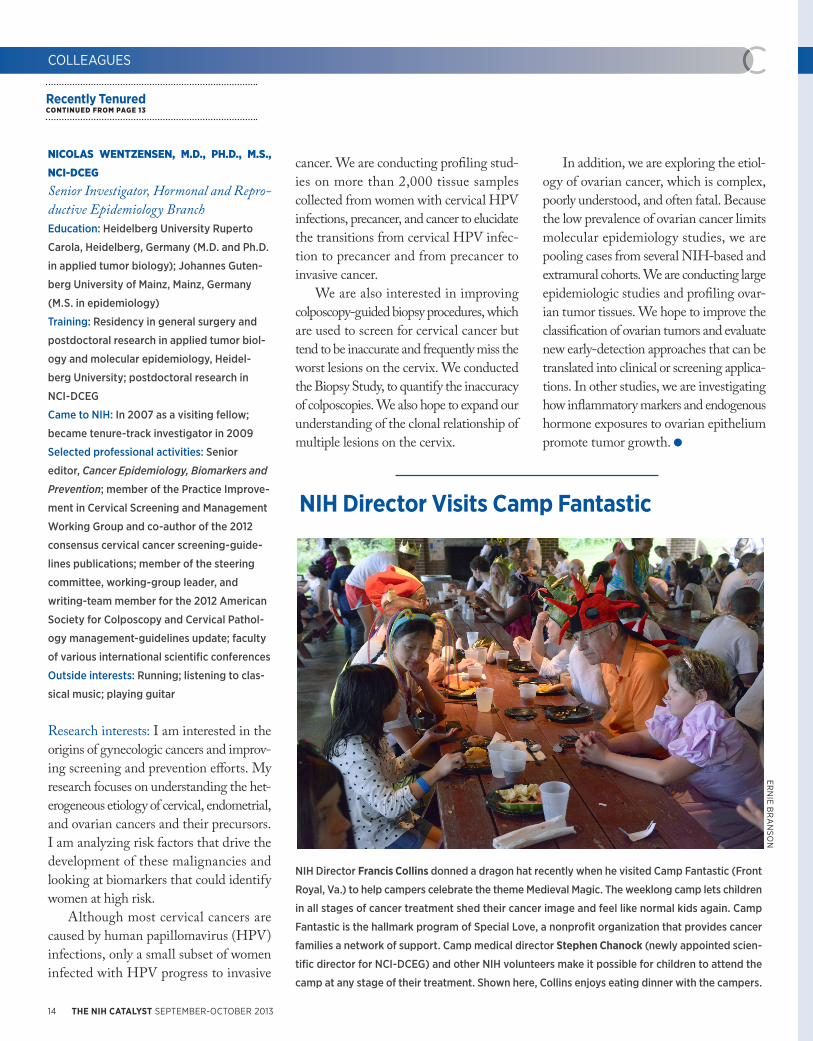
NIH Director Francis Collins donned a dragon hat recently when he visited Camp Fantastic (Front
Royal, Va.) to help campers celebrate the theme Medieval Magic. The weeklong camp lets children
in all stages of cancer treatment shed their cancer image and feel like normal kids again. Camp
Fantastic is the hallmark program of Special Love, a nonprofit organization that provides cancer
families a network of support. Camp medical director Stephen Chanock (newly appointed scien-
tific director for NCI-DCEG) and other NIH volunteers make it possible for children to attend the
camp at any stage of their treatment. Shown here, Collins enjoys eating dinner with the campers.
ERN
IE BR
AN
SON

http://irp.nih.gov/catalyst 15
MONDAY, September 9: Cori Bargmann (The
Rockefeller University), “Neuromodulatory
Circuits and Motivated Behavior”
September 11: Jeffrey Esko (University of Cal-
ifornia, San Diego), ”Proteoglycans: Arbiters
of Lipoprotein Metabolism”
September 25: Moyses Szklo (Johns Hopkins
Bloomberg School of Public Health), ”Epide-
miology: Back to Translation”
October 2: Taekjip Ha (University of Illinois at
Urbana-Champaign), ”Genome Maintenance
up Close and Personal: Eavesdropping on
Single Molecular Conversations”
October 3 (Thursday): Shinya Yamanaka
(Kyoto University and Gladstone Insti-
tutes), ”Recent Progress in iPS Cell Research
towards Regenerative Medicine”
CARDIOVASCULAR REGENERATIVE
MEDICINE
September 25–26, 2013
Natcher Conference Center (Building 45)
The NHLBI Symposium on Cardiovascular
Regenerative Medicine, will bring together
experts in basic stem cell biology, as well
as clinical cardiovascular medicine, to dis-
cuss advances and potential clinical appli-
cations. For more information and to regis-
ter, go to http://www.nhlbi.nih.gov/news/
events/2013-nhlbi-cvregenmed.
NIH RESEARCH FESTIVAL
October 7–11, 2013
FAES Academic Center (Building 10)
Masur Auditorium (Building 10)
Find out what’s going on in NIH research:
Attend lectures, minisymposia, poster
sessions, and more during this weeklong
festival. Don’t miss the special poster ses-
sion on Monday, October 7, at which the
scientific directors will showcase their work
(4:00-6:00 p.m.). For more information and
schedules, visit http://researchfestival.nih.
gov or contact Jacqueline Roberts (301-594-
6747 or [email protected]).
NATIONAL GRADUATE STUDENT RESEARCH
CONFERENCE
October 6–8, 2013
FAES Academic Center (Building 10)
Natcher Conference Center (Building 45)
Ninety advanced graduate students from
across the United States will share their own
cutting-edge research and learn about scien-
tific advances being made at NIH. NIH inves-
tigators will have the opportunity to recruit
participants as postdoctoral fellows. Agenda:
career- and professional-development work-
shops; a panel of former NIH trainees discuss-
ing their careers; and NIH Research Festival
poster sessions. NIH investigators and post-
docs are encouraged to visit the posters to
discuss potential collaborations and learn first-
hand about novel techniques and approaches
that could enhance their investigations. For
more information visit https://www.training.
nih.gov/events/recurring/nih_national_gradu-
ate_student_research_festival.
INTRODUCTION TO THE PRINCIPLES AND
PRACTICE OF CLINICAL RESEARCH
October 15, 2013–March 24, 2014
Monday and Tuesday evenings
5:00–6:30 p.m., Bethesda campus
Registration deadline: October 8, 2013
This free course is for physicians and other
health professionals planning a career
in clinical research. A certificate will be
awarded on successful completion of the
course. Topics include: basic epidemiologic
methods; ethical and legal issues; the role of
institutional review boards; the monitoring
of patient-oriented research; infrastructure;
developing and funding research studies;
and more. It’s suggested that participants
acquire a copy of the textbook, Principles
and Practice of Clinical Research, Third
Edition. For more information or to register,
visit http://www.cc.nih.gov/training/train-
ing/ippcr/application.html or call 301-496-
9425. An e-mail confirmation will be sent to
those accepted into the program.
http://irp.nih.gov/catalyst 15
THE ANITA B. ROBERTS FALL LECTURE
Tuesday, September 17, 2013, 1:00 p.m.
Lipsett Amphitheater (Building 10)
Wei Yang, section chief of NIDDK’s Labora-
tory of Molecular Biology, will give the fall
seminar honoring the memory of Anita B.
Roberts, former chief of NCI’s Laboratory of
Cell Regulation and Carcinogenesis. Yang’s
talk, “Seeing Is Believing: Functional Biology
at Atomic Resolution,” is sponsored by the
NIH Women Scientist Advisors Commit-
tee and the Office of Research on Women’s
Health. Individuals who need reasonable
accommodations to participate should con-
tact Margaret McBurney at 301-496-1921 and/
or Federal Relay, 1-800-877-8339, five days
before the lecture.
NIH SUPPLY CENTER BLOWOUT SALE!
Hurry! The NIH Supply Center and its Self Ser-
vices Stores (SSS) are discounting a huge of
amount of stock to make room for new items.
Office and laboratory items are being reduced
weekly. Get these products while supplies
last. Our goal is to provide more responsive
support to our customers. Self Service Store
campus locations (Monday–Friday, 8:00
a.m.–4:00 p.m.): BLDG 10, Room B2B41 (301-
496-2051); Building 31, Room B1A47 (301-496-
4430). View the list of reduced items at http://
nihsc.od.nih.gov/Promotions.aspx.
WEDNESDAY AFTERNOON LECTURE SERIES
SPECIAL: Begins Monday, September 9
Most Wednesdays, 3:00–4:00 p.m.
Masur Auditorium (Building 10)
The NIH Director’s Wednesday Afternoon
Lecture Series, colloquially known as WALS,
is the highest-profile lecture program at
the NIH. Each season includes some of the
biggest names in biomedical and behav-
ioral research. For more information, contact
Jackie Roberts (301-594-6747 or robertsjm@
od.nih.gov) or visit the WALS Web site at
http://wals.od.nih.gov. Here's a sampling:
ANNOUNCEMENTS

BILL B
RA
NSO
N
Town Hall Meeting at NIH
U.S. Secretary of Health and Human Services Kathleen Sebelius (right) visited the NIH on August 1 for a tour that included the Children’s Inn and an NCI lab; meetings with NIH leaders; and a Town Hall Meeting for NIH staff. At the Town Hall Meeting, hosted by NIH Director Francis Collins (left), Secretary Sebelius recognized the “dazzling” contributions of NIH researchers despite budget cuts and uncertainty. As she answered questions submitted in advance by members of the NIH community, she reaffirmed her commitment to funding medical research and celebrated NIH advancements including studies of “super-bug” hospital infections at the NIH Clinical Center and improved cancer treatments resulting from genomic sequencing.
CATALYTIC REACTIONS?
If you have a photo or other graphic that ref lects an aspect of l i fe at NIH (including laboratory life) or a quotation or confession that scientists might appreciate and that would be fit to print in the space to the right, why not send it via e-mail: [email protected]; fax: 301-402-4303; or mail: The NIH Catalyst, Building 1, Room 333.
Also, we welcome “letters to the editor” for publication and your reactions to anything on the NIH Catalyst pages.
FIRST-CLASS MAILPOSTAGE & FEES PAID
DHHS/NIHPermit No. G-802
Publication No. 13-6250
Official BusinessPenalty for Private Use $300
U.S. DEPARTMENT OF HEALTH AND HUMAN SERVICESNational Institutes of HealthBuilding 1, Room 333MSC 0183Bethesda, Maryland 20892
Printed on at least 20% recycled content paper and can be recycled as office white paper.
PUBLISHERMICHAEL GOTTESMAN Deputy Director for Intramural Research, OD
EDITORS JOHN I. GALLIN Director, NIH Clinical Center HENRY METZGER Scientist Emeritus
MANAGING EDITOR LAURA STEPHENSON CARTER
WRITER-EDITORCHRISTOPHER WANJEK Director of Communications, OIR
COPY EDITORSHAUNA ROBERTS
EDITORIAL INTERNJENNIFER SARGENT
CONTRIBUTING WRITERSREBECCA BAKERJANET HULSTRANDADAM KUSZAKREBECCA LAZERATIONMICHELE LYONSKATHERINE WENDELSDORF
PHOTOGRAPHERS/ILLUSTRATORS MAGGIE BARTLETT, BILL BRANSON, ERNIE BRANSON, DYLAN BURNETTE, MICHELE LYONS, MARIA NEKHAYONAK, THOMAS WELLEMS
EDITORIAL ADVISORY BOARDDAN APPELLA, NIDDK DAVID DAVIES, NIDDKMICHAEL ESPEY, NIDDK ELISE C. KOHN, NCI SUSAN LEITMAN, CC GERMAINE BUCK LOUIS, NICHD DAVID MILLER, NIEHS BERNARD MOSS, NIAIDHYUN PARK, NCI PAUL PLOTZ, NIAMS SARAH RHODES, NIMHJULIE SEGRE, NHGRI ANDY SINGLETON, NIA GISELA STORZ, NICHD RONALD SUMMERS, CCRICHARD WYATT, OIR
The NIH Catalyst is published bimonthly for and by the intramural scientists at NIH.
Address correspondence to: Building 1, Room 333, NIHBethesda, MD 20892Ph: 301-402-1449 Fax: 301-402-4303e-mail: [email protected]
The NIH Catalyst online: http://irp.nih.gov/catalyst
OTHER NEWS
More OnlineTo read expanded versions of the NIH Catalyst articles, go to (http://irp.nih.gov/catalyst/v21i5).