hormone biosynthesis, metabolism, and mechanism of action adrian quesada rojas, md
TRANSCRIPT
Hormone Biosynthesis, Metabolism, and Mechanism of Action
Adrian Quesada Rojas, MD
Definitions
Hormone: Substance produced in tissue => bloodstream => responsive cells
Provides means of communicationchemical regulatory and
signalingBloodstreamParacrine: cell to cell (contiguos)Autocrine: same cellIntracrine: same cell (unsecreted)

Nomenclature
Steroid hormonesSex steroids (Cholesterol derivatives)3 groups (number of carbon atoms)
21 carbons: pregnane nucleus19 carbons: androstane 18 carbons: estrane

Nomenclature

Lipoproteins and Cholesterol
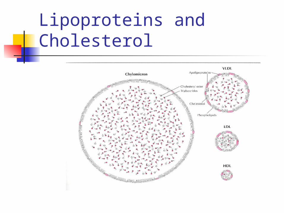
All steroid prod organs can synth (acetate)LPP: transport of non polar fat in polar solvent Chylomicrons: chlol 10, TG 90 formed in GI VLDL: more dense IDL: removal of some TG from VLDL interior LDL: end prod VLDL catabolism (50% chol) major carrier of chol in plasma HDL: highest protein content
Lipoproteins and Cholesterol

Reactions:Cleavage of side chainConv hydroxyl <=> ketones (dehydrogenase)Addition of OH group (hydroxylation)Creation of double bonds (remov of H)Addition of hydrogen (saturation)
Enz are dehydrogenases or C P450 oxidases
Steroidogenesis

Rate limit step: transfer Chol from outer to inner mitochondrial membrane (SSC)
Once Pregnenolone: two ways in ovary
pathway (3-hydroxysteroids) => DHEA
pathway (3-ketone) => 17 hydroxyP Conversion of pregnenolone to P:
3 hydroxysteroid dehydrogenase
isomerase reaction (3-OH group to ketone + transfer double bond 5-6 to 4-5)
Steroidogenesis
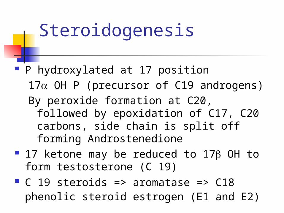
P hydroxylated at 17 position17 OH P (precursor of C19 androgens)By peroxide formation at C20, followed by
epoxidation of C17, C20 carbons, side chain is split off forming Androstenedione
17 ketone may be reduced to 17 OH to form testosterone (C 19)
C 19 steroids => aromatase => C18 phenolic steroid estrogen (E1 and E2)
Steroidogenesis

Alternative
Pregnenolone conv to 3 OH C19 steroid(DHA) by 17 OH lation followed by SCC
With ketone formation, DHA is converted into Androstenedione (C19 steroids)C 19 steroids undergo aromatization
(hydroxylation of angular 19 methyl group, followed by oxydation and loss of 19 C as formaldehyde and
dehydrogenation)
Steroidogenesis

Steroidogenesis

Two Cell System FSH receptors on granulosa FSH receptors are induced by FSH LH receptors on theca initially, as follicle
grows FSH induces LH receptors on granulosa
FSH ind aromatase activity on granulosa Actions regulated by autocrine and
paracrine factors

Two Cell System

Blood Transport of Steroids
Most E and T bound to protein carrier (SHBG) 30% is bound to albumin 1% freeHyperthyroidism, pregnancy, E adm SHBGCorticoids, A, P, GH, Insulin SHBGSHBG inversely related to weightHyperinsulinemia (low SHBG predictor of DM)
Body fat distrib (central=> hyperinsulinemia)

Blood Transport of Steroids

Estrogen Metabolism
Ovary => Estradiol / estrone Estriol => periph metab of estrone / E
not a secretory prod of ovary
conversion to less active form
Androgens are precursors of estrogensAdrenal gland (source of A, Androstenedione)

Estrogen Metabolism

E2 100-300 mg/day Androstenedione 3 mg/day ( 1% conversion)
=> 20- 30 % of E1 (produced every day)
Estrogen Metabolism

Progesterone Metabolism
NO peripheral conversion Secretion from adrenal and ovaries Preov < 1 mg /day Post ov 20-30 mg /day 10-20% of P is excreted as Pregnanediol Pre ov <1 mg/day, post ov 3-6 mg/d
(home ovulation test)

Androgen Metabolism Ovary: DHA, Androstenedione, testosterone Adrenal cortex: Gluco, mineralo, sex steroid
Sex steroids (less than ovary) DHA (½ adrenal, ¼ ovary, ¼ periph) Androstenedione (½ adrenal, ½ ovary) Testost 0.2-0.3 mg/day
50% periph conv of androstenedione and DHA
25% adrenal 25% ovary
Androgens Test binding capacity Androg effects depends on unbound fraction Hirsutism Test => 5reductase => DHT (princ androg) Androstenedione > Test (women)
DHT is derived from Androst and DHA DHT largely metabolized intracell
Only 1/10 of levels of Test Not all tissue requires DHT from Test
(Wolffian)
Androgen Metabolism

Cellular Mechanism of Action
Two major types of hormone actionTropic hormones (recep at cell
memb)Steroids (recep inside the cell)
Multiple cell receptors:Intracellular:
In the nucleus (transcription activity)G protein:
Single polypeptide chain (cell memb)

Ion gate channel: Cell surface, mult units (ACh)
Intrinsic enz activ:Trans memb recep with intracell component with tyrosine or serine kinase activity
Cellular Mechanism of Action

Mechanism of actionSteroid hormones
Includes: Hormone diffusion across cell memb Steroid binding to receptor protein Interaction of Horm-Recep complex with
DNA Synthesis of mRNA Transport of mRNA to ribosomes Protein synthesis in cytoplasm

Mechanism of actionSteroid hormones

Biological activityMantained only while H-R complex attached to nuclear siteDuration of exposure to hormone is as important as doseMajor factor in potency differences among various estrogens is length of time E-R complex occupies nucleus
Mechanism of actionSteroid hormones
Cortisol and P must circulate in large concentrations 2 to receptor complexes short half-lives in nucleusH-R complex after gene activation (processing)
Rapid degradation of R unbound to EMuch slower degradation of bound R
Continuous presence of E is important factor in continuing response
Mechanism of actionSteroid hormones

Estrogen Receptors
ER-rapid turnover) ER-96% homologous in DNA binding
domain and 53% homologous in the hormone binding domain (when compared to ER-)
Respond in comparable manner to same hormones

Estrogen Receptors

E receptors => nucleocytoplasmic shuttling Recep can diffuse out of nucleus and be
transported back in or undergo metabolism If shuttling impaired Receptors are
degraded rapidly E antagonists prevent nuclear translocation
and thus increase cytoplasmic degradation
Estrogen Receptors

Prior to binding R is inactive complex that includes a variety of heat shock proteins
Activation => dissociation of HSP When H binds R => conformational
change Conformational shape determines exact
message transmitted to the gene E2, raloxifene, tamoxifen induces a
different conformational shape
Estrogen Receptors

Once activated H receptor activates transcription in partnership with several groups of polypeptides
1. Transcription factors (polymerase enzyme and DNA)
2. Coactivators and corepressors (intracel prot called adaptor proteins and activate or suppress the TAF)
Estrogen Receptors

Phosphorylation Also stimulated by ligand binding Occur in specific receptor sites Increases potency of molecule to
regulate transcription cAMP and prot kinase pathways
increase transcriptional activity of E-R
Estrogen Receptors

Differences ER- and ER-ER-prevalent in brain, CV system, granulosa
cellsBreast expresses ER- and ER-ER- acts as natural suppressor of ER-
activity on breast tissueColon contains only ER-, reduction of colon
CA in postmenopausal on HRT may reflect antiproliferative effect of beta receptor
Estrogen Receptors
Progesterone Receptor Induced by E, decreased by Progestins Two major forms (A and B) A and B are expressed differently in
Breast and Endometrial tissues In most cells B positive regulator of P-
responsive genes, A inhibits B activity A and B have different molecular
functions, affecting different genes

Mice lacking of P receptors (A,B) are unable to ovulate (failure to expel a mature oocyte in a fully developed follicle)
PR-A protects against uterine and mammary gland hyperplasia induced by PR-B
Progesterone Receptor

Androgen Receptor Androgens only in one of three ways:1. T => DHT (Intracrine)2. T itself (Endocrine)3. Intracel conversion of T to E (Intracrine) Wolffian duct derivatives (T) Hair follicles, urogenital sinus
derivatives (req T => DHT)

T and DHT binds to same high affinity A-receptor (DHT has greater affinity)
Anti-A also bind to same R (20% affinity of T) A-R: two forms B (full length) and A (shorter) Functional differences yet to be determined A and P can cross react for their receptors
(pharmacologic concentrations) Progestin can act as an anti-E and anti-A
Androgen Receptor

Androgen Insensitivity SyndromeCongenital abnormality in A intracel RA-R gene is on X chromosome (Xq11-12)X-linked disorderDeletion of amino acids from steroid binding domain 2 to nucleotide alterations in gene
Androgen Receptor

Androgen Receptor

Agonists and Antagonists
Agonist => stimulates responseAntagonist => inhibits actions of an
agonistblockage of receptor
message
Examples: TamoxifenMifepristoneHistamine receptor antag
Physiologic AntagonistsP is not an E antagonistModifies E action by depleting E receptorsP can induce conversion of E2 to E1
Androgens block E actions (unclear mechan)
Agonists and Antagonists

Two groups of anti-E:Pure anti-EMixed agonists antagonists
Tamoxifen:Similar to clomipheneCompetitively inhibits E binding to receptorE affinity for receptor is 100-1000x better
than tamoxifen
Agonists and Antagonists

Agonists and Antagonists

Tamoxifen–E receptor binds to DNA Agonistic – Antagonistic actions depends
on coactivators present in specific cell types
Estrogenic actions: Lowers FSH levels Decreases Cholesterol, LDL Stimulates P receptor synthesis Maintenance of bone, vagina, endometrium
Agonists and Antagonists

Causes endometrial hyperplasia, polyps and 4x increase in endometrial cancer
Mechanism of action: TAF-1 and TAF-2 can both activate
transcription Tamoxifen agonistic ability is due to
activation of TAF-1 Antagonistic activity is due to competitive
inhibition of E-dependent activation of TAF-2
Agonists and Antagonists

Estrogen Receptors

Response to E and anti-E depends on: Nature of E receptor E response elements and promoters Cell content of protein coactivators Properties of ligand Modulation by growth factors and
agents that affect phosphorylation and protein kinases
Agonists and Antagonists
Tamoxifen reduces risk of recurrent disease in E-R positive and negative breast cancer
Besides binding to E-R (competitive inh), Inhibits protein kinase C activity (phosphor) Inhibits calmodulin-dependent cyclic AMP
phosphodiesterase (binds to calmodulin) Stim secretion of TGF- (inh growth CA cell)
Agonists and Antagonists

Tamoxifen: treatment should be for only 5 years
Emergence of tamoxifen-resistant tumors, how?Loss of E receptors
Loss of cellular control / loss of E-R expression Variant and mutant E-R Changes in coactivators Differential metabolism Resistance occur when E-R is NOT dominant in
growth of CA cells
Agonists and Antagonists

Selective E Agonist / AntagonistRaloxifeneInduce conformational shape change that
results in modified action influenced by cellular regulating proteins
Anti-E in uterus and breast
Agonist in certain tissues (bone, lipids)
Agonists and Antagonists

Antiprogestins Mifepristone Binding => different conformational change =>
prevents full gene activation Has also some agonistic activity Final response is determined by coact, corepr Affinity to P-R is 5x greater than P In absence of P can produce weak agonistic
effect Also works as weak anti-androgens

Tropic Hormones Receptor in cell membrane R functions as ion channel or as enzime Can also couple an active agent
(messenger) Cyclic AMP IP3 1,2 DG Ca Cyclic GMP

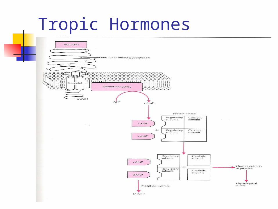
Cyclic AMPFSH, LH, hCG, TSH and ACTHLigand => R activates adenylate cyclaseConversion of ATP into cAMPcAMP binds cytoplasm receptor protein
and activates protein kinaseCatalyze phosphorilation of serine in cell
proteins => physiologic events
Tropic Hormones
Tropic Hormones

cAMP is degrated by PDE into 5’-AMP PG’s stim adenylate cyclase and cAMP accum Other tropic hormones
Oxytocin Insulin GH do not use Adenylate cyclase PRL hPL
Tropic Hormones

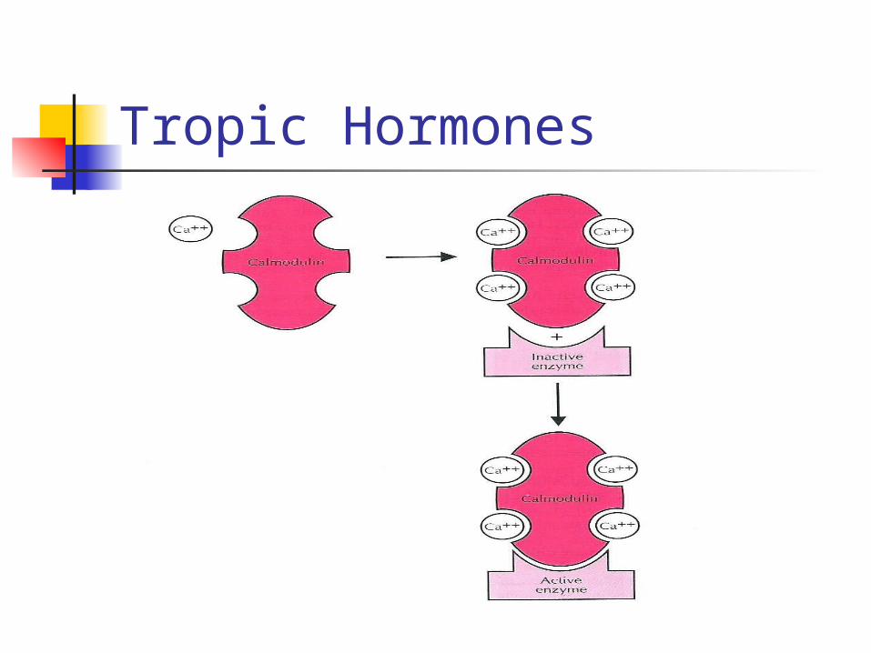
Calcium messenger systemIntracel Ca regulates cAMP, cGMP levelsCa mess system is linked to H-R function by
specific enzyme Phospholipase CCatalyzes hydrolysis of phosphatidylinositols
(cell membrane phospholipids)PLC activ generates two intracell messengers
Tropic Hormones

IP3 and DAG Both initiate functions of 2 parts of Ca system First part => Ca activ Prot kinase
Sustained cellular responses
Second part => Calmodulin (regulator)Acute cell responses
Tropic Hormones
Tropic Hormones

Kinase receptorsCell membrane receptors of
insulin, IGF, EGF, platelet-derived GF and fibroblast GF are tyrosine kinases
Extracell, transmembrane, cytoplasmic domainCytoplasmic (autophospholylation)Insulin / IGF (22 subunits)
Tropic Hormones

Tropic Hormones
Regulation of tropic hormones
Four major components Autocrine and paracrine reg factors
GF are produced by local gene expression Modulate activ in cells (nearby or producing cells) Involved in reproductive physiology (activin,
inhibin, IGF-I, IGF-II, TGF-, FGF, EGF) Also cytokines (modulate ovarian steroidogenesis)
Heterogeneity of tropic hormones (FSH, hCG) Glycoproteins are not single proteins (isoforms)

Up and down-regulation of receptors Positive or negative modulation of receptors
by homologous hormones Little is known regarding up regulation (PRL,
GnRH)
Regulation of adenylate cyclase
Regulation of tropic hormones

Regulation of Adenylate cyclase G protein system: Adenylate cyclase (composition):
Receptor Guanyl nucleotide regulatory unit (GTP
regul) Catalytic unit (converts ATP to cAMP)
Regulation of tropic hormones

Regulation of tropic hormones

Regulation of tropic hormones

Ligand => receptor => nucleotide regulatory unit (GTP uptake) => catalytic enzyme converting ATP to cAMP
Coupling of regulatory unit with catalytic unit
Enzyme activity is terminated by hydrolysis of GTP to GDP
Regulation of tropic hormones

G protein system Stimulatory nucleotide regulatory G
protein Inhibitory nucleotide regulatory G
protein Not limited to cAMP signal, can
activate other messenger-generating enzymes
Regulation of tropic hormones

Regulation of tropic hormones