hiv-1 tat-coated nanoparticles result in enhanced humoral immune responses and neutralizing...
TRANSCRIPT
Vaccine 24 (2006) 3564–3573
HIV-1 Tat-coated nanoparticles result in enhanced humoral immuneresponses and neutralizing antibodies compared to alum adjuvant
Jigna Patel a, David Galey b, Julia Jones c, Phillip Ray c, Jerold G. Woodward c,Avi Nath b, Russell J. Mumper a,∗
a Department of Pharmaceutical Sciences, College of Pharmacy, University of Kentucky, Lexington, KY 40536-0082, USAb Department of Neurology, Johns Hopkins University, Baltimore, MD 21287, USA
c Department of Microbiology, Immunology and Molecular Genetics, University of Kentucky, Lexington, KY 40536-0298, USA
Received 5 November 2005; received in revised form 25 January 2006; accepted 31 January 2006Available online 17 February 2006
Abstract
HIV-1 Tat has been identified as an attractive target for vaccine development and is currently under investigation in clinical trials asbt(reaoit©
K
1
vrh[aepwaf
0d
oth a therapeutic and preventative vaccine for HIV-1. It is well known that protein based vaccines produce poor immune responses byhemselves and therefore require adjuvants to enhance immune responses. We have previously reported on the use of anionic nanoparticlesNPs) for enhancing cellular and humoral immune responses to Tat (1–72). The purpose of this study was to further evaluate the immuneesponse of HIV-1 Tat (1–72) coated on anionic nanoparticles compared to alum using various doses of Tat (1–72). Nanoparticles wereffective at generating comparable antibody titers at both 1 and 5 �g doses of Tat (1–72), whereas the antibody titers significantly decreasedt the lower dose of Tat (1–72) using alum. Anti-sera from Tat (1–72) immunized mice reacted greatest to the N-terminal and basic regionsf Tat, with the NP groups showing stronger reactivity to these regions compared to alum. Moreover, the anti-sera from all Tat (1–72)mmunized groups contained Tat-neutralizing antibodies and were able to significantly inhibit Tat-mediated long terminal repeat (LTR)ransactivation.
2006 Elsevier Ltd. All rights reserved.
eywords: HIV-1 Tat; Protein; LTR-transactivation; Nanoparticles; Vaccine; Adjuvants
. Introduction
Ideally, an HIV vaccine would be effective at blockingiral entry and thus, provide sterilizing immunity. However,ecent failures with the envelope (Env) based HIV vaccineave demonstrated that this may be a difficult goal to attain1]. These experiences have prompted investigators to lookt alternative strategies in developing an HIV vaccine; forxample, to limit viral replication and thereby block or delayrogression on to AIDS. In pursuit of this secondary goal, it isell recognized that the candidate antigen must be conserved
mong different HIV subtypes, as this is a major drawbackor Env based vaccines. In addition, the antigen should be
∗ Corresponding author. Tel.: +1 859 257 2300x258; fax: +1 859 323 5985.E-mail address: [email protected] (R.J. Mumper).
immunogenic and play a critical role in the virus life cycle.To this end, the HIV-1 regulatory protein Tat has receivedconsiderable interest as a potential HIV vaccine or as a com-ponent in a HIV vaccine [2–5].
Tat, a small regulatory HIV protein, is encoded by twoexons, with the size of the full length protein varying from86 to 102 amino acids (aa) depending on the viral strain. Thefirst exon of Tat encodes the first 72 aa of the protein, whichincludes: the amino-terminal (aa 1–21); the cysteine-richdomain (aa 22–37) representing the transactivation domain;the core (aa 38–48); and the basic domain (aa 49–72) con-taining nuclear localization signals and regions mediatingcellular uptake of Tat [6]. This region of Tat has been shownto be necessary for Tat-mediated transactivation of HIV-1gene expression [7,8] and is highly conserved among differ-ent viral subtypes [9,10], with the cysteine-rich domain (aa
264-410X/$ – see front matter © 2006 Elsevier Ltd. All rights reserved.oi:10.1016/j.vaccine.2006.01.065
J. Patel et al. / Vaccine 24 (2006) 3564–3573 3565
25–38) being conserved among human, bovine and simianspecies [11]. While the second exon, encoding the C-terminaldomain of Tat is not required for the transactivation, it con-tains an arginine–glycine–aspartic acid (RGD) sequence thatmediates the binding of extracellular Tat to integrin receptors[12]. Additionally, the C-terminal domain of Tat is of signif-icant importance in the cellular uptake of extracellular Tat[13].
Tat plays a vital role in the viral life cycle and mediatesmany processes allowing for spread of the virus through-out the body and potentiating disease. Of these, possiblythe most significant role of Tat in the virus life cycle is theenhancement of HIV gene expression. Tat is expressed inthe very early stages of infection before the expression of thestructural components (Env, Gag, Pol) [14]. In fact, Tat is nec-essary for efficient viral gene expression and in the absence ofTat, no or only minute amounts of the structural proteins areexpressed, preventing the production of an infectious virus[15,16]. In addition to promoting viral gene expression inHIV-infected cells, extracellular Tat released from infectedT-lymphocytes has numerous effects on uninfected cells thataid in the progression of disease. Tat induces the expres-sion of the chemokine receptors CCR5 and CXCR4, whichfunction as co-receptors for HIV-1 and facilitate the trans-mission of macrophage and T-cell tropic HIV-1 strains [17].Some in vitro studies have demonstrated that Tat inducestttFapdmsoiwimaactM[
hbTpAaimn
and cellular immune responses [33]. However, conflictingdata exists on the effectiveness of a Tat-based vaccine in con-trolling infection upon challenge with a highly pathogenicstrain (SHIV-89.6P) in non-human primates [18,34–37]. Inspite of these data, many researchers agree that the potentialof Tat based vaccines warrant further investigation and PhaseI clinical trials evaluating Tat for preventative and therapeu-tic vaccines are currently ongoing in Italy [27,33]. Moreover,a Tat-toxoid (chemically modified Tat) vaccine has alreadybeen evaluated in both HIV negative and positive patientsand was shown to be safe and effective [32]. Futhermore,other ongoing clinical trials evaluating preventative HIV-1vaccines include Tat as component of the vaccine in additionto other HIV-1 antigens (IAVI Report, February 2005).
We have previously reported on the preparation and purifi-cation of the first exon of Tat protein (referred to as Tat (1–72))[13]. We have demonstrated that Tat (1–72) is immuno-genic and that Tat (1–72) coated anionic nanoparticles (NPs)were effective at generating humoral and cellular immuneresponses to Tat [38]. In the present study, the use of nanopar-ticles for generating immune responses to various doses of Tat(1–72) was further investigated. More specifically, we soughtto determine the lowest effective dose of Tat (1–72) that couldproduce immune responses when administered with anionicnanoparticles. In addition, the antibody epitopes generated inmice immunized with Tat (1–72) were mapped and the Tat-ne
2
2
btPeCfpcG(odfttwCoPRd
he production of Interferon-�, inhibiting T-cell prolifera-ion, [4] and also promotes apoptosis of T-cells by increasinghe expression of CD95L/Fas ligand on macrophages [18].anales-Belasio et al. have demonstrated that biologicallyctive Tat at high concentrations is taken up by antigenresenting cells (APCs), particularly monocyte-derived den-ritic cells, inducing expression of MHC II, co-stimulatoryolecules, and causing production of Th-1 type cytokines
uch as IL-12 [19]. On the contrary, Izmailova et al. did notbserve DC activation or maturation in the presence of HIV-nfection or Tat expression; however, they reported that Tatas involved in up-regulation of chemoattractant proteins in
mmature DC which are involved in recruiting of T-cells andacrophages thus, aiding in the spread of infection [20]. The
bility of Tat to be efficiently taken up by cells has becomettractive for the delivery of proteins into cells [21] andonjugation of proteins to Tat has been shown to be an effec-ive method in presenting exogenous proteins in context of
HC class I, and generating antigen-specific CTL responses22].
The importance of Tat in the progression to diseased statesas been demonstrated in numerous reports [23–31]. In fact,oth strong antibody [25,28,30] and CTL responses [24,31] toat have been inversely correlated with viral loads and diseaserogression. Moreover, Tat is involved in the progression ofIDS related Kaposi sarcoma and high serum levels anti-Tat
ntibodies have been correlated with reduced Kaposi sarcoman HIV infected patients [32]. Extensive studies carried out in
ice and non-human primates have demonstrated that immu-ization with Tat is safe and effective at generating humoral
eutralizing activity in the sera from immunized mice wasvaluated.
. Materials and methods
.1. Materials
Emulsifying wax, comprised of cetyl alcohol and polysor-ate 60 (molar ratio of 20:1), was purchased from Spec-rum (New Brunswick, NJ). Sodium dodecyl sulfate (SDS),BS/Tween 20 buffer, bovine serum albumin (BSA), tri-thanolamine, and mannitol were purchased from Sigmahemical Co. (St. Louis, MO). Brij 78 was purchased
rom Uniqema (New Castle, DE). Sheep anti-mouse IgG,eroxidase-linked species specific F(ab′)2 fragment was pur-hased from Amersham Pharmacia Biotech (Piscataway, NJ).oat anti-mouse IgG2a and IgG1 horseradish peroxidase
HRP) conjugates were purchased from Southern Biotechnol-gy Associates, Inc. (Birmingham, AL). Tetramethylbenzi-ine (TMB) Substrate Kit was purchased from Pierce (Rock-ord, IL). Incomplete freunds adjuvant and mycobacteriumuberculosis were purchased from Fisher Scientific (Hamp-on, NH). Lipid A from Salmonella Minnesota R595 (Re)as purchased from List Biological Laboratories (Campbell,A). HIV-1 Clade B consensus Tat peptides (15 aa) werebtained through the AIDS Research and Reference Reagentrogram (Division of AIDS, NIAID, NIH, Bethesda, MA).ecombinant HIV-1 Tat (1–72 aa) was prepared as previouslyescribed [13].
3566 J. Patel et al. / Vaccine 24 (2006) 3564–3573
2.2. Preparation of anionic NPs
Nanoparticles from oil-in-water microemulsion precur-sors were prepared as previously described with slight modi-fication. Briefly, 2 mg of emulsifying wax and 3.5 mg of Brij78 was melted and mixed at ∼60–65 ◦C. Deionized and fil-tered (0.2 �m) water (980 �L) was added to the melted waxand surfactant while stirring to form an opaque suspension.Finally, 20 �L of sodium dodecyl sulfate (50 mM) was addedto form clear microemulsions at 60–65 ◦C. The microemul-sions were cooled to room temperature, while stirring, toobtain NPs (2 mg/mL). The final concentration of compo-nents in the NP suspension was emulsifying wax (2 mg/mL),Brij 78 (3 mM), and SDS (1 mM). The NP sizes were mea-sured using a Coulter N4 Plus Sub-Micron Particle Sizer(Coulter Corporation, Miami, FL) at 90◦. The zeta poten-tial of the NPs was measured using Malvern Zeta Sizer 2000(Malvern Instruments, Ingleside, IL).
2.3. Coating of the anionic NPs with Tat
Varying amounts of Tat were added to NPs (1000 �g/mL)in 5% (v/v) mannitol. The suspension was vortexed gentlyand placed on a horizontal shaker at room temperature fora minimum of 30 min to allow for coating. The coated NPsws
2
iefSiIwcT10vi
cardiac puncture and sera were separated. All sera collectedwere stored at −20 ◦C.
In the follow up study (Study 2), mice (n = 6–8/group)were dosed on Day 0, 14 and 28 with 0.2 �g or 1 �g of Tat-coated NPs or 0.2 �g or 1 �g of Tat adjuvanted with alum.Again, the dose of NPs and alum given to animals was 100 �g.Mice were immunized with 1 �g of Tat adjuvanted with LipidA (50 �g) as a positive control. To assess the kinetics of Tatspecific antibodies generated using the different treatments,mice were bled on Days 13 and 34 by tail vein and sera werecollected. On Day 42, all mice were bled by cardiac punctureand sera were collected. All sera were stored at −20 ◦C.
2.5. Determination of antibody titers
Tat-specific serum IgG, IgG1 and IgG2a antibody titerwere determined using an ELISA. Briefly, 96-well plates(costar) were coated with 50 �L of Tat (5–8 �g/mL in 0.01 Mphosphate buffered saline, pH 7.4) overnight at 4 ◦C. Theplates were blocked for 1 h at 37 ◦C with 200 �L of 4% BSAprepared in PBS/Tween 20. The plates were then incubatedwith 50 �L per well of mouse serum diluted appropriatelyin 4%BSA/PBS/Tween 20 for 2 h at 37 ◦C. The plates werewashed with PBS/Tween 20 and incubated with 50 �L/wellanti-mouse IgG HRP F(ab′)2 fragment from sheep (1:3000i ◦IwHtaa1aTmu0
2
tp
TE
S
S�g�g
S1 �g1 �g1 �g
ere diluted appropriately in de-ionized water for particleize and zeta potential measurements.
.4. Mouse immunization study
Two animal studies were carried out to determine themmune response to different doses of Tat. A summary of thexperimental design is presented in Table 1. For both studies,emale BALB/c mice (8–10 weeks old) obtained from Harlanprague–Dawley Laboratories (Indianapolis, Indiana) were
mmunized subcutaneously with 100 �L of the formulations.n the initial mouse study (Study 1), mice (n = 5–6/group)ere dosed on Day 0, 21 and 28 with 1 �g or 5 �g of Tat-
oated NPs or 1 �g or 5 �g of Tat adjuvanted with alum.he dose of NPs and alum administered in both cases was00 �g. As a positive control, mice were immunized on Daywith 5 �g of Tat adjuvanted with complete freunds adju-
ant (CFA) followed by boost with 5 �g Tat adjuvanted withncomplete freunds adjuvant. On Day 35, mice were bled by
able 1xperimental design for mouse immunization study
tudy Treatment Tat dose
tudy 1
Naı̈ve –NPs + Tat 1 and 5Alum + Tat 1 and 5CFA + Tat 5 �g
tudy 2
Naı̈ve –NPs + Tat 0.2 andAlum + Tat 0.2 andLipid A + Tat 0.2 and
n 1%BSA/PBS/Tween 20) for 1 h at 37 C. For IgG1 andgG2a determination, the plates were similarly incubatedith goat anti-mouse IgG1-HRP or goat anti-mouse IgG2a-RP diluted 1:8000 (1%BSA/PBS/Tween 20). After washing
he plates with PBS/Tween 20, the plate was developed bydding 100 �L of TMB substrate and incubating for 30 mint RT. The color development was stopped by addition of00 �L of 2M H2SO4 and the OD at 450 nm was read usingUniversal Microplate Reader (Bio-Tek Instruments, Inc.).he OD at 450 nm versus log serum dilution for each ani-al was plotted and fit to a four parameter logistic equation
sing GraphPad Prism software. The titer was defined as.5 × ODmax.
.6. B-Cell epitope mapping
Tat anti-sera were tested by ELISA to determine reactivityo various regions of Tat. Tat peptides (50 �L of 1 �g/mL Tateptide in 0.05 M carbonate buffer, pH 9.6) were coated onto
Immunization schedule Sera collected
–
Day 35Day 0, 21, 28
–
Day 13, 34, 42Day 0, 14, 28
J. Patel et al. / Vaccine 24 (2006) 3564–3573 3567
96-well costar plates by incubating overnight at 4 ◦C. Theplates were blocked for 1 h at 37 ◦C with 200 �L of 4% BSAprepared in PBS/Tween 20. Tat anti-sera diluted at 1:100 in4%BSA/PBS/Tween 20 were added to the wells and incu-bated for 2 h at 37 ◦C. The wells were washed, reacted withanti-mouse IgG HRP F(ab′)2 fragment from sheep, and devel-oped as described for total IgG titer.
2.7. Tat-mediated LTR-transactivation assay
SVGA LTR-chloramphenicol acetyltransferase (CAT)was produced by stable transfection of the astrocytic cellline SVGA with pHIV-CAT [39]. SVGA LTR-CAT cellswere seeded into 6-well plates a minimum of 24 h prior touse in Dulbecco’s Modified Eagle Medium (DMEM; Gibco-BRL) with 10% heat-inactivated fetal bovine serum (FBS;Sigma) and 1% antibiotic–antimycotic solution (penicillin Gsodium, streptomycin sulfate, and amphotericin B in 0.85%saline; GibcoBRL) (DMEM + 10%FBS + Ab). Ten micro-liters of sera was mixed with 2 �L (1000 ng) recombinantTat (1–72) derived from the first exon of the HIV-1 tat geneand incubated for 30 min at 37 ◦C in a water bath. During theincubation, medium was replaced on the SVGA LTR-CATcells with 2 mL fresh DMEM + 10%FBS + Ab. The sera/Tatmixtures were then added to the cells. Cells were then lysedat 24 h post-treatment and CAT levels were quantitated byE
2
suP
3
TcnatOntnpicrprbe
Table 2Physical properties of anionic nanoparticles coated with HIV Tat (1–72)
Sample (n = 3) Mean size (nm) Mean charge (mV)
Anionic NPs 102.5 ± 7.6 −55.7 ± 0.7Tat-coated NPs (1:500, w/w) 130.6 ± 3.3 −48.6 ± 3.7Tat-coated NPs (1:100, w/w) 110.9 ± 3.3 −43.9 ± 1.9Tat-coated NPs (1:20, w/w) 111.6 ± 5.1 −43.6 ± 1.7
tage of being prepared in a single step, one vessel processwith relative ease in modifying the physical characteristicsof the particles by using appropriate surfactants. We recentlyreported the use of anionic NPs as effective delivery systemsfor enhancing immune responses to Tat (1–72) [38]. In thepresent studies, the dose-response to Tat (1–72) using NPscompared to standard alum adjuvanted protein was furtherevaluated. In addition, the humoral immune responses to Tat(1–72) were characterized for reactivity to various regions ofTat and for activity in a LTR-transactivation assay.
3.1. Adsorption of Tat (1–72) on anionic nanoparticles
The NPs prepared in these present studies were stabilizedby the inclusion of Brij 78 and made to be negatively-chargedby the use of the anionic surfactant SDS. The net charge ofthe NPs prior to coating with Tat was approximately −56 mV(Table 2). Tat, a cationic protein, is expected to be coated onthe surface of the NPs via ionic interactions. It is thought thatthe negatively charged amino acids of Tat are exposed onthe surface of NPs while the positively charged amino acidsinteract with the negatively charged NPs. As demonstratedin Table 1, there was a slight increase in the zeta potentialwith increasing amounts of Tat coated on NPs; however, theTat-coated NPs continued to have a net negative charge pos-sibly due to the exposed negatively charged amino acids. Thecas
3
NAwmt1wvFwNiacu
LISA according to the manufacturer’s directions (Roche).
.8. Statistical analysis
Statistical analysis was performed using one-way analy-is of variances (ANOVA) followed by pair-wise comparisonssing Newman–Keuls multiple comparison test using Graph-ad Prism software.
. Results and discussion
Many researchers have reported on the potential of HIV-1at based vaccine or supported the idea including Tat as aomponent in a cocktail for vaccine development [2–5]. Asew targets for HIV vaccine development are identified, it islso important to investigate novel delivery systems for effec-ively enhancing immune responses to the target antigen(s).ur laboratory has reported on the use of anionic and cationicanoparticles for effectively enhancing immune responseso plasmid DNA and protein based vaccines [40–42]. Theseanoparticles are prepared from oil-in-water microemulsionrecursors and form solid stable particles. The emulsify-ng wax oil phase used to prepare these nanoparticles isomprised of cetyl alcohol and polysorbate (20:1 molaratio) which are both employed as components in parenteralroducts and are potentially non-toxic materials. We haveecently shown that these nanoparticles are hemocompati-le and metabolized via endogenous alcohol dehydrogenasenzyme systems [43]. In addition, these NPs have the advan-
oating of Tat on NPs at a 1:100 (w/w) ratio was found to bepproximately 85% by SDS–PAGE/densitometry (data nothown).
.2. Dose response to Tat in immunized mice
We previously reported that Tat (5 �g) coated on anionicPs result in similar Tat-specific IgG levels as alum and lipidadjuvanted with Tat [38]. Thus, a dose response of Tat
as evaluated in these present studies to determine the mini-um Tat dose that could be administered while maintaining
he Tat-specific IgG response. In the first study, the use ofand 5 �g of Tat coated on NPs or adjuvanted with alumas investigated. Tat (5 �g) with complete freunds adju-ant was used as a positive control. The results shown inig. 1A demonstrate that Tat-specific IgG antibody titersere maintained at both the 1 and 5 �g of Tat coated onPs, while Tat (1 �g) adjuvanted with alum produced signif-
cantly lower titers compared to Tat (5 �g) adjuvanted withlum. Moreover, both the 1 and 5 �g Tat-coated NPs producedomparable Tat-specific IgG titers compared to complete fre-nds adjuvant. Only the higher dose of Tat adjuvanted with
3568 J. Patel et al. / Vaccine 24 (2006) 3564–3573
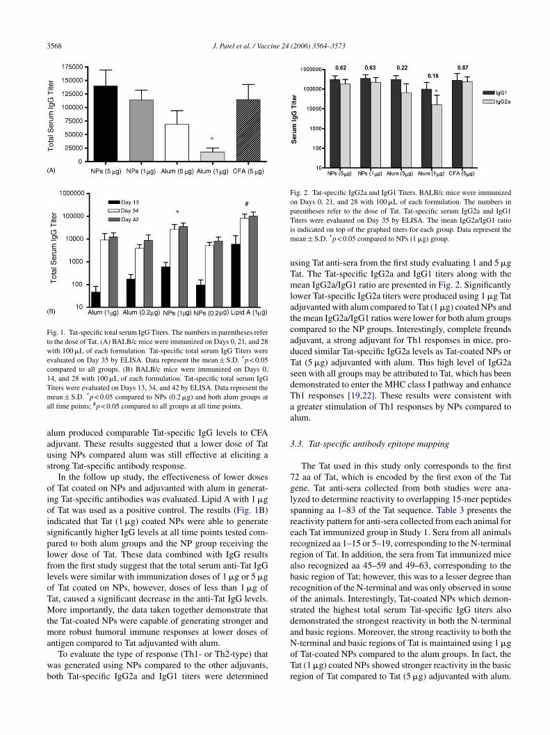
Fig. 1. Tat-specific total serum IgG Titers. The numbers in parentheses referto the dose of Tat. (A) BALB/c mice were immunized on Days 0, 21, and 28with 100 �L of each formulation. Tat-specific total serum IgG Titers wereevaluated on Day 35 by ELISA. Data represent the mean ± S.D. *p < 0.05compared to all groups. (B) BALB/c mice were immunized on Days 0,14, and 28 with 100 �L of each formulation. Tat-specific total serum IgGTiters were evaluated on Days 13, 34, and 42 by ELISA. Data represent themean ± S.D. *p < 0.05 compared to NPs (0.2 �g) and both alum groups atall time points; #p < 0.05 compared to all groups at all time points.
alum produced comparable Tat-specific IgG levels to CFAadjuvant. These results suggested that a lower dose of Tatusing NPs compared alum was still effective at eliciting astrong Tat-specific antibody response.
In the follow up study, the effectiveness of lower dosesof Tat coated on NPs and adjuvanted with alum in generat-ing Tat-specific antibodies was evaluated. Lipid A with 1 �gof Tat was used as a positive control. The results (Fig. 1B)indicated that Tat (1 �g) coated NPs were able to generatesignificantly higher IgG levels at all time points tested com-pared to both alum groups and the NP group receiving thelower dose of Tat. These data combined with IgG resultsfrom the first study suggest that the total serum anti-Tat IgGlevels were similar with immunization doses of 1 �g or 5 �gof Tat coated on NPs, however, doses of less than 1 �g ofTat, caused a significant decrease in the anti-Tat IgG levels.More importantly, the data taken together demonstrate thatthe Tat-coated NPs were capable of generating stronger andmore robust humoral immune responses at lower doses ofantigen compared to Tat adjuvanted with alum.
To evaluate the type of response (Th1- or Th2-type) thatwas generated using NPs compared to the other adjuvants,both Tat-specific IgG2a and IgG1 titers were determined
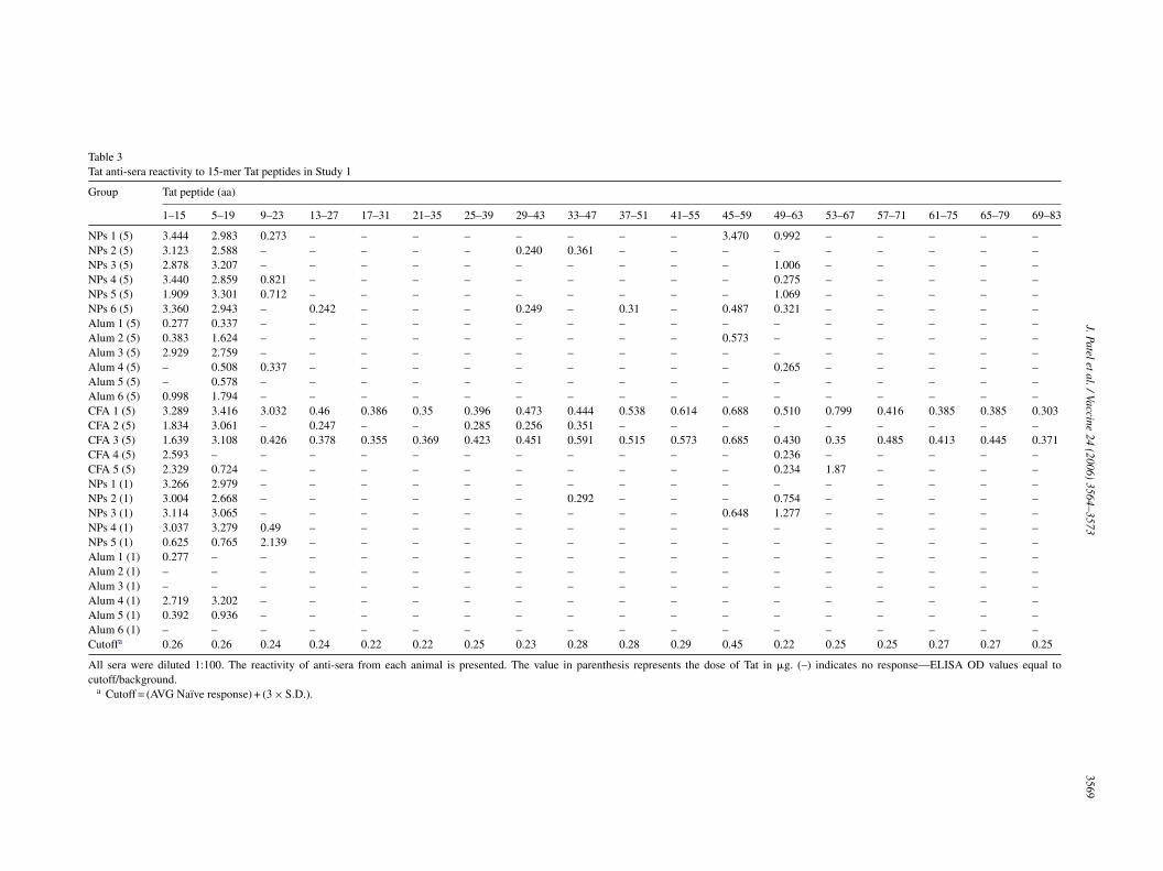
Fig. 2. Tat-specific IgG2a and IgG1 Titers. BALB/c mice were immunizedon Days 0, 21, and 28 with 100 �L of each formulation. The numbers inparentheses refer to the dose of Tat. Tat-specific serum IgG2a and IgG1Titers were evaluated on Day 35 by ELISA. The mean IgG2a/IgG1 ratiois indicated on top of the graphed titers for each group. Data represent themean ± S.D. *p < 0.05 compared to NPs (1 �g) group.
using Tat anti-sera from the first study evaluating 1 and 5 �gTat. The Tat-specific IgG2a and IgG1 titers along with themean IgG2a/IgG1 ratio are presented in Fig. 2. Significantlylower Tat-specific IgG2a titers were produced using 1 �g Tatadjuvanted with alum compared to Tat (1 �g) coated NPs andthe mean IgG2a/IgG1 ratios were lower for both alum groupscompared to the NP groups. Interestingly, complete freundsadjuvant, a strong adjuvant for Th1 responses in mice, pro-duced similar Tat-specific IgG2a levels as Tat-coated NPs orTat (5 �g) adjuvanted with alum. This high level of IgG2aseen with all groups may be attributed to Tat, which has beendemonstrated to enter the MHC class I pathway and enhanceTh1 responses [19,22]. These results were consistent witha greater stimulation of Th1 responses by NPs compared toalum.
3.3. Tat-specific antibody epitope mapping
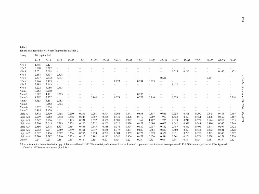
The Tat used in this study only corresponds to the first72 aa of Tat, which is encoded by the first exon of the Tatgene. Tat anti-sera collected from both studies were ana-lyzed to determine reactivity to overlapping 15-mer peptidesspanning aa 1–83 of the Tat sequence. Table 3 presents thereactivity pattern for anti-sera collected from each animal foreach Tat immunized group in Study 1. Sera from all animalsrrabrosdaNoTr
ecognized aa 1–15 or 5–19, corresponding to the N-terminalegion of Tat. In addition, the sera from Tat immunized micelso recognized aa 45–59 and 49–63, corresponding to theasic region of Tat; however, this was to a lesser degree thanecognition of the N-terminal and was only observed in somef the animals. Interestingly, Tat-coated NPs which demon-trated the highest total serum Tat-specific IgG titers alsoemonstrated the strongest reactivity in both the N-terminalnd basic regions. Moreover, the strong reactivity to both the-terminal and basic regions of Tat is maintained using 1 �gf Tat-coated NPs compared to the alum groups. In fact, theat (1 �g) coated NPs showed stronger reactivity in the basicegion of Tat compared to Tat (5 �g) adjuvanted with alum.
J.Pateletal./Vaccine24
(2006)3564–3573
3569
Table 3Tat anti-sera reactivity to 15-mer Tat peptides in Study 1
Group Tat peptide (aa)
1–15 5–19 9–23 13–27 17–31 21–35 25–39 29–43 33–47 37–51 41–55 45–59 49–63 53–67 57–71 61–75 65–79 69–83
NPs 1 (5) 3.444 2.983 0.273 – – – – – – – – 3.470 0.992 – – – – –NPs 2 (5) 3.123 2.588 – – – – – 0.240 0.361 – – – – – – – – –NPs 3 (5) 2.878 3.207 – – – – – – – – – – 1.006 – – – – –NPs 4 (5) 3.440 2.859 0.821 – – – – – – – – – 0.275 – – – – –NPs 5 (5) 1.909 3.301 0.712 – – – – – – – – – 1.069 – – – – –NPs 6 (5) 3.360 2.943 – 0.242 – – – 0.249 – 0.31 – 0.487 0.321 – – – – –Alum 1 (5) 0.277 0.337 – – – – – – – – – – – – – – – –Alum 2 (5) 0.383 1.624 – – – – – – – – – 0.573 – – – – – –Alum 3 (5) 2.929 2.759 – – – – – – – – – – – – – – – –Alum 4 (5) – 0.508 0.337 – – – – – – – – – 0.265 – – – – –Alum 5 (5) – 0.578 – – – – – – – – – – – – – – – –Alum 6 (5) 0.998 1.794 – – – – – – – – – – – – – – – –CFA 1 (5) 3.289 3.416 3.032 0.46 0.386 0.35 0.396 0.473 0.444 0.538 0.614 0.688 0.510 0.799 0.416 0.385 0.385 0.303CFA 2 (5) 1.834 3.061 – 0.247 – – 0.285 0.256 0.351 – – – – – – – – –CFA 3 (5) 1.639 3.108 0.426 0.378 0.355 0.369 0.423 0.451 0.591 0.515 0.573 0.685 0.430 0.35 0.485 0.413 0.445 0.371CFA 4 (5) 2.593 – – – – – – – – – – – 0.236 – – – – –CFA 5 (5) 2.329 0.724 – – – – – – – – – – 0.234 1.87 – – – –NPs 1 (1) 3.266 2.979 – – – – – – – – – – – – – – – –NPs 2 (1) 3.004 2.668 – – – – – – 0.292 – – – 0.754 – – – – –NPs 3 (1) 3.114 3.065 – – – – – – – – – 0.648 1.277 – – – – –NPs 4 (1) 3.037 3.279 0.49 – – – – – – – – – – – – – – –NPs 5 (1) 0.625 0.765 2.139 – – – – – – – – – – – – – – –Alum 1 (1) 0.277 – – – – – – – – – – – – – – – – –Alum 2 (1) – – – – – – – – – – – – – – – – – –Alum 3 (1) – – – – – – – – – – – – – – – – – –Alum 4 (1) 2.719 3.202 – – – – – – – – – – – – – – – –Alum 5 (1) 0.392 0.936 – – – – – – – – – – – – – – – –Alum 6 (1) – – – – – – – – – – – – – – – – – –Cutoffa 0.26 0.26 0.24 0.24 0.22 0.22 0.25 0.23 0.28 0.28 0.29 0.45 0.22 0.25 0.25 0.27 0.27 0.25
All sera were diluted 1:100. The reactivity of anti-sera from each animal is presented. The value in parenthesis represents the dose of Tat in �g. (–) indicates no response—ELISA OD values equal tocutoff/background.
a Cutoff = (AVG Naı̈ve response) + (3 × S.D.).
3570J.Pateletal./Vaccine
24(2006)
3564–3573
Table 4Tat anti-sera reactivity to 15-mer Tat peptides in Study 2
Group Tat peptide (aa)
1–15 5–19 9–23 13–27 17–31 21–35 25–39 29–43 33–47 37–51 41–55 45–59 49–63 53–67 57–71 61–75 65–79 69–83
NPs 1 1.395 2.121 – – – – – – – – – – – – – – – –NPs 2 0.828 3.262 – – – – – – – – – – – – – – – –NPs 3 3.071 3.008 – – – – – – – – – – 0.555 0.242 – – 0.165 .172NPs 4 2.194 3.337 2.828 – – – – – – – – – – – – – – –NPs 5 2.537 2.872 3.044 – – – – – – – – 0.621 – – – 0.181 – –NPs 6 2.946 2.423 – – – – – 0.173 – 0.294 0.373 – – – – – – –NPs 7 2.998 2.413 – – – – – – – – – – 1.422 – – – – –NPs 8 3.232 3.086 0.693 – – – – – – – – – – – – – – –Alum 1 0.253 3.210 – – – – – – – – – – – – – – –Alum 2 0.562 1.471 0.205 – – – – – – 0.233 – – – – – – – –Alum 3 1.207 2.377 – – – 0.164 – 0.273 – 0.772 0.746 – 0.778 – – – – 0.214Alum 4 3.353 3.191 2.983 – – – – – – – – – – – – – – –Alum 5 – 0.452 0.803 – – – – – – – – – – – – – – –Alum 6 0.357 0.354 – – – – – – – – – – – – – – – –Alum 7 0.805 2.479 – – – – – – – – – – – – – – – –Lipid A 1 3.542 2.839 0.458 0.290 0.296 0.291 0.304 0.364 0.441 0.638 0.817 0.646 0.953 0.378 0.390 0.345 0.483 0.407Lipid A 2 2.919 3.282 0.512 0.348 0.348 0.357 0.375 0.440 0.498 0.719 0.989 1.967 1.423 0.397 0.464 0.438 0.569 0.487Lipid A 3 3.347 2.996 0.831 0.495 0.531 0.557 0.566 0.585 0.722 1.169 1.707 1.736 2.019 0.712 0.771 0.644 0.912 0.555Lipid A 4 3.506 3.045 0.383 0.228 0.220 0.222 0.262 0.320 0.429 0.473 0.696 0.603 1.042 0.370 0.348 0.310 0.365 0.269Lipid A 5 2.556 2.378 1.337 0.364 0.435 0.345 0.338 0.378 0.494 0.880 0.907 0.882 2.487 0.463 0.491 0.441 0.597 0.422Lipid A 6 2.912 2.941 2.369 0.349 0.401 0.347 0.336 0.377 0.404 0.680 0.884 0.810 0.802 0.397 0.432 0.392 0.521 0.428Lipid A 7 2.627 2.496 2.562 0.274 0.286 0.258 0.280 0.304 0.369 0.572 0.579 0.522 0.831 0.287 0.325 0.305 0.346 0.322Lipid A 8 2.590 2.597 0.310 0.233 0.212 0.192 0.215 0.240 0.306 0.473 0.670 0.584 0.561 0.251 0.271 0.239 0.271 0.239Cutoffa 0.24 0.21 0.16 0.19 0.18 0.15 0.20 0.15 0.22 0.23 0.33 0.61 0.34 0.14 0.15 0.15 0.15 0.12
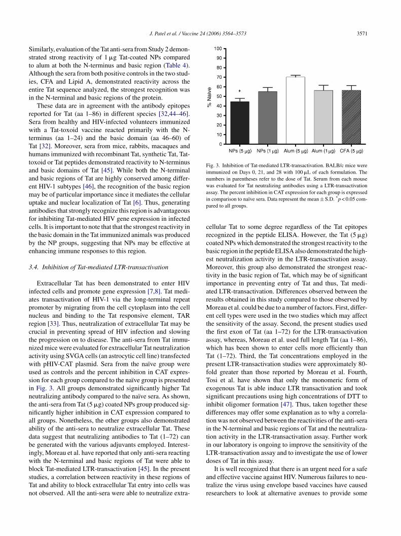
All sera from mice immunized with 1 �g of Tat were diluted 1:100. The reactivity of anti-sera from each animal is presented. (–) indicates no response—ELISA OD values equal to cutoff/background.a Cutoff = (AVG naı̈ve response) + (3 × S.D.).
J. Patel et al. / Vaccine 24 (2006) 3564–3573 3571
Similarly, evaluation of the Tat anti-sera from Study 2 demon-strated strong reactivity of 1 �g Tat-coated NPs comparedto alum at both the N-terminus and basic region (Table 4).Although the sera from both positive controls in the two stud-ies, CFA and Lipid A, demonstrated reactivity across theentire Tat sequence analyzed, the strongest recognition wasin the N-terminal and basic regions of the protein.
These data are in agreement with the antibody epitopesreported for Tat (aa 1–86) in different species [32,44–46].Sera from healthy and HIV-infected volunteers immunizedwith a Tat-toxoid vaccine reacted primarily with the N-terminus (aa 1–24) and the basic domain (aa 46–60) ofTat [32]. Moreover, sera from mice, rabbits, macaques andhumans immunized with recombinant Tat, synthetic Tat, Tat-toxoid or Tat peptides demonstrated reactivity to N-terminusand basic domains of Tat [45]. While both the N-terminaland basic regions of Tat are highly conserved among differ-ent HIV-1 subtypes [46], the recognition of the basic regionmay be of particular importance since it mediates the cellularuptake and nuclear localization of Tat [6]. Thus, generatingantibodies that strongly recognize this region is advantageousfor inhibiting Tat-mediated HIV gene expression in infectedcells. It is important to note that that the strongest reactivity inthe basic domain in the Tat immunized animals was producedby the NP groups, suggesting that NPs may be effective atenhancing immune responses to this region.
3
iapnrctnawusintnaadbiwbsTn
Fig. 3. Inhibition of Tat-mediated LTR-transactivation. BALB/c mice wereimmunized on Days 0, 21, and 28 with 100 �L of each formulation. Thenumbers in parentheses refer to the dose of Tat. Serum from each mousewas evaluated for Tat neutralizing antibodies using a LTR-transactivationassay. The percent inhibition in CAT expression for each group is expressedin comparison to naı̈ve sera. Data represent the mean ± S.D. *p < 0.05 com-pared to all groups.
cellular Tat to some degree regardless of the Tat epitopesrecognized in the peptide ELISA. However, the Tat (5 �g)coated NPs which demonstrated the strongest reactivity to thebasic region in the peptide ELISA also demonstrated the high-est neutralization activity in the LTR-transactivation assay.Moreover, this group also demonstrated the strongest reac-tivity in the basic region of Tat, which may be of significantimportance in preventing entry of Tat and thus, Tat medi-ated LTR-transactivation. Differences observed between theresults obtained in this study compared to those observed byMoreau et al. could be due to a number of factors. First, differ-ent cell types were used in the two studies which may affectthe sensitivity of the assay. Second, the present studies usedthe first exon of Tat (aa 1–72) for the LTR-transactivationassay, whereas, Moreau et al. used full length Tat (aa 1–86),which has been shown to enter cells more efficiently thanTat (1–72). Third, the Tat concentrations employed in thepresent LTR-transactivation studies were approximately 80-fold greater than those reported by Moreau et al. Fourth,Tosi et al. have shown that only the monomeric form ofexogenous Tat is able induce LTR transactivation and tooksignificant precautions using high concentrations of DTT toinhibit oligomer formation [47]. Thus, taken together thesedifferences may offer some explanation as to why a correla-tion was not observed between the reactivities of the anti-serain the N-terminal and basic regions of Tat and the neutraliza-tiLd
atr
.4. Inhibition of Tat-mediated LTR-transactivation
Extracellular Tat has been demonstrated to enter HIVnfected cells and promote gene expression [7,8]. Tat medi-tes transactivation of HIV-1 via the long-terminal repeatromoter by migrating from the cell cytoplasm into the cellucleus and binding to the Tat responsive element, TARegion [33]. Thus, neutralization of extracellular Tat may berucial in preventing spread of HIV infection and slowinghe progression on to disease. The anti-sera from Tat immu-ized mice were evaluated for extracellular Tat neutralizationctivity using SVGA cells (an astrocytic cell line) transfectedith pHIV-CAT plasmid. Sera from the naı̈ve group weresed as controls and the percent inhibition in CAT expres-ion for each group compared to the naı̈ve group is presentedn Fig. 3. All groups demonstrated significantly higher Tateutralizing antibody compared to the naı̈ve sera. As shown,he anti-sera from Tat (5 �g) coated NPs group produced sig-ificantly higher inhibition in CAT expression compared toll groups. Nonetheless, the other groups also demonstratedbility of the anti-sera to neutralize extracellular Tat. Theseata suggest that neutralizing antibodies to Tat (1–72) cane generated with the various adjuvants employed. Interest-ngly, Moreau et al. have reported that only anti-sera reactingith the N-terminal and basic regions of Tat were able tolock Tat-mediated LTR-transactivation [45]. In the presenttudies, a correlation between reactivity in these regions ofat and ability to block extracellular Tat entry into cells wasot observed. All the anti-sera were able to neutralize extra-
ion activity in the LTR-transactivation assay. Further workn our laboratory is ongoing to improve the sensitivity of theTR-transactivation assay and to investigate the use of loweroses of Tat in this assay.
It is well recognized that there is an urgent need for a safend effective vaccine against HIV. Numerous failures to neu-ralize the virus using envelope based vaccines have causedesearchers to look at alternative avenues to provide some
3572 J. Patel et al. / Vaccine 24 (2006) 3564–3573
protection or to slow down the progression of the diseasein HIV infected patients by the development of therapeuticvaccines. One of the requirements for an effective HIV vac-cine will be that the antigen is conserved among the differentviral subtypes. Thus, many researchers are investigating thepotential of HIV regulatory proteins Nef, Rev, and Tat, whichare expressed early in the life cycle and are also fairly wellconserved among the different HIV subtypes, as alternativetargets for vaccine development [23,48,49]. Several studieshave demonstrated the importance of Tat in the virus lifecycle and demonstrated that anti-Tat antibodies [25,28,30]and CTL responses [24,31] correlate inversely with diseaseprogression, making Tat an attractive candidate for HIV vac-cine development.
In conclusion, we previously reported that anionic NPswere effective for enhancing immune humoral and cellularimmune responses to HIV-1 Tat [38]. The present studies fur-ther demonstrated the advantages of using NPs over alum asan adjuvant for protein based vaccine in that NPs allowedfor lower doses of Tat to be administered. Moreover, Tat-coated NPs were able to generate antibodies that stronglyrecognized both the N-terminal and basic regions of the pro-tein. Tat-mediated LTR-transactivation studies also revealedthat the antibodies generated with all Tat groups were ableto block Tat entry into cells, with Tat-coated NPs show-ing superior Tat neutralization activity over other forms ofdpd1v
A
AfPFG
R
[6] Caselli E, Betti M, Grossi MP, et al. DNA immunization withHIV-1 tat mutated in the trans activation domain induces humoraland cellular immune responses against wild-type Tat. J Immunol1999;162(9):5631–8.
[7] Frankel AD, Biancalana S, Hudson D. Activity of synthetic peptidesfrom the Tat protein of human immunodeficiency virus type 1. ProcNatl Acad Sci USA 1989;86(19):7397–401.
[8] Sodroski J, Patarca R, Rosen C, Wong-Staal F, Haseltine W. Loca-tion of the trans-activating region on the genome of human T-celllymphotropic virus type III. Science 1985;229(4708):74–7.
[9] Misumi S, Takamune N, Ohtsubo Y, Waniguchi K, Shoji S. Zn2+
binding to cysteine-rich domain of extracellular human immunodefi-ciency virus type 1 Tat protein is associated with Tat protein-inducedapoptosis. AIDS Res Hum Retroviruses 2004;20(3):297–304.
[10] Butto S, Fiorelli V, Tripiciano A, et al. Sequence conservationand antibody cross-recognition of clade B human immunodeficiencyvirus (HIV) type 1 Tat protein in HIV-1-infected Italians, Ugandans,and South Africans. J Infect Dis 2003;188(8):1171–80.
[11] Huang HW, Wang KT. Structural characterization of the metal bind-ing site in the cysteine-rich region of HIV-1 Tat protein. BiochemBiophys Res Commun 1996;227(2):615–21.
[12] Chang HC, Samaniego F, Nair BC, Buonaguro L, Ensoli B. HIV-1Tat protein exits from cells via a leaderless secretory pathway andbinds to extracellular matrix-associated heparan sulfate proteoglycansthrough its basic region. Aids 1997;11(12):1421–31.
[13] Ma M, Nath A. Molecular determinants for cellular uptake of Tatprotein of human immunodeficiency virus type 1 in brain cells. JVirol 1997;71(3):2495–9.
[14] Fisher AG, Feinberg MB, Josephs SF, et al. The trans-activator geneof HTLV-III is essential for virus replication. Nature 1986;320(6060):367–71.
[
[
[
[
[
[
[
[
[
[
[
elivery. Together the data presented here demonstrate theotential of these novel anionic NPs as effective vaccineelivery systems for enhancing immune responses to HIV-Tat based vaccines and possibly other HIV protein based
accines.
cknowledgements
This research was funded by NIH-NIAID AI051147 andI058842. J. Patel was supported, in part, by a pre-doctoral
ellowship received from the American Foundation forharmaceutical Education and the 2005 Dissertation Yearellowship received from the University of Kentuckyraduate School.
eferences
[1] Cohen J. Public health. AIDS vaccine trial produces disappointmentand confusion. Science 2003;299(5611):1290–1.
[2] Agwale SM, Shata MT, Reitz Jr MS, et al. A Tat subunit vaccineconfers protective immunity against the immune-modulating activityof the human immunodeficiency virus type-1 Tat protein in mice.Proc Natl Acad Sci USA 2002;99(15):10037–41.
[3] Fanales-Belasio E, Cafaro A, Cara A, et al. HIV-1 Tat-based vac-cines: from basic science to clinical trials. DNA Cell Biol 2002;21(9):599–610.
[4] Gallo RC, Burny A, Zagury D. Targeting Tat and IFN(alpha) as atherapeutic AIDS vaccine. DNA Cell Biol 2002;21(9):611–8.
[5] Goldstein G. HIV-1 Tat protein as a potential AIDS vaccine. NatMed 1996;2(9):960–4.
15] Popik W, Pitha PM. Role of tumor necrosis factor alpha in activationand replication of the tat-defective human immunodeficiency virustype 1. J Virol 1993;67(2):1094–9.
16] Feinberg MB, Baltimore D, Frankel AD. The role of Tat inthe human immunodeficiency virus life cycle indicates a primaryeffect on transcriptional elongation. Proc Natl Acad Sci USA1991;88(9):4045–9.
17] Ensoli B, Cafaro A. HIV-1 Tat vaccines. Virus Res 2002;82(1–2):91–101.
18] Pauza CD, Trivedi P, Wallace M, et al. Vaccination with tat tox-oid attenuates disease in simian/HIV-challenged macaques. Proc NatlAcad Sci USA 2000;97(7):3515–9.
19] Fanales-Belasio E, Moretti S, Nappi F, et al. Native HIV-1 Tat proteintargets monocyte-derived dendritic cells and enhances their matu-ration, function, and antigen-specific T cell responses. J Immunol2002;168(1):197–206.
20] Izmailova E, Bertley FM, Huang Q, et al. HIV-1 Tat reprogramsimmature dendritic cells to express chemoattractants for activated Tcells and macrophages. Nat Med 2003;9(2):191–7.
21] Fawell S, Seery J, Daikh Y, et al. Tat-mediated delivery of heterolo-gous proteins into cells. Proc Natl Acad Sci USA 1994;91(2):664–8.
22] Kim DT, Mitchell DJ, Brockstedt DG, et al. Introduction of solubleproteins into the MHC class I pathway by conjugation to an HIVtat peptide. J Immunol 1997;159(4):1666–8.
23] Addo MM, Altfeld M, Rosenberg ES, et al. The HIV-1 regulatoryproteins Tat and Rev are frequently targeted by cytotoxic T lympho-cytes derived from HIV-1-infected individuals. Proc Natl Acad SciUSA 2001;98(4):1781–6.
24] van Baalen CA, Pontesilli O, Huisman RC, et al. Human immunod-eficiency virus type 1 Rev- and Tat-specific cytotoxic T lymphocytefrequencies inversely correlate with rapid progression to AIDS. JGen Virol 1997;78(Pt 8):1913–8.
25] Zagury JF, Sill A, Blattner W, et al. Antibodies to the HIV-1 Tatprotein correlated with nonprogression to AIDS: a rationale for theuse of Tat toxoid as an HIV-1 vaccine. J Hum Virol 1998;1(4):282–92.
J. Patel et al. / Vaccine 24 (2006) 3564–3573 3573
[26] Re MC, Furlini G, Vignoli M, et al. Effect of antibody to HIV-1Tat protein on viral replication in vitro and progression of HIV-1 disease in vivo. J Acquir Immune Defic Syndr Hum Retrovirol1995;10(4):408–16.
[27] Rezza G, Fiorelli V, Dorrucci M, et al. The presence of anti-Tat anti-bodies is predictive of long-term nonprogression to AIDS or severeimmunodeficiency: findings in a cohort of HIV-1 seroconverters. JInfect Dis 2005;191(8):1321–4.
[28] Re MC, Gibellini D, Furlini G, et al. Relationships between the pres-ence of anti-Tat antibody, DNA and RNA viral load. New Microbiol2001;24(3):207–15.
[29] Re MC, Gibellini D, Vitone F, La Placa M. Antibody to HIV-1 Tatprotein, a key molecule in HIV-1 pathogenesis. A brief review. NewMicrobiol 2001;24(2):197–205.
[30] Re MC, Vignoli M, Furlini G, et al. Antibodies against full-lengthTat protein and some low-molecular-weight Tat-peptides correlatewith low or undetectable viral load in HIV-1 seropositive patients. JClin Virol 2001;21(1):81–9.
[31] Allen TM, O’Connor DH, Jing P, et al. Tat-specific cytotoxic T lym-phocytes select for SIV escape variants during resolution of primaryviraemia. Nature 2000;407(6802):386–90.
[32] Noonan DM, Gringeri A, Meazza R, et al. Identification of immun-odominant epitopes in inactivated Tat-vaccinated healthy and HIV-1-infected volunteers. J Acquir Immune Defic Syndr 2003;33(1):47–55.
[33] Caputo A, Gavioli R, Ensoli B. Recent advances in the developmentof HIV-1 Tat-based vaccines. Curr HIV Res 2004;2(4):357–76.
[34] Maggiorella MT, Baroncelli S, Michelini Z, et al. Long-term protec-tion against SHIV89.6P replication in HIV-1 Tat vaccinated cynomol-gus monkeys. Vaccine 2004;22(25–26):3258–69.
[35] Silvera P, Richardson MW, Greenhouse J, et al. Outcome of simian-human immunodeficiency virus strain 89.6p challenge following
[
[
[38] Cui Z, Patel J, Tuzova M, et al. Strong T cell type-1 immuneresponses to HIV-1 Tat (1–72) protein-coated nanoparticles. Vaccine2004;22(20):2631–40.
[39] Nabel G, Baltimore D. An inducible transcription factor activatesexpression of human immunodeficiency virus in T cells. Nature1987;326(6114):711–3.
[40] Cui Z, Mumper RJ. Topical immunization using nanoengineeredgenetic vaccines. J Control Release 2002;81(1–2):173–84.
[41] Cui Z, Mumper RJ. Coating of cationized protein on engineerednanoparticles results in enhanced immune responses. Int J Pharm2002;238(1–2):229–39.
[42] Cui Z, Mumper RJ. Genetic immunization using nanoparti-cles engineered from microemulsion precursors. Pharm Res2002;19(7):939–46.
[43] Koziara JM, Oh JJ, Akers WS, Ferraris SP, Mumper RJ. Blood com-patibility of cetyl alcohol/polysorbate-based nanoparticles. PharmRes 2005;22(11):1821–8.
[44] Marinaro M, Riccomi A, Rappuoli R, et al. Mucosal delivery of thehuman immunodeficiency virus-1 Tat protein in mice elicits systemicneutralizing antibodies, cytotoxic T lymphocytes and mucosal IgA.Vaccine 2003;21(25–26):3972–81.
[45] Moreau E, Belliard G, Partidos CD, et al. Important B-cell epitopesfor neutralization of human immunodeficiency virus type 1 Tat inserum samples of humans and different animal species immunizedwith Tat protein or peptides. J Gen Virol 2004;85(Pt 10):2893–901.
[46] Goldstein G, Tribbick G, Manson K. Two B cell epitopes of HIV-1Tat protein have limited antigenic polymorphism in geographicallydiverse HIV-1 strains. Vaccine 2001;19(13–14):1738–46.
[47] Tosi G, Meazza R, De Lerma Barbaro A, et al. Highly stableoligomerization forms of HIV-1 Tat detected by monoclonal anti-bodies and requirement of monomeric forms for the transactivating
[
[
vaccination of rhesus macaques with human immunodeficiency virusTat protein. J Virol 2002;76(8):3800–9.
36] Belliard G, Hurtrel B, Moreau E, et al. Tat-neutralizing versusTat-protecting antibodies in rhesus macaques vaccinated with Tatpeptides. Vaccine 2005;23(11):1399–407.
37] Cafaro A, Caputo A, Fracasso C, et al. Control of SHIV-89.6P-infection of cynomolgus monkeys by HIV-1 Tat protein vaccine.Nat Med 1999;5(6):643–50.
function on the HIV-1 LTR. Eur J Immunol 2000;30(4):1120–6.48] Addo MM, Yu XG, Rosenberg ES, Walker BD, Altfeld M. Cytotoxic
T-lymphocyte (CTL) responses directed against regulatory and acces-sory proteins in HIV-1 infection. DNA Cell Biol 2002;21(9):671–8.
49] Yamada T, Iwamoto A. Comparison of proviral accessory genesbetween long-term nonprogressors and progressors of humanimmunodeficiency virus type 1 infection. Arch Virol 2000;145(5):1021–7.