history of chest pain
TRANSCRIPT
OSC
E M
AST
ER
1

OSC
E M
AST
ER
2
OSC
E M
AST
ER
3
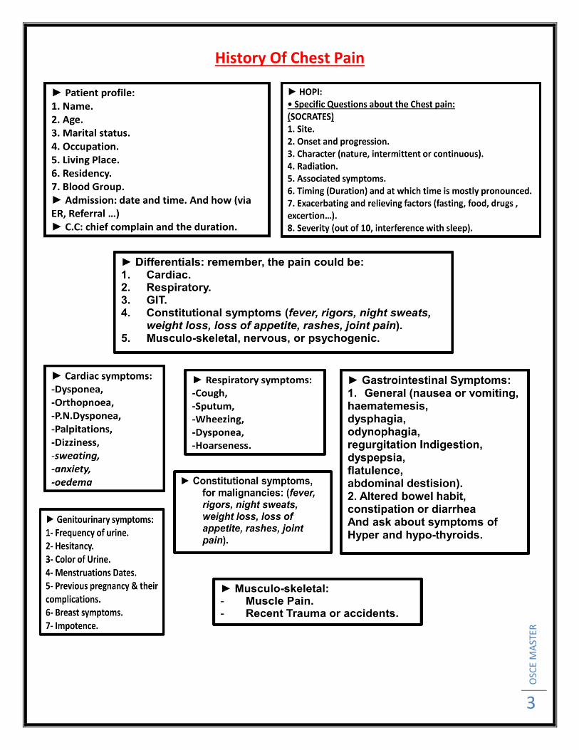
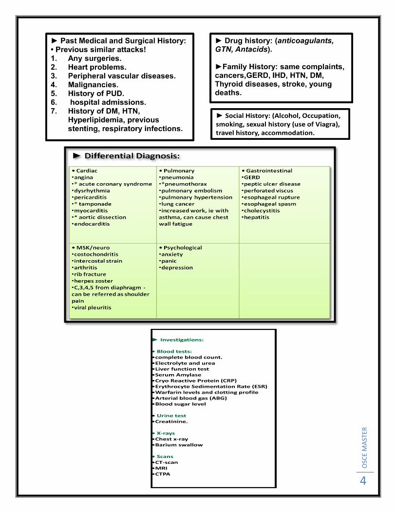
History Of Chest Pain
OSC
E M
AST
ER
4
OSC
E M
AST
ER
5
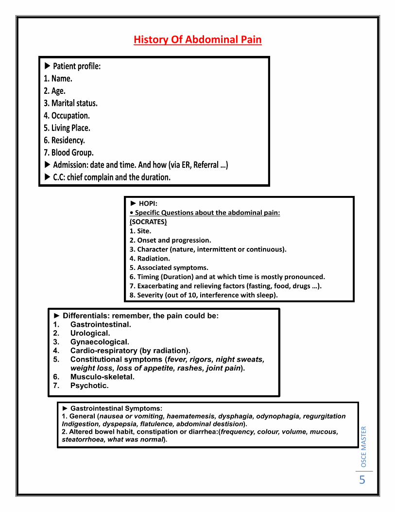
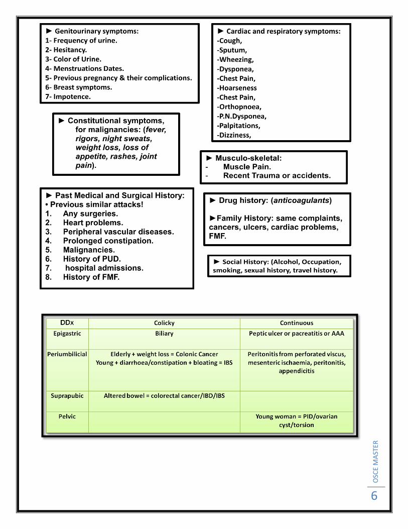
History Of Abdominal Pain
OSC
E M
AST
ER
6
OSC
E M
AST
ER
7
OSC
E M
AST
ER
8
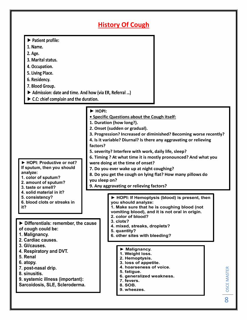
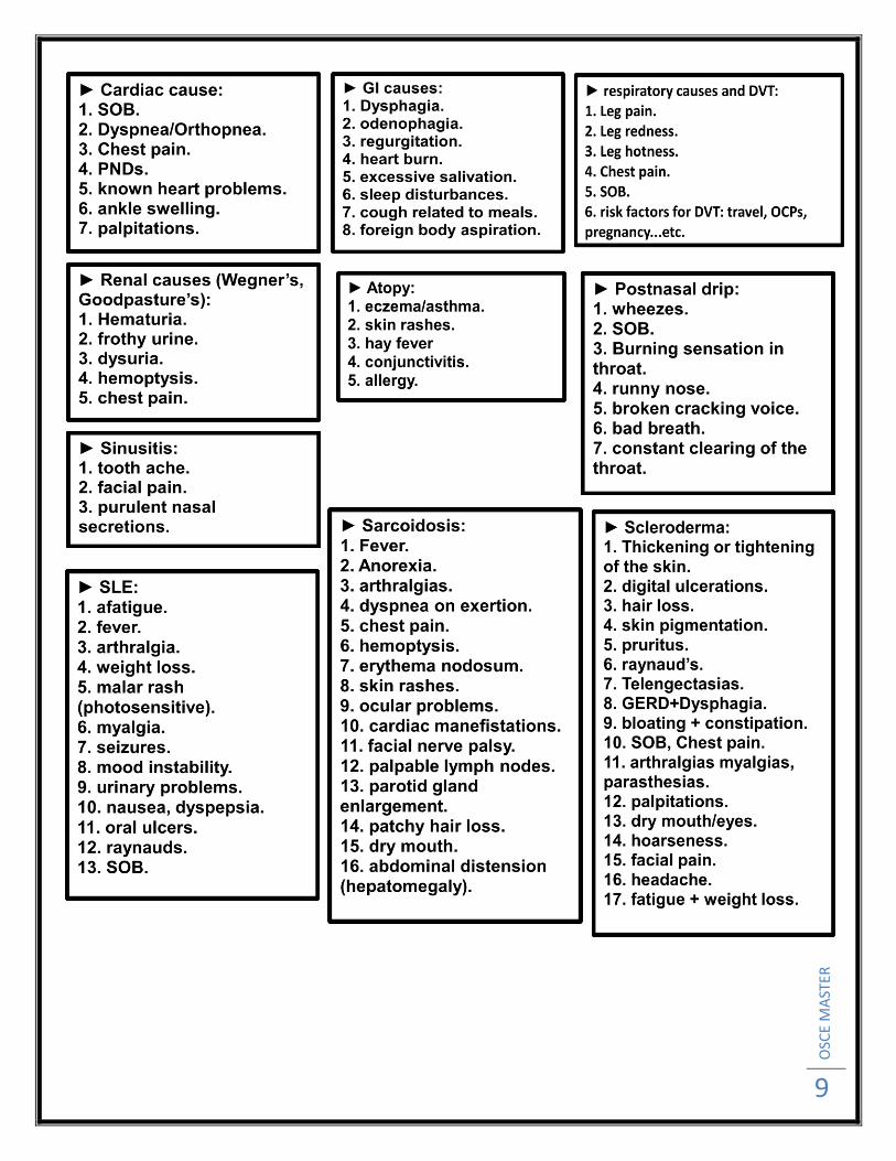
History Of Cough
OSC
E M
AST
ER
9
OSC
E M
AST
ER
10
OSC
E M
AST
ER
11
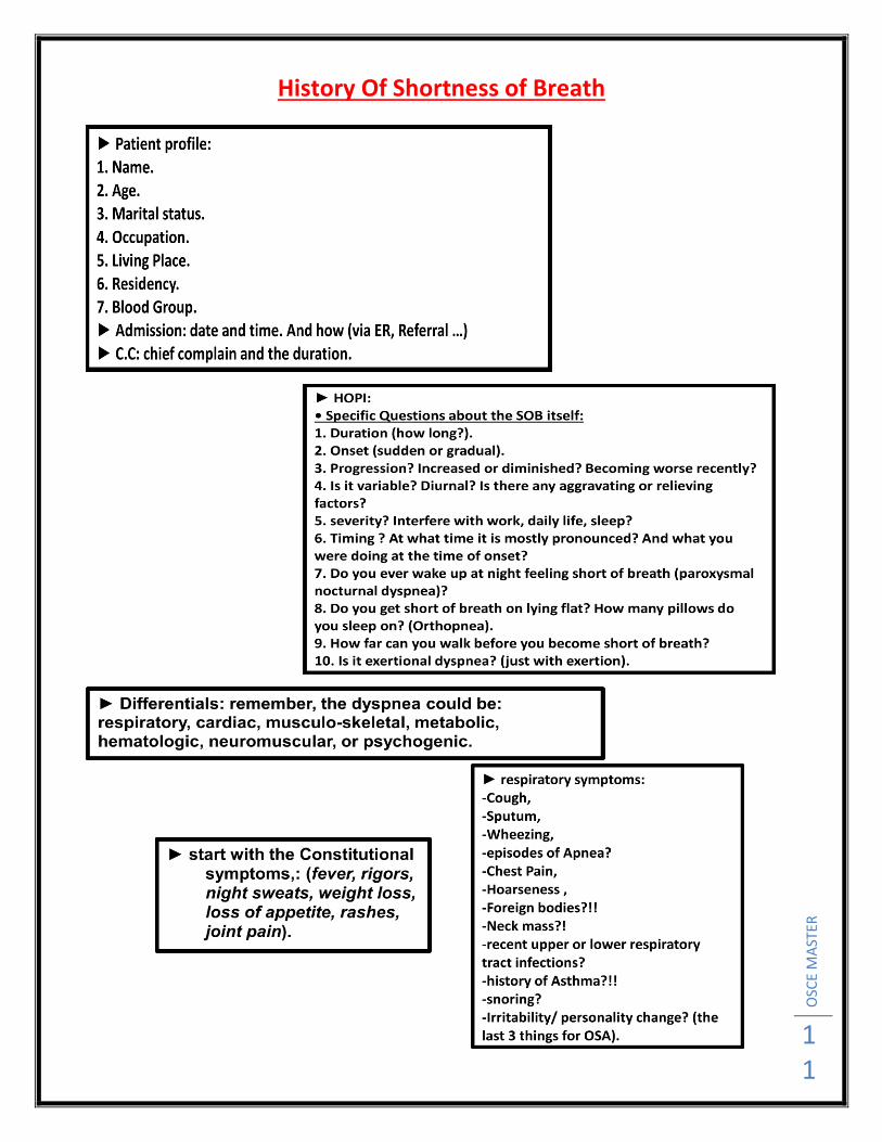
History Of Shortness of Breath

OSC
E M
AST
ER
12
OSC
E M
AST
ER
13
OSC
E M
AST
ER
14
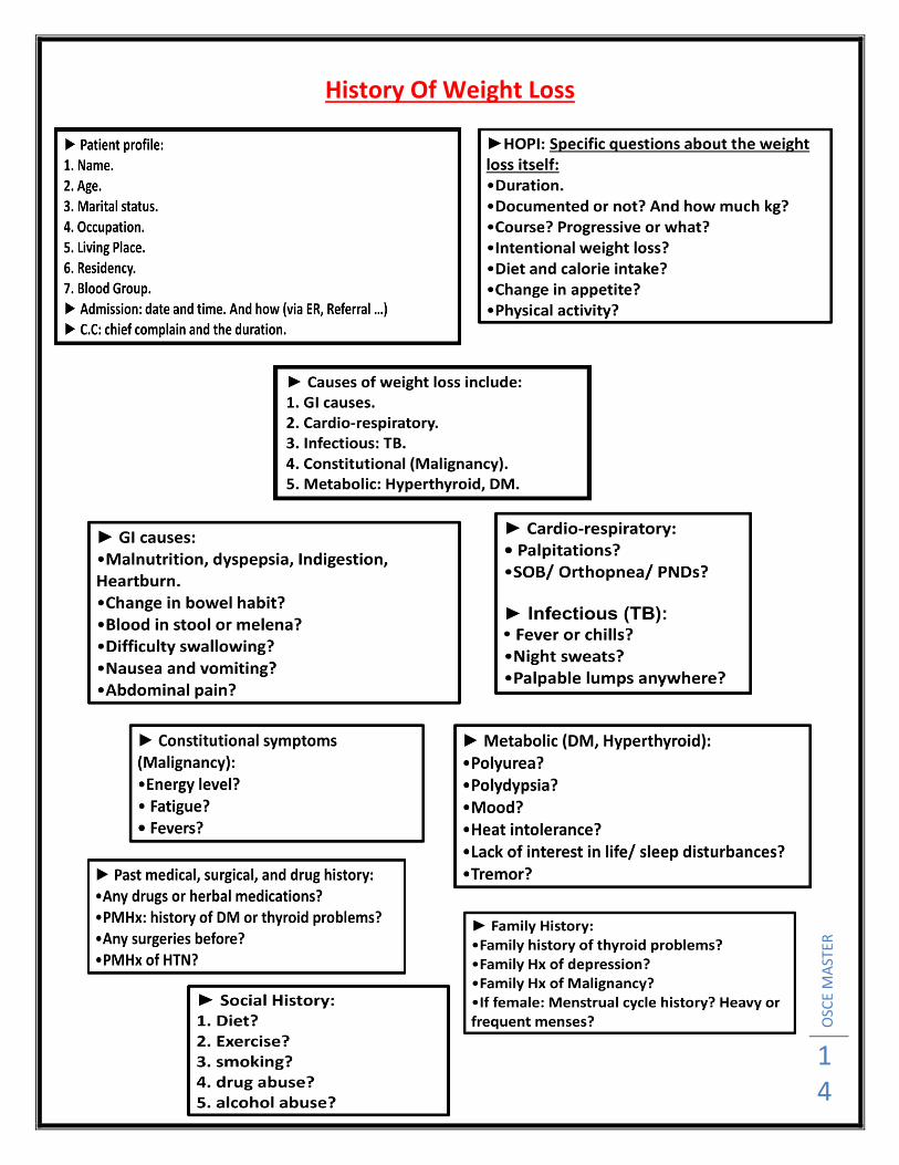
History Of Weight Loss
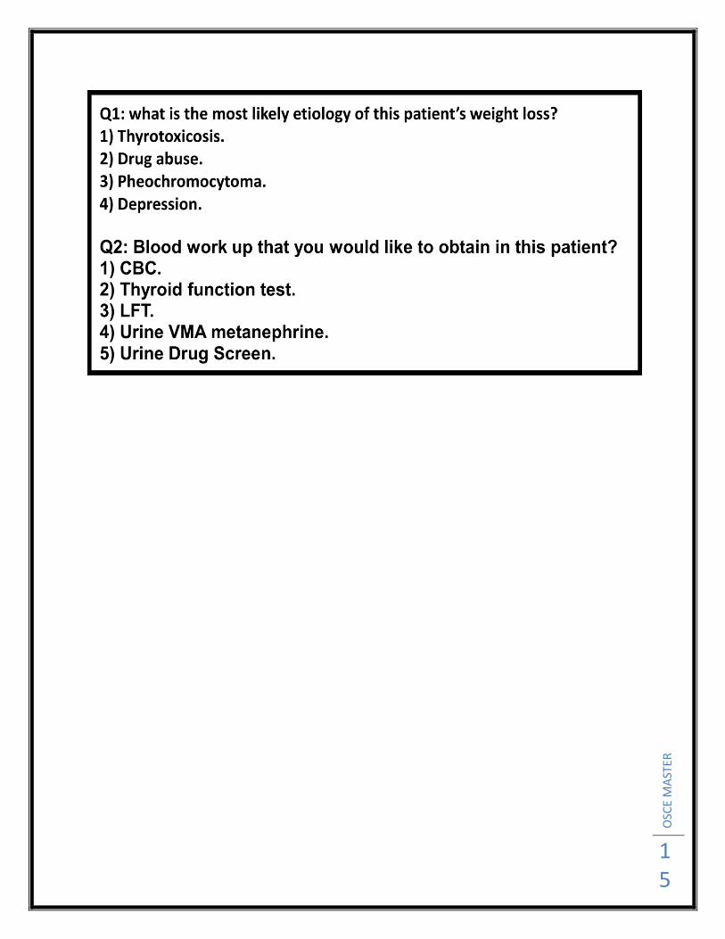
OSC
E M
AST
ER
15

OSC
E M
AST
ER
16
History Of Thyroid disease case

OSC
E M
AST
ER
17
OSC
E M
AST
ER
18
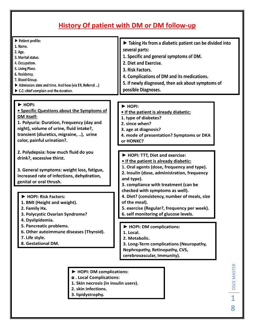
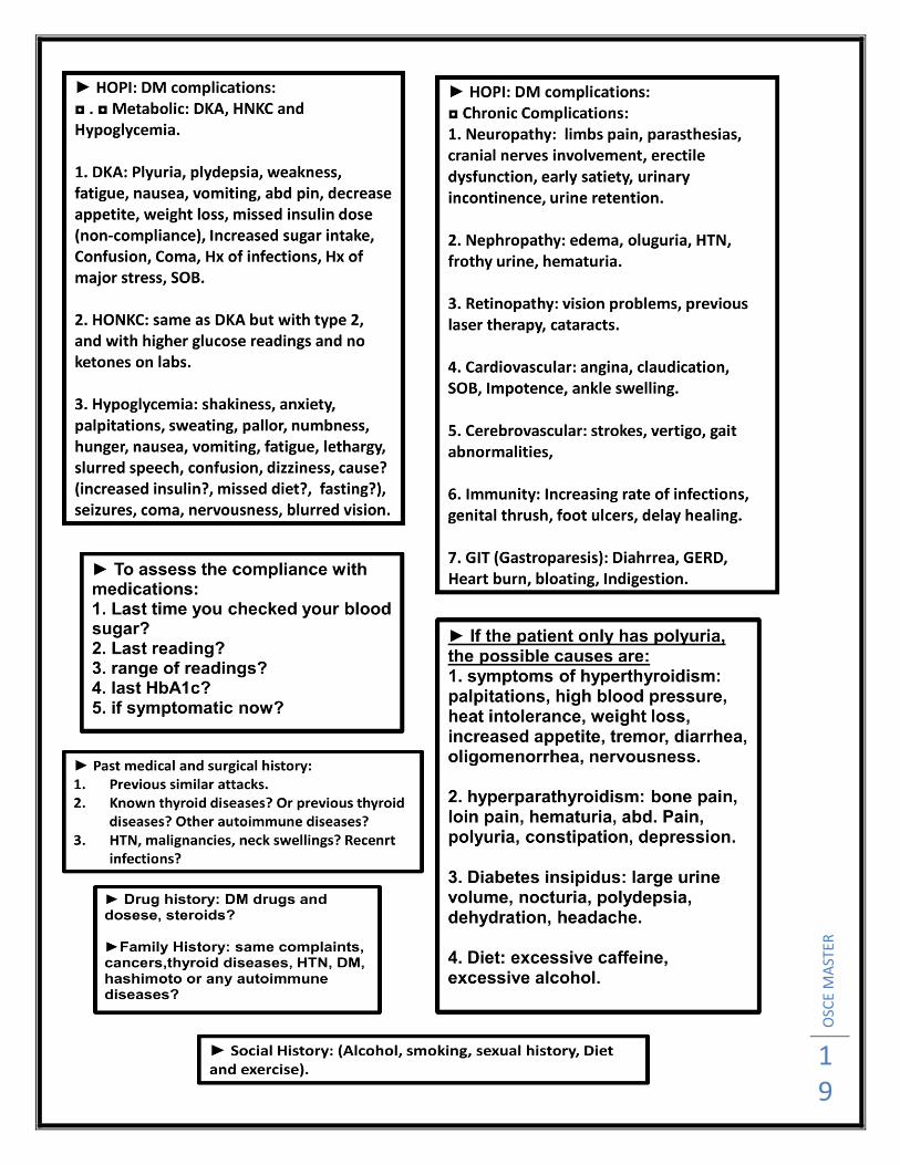
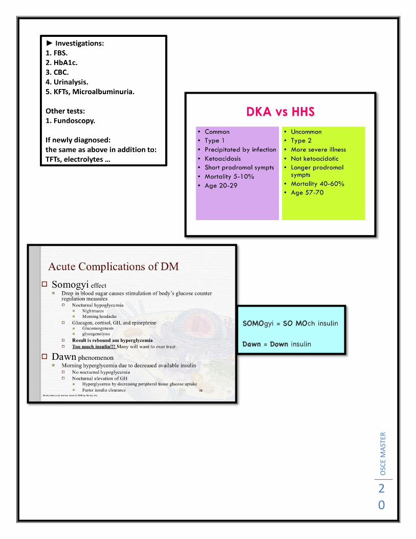
History Of patient with DM or DM follow-up
OSC
E M
AST
ER
19
OSC
E M
AST
ER
20

OSC
E M
AST
ER
21
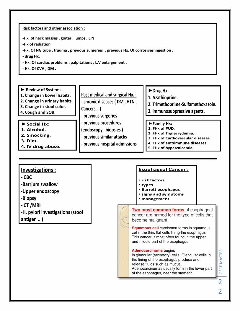
History Of Dysphagia
History of presenting illness :
1. For solids/ liquids / both
2. Which type of food, and which help in easing
the problem
3. Level of stuck
4. Pain (odenophagia)
5. Intermittent /all the time
6. Progression over time
7. Has food ever gone down the wrong way
8. Previous similar attacks
Differential diagnosis :
- PUD -malignancy -CREST -Myasthenia Gravis
-GERD -pharyngeal pouch -plummer Vinson syndrome -esophagitis
-Chagas disease
- ask about risk factors and complications .
PUD :
- indigestion
- heartburn
- regurgitation
- abd. Pain
Malignancy :
- early statiety
- fever , weight loss , loss of appetite
, fatigue .
-hematemesis , melena
GERD , Pharyngeal pouch :
- heartburn
- hoarseness of voice
- post nasal drip
- when drinking gurgle
Complications :
- chest pain / cough / hemoptysis / SOB -halitosis
- wheezes /breathing sounds - aspiration of food
- fever - sore throat
- hoarsness of voice
Plummer Vinson synd.
-on iron tablet
Hx. Of iron def. anemia
- glossitis
( triad of dysphagia +
glossitis + iron def.
anemia)
Esophagitis
-Hx of chronic
diseases like
DM , HIV ,
cancer , or on
steroids
(candida)
- drug Hx.
CREST synd
-dysphagia
- skin tightness
- raynaud
- finger tip ulceration
- telangectasia
- sclerodactylel
Myasthenia Gravis
. at the end of day
become worse ,
more fatigue
.ptosis/ diplopia
Chagas
disease
Hx of
recent
foreign
travel
OSC
E M
AST
ER
22
Risk factors and other association :
-Hx .of neck masses , goiter , lumps , L.N
-Hx of radiation
-Hx. Of NG tube , trauma , previous surgeries , previous Hx. Of corrosives ingestion .
- drug Hx.
- Hx. Of cardiac problems , palpitations , L.V enlargement .
- Hx. Of CVA , DM .
OSC
E M
AST
ER
23
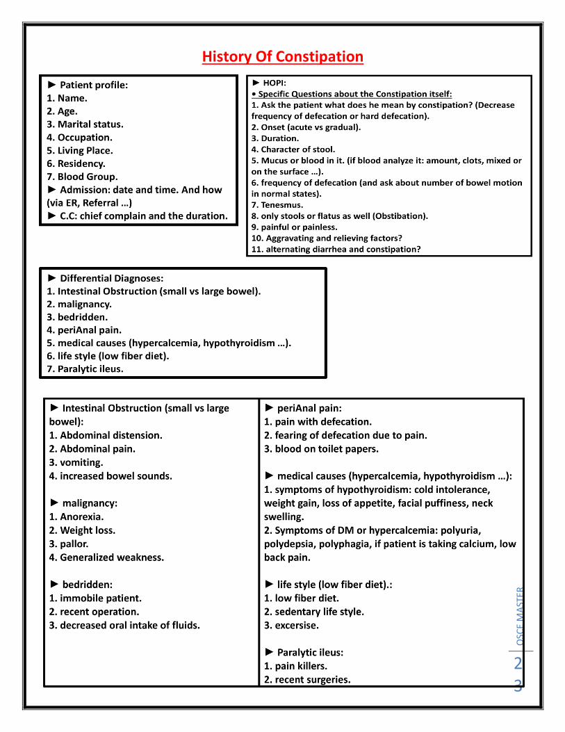
History Of Constipation
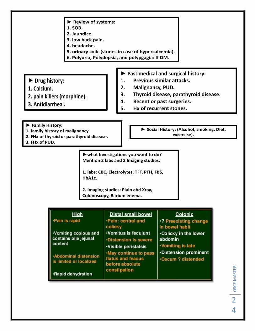
OSC
E M
AST
ER
24

OSC
E M
AST
ER
25

OSC
E M
AST
ER
26
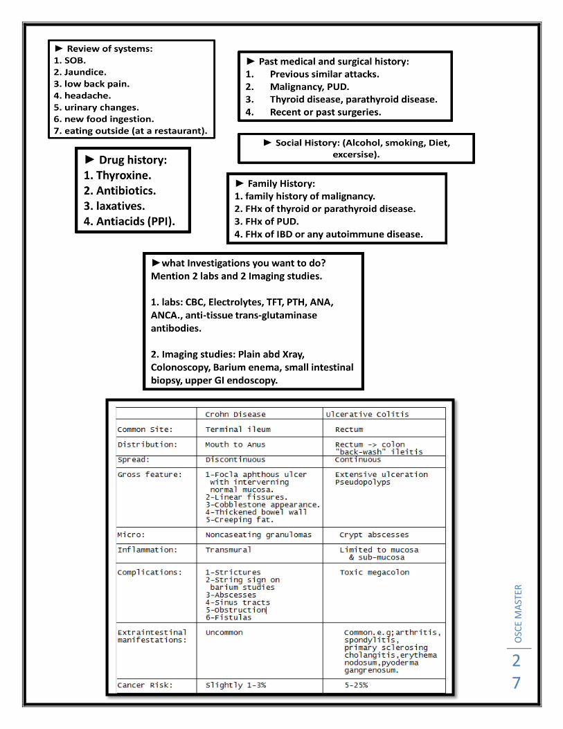
History Of Diarrhea
OSC
E M
AST
ER
27
OSC
E M
AST
ER
28
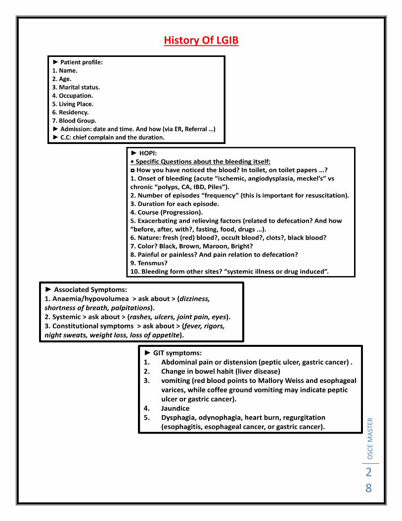
History Of LGIB
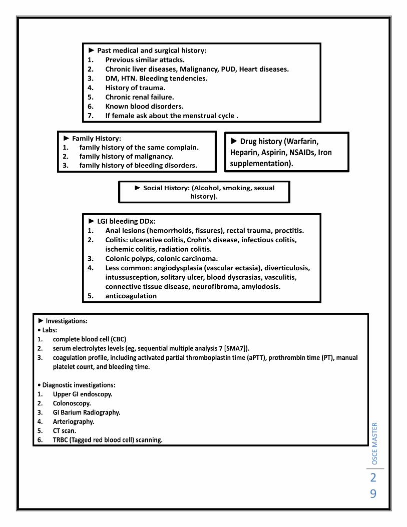
OSC
E M
AST
ER
29

OSC
E M
AST
ER
30
History Of UGIB
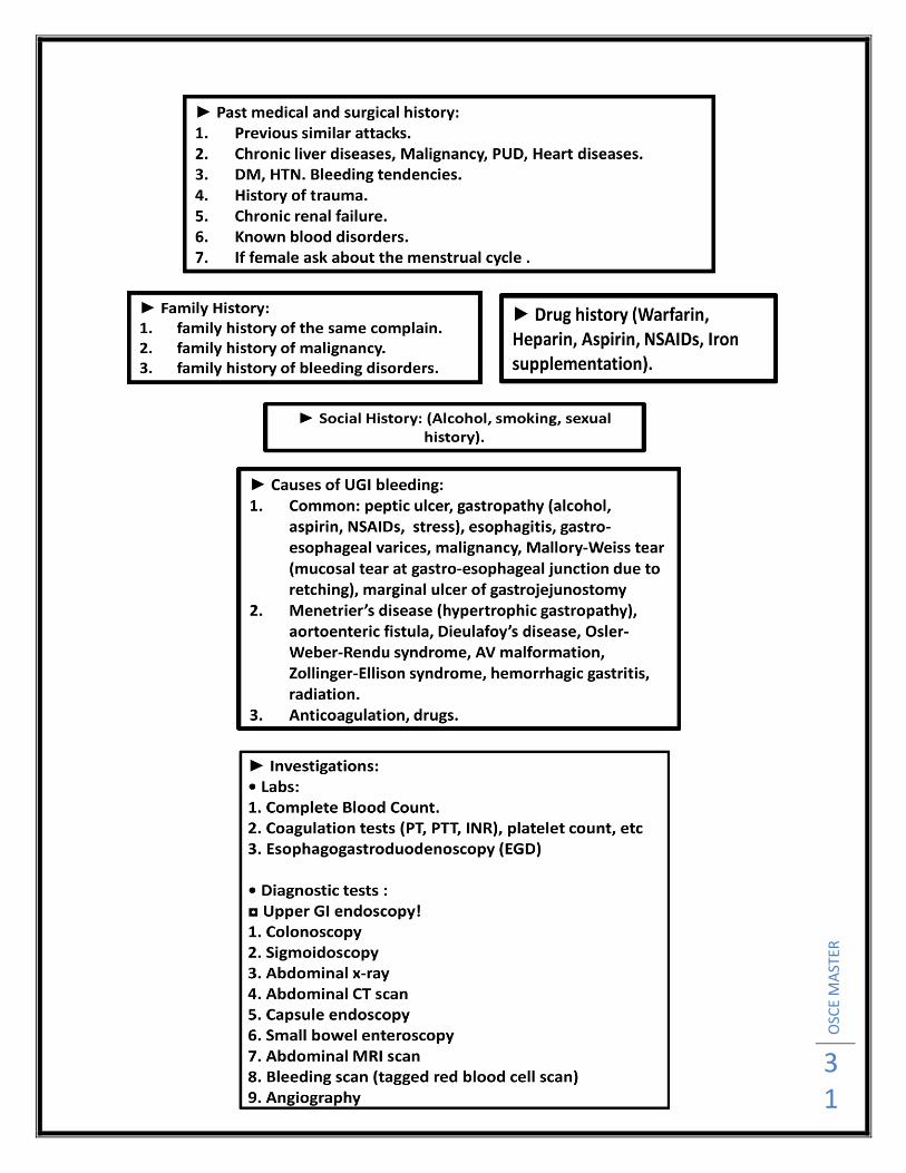
OSC
E M
AST
ER
31
OSC
E M
AST
ER
32
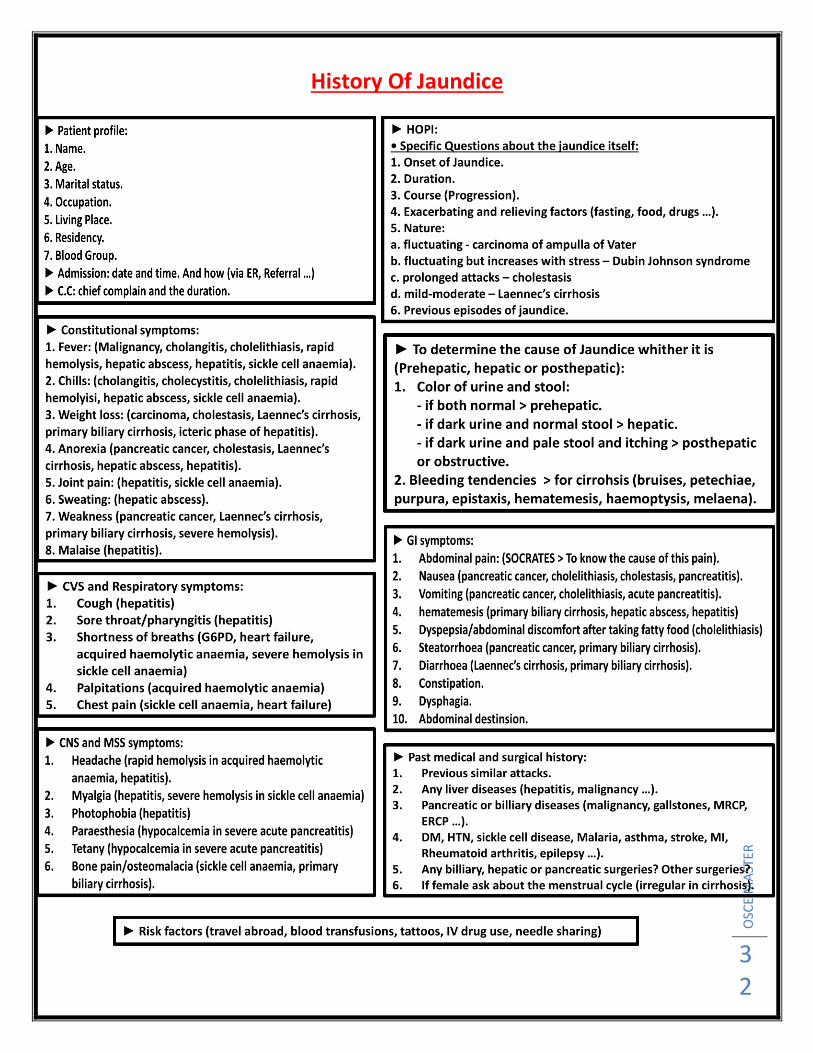
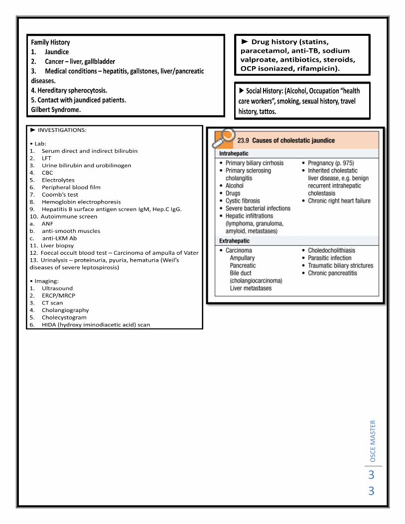
History Of Jaundice
OSC
E M
AST
ER
33
OSC
E M
AST
ER
34
History Of Joint Pain
OSC
E M
AST
ER
35
OSC
E M
AST
ER
36
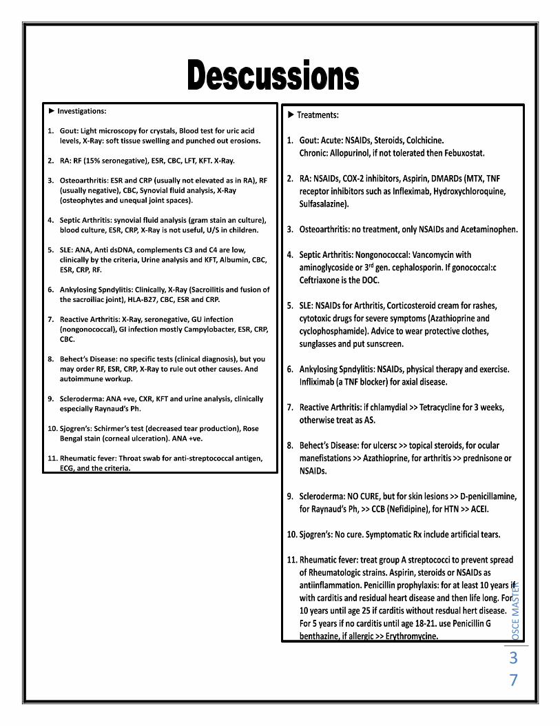
OSC
E M
AST
ER
37

OSC
E M
AST
ER
38
OSC
E M
AST
ER
39
History Of Lower Limbs Swelling
OSC
E M
AST
ER
40
OSC
E M
AST
ER
41

OSC
E M
AST
ER
42

OSC
E M
AST
ER
43

OSC
E M
AST
ER
44
Abdominal Examination Station
1. Introduce yourself.
2. Ask for permission.
3. Ask for a chaperon.
4. Ensure the privacy.
5. Explain to the patient what you want to do.
6. Proper exposure? (From the nipples to mid thighs).
7. Position of the patient? It should be flat.
8. General observation of the patient (looks well? , conscious? , oriented? , breath
comfortably? , not in pain? , not pale, jaundiced or cyanosed? , if there is any IV lines,
dressings, masks or drains? .. ).
9. Inspection (from the foot of the bed):
move with respiration? ,
visible pulsations? ,
any deformity (scoliosis or kyphoscoliosis)? ,
any scars? ,
change in color of the skin? ,
caput medusa? ,
dilated visible veins? ,
visible masses or swellings? ,
Distended abdomen or full flanks? ,
symmetrical? ,
umbilicus (central and inverted)?! ,
striae? (sign of weight loss) ,
Stomas? ,
gynecomastia and spider neavi in liver disease? ,
Peripheral Odema?
OSC
E M
AST
ER
45
►After this step you have to mention that you should take the vital signs (Temp., BP, Pulse
Rate, and respiratory rate). You also have to say that you want to do an examination of the
hands (signs of liver failure: jaundice, clubbing, dupetryn contracture, thenar and hypothenar
wasting, flapping tremor, palmar erythema, leukonychia..), and also the mouth and eyes
(ulcers, cyanosis, haydration status (pink tounge and sunken eyes), jaundice, conjunctiva
palor .. etc).
10. now, you have to proceed with “Palpation of the abdomen”:
Before starting, you have to warm your hands, then ask the patient if there is any pain and in
which areas, if so, examine them the last!
Start from the right iliac fossa, clock wise, and don’t forget to maintain eye contact with the
patient!
You are doing palpation to check for: any masses (superficial and deep), any tenderness, any
guarding or rigidity, any fluids accumulation, any enlargement of the internal organs, and the
temperature.
There are 3 types of palpation:
A- Superficial palpation: here you are looking for any superficial masses or tenderness.
B- Deep palpation: here you have to tell the patient that you want to press deeper. You are
looking here for any tenderness or deep masses.
Note: if there is abdominal guarding, you can overcome it by tact, or by flexion of the neck or
the knees, so the abdominal muscles will be relaxed.
You can ask the patient to strain or to extend his neck, so you can differentiate between
deep and superficial masses.
C- Organomegaly: for the liver, spleen and kidneys.
for liver: start from the right iliac fossa, going upward, with deep press with each inspiration
and moving up 2 cm after each press (say that you can’t feel the edge of the liver).
It is possible to do the liver span at this stage! (by percussion).
For spleen: start from the right iliac fossa, and go diagonally as the same way for the liver.
The spleen should be enlarged as 3 times as its original size to be palpable.
For the kidney: use your both hands (bimanual method).
After finishing palpation you have to summarize! No masses, no tenderness, and no
organomegaly.
How to differentiate between the kidney and the spleen by palpation: (A very common question!! ) 1. kidney is BALOTTABLE, spleen is NOT 2. NOTCH ON ANTERIOR BORDER - palpable in spleen, not in kidney

OSC
E M
AST
ER
46
3. Spleen enlarges diagonally towards RLQ, while the kidney enlarges inferiorly 4. Kidney can be resonant to percussion (d/t overlying bowel), spleen should be DULL 5. UPPER EDGE of spleen NOT palpable, upper edge of kidney is 6. SPLENIC RUB on auscultation (have patient breath in and out) and kidney it’s not
11. Percussion of the abdomen: percuss all over the abdomen, the percussion note should be “Tympanic”, and if you suspect ascitis you have to do shifting dullness and transmitting thrill. You can do the liver span in this step!
12. now Auscultation: inferior and lateral to the umbilicus, wait for 15-30 seconds, if you didn’t hear anything, wait for 1 min, then 2 min >> you have to hear at least 1 time for bowel sounds. You also have to auscultate for aortic bruits (above the umbilicus0, renal artery bruits (above and lateral to the umbilicus), iliac bruits (below and lateral to the umbilicus), and hepatic and splenic rub or bruits. do “succession splash” if you suspect delayed gastric emptying! Place your hands on the pelvis ans shake the abdomen. Here you have to summarize after finishing this (normal bowel sounds, no arterial bruits, and no rubs).
► NOTE: before finishing your exam, you should say: “I have to do DRE (digital rectal examination), and also examine the hernial orifices and the genitals, peripheral edema and lymph nodes”.
By doing the previous steps, you have accomplished at least 20/25 marks Ensha’allah
OSC
E M
AST
ER
47
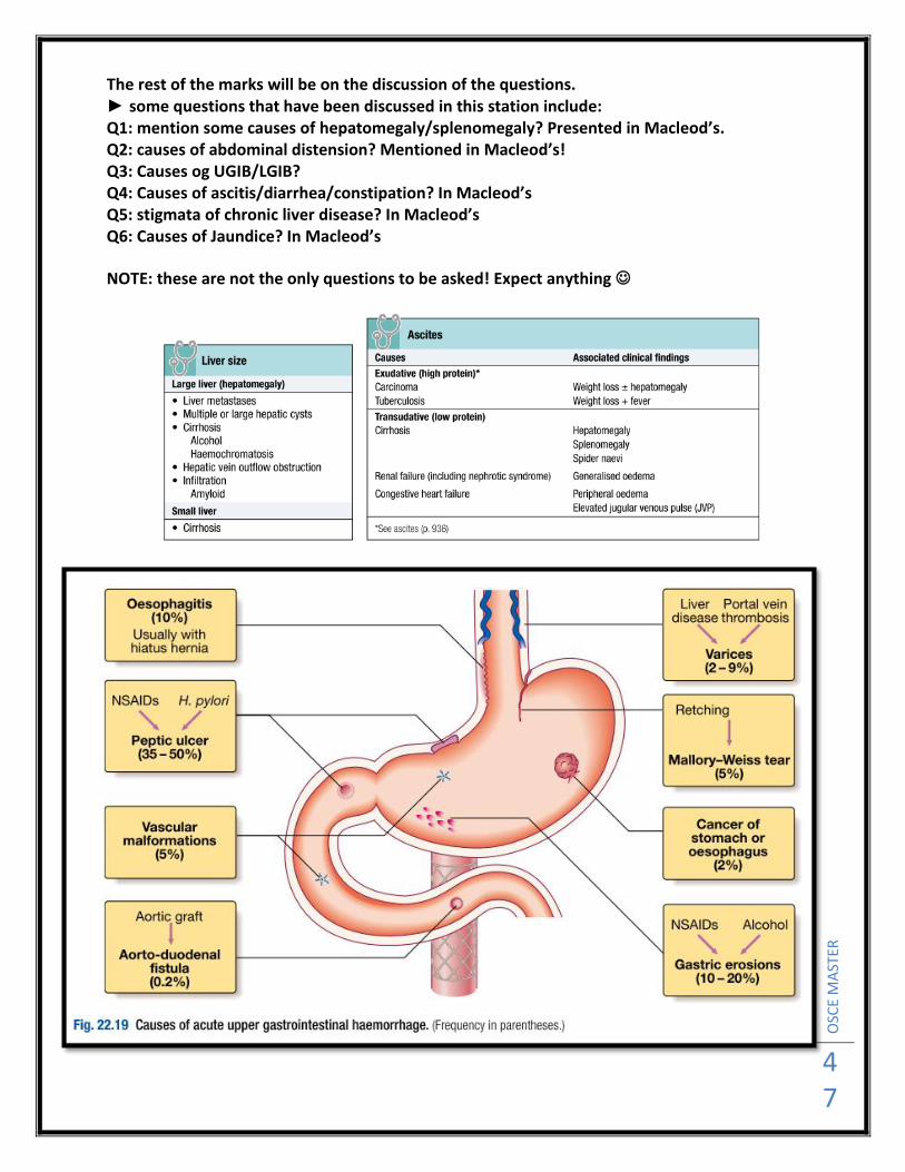
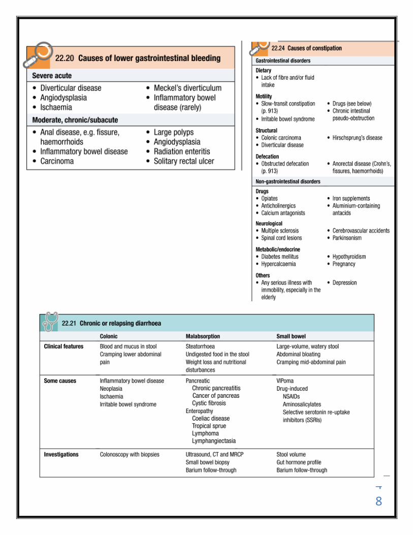
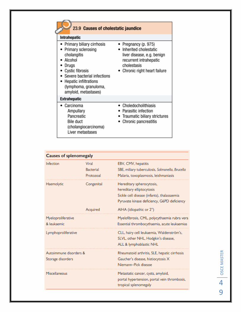
The rest of the marks will be on the discussion of the questions. ► some questions that have been discussed in this station include: Q1: mention some causes of hepatomegaly/splenomegaly? Presented in Macleod’s. Q2: causes of abdominal distension? Mentioned in Macleod’s! Q3: Causes og UGIB/LGIB? Q4: Causes of ascitis/diarrhea/constipation? In Macleod’s Q5: stigmata of chronic liver disease? In Macleod’s Q6: Causes of Jaundice? In Macleod’s
NOTE: these are not the only questions to be asked! Expect anything
OSC
E M
AST
ER
48
OSC
E M
AST
ER
49

OSC
E M
AST
ER
50
OSC
E M
AST
ER
51
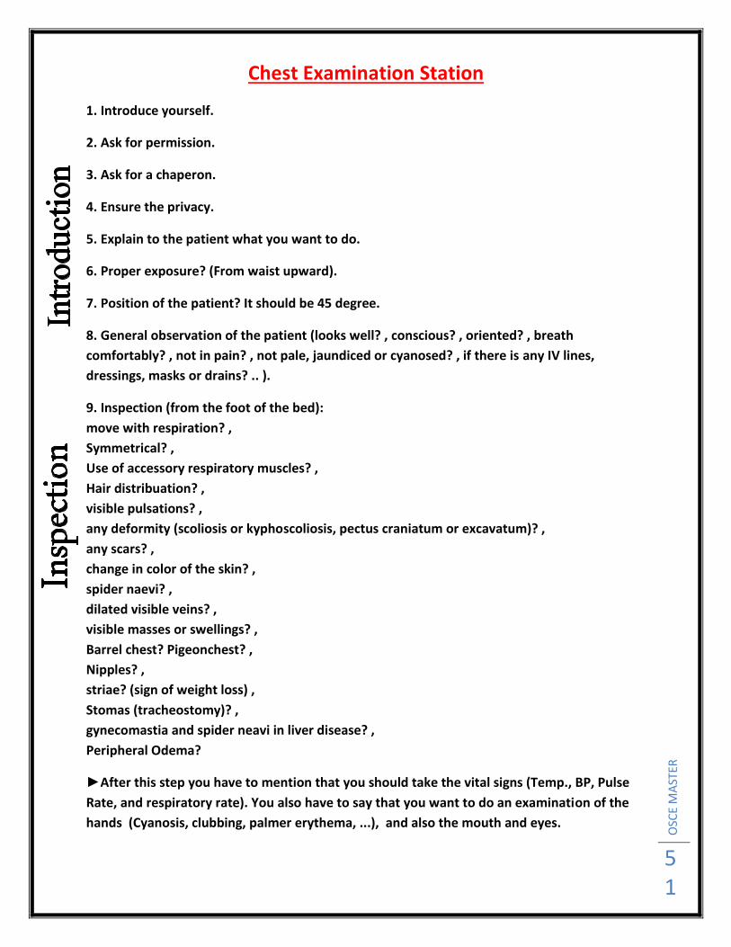
Chest Examination Station
1. Introduce yourself.
2. Ask for permission.
3. Ask for a chaperon.
4. Ensure the privacy.
5. Explain to the patient what you want to do.
6. Proper exposure? (From waist upward).
7. Position of the patient? It should be 45 degree.
8. General observation of the patient (looks well? , conscious? , oriented? , breath
comfortably? , not in pain? , not pale, jaundiced or cyanosed? , if there is any IV lines,
dressings, masks or drains? .. ).
9. Inspection (from the foot of the bed):
move with respiration? ,
Symmetrical? ,
Use of accessory respiratory muscles? ,
Hair distribuation? ,
visible pulsations? ,
any deformity (scoliosis or kyphoscoliosis, pectus craniatum or excavatum)? ,
any scars? ,
change in color of the skin? ,
spider naevi? ,
dilated visible veins? ,
visible masses or swellings? ,
Barrel chest? Pigeonchest? ,
Nipples? ,
striae? (sign of weight loss) ,
Stomas (tracheostomy)? ,
gynecomastia and spider neavi in liver disease? ,
Peripheral Odema?
►After this step you have to mention that you should take the vital signs (Temp., BP, Pulse
Rate, and respiratory rate). You also have to say that you want to do an examination of the
hands (Cyanosis, clubbing, palmer erythema, ...), and also the mouth and eyes.

OSC
E M
AST
ER
52
► Palpation:
• do the examination of head and neck lymph nodes (they are 7 groups: supraclavicular,
anterior, middle and posterior in the neck, submental, submandibular, postaricular and
occipital).
• superfacial palpation: look for masses, subcutaneous emphysema, tenderness, apex beat.
• check for Tactile Vocal Fremitus, compare both sides each one with the other. (comment on
any abnormality).
• Chest expansion (enclose the chest by both of your hands and make 2 skin folds, then ask
the patient to take deep breath – it should move 3-5 cm). remember to do all things
anteriorly and posteriorly. (comment if there is decrease or limitation).
• examination for tracheal deviation (use your middle 3 fingers).
► Percussion: lung Apices, over the clavicles, and 4-5 areas on the chest and the axilla.
(compare each side with the other). comment if there is any dullness, or hyperresonance).
► Auscultation: auscultate the lungs on 7 areas. (comment if it is vesicular or bronchial
breathing, and if there is decrease air entry).
► by doing these steps you have achieved at least 20/25 of this station. Now I will let you
read some questions that have been asked in this station:
Q1: what is the differences between Asthma and COPD?
Q2: what is the presentation of Pancost tumor?
Q3: what is the difference between bronchial and vesicular breathing?
Q4: Causes of pleural effusion?
Q5: Causes of Asterixis?
In addition: COPD >> sputum
Asthma >> usually Dry
COPD >> more associated with
smoking
Asthma >> not always
COPD >> Progressive
Asthma >> intermittent

OSC
E M
AST
ER
53

OSC
E M
AST
ER
54
OSC
E M
AST
ER
55
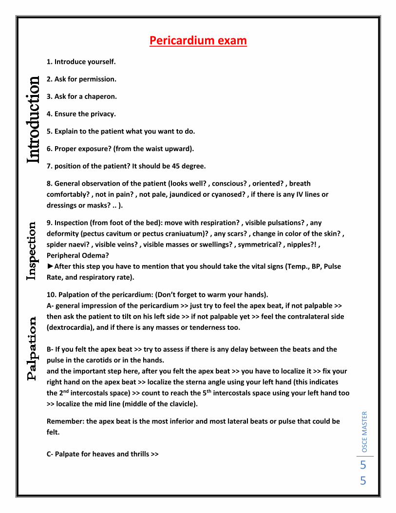
Pericardium exam
1. Introduce yourself.
2. Ask for permission.
3. Ask for a chaperon.
4. Ensure the privacy.
5. Explain to the patient what you want to do.
6. Proper exposure? (from the waist upward).
7. position of the patient? It should be 45 degree.
8. General observation of the patient (looks well? , conscious? , oriented? , breath
comfortably? , not in pain? , not pale, jaundiced or cyanosed? , if there is any IV lines or
dressings or masks? .. ).
9. Inspection (from foot of the bed): move with respiration? , visible pulsations? , any
deformity (pectus cavitum or pectus craniuatum)? , any scars? , change in color of the skin? ,
spider naevi? , visible veins? , visible masses or swellings? , symmetrical? , nipples?! ,
Peripheral Odema?
►After this step you have to mention that you should take the vital signs (Temp., BP, Pulse
Rate, and respiratory rate).
10. Palpation of the pericardium: (Don’t forget to warm your hands).
A- general impression of the pericardium >> just try to feel the apex beat, if not palpable >>
then ask the patient to tilt on his left side >> if not palpable yet >> feel the contralateral side
(dextrocardia), and if there is any masses or tenderness too.
B- If you felt the apex beat >> try to assess if there is any delay between the beats and the
pulse in the carotids or in the hands.
and the important step here, after you felt the apex beat >> you have to localize it >> fix your
right hand on the apex beat >> localize the sterna angle using your left hand (this indicates
the 2nd intercostals space) >> count to reach the 5th intercostals space using your left hand too
>> localize the mid line (middle of the clavicle).
Remember: the apex beat is the most inferior and most lateral beats or pulse that could be
felt.
C- Palpate for heaves and thrills >>
OSC
E M
AST
ER
56
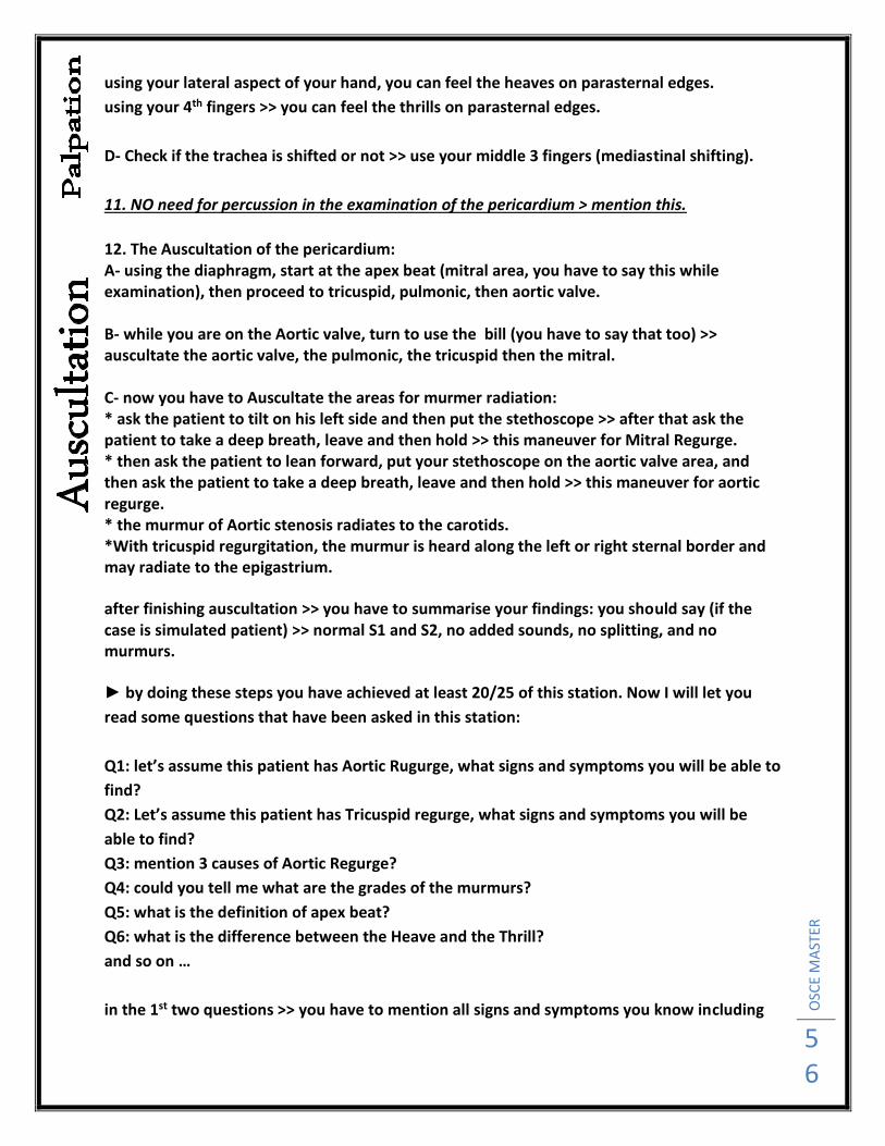
using your lateral aspect of your hand, you can feel the heaves on parasternal edges.
using your 4th fingers >> you can feel the thrills on parasternal edges.
D- Check if the trachea is shifted or not >> use your middle 3 fingers (mediastinal shifting).
11. NO need for percussion in the examination of the pericardium > mention this.
12. The Auscultation of the pericardium: A- using the diaphragm, start at the apex beat (mitral area, you have to say this while examination), then proceed to tricuspid, pulmonic, then aortic valve. B- while you are on the Aortic valve, turn to use the bill (you have to say that too) >> auscultate the aortic valve, the pulmonic, the tricuspid then the mitral. C- now you have to Auscultate the areas for murmer radiation: * ask the patient to tilt on his left side and then put the stethoscope >> after that ask the patient to take a deep breath, leave and then hold >> this maneuver for Mitral Regurge. * then ask the patient to lean forward, put your stethoscope on the aortic valve area, and then ask the patient to take a deep breath, leave and then hold >> this maneuver for aortic regurge. * the murmur of Aortic stenosis radiates to the carotids. *With tricuspid regurgitation, the murmur is heard along the left or right sternal border and may radiate to the epigastrium.
after finishing auscultation >> you have to summarise your findings: you should say (if the case is simulated patient) >> normal S1 and S2, no added sounds, no splitting, and no murmurs.
► by doing these steps you have achieved at least 20/25 of this station. Now I will let you
read some questions that have been asked in this station:
Q1: let’s assume this patient has Aortic Rugurge, what signs and symptoms you will be able to
find?
Q2: Let’s assume this patient has Tricuspid regurge, what signs and symptoms you will be
able to find?
Q3: mention 3 causes of Aortic Regurge?
Q4: could you tell me what are the grades of the murmurs?
Q5: what is the definition of apex beat?
Q6: what is the difference between the Heave and the Thrill?
and so on …
in the 1st two questions >> you have to mention all signs and symptoms you know including
OSC
E M
AST
ER
57
the type of the murmur! And you might be asked “where do you best hear this murmur?”, so
save them well
these questions are not specific for this station and not the only questions! But they are the
most common to be asked so study everything about the HEART!
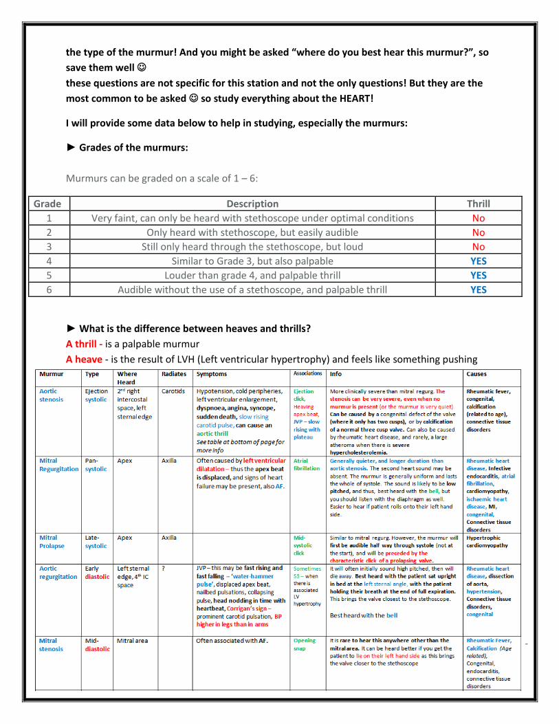
I will provide some data below to help in studying, especially the murmurs:
► Grades of the murmurs:
Murmurs can be graded on a scale of 1 – 6:
Grade Description Thrill
1 Very faint, can only be heard with stethoscope under optimal conditions No
2 Only heard with stethoscope, but easily audible No
3 Still only heard through the stethoscope, but loud No
4 Similar to Grade 3, but also palpable YES
5 Louder than grade 4, and palpable thrill YES
6 Audible without the use of a stethoscope, and palpable thrill YES
► What is the difference between heaves and thrills?
A thrill - is a palpable murmur
A heave - is the result of LVH (Left ventricular hypertrophy) and feels like something pushing
your hand off the chest

OSC
E M
AST
ER
58
► Remember: Austin Flint Murmur >> presents with Aortic Regurgitation.
► Symptoms and Signs of tricuspid Regurge:
Symptoms
Symptoms are generally those of right-sided heart failure, such as ascites, hepatomegaly,edema and jugular venous distension Vague upper abdominal discomfort (from a congested liver), and fatigue (due to diminished cardiac output) can all be present to some degree.
Signs
On examination, the jugular venous pressure is usually elevated, and 'CV' waves can be seen.
The liver may be enlarged and is often pulsatile (the latter finding being virtually diagnostic of tricuspid insufficiency). Peripheral edema is often found. In severe cases, there may be ascites and even cirrhosis (so-called 'cardiac cirrhosis').
Tricuspid insufficiency may lead to the presence of a pansystolic heart murmur. Such a murmur is usually of low frequency and best heard low on the lower left sternal border (on the tricuspid area). It tends to increase with inspiration, and decrease with expiration and Valsalva maneuver.
However, the murmur may be inaudible reflecting the relatively low pressures in the right side of the heart. A third heart sound may also be present, also heard best with inspiration at the left lower sternal border. Parasternal heave may be felt along the left lower sternal border as well.
Atrial fibrillation is usually present.
► Murmurs and certain Maneuvers:
Inspiration: Inspiration leads to a decrease in the intrathoracic pressure with an increase in venous return to the right side of the heart. The murmurs generated from the right side of the heart increase in intensity with inspiration.
Expiration: Expiration has the opposite effect as inspiration. There is an increase in the intrathoracic pressure and a decrease in venous return to the right side of the heart. Blood in the lung is “forced” into the left heart. Hence, murmurs arising from the left side of the heart become more prominent with expiration.
Standing up: This causes a peripheral pooling of blood and a net decrease in venous return. Most murmurs are thus decreased in intensity upon standing, except that of hypertrophic obstructive cardiomyopathy (HOCM) and MVP, which become more prominent.
Squatting: Squatting causes an increase in the afterload and venous return (ie, preload). The net effect is an increase in intensity of all the murmurs, except those associated with MVP and HOCM, which become less prominent with squatting.
OSC
E M
AST
ER
59
Straight leg raising: Passive straight leg raising increases venous return (ie, preload) and has an effect similar to brisk squatting. All murmurs increase in intensity except those of HOCM and MVP, which decrease in intensity with this maneuver.
Hand grip: Hand grip is a form of isometric exercise and increases the afterload, arterial pressure, LV volume, and LV pressure. The net effect of these changes is complex and variable. Murmurs of MR, AR, and VSD worsen with hand grip, while those of HOCM and MVP are less prominent.
Valsalva maneuver: Valsalva maneuver involves asking the patient to strain, which increases the intrathoracic pressure, thus causing a net decrease in preload. Most heart murmurs decrease in intensity with Valsalva, except those of HOCM and MVP, which are more prominent.
Amyl nitrate inhalation: Amyl nitrate is an arteriolar vasodilator and initially causes decreased afterload followed by reflex tachycardia. During the initial phase, because of reduced afterload, the murmurs of AR, MR, and VSD diminish, while those of AS are accentuated. Later on, during the tachycardic phase, the murmur of MS is accentuated.
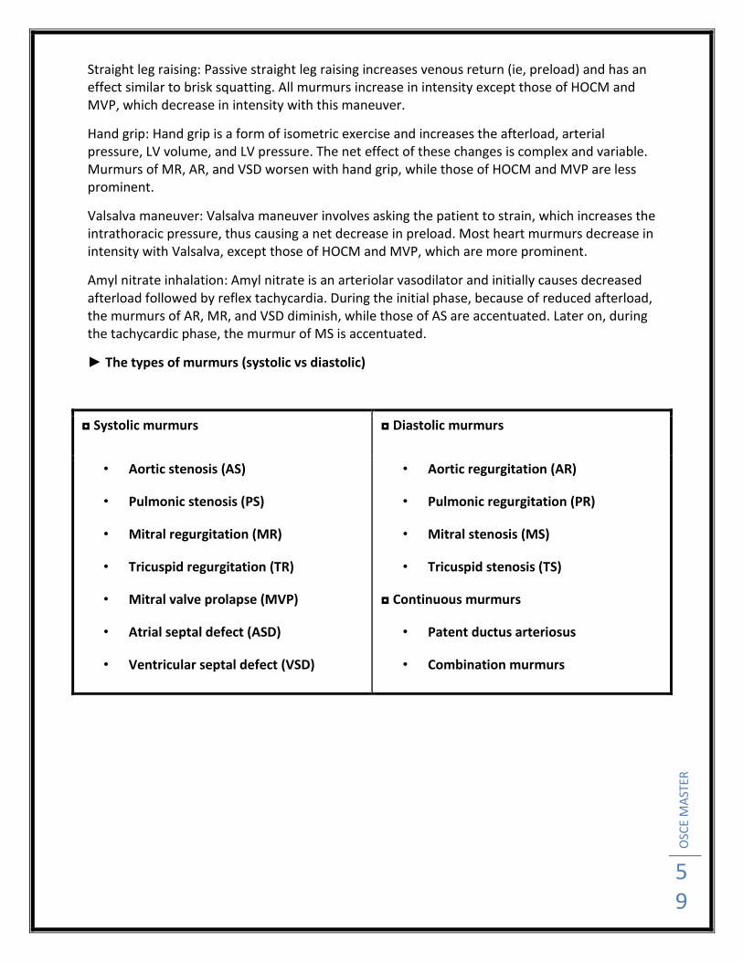
► The types of murmurs (systolic vs diastolic)
◘ Systolic murmurs ◘ Diastolic murmurs
• Aortic stenosis (AS)
• Pulmonic stenosis (PS)
• Mitral regurgitation (MR)
• Tricuspid regurgitation (TR)
• Mitral valve prolapse (MVP)
• Atrial septal defect (ASD)
• Ventricular septal defect (VSD)
• Aortic regurgitation (AR)
• Pulmonic regurgitation (PR)
• Mitral stenosis (MS)
• Tricuspid stenosis (TS)
◘ Continuous murmurs
• Patent ductus arteriosus
• Combination murmurs
OSC
E M
AST
ER
60
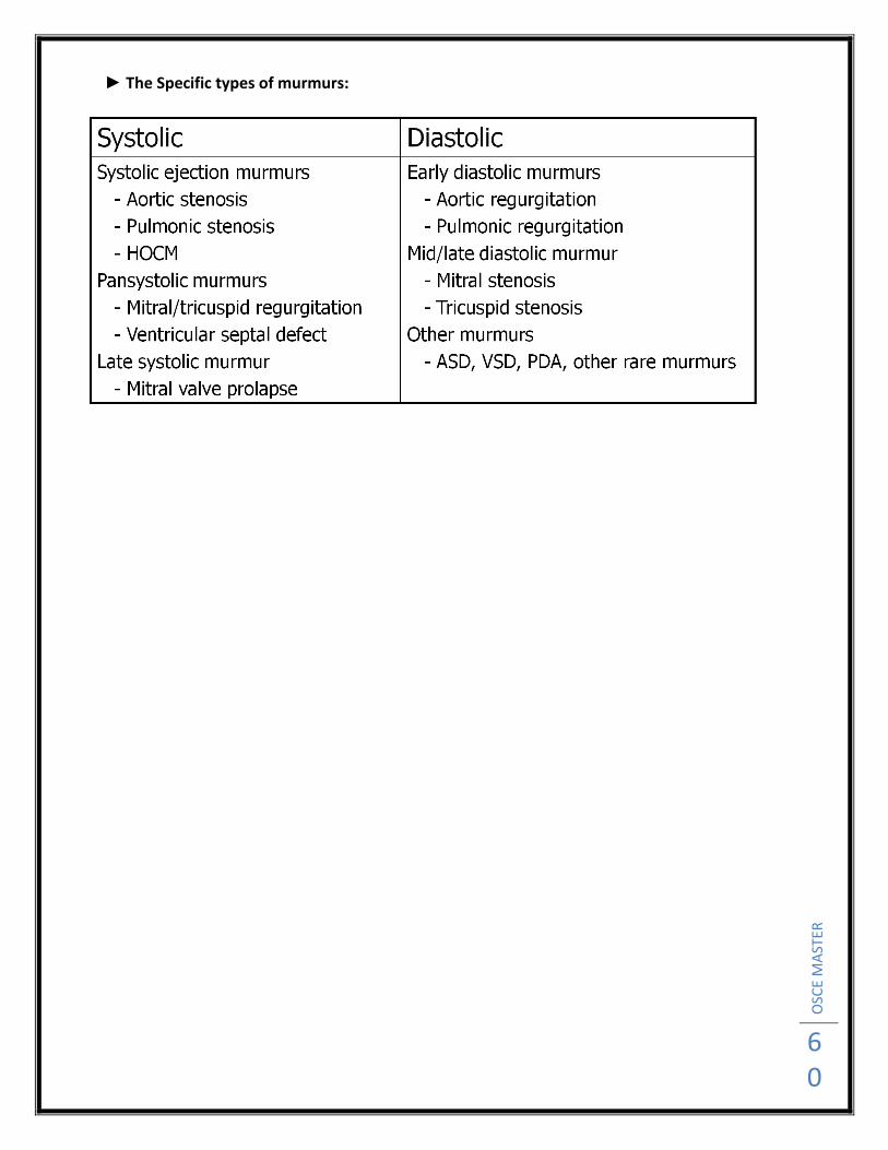
► The Specific types of murmurs:
OSC
E M
AST
ER
61
Lower Limb exam
1. Introduce yourself.
2. Ask for permission.
3. Ask for a chaperon.
4. Ensure the privacy.
5. Explain to the patient what you want to do.
6. Proper exposure? (The whole lower limbs bilaterally, but for privacy issues and the exam
bilateral lower limb exposure to the knees).
7. Position of the patient? Flat at this point.
8. General observation of the patient (looks well? , conscious? , oriented? , breath
comfortably (short of breath or chest pain may indicate PE after DVT)? , not in pain? , not
pale, jaundiced or cyanosed (Liver Failure, Heart Failure)? , if there is any IV lines or dressings
or masks? .. ).
►After this step you have to mention that you should take the vital signs (Temp., BP, Pulse
Rate, and respiratory rate).
9. Inspection (from foot of the bed): Inspect both legs and compare them: if there are ulcers,
Redness, apparent swelling, scars, hair distribution, dilated veins (leads to venous stasis
which is a risk factor for DVT), pigmentation, lesions, masses, amputation and the nails
after this you have to say that you need to inspect the dorsum of the legs and feet for the
same things. Say that you have to do this for the thighs as well.
Inspect between the toes.
10. Palpation:
A- use the dorsum of your hands to feel the temperature of the legs bilaterally in the same
time from the toes to the knees. Then ask the patient if there is any pain in his legs, if no pain
then palpate for any tenderness each leg separately. After that you have to check the pulses
(Dorsalis pedis, Tibialis posterior, and Popliteal).
Dordsalis pedis: on the dorsum of the foot lateral to the extensor hallucis longus, against the
prominent part of the navicular bone (abscent in 2-3% of young healthy individuals).
Tibialis posterior: 2 cm posterior and inferior to the medial malleolus (never abscent in
healthy).
check for odema by pressing against the tibial shaft for few seconds (bilaterally).
OSC
E M
AST
ER
62
Check capillary refill by pressing on one of the nails bilaterally (capillary refill should be less
than 2 seconds)
• Homan’s Sign: only mention this and say it is not used anymore because it has low
specificity and may increase the risk of the thrombus to be dislodged, so increase the risk of
PE. How it is done: with the leg extended at the knee joint, make a forceful dorsiflexion of the
foot at the ankle, this may elicit a pain in the calf which may indicate DVT.
If there is any ulcer you have to examine it or at least mention that, if the Dr. tell you skip
then skip, if not examine it by inspection (site, size, shape, depth, color, surrounding skin, any
discharge and its color, and then palpation for tenderness and milking for discharge).
B- The next step is to measure the diameter of the legs. First you have to choose a reference
point. Here there are two methods: the first one is to localize the tibial tuberosity and using
the tape to measure 10 down from the tibial tuberosity and then measuring the diameter at
that point using the tape. Do the same thing bilaterally.
the second method: is to choose a refrrence point between the tibial tuberosity and the
medial malleolus using the tape. Measure the distance between the tibial tuberosity after
localizing it and the medial malleolus, then go 10 cm either from the top or from the bottom
of the tape (preferrably from the top, i.e. tibial tuberosity) and measure the leg diameter at
that point. Do the same bilaterally.
Keep in mind that a difference of 3 cm or more is significant.
Say you want to do the same for the thighs.
After that, you have to say: to complete my examination I have to examine the nerves
(sensations and power mainly), and the range of motion (Rheumatological diseases).
Questions that might be asked in discussion:
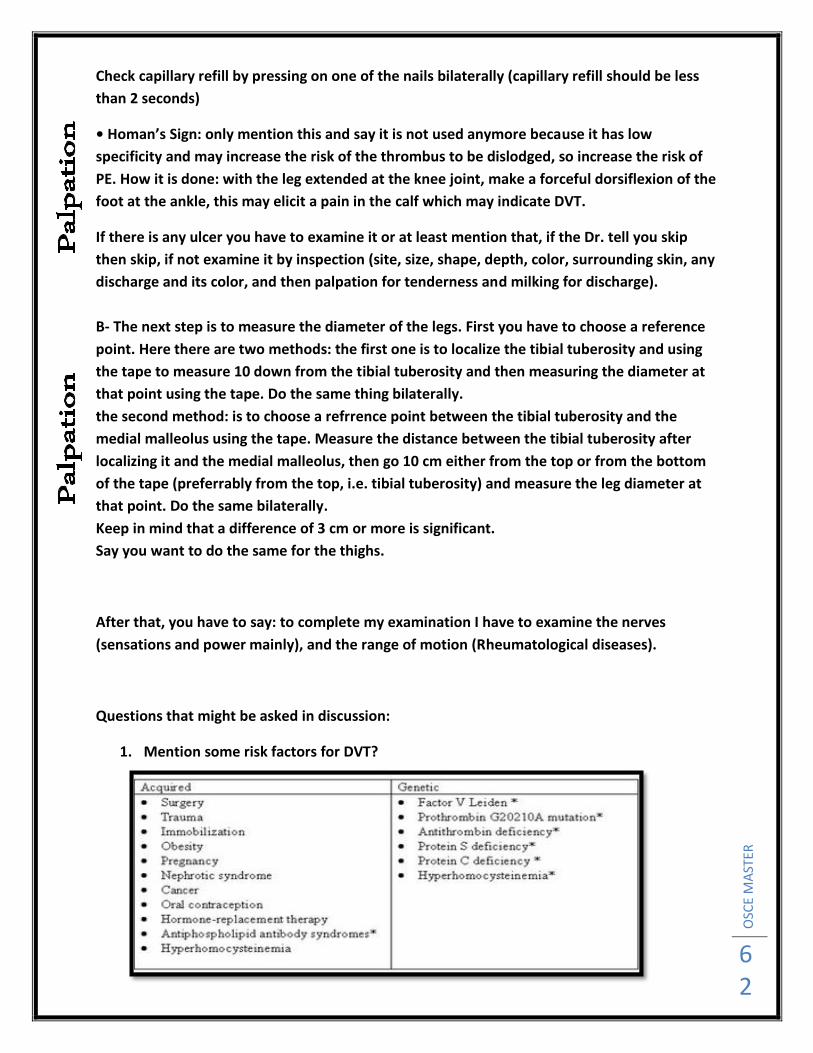
1. Mention some risk factors for DVT?
OSC
E M
AST
ER
63
2. Risks of recurrent DVT?
Bed ridden, malignancy, antiphospholipid syndrome, and antithrombin protein S and
C deficiencies.
3. DDx of lower limb swelling?
Bilateral: Heart failure, Renal Failure, liver Failure, Drug induced (e.g. CCB), Bilateral
DVT (Rare).
Unilateral: DVT, insect bite, cellulitis, muscle injury, popliteal cyst.
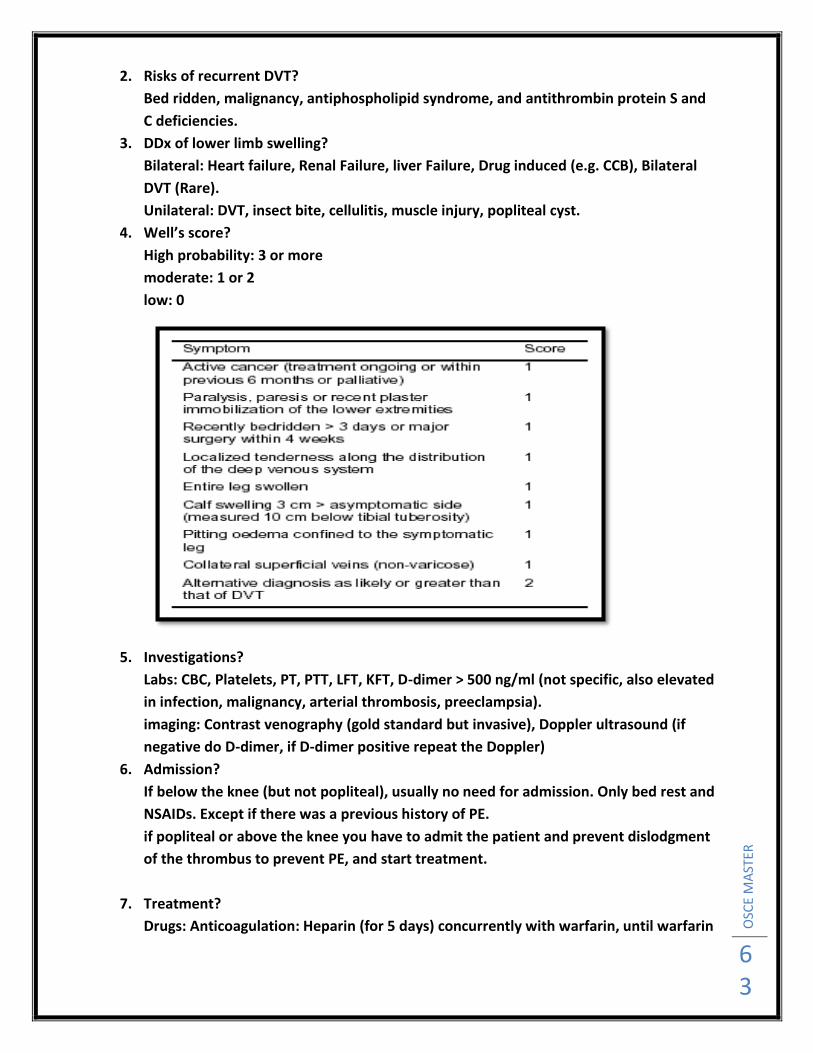
4. Well’s score?
High probability: 3 or more
moderate: 1 or 2
low: 0
5. Investigations?
Labs: CBC, Platelets, PT, PTT, LFT, KFT, D-dimer > 500 ng/ml (not specific, also elevated
in infection, malignancy, arterial thrombosis, preeclampsia).
imaging: Contrast venography (gold standard but invasive), Doppler ultrasound (if
negative do D-dimer, if D-dimer positive repeat the Doppler)
6. Admission?
If below the knee (but not popliteal), usually no need for admission. Only bed rest and
NSAIDs. Except if there was a previous history of PE.
if popliteal or above the knee you have to admit the patient and prevent dislodgment
of the thrombus to prevent PE, and start treatment.
7. Treatment?
Drugs: Anticoagulation: Heparin (for 5 days) concurrently with warfarin, until warfarin

OSC
E M
AST
ER
64
works (in the first few days warfarin may cause thrombosis).
Anticoagulation is contraindicated in hemodynamically unstable patients:
Intracerebral hemorrhage, Uncontrolled HTN, recent surgery, PUD, liver disease.
If the patient starts to bleed: stop warfarin for 3-4 days, if still bleeding give fresh
frozen plasma rather than Vit. K cause it needs time to start working and may cause
thrombosis.
Thrombolytic therapy: e.g. Streptokinase, only degrades pre-formed clot, don’t
prevent further clot formation, so you have to give with it anticoagulation.
Surgery: Thrombectomy: in patients where anticoagulation contraindicated.
IVC filter, especially in patients who failed medical therapy or have recurrent emboli.
Duration of treatment: at least 3 months.
8. Prophylaxis: walking, compression stocking.
9. Most important complication? PE
diagnosed clinically with lab investigations (D-dimer), Ventilation-perfusion scan,
Chest X-ray, ECG (S1Q3T3). Pulmonary angiography (gold standard).
in chest X-Ray you see:

OSC
E M
AST
ER
65
1. Wash hands (or wear gloves).
2. Introduce yourself.
3. Explain what you want to do and gain consent.
4. Ensure the privacy and ask for a chaperon.
5. Exposure should be from the shoulders upward.
6. General observation of the patient (looks well/ill/Anxious/Restless/irritable?,
conscious?, oriented?, not in pain?, not in respiratory distress/tachypnic due to
thyroid enlargement compressing the trachea?, sweaty?, wasted (weight loss)? Hair
loss?
7. Say you need to take the vital signs. (Tchycardia and increased BP in hyperthyroid).
Thyroid gland examination is of 2 parts:
Examination of the thyroid status and examination of the thyroid as a mass.
First, we start with the thyroid status:
8. Hands:
a. Examine the pulse (tachy/bradycardia, Irregular irregularity may indicates AF, a
complication of thyrotoxicosis).
b. moisture (sweaty), palmar erythema, warmth.
c. fine tremor.
d. reflexes in the arm (exaggerated in hyper, slow in hypo).
9. Eyes:
a. Lid retraction.
b. lid lag (ask the patient to close and open his eyes once, and follow the lids
movement, it will be slow).
c. Exopthalmus (bulging of the eyes, the patient can look upward without wrinkles).
d. opthalmoplegia.
e. chemosis (redness of the eyes).
10. Legs:
a. pretibial myxedema (red, thickened swelling above the lateral malleoli).
b. reflexes in the legs.
Now, we start with the thyroid exam as a Mass:
11. You start with Inspection, if there is swelling in the neck you have to comment on it
regarding:
OSC
E M
AST
ER
66
a. site of the swelling.
b. approximate size.
c. shape of it, if it can be assessed.
d. color of the overlying skin.
e. movement of it with swelling and tongue protrusion.
f. if there is any lumps or masses anywhere else.
12. Now we move to Palpation: start anteriorly and then from behind the patient:
a. Size of the swelling or lump.
b. Site of the swelling and if it extends anywhere (to the mediastinum for example).
c. Shape of the mass: symmetrical or not, spherical, oval, regular or irregular.
d. Surface: smooth, rough, bosselated, irregular.
e. Temperature.
f. Tenderness.
g. Translucency (Transillumination).
h. Thrill or pulsation.
i. Fluctuation.
j. Mobility/Fixation.
k. Consistency: stony hard, firm, rubbery, spongy, soft.
l. if the trachea can be palpated, then assess it for any deviation.
► Then posteriorly:
a. flex the head and examine the mass again.
b. examine the lymph nodes: submental, submandibular, anterior, middle and
posterior cervical, and supraclavicular.
13. Percussion:
a. for extension of the mass into the chest (retrosternal extension).
b. percussion over the mass itself (dull if fluid filled cysts or solid mass, resonant if gas
filled cysts).
14. Auscultation: listen to the lump using the stethoscope for any possible bruits (a bruit
may indicate AV fistula).
4S , 4T , FMC
OSC
E M
AST
ER
67
1. Investigations you want to do?
a. CBC
b. TSH, fT4
c. Autoantibodies (antimicrosomal, anti-peroxidase, and
antithyroglobuline antibodies, anti-TSH antibodies).
d. Ultrasound.
e. FNA.
f. Radioactive Iodine.
g. CT or MRI (for mets and extension).
h. CXR
i. ECG

OSC
E M
AST
ER
68