historical dental investigations - elsevier...7 historical dental investigations was appointed...
TRANSCRIPT

CHAPTER 1
1Forensic Dental Evidence. DOI: 10.116/B978-0-12-382000-6.00001-9Copyright © 2011 by Elsevier Ltd. All rights of reproduction in any form reserved.
C. Michael Bowers Associate Clinical Professor, Herman Ostrow School of Dentistry, University of Southern California, Los Angeles, CA
Historical Dental Investigations
Marie Svoboda Associate Conservator, Antiquities Conservation, J. Paul Getty Museum, Los Angeles, CA
C. Michael Bowers Associate Clinical Professor, Herman Ostrow School of Dentistry, University of Southern
California, Los Angeles, CA
Dental Aging Analysis of Ancient Human Remains: Herakleides from
the First Century AD
Overview A death investigation of unidentified human remains requires professional determination of all physical evidence available from the body. The unknown person's gender, age, medical status and cause of death are vital information for a forensic autopsy report. The following case explains details of the life

2
Forensic Dental Evidence
and death of a young man whose body was originally found in Egypt. Dental information was important to confirm his age and give insight to his health history at the time of his death.
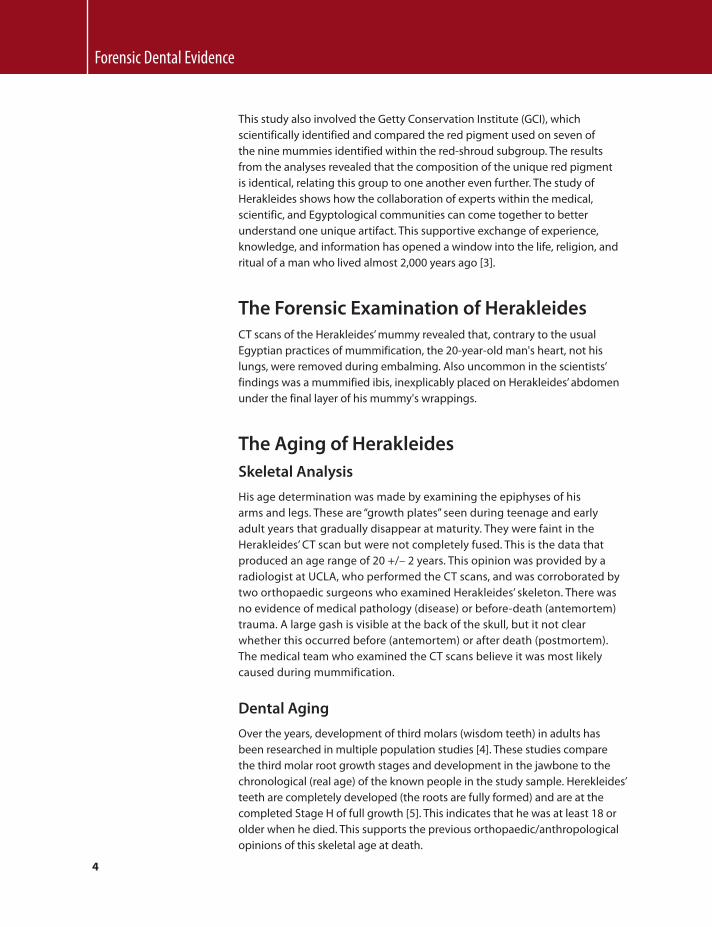
In 2003, the J. Paul Getty Museum Antiquities Conservation Department initiated the study of a Romano-Egyptian red-shroud mummy (91.AP.6) in the museum's collection. The mummy is known as Herakleides from a painted inscription on top of the wrapped feet. Dating to the first century AD, the mummy incorporates a portrait panel depicting a young man in his early twenties. The mummy, measuring 175 cm in length, was wrapped in one large outer shroud that had been painted red and decorated with Egyptian funerary images. The beautifully executed portrait, the quality of the wrappings, and the elaborate use of gold, on both the panel and the shroud, attest to the prominent status of this individual.
The mummy of Herakleides belongs to a group of Romano-Egyptian mummies called “portrait mummies” and to a subgroup within that category known as “red-shroud portrait mummies.” The designation of such complete mummies with added classically painted portraits is credited to Flinders Petrie, the archaeologist who was the first to scientifically excavate and document them in the late nineteenth century. The unique style and color of Herakleides’ shroud classifies it as red-shroud [ 1 ]. These mummified bodies are described as being completely wrapped in a single cloth painted red and decorated with either funerary or daily dress motifs.
The investigation of Herakleides’ mummy began with the conservation treatment of the fragile foot area, which was damaged and unstable [ 2 ]. The conservation treatment was in preparation for the mummy's first public display at the Getty Villa in 2005, where its presence in the gallery contextualizes the now detached Romano-Egyptian portraits in the collection by illustrating their mortuary function. From here a full study of the body (human remains) and the materials used for the mummification and decoration of Herakleides evolved. The aim of this study was to better understand the person within the wrappings and the ancient techniques employed in its fabrication and adornment process. Imaging technology such as computerized tomography (CT) and infrared photographic techniques revealed secrets such as his complete name and the curious inclusion of a mummified ibis within the wrappings. Examination of the skeleton by orthopedic surgeons and a forensic dentist established his age, health, and height at the time of death. Radiocarbon dating (carbon 14) provided a secure date for the materials used in the mummification process and a likely time frame for when he lived. Motifs and religious iconography were studied and documented to better understand their meaning. The lack of clothing on the youth's shoulders suggests he was an ephebe, or adolescent male of social standing. His presumed nudity, a symbol of rebirth, indicates he may have been an initiate in the cult of the Egyptian goddess Isis.

3
Historical Dental Investigations
FIGURE 1.1 The mummy of Herakleides: “Mummy and Portrait on wooden panel.” © The J. Paul Getty Museum, Villa Collection, Malibu, California (91.AP.6).
4
Forensic Dental Evidence
This study also involved the Getty Conservation Institute (GCI), which scientifically identified and compared the red pigment used on seven of the nine mummies identified within the red-shroud subgroup. The results from the analyses revealed that the composition of the unique red pigment is identical, relating this group to one another even further. The study of Herakleides shows how the collaboration of experts within the medical, scientific, and Egyptological communities can come together to better understand one unique artifact. This supportive exchange of experience, knowledge, and information has opened a window into the life, religion, and ritual of a man who lived almost 2,000 years ago [ 3 ].
The Forensic Examination of Herakleides CT scans of the Herakleides’ mummy revealed that, contrary to the usual Egyptian practices of mummification, the 20-year-old man's heart, not his lungs, were removed during embalming. Also uncommon in the scientists’ findings was a mummified ibis, inexplicably placed on Herakleides’ abdomen under the final layer of his mummy's wrappings.
The Aging of Herakleides Skeletal Analysis
His age determination was made by examining the epiphyses of his arms and legs. These are “growth plates” seen during teenage and early adult years that gradually disappear at maturity. They were faint in the Herakleides’ CT scan but were not completely fused. This is the data that produced an age range of 20 +/– 2 years. This opinion was provided by a radiologist at UCLA, who performed the CT scans, and was corroborated by two orthopaedic surgeons who examined Herakleides’ skeleton. There was no evidence of medical pathology (disease) or before-death (antemortem) trauma. A large gash is visible at the back of the skull, but it not clear whether this occurred before (antemortem) or after death (postmortem). The medical team who examined the CT scans believe it was most likely caused during mummification.
Dental Aging
Over the years, development of third molars (wisdom teeth) in adults has been researched in multiple population studies [ 4 ]. These studies compare the third molar root growth stages and development in the jawbone to the chronological (real age) of the known people in the study sample. Herekleides’ teeth are completely developed (the roots are fully formed) and are at the completed Stage H of full growth [ 5 ]. This indicates that he was at least 18 or older when he died. This supports the previous orthopaedic/anthropological opinions of this skeletal age at death.

5
Historical Dental Investigations
Acknowledgments Photo: The J. Paul Getty Museum, Villa Collection, Malibu, California
References [1] L. Corcoran , Portrait Mummies from Roman Egypt (I-IV Centuries A.D.),
with a catalogue of portrait mummies in Egyptian Museums , Chicago Press , 1995 , p. 7 .
[2] http://getty.edu/art/videos/mummification_process/mummification_process.html [3] L. Corcoran , M. Svoboda , Herakleides: A Red-Shroud Portrait Mummy from Roman
Egypt , J. Paul Getty Museum , Los Angeles , 2010 . [4] C.M. Bowers , Determining Age from Teeth: The Estimation of Age From Dental
Development , in: C.M. Bowers , G.L. Bell (Eds.), Manual of Forensic Odontology , third ed. , ASFO , 1995 , pp. 74 – 105 .
[5] Ibid. p. 88.
The Odontological Identification of Adolf Hitler, Using
Cinematographic Documents Michel Perrier Forensic Odontology Unit, Centre Universitaire Romand de Médecine Légale, University de Lausanne, Switzerland
Introduction A “toothy” antemortem photograph can be invaluable when investigating the identity of unknown human remains. Pictures can show dental characteristics that can be very helpful to the forensic odontologist in comparing antemortem and postmortem data. Such documents can give a lot of information, but how much information is necessary for a positive identification? This paper gives investigators answers to this question from casework that combines both the photographic and dental evidence that is necessary for a positive determination of identity.
Forty-eight members of the sect of the Solar Temple were found dead in two different villages in October 1993. Their guru, Luc Jouret, was among them and was severely cremated. He was odontologically identified, and a picture published by the press was an additional contribution to the identification procedure [ 1 ].
The following account is a good example of how photographic documents may contribute to postulate an identity [ 2 ]. In October 2001, a Crossair plane crashed near the airport in Zurich, Switzerland. Eleven of the 24 passengers

6
Forensic Dental Evidence
died in this mass disaster. The examination of the dental work of one passenger showed ceramic restorations that appeared unusual both in shape and shade. Before receiving any postmortem data of this otherwise cremated passenger, investigators were struck by an aspect of the dental work that led to the possibility that the remains were of a well-known entertainer. A search on the Internet provided an account of the passenger's travel plans on the day of the crash.
In 1973, Sognnaes and Ström published an article on the identification of Adolf Hitler [ 3 ]. The major portion of this article compares more recently released postmortem dental remnants of Adolf Hitler with his cinematographic facial phtotographs. Photographic documents can contribute to odontological identification of human remains, provided that the teeth and their characteristics are sufficiently visible. But how do motion pictures help in the same way? A historical figure such as Adolf Hitler was identified odontologically thanks to information provided by his dentist and radiographic plates found in the U.S. National Archives [ 3 ].
A Short Biography of Adolf Hitler According to Kershaw [ 4 ], the first of many strokes of good fortune for Adolf Hitler took place 13 years before he was born. In 1876, the man who was to become his father changed his name from Alois Schickelgruber to Alois Hitler. Certainly, “Heil Schickelgruber” would have been an unlikely salutation to a national hero.
Hitler was born on April 20, 1889, in the little town of Braunau am Inn in Austria. He had three brothers and two sisters, as well as four half-brothers and half-sisters from his father's first marriage. Hitler loved his mother dearly, and he carried her picture with him until his final days in the Berlin bunker. The violence of Hitler's father would turn against him and the rest of the family. His childhood was also punctuated with several moves, and for much of his childhood, he lived in Linz and Vienna, Austria.
Hitler wanted to be a painter, but in 1907 he failed the entrance examination for the Academy of Fine Arts in Vienna. Many members of the faculty were Jewish. He moved to Munich in 1913. While serving in the German army during World War I, he was wounded. He began his political career as an army political agent in the German Workers’ (later Nazi) Party in 1919 and became head of its propaganda arm in 1920.
Hitler was made president of the party in 1921 and began his creation of mass movement and his climb to power. After the abortive Munich (Beer Hall) Putsch in 1923, he served nine months in prison and began writing Mein Kampf, a book in which he condemned democratic government and expressed his hatred and fear of Jews.
Throughout the 1920s, Hitler continued to gain strength. He unsuccessfully opposed Paul von Hindenburg in the presidential election of 1932, but he

7
Historical Dental Investigations
was appointed chancellor in 1933. The offices of president and chancellor were merged in 1934, a move that was supported later by a popular vote. As dictator, Hitler then turned his attention to foreign policy and World War II. His “new order” for Europe called for indiscriminate extermination of entire peoples. The Jews of Europe were the most numerous among his victims of barbarism [ 4 ]. Hitler retreated to the chancellory in Berlin in January 1945, and, in the face of impending defeat, he committed suicide.
Hitler's Death In broad daylight on February 3, a huge American fleet of bombers unleashed a hail of destruction from the skies in the heaviest raid of the war on Berlin, the Reich's capital. Most of the Reich's headquarters were severely damaged. The whole area was a mass of rubble. For a time, there was a complete power failure, and water was unavailable [ 4 ].
Hitler's apartments in the Reich Chancellery were largely gutted by incendiaries. He now moved underground for much of the time to the Führer Bunker, a two-story construction deep below the garden of the Reich's Chancellery [ 5 , 6 ]. The enormous bunker complex had been deepened in 1943, extending an earlier bunker (originally meant for possible future use as an air-raid shelter) dating from 1936, and heavily reinforced during Hitler's stay at his headquarters. The complex was built beneath the Chancellery gardens. It was completely self-contained, with its own heating, lighting, and water pumps that operated from a diesel generator [ 6 ]. From now on, it would provide a macabre home for the remaining weeks of Hitler's life.
The bunker was far from the palatial surrounds to which he had been accustomed since 1933. At first, even after he had moved his living quarters into the bunker, Hitler continued to spend part of the day in the undamaged wing of the Reich Chancellery. Over the next weeks, he transferred almost all of his activities to the bunker, leaving it only for occasional snatches of fresh air, to let his dog Blondi out for a few minutes in the garden, or to take lunch with his secretaries above ground. He was there with Eva Braun, his valet Heinz Linge, Martin Bormann, Josef Goebbels, personal adjutants, secretaries, servants, a cook and frequent visitors [ 7 ].
During this period, he was constantly informed of the evolution of the situation: the Yalta conference where Roosevelt, Stalin, and Churchill defined the postwar shape of Germany and Europe; the destruction of Dresden; the bad news from the Eastern front; the imminent collapse of the German economy; civilians embracing American soldiers; and troops fleeing or surrendering on the western front.
With both the present and the future so bleak, he had begun to take refuge in endlessly speaking and recalling the “triumphs” of the past, his career, the atrocities of the “Jewish Bolshevism.” His physical condition deteriorated sharply during that time. He was haggard, aged, and stooped. His left hand

8
Forensic Dental Evidence
and arm trembled uncontrollably. He took daily concoctions of pills, potions, and injections that included both stimulants and sedatives . He seldom went out in public.
In his characteristic “either-or” way of thinking, he invariably posed total destruction as the alternative to the total victory for which he had aspired. Ultimately, the existence of the German people—if they showed themselves incapable of defeating their enemies—was less important to him than the refusal to capitulate.
The atmosphere in the bunker on April 20, 1945—Hitler's fifty-sixth birthday—was more funereal than celebratory. The assault on Berlin by the Soviets was imminent. The storm burst on April 22. Hitler realized that the war was lost. He allegedly said, “Es ist alles verloren, hoffnungslos verloren” (“It's all lost, desperately lost“).
The next day, Hitler had a discussion with his armament minister, Albert Speer. They both came to the conclusion that it would be better to end his life as Führer in the Reich's capital than in his Bavarian “weekend house.” But there was the danger that he would be captured alive. He was afraid that his body might fall into the hands of his enemy to be displayed as a trophy. He gave orders that his body should be cremated. His mistress, Eva Braun, would die alongside him.
Not long after midnight on April 29, in the most macabre surroundings, with the bunker shaking from nearby explosions, Hitler and Eva Braun exchanged marriage vows before one of Goebbels's minor officials, city councilor Walter Wagner. Goebbels and Bormann served as witnesses. Hitler then wrote his private and political testaments. He disposed his possessions to the state, and he appointed Grand Admiral Karl Dönitz as Reich president. Bormann and two emissaries left the bunker on what was to be a fruitless mission to deliver the testaments to the headquarters in Munich.
Early that morning, Dr. Ludwig Stumpfegger, an SS surgeon, distributed to the secretaries, adjutants, and any others who wanted them brass-cased ampoules containing prussic acid (cyanhydric acid). On April 30, Hitler sent for Bormann and told him the time had come. He would shoot himself that afternoon, and Eva Braun would also commit suicide. He wanted their bodies to be burned with gasoline that his chauffeur, Erich Kempka, would obtain. In the bunker were Hitler, Eva Braun, General Burgdorf, General Krebs, Hitler's secretaries, his dietician, adjutants, Borman, and Goebbels, his wife Magda, and six of their seven children. Hitler retreated behind the doors of his study, and Eva Braun followed him almost immediately.
Some 10 minutes later, the valet, Heinz Linge, and Martin Bormann opened the door cautiously. They found Hitler and Eva Braun sitting alongside on a small sofa. Eva Braun was slumped to Hitler's left. A strong whiff of bitter almonds—the distinctive smell of prussic acid—drifted up from her body. Hitler's head drooped lifelessly. Blood dripped from a bullet hole in his right temple. His 7.65 Walther pistol lay by his foot [ 5 , 6 , 7 ].

9
Historical Dental Investigations
Within minutes, the bodies of Adolf Hitler and his wife of 18 hours were wrapped in blankets. The corpses were then lifted from the sofa and carried through the bunker, up 25 feet of stairs, and into the garden of the Reich Chancellery. Heinz Linge brought out Hitler's body, his head covered with a blanket, his lower legs protruding. Martin Bormann carried Eva Braun's body into the corridor, where Erich Klempka, Hitler's chauffeur, relieved him of his burden.
Otto Günsche, Hitler's personal adjutant, who had been commissioned with overseeing the burning of the bodies, laid the bodies outside in the garden side by side on a piece of flat, open, sandy ground only about 3 meters from the door down to the bunker. It was a suitable spot, close to the bunker, though extremely hazardous, since an unceasing rain of shells from the Soviet barrage continued to bombard the whole area. Almost 200 liters of gasoline had been gathered in the bunker in readiness. It was swiftly poured over the bodies and ignited. Later, two SS men of the Führer Escort Squad ensured that the bodies had been completely burned and reported it to Otto Günsche.
Little remained of Hitler's and Eva's bodies. The intense bombardment that continued for another 24 hours played its own part in destroying and scattering the human remains strewn around the Chancellery garden. When the Soviet victors arrived there on May 2, they immediately began a vigorous search for the bodies. Nine days later, they showed Fritz Echtmann, a dental technician who had worked for Hitler's dentist, a cigar box containing part of a mandibular bone with two dental bridges and one isolated dental bridge. Echtmann was able to identify from his records the dental work of Hitler [ 5 , 6 , 7 ].
Remains and X-Rays The death of Adolf Hitler in April 1945 was a mystery until 1968, when the Russian writer Lev Brezymenski revealed documents from Soviet archives established during the identification procedures [ 8 ]. This somewhat unprecise book included descriptive information concerning Hitler's alleged corpse, with photographs of remaining dental restorations and some of his natural teeth still in the mandible. The wartime autopsy documents from Soviet archives report the forensic examination of a male corpse disfigured by fire.
The author of this section was able to examine Hitler's remains at the Russian national archives ( Figure 1 ). The photographic documents shown in Figures 1 to 6 were taken by Mark Benecke, a forensic biologist. What was left of the upper arch was a nine-tooth gold bridge with crown, abutments on each of the four remaining natural teeth, one intermediate pontic (replacement tooth), and a double cantilevered pontic (replacement tooth attached to two crowns) at each end. The entire bridge consisted of four upper incisors, two canine teeth, the first left bicuspid, and the first and second right bicuspids ( Figure 2 ).
The first left incisor had a gold crown with a white facing, with cracks and a black spot in the exposed tooth enamel at the bottom. The other teeth of the left side,

10
Forensic Dental Evidence
FIGURE 1 Hitler's remnants, as kept at the Russian archives in Moscow.
FIGURE 2 Hitler's repaired bridge on the upper arch.
FIGURE 3 The lower left bridge.

11
Historical Dental Investigations
as well as the first and second incisors, and the first bicuspid on the right side were porcelain plated. The upper right canine tooth was fully gold-capped. The maxillary bridge was vertically sawed off behind the second left bicuspid.
In the mandible, several of the natural teeth were well preserved. On the left quadrant, three abutments (canine, second bicuspid, probably third molar) supported a six-unit gold bridge ( Figure 3 ). On the right quadrant, a three-unit bridge (canine, second bicuspid) bypassed the first bicuspid using an unusual device ( Figures 4 and 5 ). The incisors showed advanced periodontitis as well as signs of buccal erosion and abrasion ( Figure 6 ).
It is not the aim of this section to give a complete list of the comparative conclusions regarding Adolf Hitler's dental condition. This topic has been fully covered by Sognnaes and Ström in 1973 [ 3 ]. The American Archives provided the detailed questioning of Hitler's dentist. Dr. Hugo Johannes Blaschke was a dental school graduate of the University of Pennsylvania. He was an outstanding student, graduating fourth in his class of over 100 classmates. From the 1911 yearbook and from recollections of some of his classmates (who called him “the count”), Blaschke was also well thought of as a person and colleague—skilled, meticulous, and dedicated to dentistry. He returned to his native Germany to open his dental practice in Berlin. One of his first well-known patients was Hermann Göring, who introduced Blaschke to Hitler, and in turn Dr. Blaschke also became the dentist of Eva Braun, Bormann, and many other high-ranking Nazis [ 9 ]. He treated Hitler from 1934 to 1945. Later that year, he was captured and questioned by U.S. Army officers and also briefly called upon as a witness during the Nuremberg trials in connection with the so-called Pohl process and the Dr. Pook case. After his release in 1948, Hugo Blachke continued to practice as a dentist in Nuremberg. He died in 1957 at age 78 [ 3 , 7 , 9 ].
After his capture in 1945, Blaschke described the characteristics of Hitler's teeth and treatment history, which were found to be compatible with the odontological examination:
• Dr. Blaschke had started a root canal treatment on the lower left lateral incisor.
• A single fixed bridge had been constructed to replace two defective upper left and right bridges.
• No prosthetic treatment was performed by Blaschke on the lower jaw. • Extensive caries and pulpal involvement had existed for several years
on the left central incisor (a “window crown”) that could not be properly treated due to Hitler's impatience.
• The existence of a porcelain cement filling on the left lower lateral incisor.
• Due to periodontitis of the upper left second premolar, the distal left portion of the upper bridge was removed in October 1944 by cutting the bridge between the first and the second bicuspids.
• All files and x-rays were lost while being sent by a transport baggage plane from Berlin to Salzburg (April 1945).

12
Forensic Dental Evidence
FIGURE 6 The remains of four natural incisors.
FIGURE 4 The lower left bridge, facial view.
FIGURE 5 The lower left bridge with metallic link, lingual view.

13
Historical Dental Investigations
Five x-rays found in the U.S. National Archives had been taken by Hitler's physicians after the assassination attempt on July 20, 1944, and had been made to assist in diagnosing pain complained of in the sinus regions ( Figure 7 ). They showed findings compatible with those recorded at the autopsy and with the descriptions provided by Hitler's dentist, including the following:
• Most of the posterior teeth on the right side were missing. • Very radioopaque profiles were observed on the left side of the jaw. • A striking feature in the anterior portion was a radiolucent zone in the
front portion of the upper left incisor, suggesting a “window” crown surrounded by radio-opaque material.
• The metallic link between the lower right canine and the second bicuspid.
• No prosthetic involvement on the lower incisors. • Signs of periodontal bone loss around the anterior lower teeth. • Two short metallic posts extending into the root canals of the upper right
incisor and the left lateral incisor. • A shift in the midline relationship between the upper and lower jaw. The
midpoint represented by the mesial contact between the upper central incisors intersected with the mesial surface of the lower right lateral incisor rather than with the space between the central ones.
It could be convincingly established from these various data that showed a total of 26 concordant points that is far in excess of what would be required even from fingerprints, that Hitler's postmortem remains had, in fact, been recovered and autopsied following the fall of Berlin in early May 1945 [ 2 ].
FIGURE 7 X-rays showing dental features.

14
Forensic Dental Evidence
Cinematographic Documents
The oldest cinematographic document in which Hitler appears goes back to 1920, but there is no opportunity to see his teeth, since the quality of the motion picture is poor and does not contain any close-ups. In the preparation of this article, most of the examined stills or static photographs of Adolf Hitler provided no relevant information because they did not show any “toothy” features. A search in the cinematographic archives of the Swiss National Film Archives (Cinémathèque Suisse) did, however, bring to light documents where Hitler showed his teeth while giving a speech or smiling. On the other hand, in order to examine Hitler's antemortem dental status, it was important to gather documents shot at a time that was not too long before his death.
The documents examined covered the period between 1934 and 1944, when, according to statements made by his dentist, Hitler underwent no further major dental treatment other than that found at the time of his death. The stills were selected from German newsreels, motion pictures on Hitler's life, and Leni Riefenstahl's propaganda films Triumph of the Will and Olympic Games 1936. Each still selected from the different motion pictures was chosen according to the degree of tooth visibility and was then digitalized by imagery in order to enhance the quality of the documents.
High and Moderate Degrees of Concordance It must be kept in mind that the questionable quality of the documents examined was due to the visual technology available at the time the films were shot and to deterioration over the ensuing years. Among the different concordant points previously established, those visible on the pictures have been cited and divided into two categories: category A, which is a high degree of concordance, and category B, which is a moderate degree of concordance.
A. High Degree of Concordance
A high degree of concordance is based on the frequency of the appearance of the feature and the relative distinctness of the feature:
1. Upper left central incisor (“window” tooth): most recorded documents show evidence of a darkened tooth ( Figure 8 )
2. Presence of four natural lower incisors ( Figures 9 and 10 ) 3. Periodontal involvement of the lower incisors ( Figure 9 ) 4. Signs of buccal erosion of the lower incisors ( Figures 9 and 10 )

15
Historical Dental Investigations
B. Moderate Degree of Concordance
A moderate degree of concordance is based on (1) a lack of sufficient distinctness of the feature, (2) only occasional appearances of the feature, and (3) compatibility and interpretation in conjunction with previous findings (subjective interpretation):
1. Diastema between second left mandibular incisor and canine ( Figures 9 and 10 )
2. Reflection of the gold caps of the lower left canine and lower right second bicuspid ( Figure 11 )
3. Reflection of the gold capping of the upper right canine ( Figure 12 ) 4. Suggestion of a lateral lower left bridgework, ( Figure 10 )
FIGURE 8 Evidence of a darkened central incisor. FIGURE 9 Four natural lower incisors with signs of periodontal disease and buccal erosion.
FIGURE 10 Diastema between second left mandibular incisor and canine.

16
Forensic Dental Evidence
5. Suggestion of lateral lower right gold capping on second bicuspid and first molar.
There were no factors of exclusion, except for some stills taken from Leni Riefenstahl's motion picture Triumph of the Will. This film was made in 1934, probably shortly before Hitler's maxillary bridgework replaced former dental appliances, as described by Dr. Blaschke.
Conclusions The identification of Adolf Hitler has already been demonstrated by Sognnaes and Ström in 1973 [ 3 ], although they never had any access to Hitler's skeletal remnants. These authors used several documentary evidence sources:
1. Complete testimonies by Hitler's dentist and physicians 2. Five x-ray plates taken in 1944 revealing several very characteristic dental
features 3. The observations in items 1 and 2 were compared with dental features
contained in the Russian autopsy report.
In the present study, cinematographic documents were used, and the findings were compared with previously made observations and Hitler's remnants kept at the Russian archives in Moscow. Because these would have been the only antemortem documents available to compare with the Russian autopsy report, could Hitler's identity be postulated?
The upper left darkened central incisor, the four lower incisors with periodontal involvement, and signs of erosions show high degrees of concordance. Taken together, they show no factors of exclusion, but do they represent enough
FIGURE 11 Reflection of the gold cappings of the lower canines.
FIGURE 12 Reflection of the gold capping of the upper right canine.

17
Historical Dental Investigations
evidence of concordance? When adding and interpreting the four elements of moderate concordance, there are still no signs of exclusion.
It should be kept in mind that, tempting as it may be, the second category represents occasional appearances, the distinctness of which is not sufficient to make major conclusions. It may also induce subjective interpretation. Thus, though the second category does not add exclusion factors, it does not, by itself, add any certitude either. In other words, based on previous data and on our subjective assessment of their rarity in a reference population, the concordant features support the hypothesis that the odontological characteristics described by the autopsy report, Hitler's dentist's testimony, and the American archives came from Adolf Hitler.
This contribution to the identification of Adolf Hitler using cinematographic documents represents a complementary approach to the methods of investigation used by Sognnaes and Ström [ 3 ]. Though it does not by itself provide sufficient data to postulate Hitler's identity with the highest degree of certainty, it reconfirms, with no signs of exclusion, previously made findings and demonstrates the possibility of using cinematographic material as a complement for identification purposes.
References [1] T. Krompecher , C. Brandt-Casadevall , B. Horisberger , M. Perrier , U. Zollinger , The
challenge of identification following the tragedy of the Solar Temple (Cheiry/Salvan, Switzerland) , Forensic Sci. Int. 110 (3) (2000) 215 – 226 .
[2] M. Perrier , Oral communication; L’identification en odontologie médico-légale , University of Lausanne , Lausanne , 2009 (unpublished) .
[3] R.F. Sognnaes , F. Ström , The odontological identification of Adolf Hitler: Definitive documentation by x-rays, interrogations and autopsy findings , Acta Odontol. Scand. 31 (1) (1973) 43 – 69 .
[4] I. Kershaw , Hitler. 1889–1936, Hubris , The Penguin Press , Allen Lane , 1998 . [5] A. Joachimsthaler , The last day of Hitler: The legends—the evidence—the truth ,
Cassell , London , 1996 . [6] A. Petrova , P. Watson , The death of Hitler: The final words from Russia's secret
archives , WW Norton , New York - London , 1995 . [7] I. Kershaw , Hitler. 1936–1945, Nemesis , The Penguin Press , Allen Lane , 2000 . [8] L. Bezymenski , Der Tod des Adolf Hitler: Unbekannte Dokumente aus Moskauer
Archiven , Christian Wegner Verlag , Hamburg, BRD , 1968 . [9] R.M.W. Kempner , Das dritte Reich im Kreuzverhör , Aus den unveröffentlichen
Vernehmungsprotokollen des Anklägers Robert M.W. Klempner , München-Esslingen , 1969 .

18
Forensic Dental Evidence
Scott Swank National Dental Museum, Baltimore, MD
Humans have probably recognized or identified one another from facial features since the origins of the species. While the eyes and nose play an obvious role in this identification, it appears that the mouth, especially the teeth, plays an equally or even more important role. Visitors to the Dr. Samuel D. Harris National Museum of Dentistry in Baltimore, Maryland, can interact with a facial recognition program to identify celebrities based solely on a picture of their smile. Most visitors score very well and are able to identify the majority of presented celebrities just by their smiles.
Identifying family, friends, and acquaintances from facial features occurs predominantly during life, but what happens after a person dies? With an intact body, most of the facial features remain, allowing for those who are familiar with the deceased to continue to identify the individual. However, the soft tissue elements allowing for the identification are absent in instances where the soft tissues of the body have decayed or have been destroyed. Fortunately for the forensic odontologist, the teeth survive, largely intact, through the conditions that render a body unrecognizable.
Hesi-Ré: The First Dentist When the first body was identified from the remaining dentition is anyone's guess. The first person to be labeled as a “dentist” was the Egyptian Hesi-Ré in an inscription dating to 2650 BC. Egyptian medicine divided most of the body parts and functions into subspecialties of medicine, and Hesi-Ré is described as a physician being “the greatest of those who deal with the teeth.” No records exist stating that Hesi-Ré ever identified a body from its teeth.
Lollia Paulina: The First Record of Forensic Dental Identification The earliest record of a body being identified by its teeth would not happen until the Roman Age. Dion Cassius published his history of Rome over 150 years after Emperor Nero's death, making the record of the identification more of a legend than a transcription of actual events. The story proceeds thusly: Agrippina married Emperor Claudius around AD 49. Agrippina was the mother of Nero and wanted to secure not only her position but also
Dental Forensic Identifications: The Beginnings to the Nineteenth Century

19
Historical Dental Investigations
that of her son. Claudius had recently divorced Lollia Paulina. Fearing that Lollia Paulina could still rival her for her new husband's attention, Agrippina persuaded Claudius to banish Lollia Paulina from Rome. Ever paranoid of her position within Roman society, Agrippina decide that the only way to be certain that Lollia Paulina would never have influence over Claudius again would be to have Lollia Paulina killed. Agrippina sent her own soldiers to kill Lollia Paulina, perhaps fearing reprisals from Claudius should he ever find out. Agrippina further instructed her soldiers to return with Lollia Paulina's head so she could confirm the death of her perceived principal rival. Apparently, the ensuing delay in returning with Lollia Paulina's head was enough to render the face unidentifiable based on the soft tissues. Undeterred, Agrippina parted the lips of Lollia Paulina's severed head and identified the head based on the teeth, as Lollia Paulina was known to have distinctive features in her dentition [ 1 ].
Dr. Joseph Warren: The First Forensic Dental Identification in the United States It would be almost two centuries later until another identification based on dental forensic evidence occurred, and this time it took place in a fledgling country by one of its earliest patriots. Paul Revere is well known as an American Colonial silversmith and patriot. Very few know, however, that Revere also practiced dentistry.
Evidence that Revere practiced dentistry comes from announcements published in the Boston Gazette . In these, Revere never refers to himself as a “dentist,” but he states that he continues the business of a dentist and cleanses and fixes teeth “as well as any Surgeon Dentist … from London” [ 2 ]. The ad also mentions that Revere has two years of experience, during which he had allegedly fixed hundreds of teeth.
A few inferences can be made from the ads. Notably, Revere seems to have studied dentistry under an already established dentist, which was only one of three ways to become a dentist before the advent of dental schools. Two years of study, observation, and practice under a preceptor was fairly standard. The other ways to become a dentist were to just start a practice after purchasing the necessary instruments or to practice dentistry in addition to a medical practice. Most trained and degreed physicians also practiced dentistry, with some practicing dentistry almost exclusively. Revere may have trained under John Baker, an English physician and dentist, who was the first or second dentist to immigrate to the United States.
By most accounts, Revere practiced dentistry for only a few years, with the bulk of his practice being what would be called “prosthetic dentistry” today. His mark on dentistry would not have even been a footnote in American dental history except for the fact that one of his patients was Dr. Joseph Warren, a prominent Boston physician.

20
Forensic Dental Evidence
Dr. Warren was a Harvard graduate and came from a very distinguished family. He was also one of the leading revolutionaries in the Boston area. Dr. Warren was a Grand Master Mason, a leader of the Sons of Liberty, helped to organize the Boston Tea Party, and sent Revere on his ride to alert the militia made famous in Longfellow's poem “Paul Revere's Ride” [ 3 , 4 ]. Dr. Warren turned down the opportunity to serve as surgeon-in-chief of the Continental Army, instead requesting to be commissioned as a line officer [ 5 ]. He was elected a major general, becoming only the second person to receive this rank in the Continental Army [ 6 ]. Dr. Warren refused to command from the rear and was present with his men at the Battle of Breed's Hill, more commonly referred to as Bunker Hill [ 7 ]. During the battle he received a fatal bullet wound. The projectile entered the left maxilla just superior to the teeth roots and exited the posterior of the skull at the junction of the parietal and occipital bones.
The Battle of Breed's Hill took place in mid-June of 1775 and ended in a victory for the British forces. Due to the summer heat and exigencies of war, the British stripped the deceased Colonials of anything useful and buried them in shallow mass graves. It would be 10 months before British forces withdrew, allowing local citizens the opportunity to try and identify those Colonials who had died in the battle. Dr. Warren's fine uniform had been stripped from his body, making identification almost impossible. The family, knowing Revere had replaced one of Dr. Warren's maxillary bicuspid teeth with an artificial one held in place with silver wire, called upon the silversmith and dentist to identify Dr. Warren's body. Revere went to the area where some of the bodies that could have been Dr. Warren's were located and subsequently identified his patient and friend from the fixed bridge he had inserted just a few months prior [ 8 ]. The identification of Dr. Warren by Paul Revere is thought to be the first instance of human remains being identified via dental forensics in the United States.
Edinburgh 1814: The First Use of Dental Evidence in a Court Case Medical education faced quite a quandary in the early nineteenth century [ 9 ]. Medical school professors, especially those teaching anatomy, required human corpses for instructing students. However, the populace, in general, viewed the practice with emotions ranging from mere disdain to open hostility. As an example, the townhouse where the first students of the newly founded University of Maryland Medical School (1807) were viewing and performing anatomical dissections was burned by some of the citizens of Baltimore, prompting the school to build a substantial brick medical school building on “the outskirts of Baltimore” in 1812.
The lack of suitable bodies for study resulted in the digging up of newly interred bodies. “Body-snatching” or grave robbing became a lucrative business for some. Medical students even resorted to the practice, referring to themselves as “Resurrectionists.” In June of 1814, Drs. Granville Sharp Pattison and Andrew Russel, along with students Robert Munro and John McClean,

21
Historical Dental Investigations
were put on trial in Edinburgh, Scotland, for robbing the grave of Mrs. Janet McAllister of Glasgow. During the trial, dental evidence in the form of a maxillary denture was used to support the charge that one of the heads found in the dissecting room operated by Russel and Pattison, as lecturers of the College Street Medical School, was that of Mrs. McAllister.
Dr. James Alexander, Mrs. McAllister's dentist and a witness for the prosecution, testified that a set of her dentures fit one of the heads in the dissection room. However, Dr. John Gibson, a surgeon who had taken care of Mrs. McAllister until her death and who was also a witness for the prosecution, believed that although the teeth fit, the profile of the head was not Mrs. McAllister.
Dr. Watt, a Glasgow physician, testified for the defense that he witnessed Dr. Alexander's attempt to fit the denture to the head, but he felt the denture did not fit very well on Thursday. However, when Dr. Alexander attempted to fit the denture on Friday, it seemed to fit better. Two other area dentists testified that since Dr. Alexander was placing the dentures against soft tissue, an accurate fit could not be determined. One of the dentists also reported that he witnessed light between the denture and the jaw. Upon cross-examination, however, this “expert” informed the court that the fit of the denture could have been altered because they were dry and a more proper fit might be obtained if the denture was kept wet.
The jury returned verdicts of “not guilty” and “charge not proved.” In reviewing the case, it appears that while the dental evidence was questioned, the charges were affected more from the crime scene not being sealed during the investigation. The dissection room that contained all of the evidence changed each day of the investigation in terms of where and the amount of evidence present. Mrs. McAllister's head and other body parts were probably in the dissection at some time during the investigation, but confusion arising from the number of heads and other body parts and their location on subsequent investigative visits left the jury with a reasonable doubt of any of the body parts being those of Mrs. McAllister.
Tooth Eruption Patterns as an Age Determinant During the early nineteenth century, the Industrial Revolution in England was gaining momentum. Younger and younger children were being forced to work in horrible conditions. The Peel's Act of 1819 forbade the use of children under age 9 from working in certain mills and factories, and children aged 9 to 13 could work only 48 hours maximum. There were two problems with the Act, however: One was that there were no birth registrations in England at the time, so height was used to determine age.
In 1837, Dr. Edwin Saunders examined 1,046 children and proposed that the eruption pattern of teeth was a more accurate age determinant in children

22
Forensic Dental Evidence
than height, and he succeeded in getting the official method for determining age changed from height to an examination of the teeth [ 10 ]. Dr. Saunders would receive an honorary Doctor of Dental Surgery Degree in 1845 from the Baltimore College of Dental Surgery (BCDS), the first dental college in the world established in 1840. On page 29 of the college's 81st Annual Catalogue, Dr. Saunders is listed as “Sir E. Saunders, FRCS, England.” This entry indicates that Dr. Saunders was a Fellow of the Royal College of Surgeons and that he had been knighted. He was the first dentist to be so honored. In fact, Dr. Saunders served as Queen Victoria's personal dentist from 1847 to 1883.
Parkman/Webster Murder Trial, Boston 1850: The First Court Case Largely Built on Dental Evidence Several events had taken place during the intervening years between the McAllister case in 1814 and the Parkman/Webster case in 1850, raising dentistry to the level of a profession equal to that of medicine [ 11 ]. As mentioned, previously, the Baltimore College of Dental Surgery had been founded in 1840. It graduated two students in 1842, making them the first persons ever to receive earned dental degrees from an accredited institution. The first national dental organization was also founded in 1840 as the American Society of Dental Surgeons and in 1839 The American Journal of Dental Science , the first dental journal in the world, was published in New York.
The 1850 Parkman/Webster murder trial shook Boston society to its very core. The local press followed the case in great detail, the Harvard University Medical College had to be closed due to the large number of sightseers clamoring for a glimpse of the murder scene, and special trains and stagecoaches brought spectators from the outlying areas into Boston, resulting in such a large number that visitors were brought in and out of the courtroom in 10-minute intervals.
Dr. George Parkman was an 1813 graduate of the Harvard University Medical College and a member of Boston's social elite. He had inherited a considerable fortune and, in turn, had given Harvard land while also sponsoring building projects. He was immediately recognizable due to a prominent mandibular prognathism being referred to as the “Chin” by Boston's lower class.
Dr. John White Webster earned both masters of arts and doctor of medicine degrees from Harvard. He was the son of a wealthy apothecary and had attended Harvard's medical school with Dr. Parkman. Eventually he held the chair of Chemistry and Mineralogy at Harvard. Dr. Webster inherited $50,000 upon the death of his father, and, wishing to improve his social standing, built a huge house where he and his wife threw large parties. Maintaining this lavish lifestyle was impossible, however, on Webster's $2,000 annual income.

23
Historical Dental Investigations
To maintain the lifestyle to which he and his wife had grown accustomed, Dr. Webster began to borrow money. He borrowed an initial $400 from Dr. Parkman in 1842, but by 1847 he owed Parkman $2,432, even using his valuable mineral collection as collateral for one of the loans. Dr. Parkman, however, was not the only person from whom Dr. Webster borrowed money. He also borrowed money from Robert Gould Shaw Jr., and had secured the loan with the same mineral collection that he had used to secure one of his Parkman loans. Unfortunately for Dr. Webster, Mr. Shaw just happened to be Dr. Parkman’s brother-in-law.
It was only a matter of time before Dr. Parkman found out about Dr. Webster's deceitful borrowing practices. After that, Dr. Parkman pursued Dr. Webster incessantly, demanding the return of his money. Dr. Parkman even went so far as to threaten Dr. Webster with having him dismissed from the Harvard medical faculty and discrediting him publicly.
On Friday, November 3, 1849, Dr. Parkman encountered Dr. Webster once again during one of his frequent walks. It was the last day that Dr. Parkman was seen alive. The subsequent search for Dr. Parkman has striking similarities to searches performed today. On Saturday, a wide-ranging search organized by Boston city marshall Francis Tukey was underway. Newspaper notices offered $3,000 if Dr. Parkman was found alive or $1,000 if he was found dead. Several thousand flyers were distributed. Large wooded areas were searched, and the Charles River was dragged. Dr. Parkman owned several apartment buildings in the poorer sections of Boston, and these were searched thoroughly for any sign of the missing doctor. All members of the Harvard Medical School faculty were questioned after two boys reported seeing Dr. Parkman walking toward the medical college.
On Monday, Marshall Tukey searched the medical college along with several officers to rule out the possibility of Dr. Parkman’s body being dismembered for use in anatomic dissections. Dr. Oliver Wendell Holmes Sr. was the professor of anatomy and physiology at the medical college, and unlike the McAllister case that took place in Glasgow, Scotland, in 1814, he was able to provide evidence to the investigators that every body part present had been properly ticketed and no unidentified parts were present. Ironically, Dr. Holmes's position had been endowed by Dr. Parkman. He was the Parkman Professor of Anatomy and Physiology. When one of the investigators attempted to open Dr. Webster's laboratory, Webster promptly informed the investigator that the room contained dangerous chemicals that could explode, and the room was never searched. Dr. Parkman’s real estate agent even convinced Marshall Tukey to search the medical college, its laboratories, and the lecture rooms a second time, but nothing was found.
The investigation would turn upon evidence discovered by an unconvinced and tenacious janitor. Ephraim Littlefield lived in the college's basement with his wife. While contemplating Dr. Parkman’s disappearance, he remembered several events that seemed innocuous at the time but when

24
Forensic Dental Evidence
viewed together brought suspicion upon Dr. Webster. Littlefield had heard Parkman and Webster arguing earlier in the day that Dr. Parkman disappeared. Later that same day, Dr. Webster refused to allow the janitor into the laboratory to clean it. The next day Littlefield had seen heavy smoke coming out of the chimney associated with Dr. Webster's laboratory. He thought that this was an unusual sight, since the medical building was rarely occupied on weekends. Upon investigating, he found Dr. Webster's laboratory door locked once again, and this time he noticed that the water was running continuously.
Littlefield surreptitiously investigated the laboratory when Dr. Webster was away, discovering that the indoor privy was locked and the key missing. Undeterred, he descended into the subbasement and eventually broke through the wall of the privy's shaft, whereupon he discovered two femurs and a pelvis with some flesh still attached.
Knowing that Dr. Holmes would never allow a cadaver to be disposed of down a privy chute, Littlefield informed the police of his discovery. This search, focusing on the lecture rooms and laboratory under Dr. Webster's supervision, uncovered other body parts packed in a tea chest and human bones and a denture inside the assay furnace. Dr. Webster swallowed a strychnine pill upon his arrest, but the poison did not kill him. Afterward, he professed his innocence.
The subsequent 12-day trial of Dr. Webster would prove to be a landmark case in the use of dental forensic evidence to convict a suspect. A total of 121 witnesses were called to the stand. Among them would be several dental professionals. Dr. Nathan Cooley Keep testified that he had been Dr. Parkman’s dentist since 1825. More damning for the defense was his positive identification of the pieces of a porcelain denture removed from the assay furnace as being those of Dr. Parkman’s denture, which he had fabricated for the victim four years before his death. Dr. Keep detailed how impressions had been made of Dr. Parkman’s mouth, and he presented the original models made from those impressions. Dr. Keep went on to demonstrate how all five pieces of the denture recovered from the furnace fit the model exactly. Dr. Keep also testified that the denture must have been placed inside the assay furnace while still in the victim's head because otherwise, since the denture was made from porcelain, it would have exploded in the intense heat of the assay furnace fire. Dr. Lester Noble, an 1850 graduate of the Baltimore College of Dental Surgery [ 12 ], who had been Dr. Keep's dental assistant from 1846 until 1849, corroborated the testimony that the recovered porcelain denture pieces fit Dr. Parkman’s model.
Dr. Noble was known as a skilled manufacturer of artificial teeth and would become a “demonstrator of mechanical dentistry” at the Baltimore College of Dental Surgery (BCDS) from 1851 to 1852 [ 13 ]. Dr. Keep was both a dentist and a physician. As a dentist he was preceptor trained. As a physician he was an 1827 graduate of the Harvard University Medical College. He was awarded an

25
Historical Dental Investigations
honorary Doctor of Dental Surgery degree from the BCDS in 1843 [ 14 ]. In 1868 Dr. Keep would become the first dean of the Harvard University School of Dental Medicine, which was the first dental school affiliated with a university and was also the first dental school to graduate an African American, Dr. Robert Tanner Freeman, in 1869.
Another famous dentist would testify for the defense. Dr. William Thomas Green Morton of Connecticut, a physician and dentist who discovered the anesthetic properties of ether in 1846, testified that he was unable to detect any clues on the recovered teeth that might identify the maker. Upon cross-examination, however, he agreed that the teeth had been ground down after finishing (a point made by Drs. Noble and Keep in previous testimony) and that he could identify his own dental work, admitting that a dentist who takes considerable time to fabricate a denture for an unusual case (Parkman’s noted mandibular prognathism) could probably identify his own workmanship. Morton continued to imply, however, that the denture pieces in question could fit another mouth. Drs. Daniel Harwood, Joshua Tucker, and Willard W. Codman, who were also dentists and physicians, all testified for the prosecution, stating that dental practitioners could definitely identify their own work.
In charging the jury, the judge advised them that since there were no eyewitnesses to the crime, the recovered bones alone were inconclusive evidence upon which to base a guilty charge. Therefore, the jury would have to decide if the dental evidence presented was credible enough to render a guilty verdict. Dr. Webster was found guilty of first degree murder after only three hours of jury deliberation. His sentence was death by hanging.
John Wilkes Booth: Identification of the Infamous Assassin Following John Wilkes Booth’s death, his body was sent to the Navy Yard in Washington, D.C., where an autopsy was performed. His body was subsequently “buried nine feet below the surface in the old penitentiary (now Ft. McNair) where the sun would never shine on his grave” [ 15 ]. In 1869 the body was disinterred at the request of the Booth family. His brothers identified the body on the basis of a “plugged tooth” [ 16 , 17 ].
The “Bazar de la Charité” Disaster Results in the World’s First Forensic Odontology Text During the late nineteenth century, Paris’s socially elite women held an annual bazaar to raise money in support of their projects for the poor [ 18 ]. The venue was a long wooden structure (72 m long by 20 m wide) with poor ingress and

26
Forensic Dental Evidence
egress and a tarred cardboard roof. The structure was lavishly decorated every year, which made for a beautiful event, but all of the fabrics and decorations also made perfect fuel for a fire.
On May 4, 1897, the venue caught fire, supposedly during the refueling of an ether lamp used in a cinematographic presentation [ 19 ]. While small in the beginning, the fire quickly spread to the walls, roof, and elaborately decorated vendor stalls. Within just a very few minutes, 126 people had lost their lives. Most of the victims were elaborately dressed women. The bodies were relocated to a hall at the Palais de l’Industrie.
Dr. Oscar Amoëdo, who was a professor at the Paris dental school, in a presentation to the International Medical Congress of Moscow, Dental Section, described the scene at the hall:
The hall to which the bodies had been transported presented a terrifying aspect. The corpses, all horribly mutilated, carbonized, shapeless, a great number entirely nude, had been placed side by side upon planks. Some had lost their arms, others had had a leg completely calcined; all bore upon their faces an expression of fearful terror. Many had the cranium entirely denuded and the integuments of the face blackened and hardened by the fire. The skin of the abdomen had burst from the intense heat, allowing the intestines of the unhappy victims to fall out. In one corner lay shoes, and arms and legs detached from their trunks.
Another witness ended his description with: “… at least only the teeth remained.”
Despite the carnage, the identification process proceeded fairly rapidly the next day. Undoubtedly, most of the bodies were identified by what clothing, jewelry, and other accessories had been associated with each body, as these were Paris’s elite, and such items would have been many. By midday, only 30 corpses remained unidentified. At this point, the Paraguay Counsel, M. Albert Hans, put forth the idea of having the dentists who were most likely to have treated the victims assist with the identifications. Several dentists were summoned, but the identification process ran into an immediate problem: All of the facial muscles had contracted to such an extent that none of the jaws could be opened, making examination of the teeth impossible. The situation was made even worse by city officials forbidding the sectioning of the faces. Fortunately, each dentist present kept detailed records of their patients mouths and treatments. The dentists pressed their case for being allowed to examine the remaining victim’s dentition, and permission was finally granted. Most of the remaining victims were identified through the resulting dental examinations.
Many of the dentists noted during their examinations that many of the gums had been protected from the fire by the cheeks. Amoëdo noted that “the teeth are the parts of an individual that last the longest after all other signs have disappeared. They have a considerable value from the point of view of

27
Historical Dental Investigations
identification, and the knowledge that the dentist possesses of the dental system of his client, with the register that he keeps, are means of recognition that one ought not to neglect.”
Amoëdo ended his presentation before the International Medical Congress of Moscow, Dental Section, with two calls to action. In the first, he called for soldiers and criminals to have their teeth summarily examined, with the data to be used for their identification instead of other physical characteristics. In the second, he charged the dental profession, of all countries, to adopt a uniform universal nomenclature.
Dr. Amoëdo’s address was reprinted in the May 1897 issue of Dental Cosmos . Further research is needed to determine if Dr. Amoëdo’s address is the first delivered on forensic dentistry. During the address he stated, “I obtained much information as to the precise results obtained by these examinations, and I am in possession of numerous documents and the greater part of the registers that they used. These I am keeping for a work I have in preparation.” The work to which Dr. Amoëdo refers is L’Art Dentaire en Médecine Légale, published in 1898. This book is regarded as the first book on forensic dentistry.
Postscript
The last half of the nineteenth century was propitious for both dentistry as a profession and for forensic dentistry/forensic odontology as a subspecialty of dentistry. By the end of the century, the dental profession saw the formation of accredited dental schools, the publication of scholarly journals, and the formation of national and international associations and societies. Dedicated forensic odontology societies, associations, and journals would come later, but the groundwork had been established; forensic evidence had been successfully entered into a criminal case, the nexus for identifying disaster casualties had been worked out, and the first textbook on forensic dentistry had been published.
References [1] S.M. Stimson (Ed.), Forensic Dentistry: Legal Obligations and Methods of
Identification for the Practitioner, The Dental Clinics of North America , vol. 21 , W. B. Saunders Company , Philadelphia , 1977 , pp. 7 – 8 .
[2] B.W. Weinberger , An Introduction to the History of Dentistry in America (Vol. II) , C. V. Mosby Company , St. Louis , 1948 .
[3] S.M. Stimson, op cit. [4] L.L. Luntz , P. Luntz , Handbook for Dental Identification Techniques in Forensic
Dentistry , J. B. Lippincott Company , Philadelphia , 1973 , pp. 2 – 3 . [5] S.M. Stimson, p. 10. [6] Ibid. [7] Ibid. [8] B.W. Weinberger, pp. 116–117.

28
Forensic Dental Evidence
[9] A. Kowitz , The Earliest Use of Dental Evidence in a Courtroom , Dental History Magazine 2 (1) , Spring (2008) 6 – 7 .
[10] J.A. Cottone , S.M. Standish (Ed.), Outline of Forensic Dentistry , Year Book Medical Publishers, Inc , 1982 , pp. 23 – 24 .
[11] A.G. Christen , J.A. Christen , The 1850 Webster/Parkman Trial Dr. Keep’s Forensic Evidence , J Hist Dent 51 (1) (2003) 5 – 12 .
[12] 81st Annual Catalogue of the Baltimore College of Dental Surgery, p. 18. [13] Alumni file of the Baltimore College of Dental Surgery, University of Maryland
Medical School. Archives of The Dr. Samuel D. Harris National Museum of Dentistry. Baltimore
[14] 81st Annual Catalogue, p. 29. [15] B. Taylor , Medical Aspects of the Lincoln Assassination , Md. State Med. J. 26 (7)
(1997) 61 . [16] Ibid. [17] Cattone, Standish, p. 24. [18] O. Amoëdo , The Role of the Dentists in the Identification of the Victims of the
Catastrophe of the ‘Bazar de la Charité’ Paris, 4 May , 1897 , Dental Cosmos. 39 906 . [19] http://fr.wikipedia.org/wiki/Bazar_de_la_Charit .