histology tutorial fall semester body systems stratified
TRANSCRIPT
Histology Tutorial – Fall Semester Body Systems
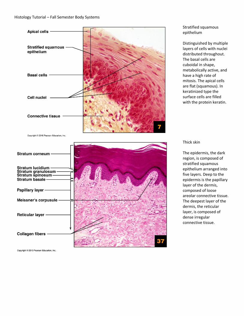
Stratified squamous epithelium Distinguished by multiple layers of cells with nuclei distributed throughout. The basal cells are cuboidal in shape, metabolically active, and have a high rate of mitosis. The apical cells are flat (squamous). In keratinized type the surface cells are filled with the protein keratin.
Thick skin The epidermis, the dark region, is composed of stratified squamous epithelium arranged into five layers. Deep to the epidermis is the papillary layer of the dermis, composed of loose areolar connective tissue. The deepest layer of the dermis, the reticular layer, is composed of dense irregular connective tissue.

Adipose connective tissue Distinguished by closely packer adipocytes (fat cells) with sparse matrix. Each adipocyte is filled with a large fat droplet causing the nucleus to be pushed to the edge of the cell.
Areolar connective tissue Matrix contains all three fiber types (collagen, elastic and reticular) within a gel-like ground substance.

Compact bone Tissue composed of hard, calcified matrix containing collagen fibers. This densely packed bone tissue is organized in lamellae (layers of bone tissue) and osteons (concentric rings of bone).
Hyaline cartilage Cartilage cells (chondrocytes) located within spaces (lacunae) in the tissue matrix. The matrix is a firm, gel-like ground substance embedded with collagen fibrils (not visible)

Elastic cartilage As in hyaline cartilage, the chondrocytes sit in spaces (lacunae) within the tissue matrix. The matrix contains a firm gel-like ground substance and both collagen fibrils and elastic fibers.
Fibrocartilage The gelatinous matrix is densely packed with thick collagen fibers. Chondrocytes are located in lacunae. This feature distinguishes this tissue from dense irregular connective tissue. This is the strongest type of cartilage.

Skeletal muscle Long cylindrical cells (fibers), multinucleated, obvious striations running perpendicular to fiber direction.
Dense regular connective tissue Densely packed fibers, primarily collagen, arranged parallel to each other. The nuclei of the fibroblasts are also aligned in parallel. This is an important feature for differentiating this tissue from smooth muscle.
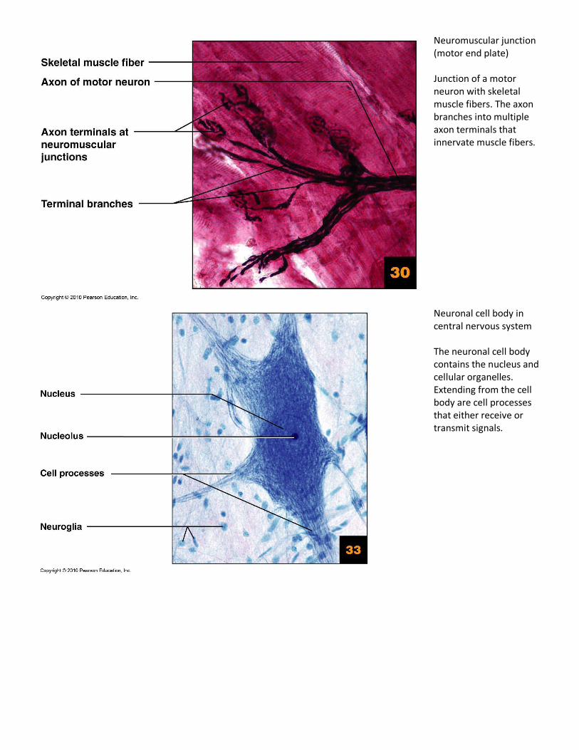
Neuromuscular junction (motor end plate) Junction of a motor neuron with skeletal muscle fibers. The axon branches into multiple axon terminals that innervate muscle fibers.
Neuronal cell body in central nervous system The neuronal cell body contains the nucleus and cellular organelles. Extending from the cell body are cell processes that either receive or transmit signals.