histology - jufilesjufiles.com/wp-content/uploads/2016/05/06.animal-tissue.lab... · histologyis...
TRANSCRIPT

is the study of tissuesHistology
A TISSUE is a group of cells, usually of one kind, & their intercellular substance (e.g. intercellular matrix in animal) which are linked together & perform a particular function
Tissues organs system organism
pg151
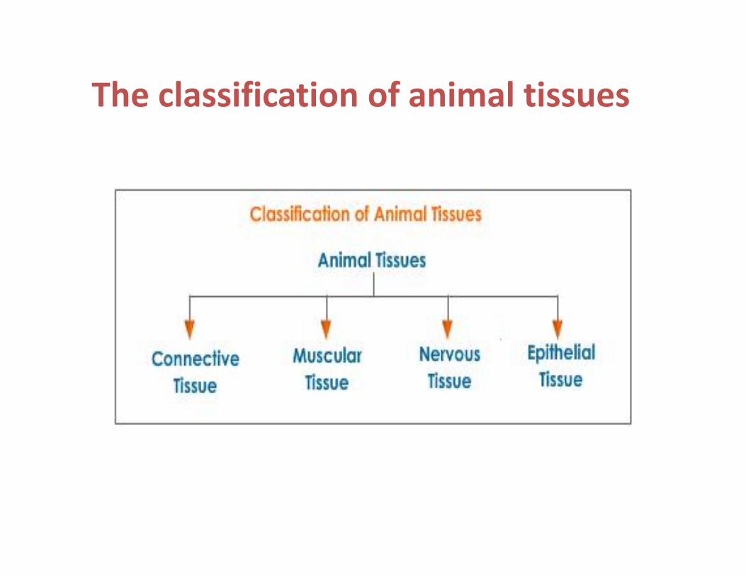
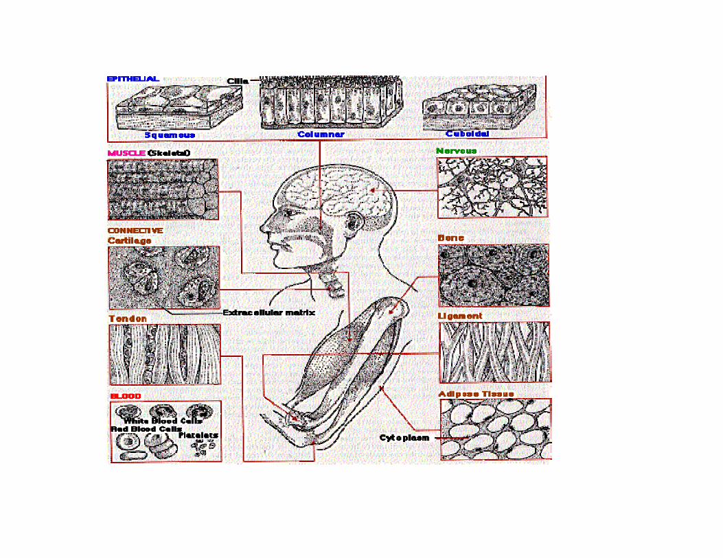
The classification of animal tissues
Epithelial tissues•Arranged in single or multilayered sheets•Cells tightly junked together
•Covered the internal and external surfaces ofthe body Lines, covers, and protects other tissuesand organs.
•Lack blood vessels
-epithelium shows a very rapid rate of cell
division so that lost cells are replaced speedily.
•pg152

*Have one free surface*Bottom layer of cells rest on a basement membrane consisting of a network of collagenous fibrespg152

Epithelial tissuesFunctions:
- protect underlying structures from injury through
pressure and from infection.
-secretion
-absorption
-sensation
-reproductive
Pg 152
**True epithelium is classified into
•simple (one cell thick) and
•compound (more than one cell thick)epithelium
Named by Cell shape
Squamous, Cuboidal, and Columnar
pg153

Squamous EpitheliumCells very thin, much wider than they are thick.
Thin, flattened and contain little cytoplasm.
The margins of the cells are irregular (tessellated)
EpitheliumSquamousSimple Air sacs of respiratoryLining of blood vessels, heart and lymphatic tubes
EpitheliumSquamousStratified SkinEsophagusMouthPg 154
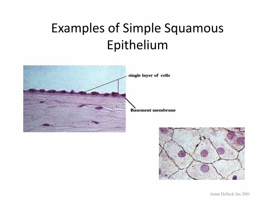
Examples of Simple Squamous Epithelium
Jenna Hellack Jan 2001
Cuboidal Epithelium SimpleCells cube shaped-functions-secretion and absorption.site-Kidney tubules-Duct and small glands-Surface of ovary
•pg154
Jenna Hellack Jan 2001
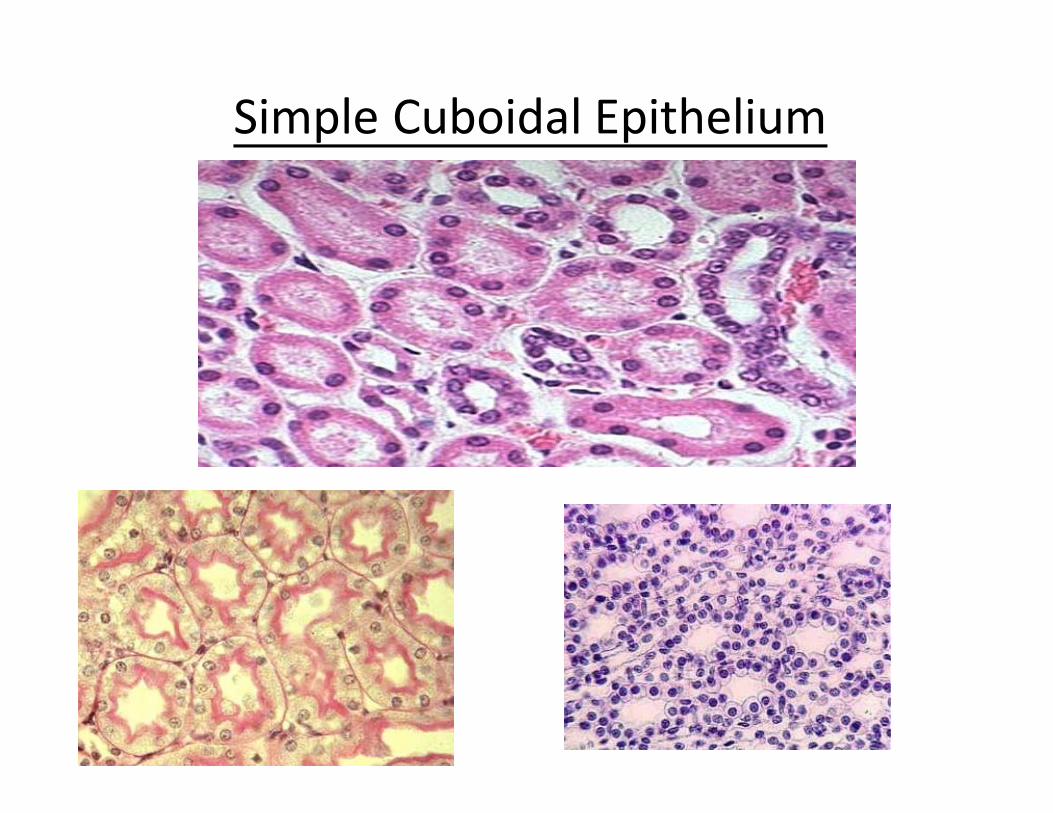
Cuboidal Epithelium Simple

Connective Tissue_Characterized by the cells widely separated from each other in a matrix that is produced by the cells.-Tissue protects and supports.-Cell Matrix composed of two regions1-GroundLiquid (sol), Gel, Gum or solid2-FibersNon-elastic (= white or Collagen)Elastic (= yellow fibers)Pg 157
Jenna Hellack Jan 2001
Types of Connective Tissue
) Connective TissueAreolarLoose (Dense Connective TissueAdiposeCartilageBoneBlood
Jenna Hellack Jan 2001
Connective tissues
1-Loose connective tissue - Areolar tissue •contains a mixture of mucin, hyaluronic acid. Scattered throughout are numerous wavy bundles of collagen fibres and elastin Gel like ground -interspersed in the matrix are a variety of different cell types: fibroblasts, macrophages, reticulo-endothelial system and mast cells
siteWraps and cushions organsUnder the skinpg158
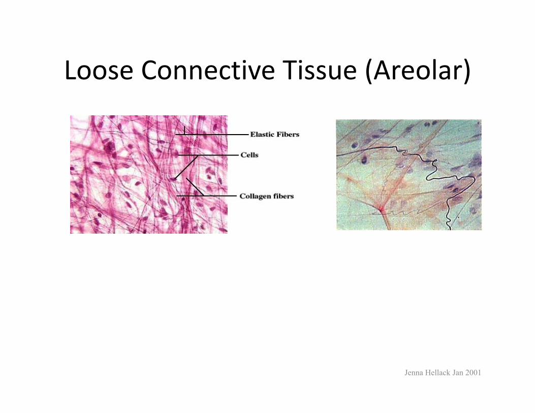
Loose Connective Tissue (Areolar)
Jenna Hellack Jan 2001
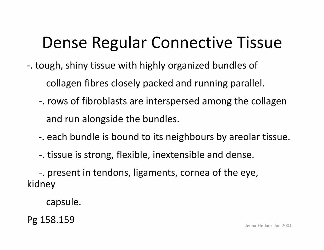
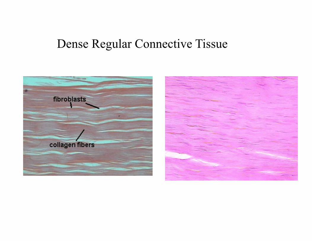
Dense Regular Connective Tissue-. tough, shiny tissue with highly organized bundles of
collagen fibres closely packed and running parallel.
-. rows of fibroblasts are interspersed among the collagen
and run alongside the bundles.
-. each bundle is bound to its neighbours by areolar tissue.
-. tissue is strong, flexible, inextensible and dense.
-. present in tendons, ligaments, cornea of the eye, kidney
capsule.
Pg 158.159Jenna Hellack Jan 2001
Dense Regular Connective Tissue
Adipose (Fat) Found in dermis of the skin, mesenteries and around the kidneys and heart
Jenna Hellack Jan 2001
Functions:Tissue provides energy reserve, acts as a shock absorber and insulates against heat loss. storage cells for adipose

•has no specific matrix and contains large number of fatcells arranged into lobules.
•each cell is filled almost entirely by a central fat dropletwhich squeezes the cytoplasm and nucleus to the periphery.
Adipose tissue
Adipose cells contain a large vacuole which in the live cell contains lipids.

Adipose tissue
Vascular Tissue (Blood)is a connective tissue of cells separated by a Blood
. Two types of cells occur. Red ) matrixplasma liquid (blood cells (erythrocytes) carry oxygen. White blood cells (leukocytes) function in the immune system. Plasma transports dissolved glucose, wastes, carbon dioxide and hormones, as well as regulating the water balance for the blood cells. Platelets are cell fragments that function in blood clotting.
Pg 159-160
Jenna Hellack Jan 2001
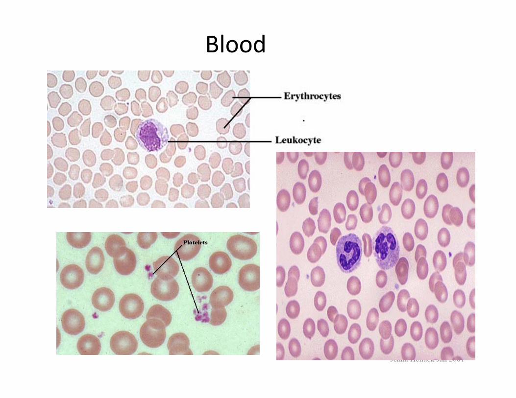
Blood
Jenna Hellack Jan 2001


CartilageGround of matrix is gum like.Cells are found in Lacunae within the matrix.Fibers may be elastic or non-elastic, or a form of non-elastic called reticular(where the non-elastic fibers of very thin)
:Types of cartilages
Hyaline cartilage: occurs in larynx, trachea and bones of embryo.
pg161
Jenna Hellack Jan 2001

Constists of cells embedded in a matrix of chondrin which is deposited by chondroblasts
The matrix consists of many fibrils and collagen
The space called lacunae encloses chondroblasts
Cartilage is hard but flexible tissue and can resist any strains. The matrix is also compressible and elastic.
pg161
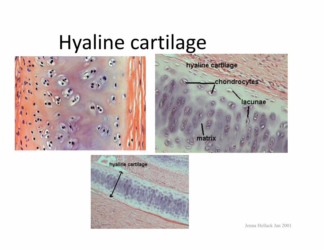
Hyaline cartilage
Jenna Hellack Jan 2001

Ground of matrix is Solid (Calcium carbonate). Organic matrix The matrix of compact bone consists of bone collagen, hydroxyapatite, magnesium carbonates, sodium carbonates and nitrates.with small amount of white fibre. Pg 162 ,163
Has blood supply and nerves running through the Haversian canal systems.
Jenna Hellack Jan 2001
Compact bone
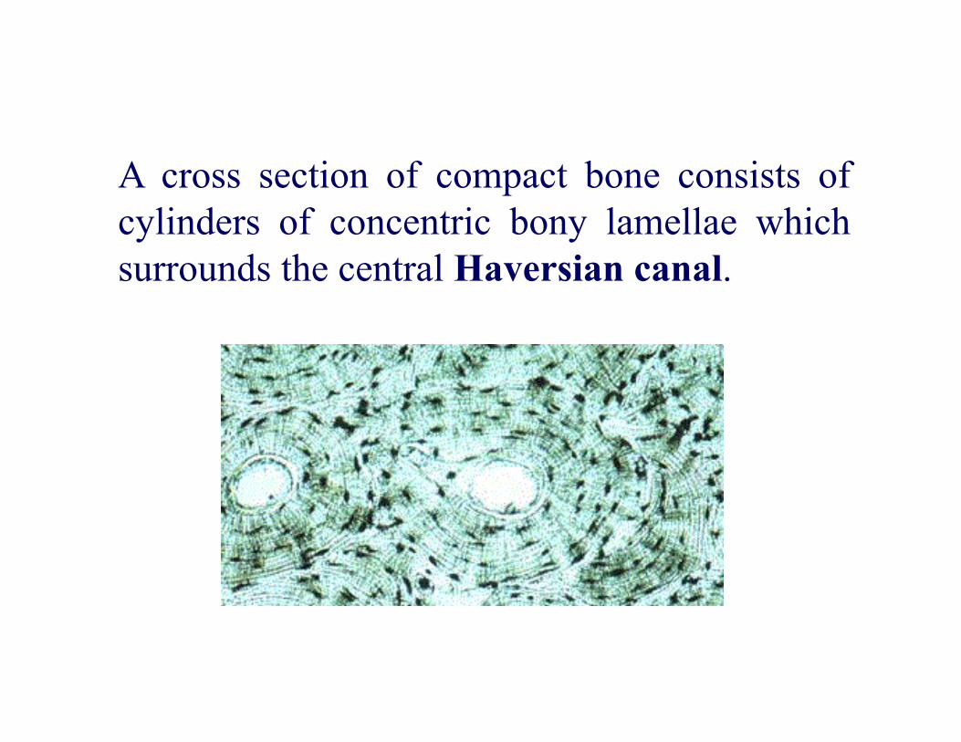
A cross section of compact bone consists ofcylinders of concentric bony lamellae whichsurrounds the central Haversian canal.

Interspersed between the lamellae are numerous lacunae containing bone cells called osteoblasts which are then termed as osteocytes when they are not active.
Radiating from each lacunae are many fine channels called canaliculi which contain cytoplasm and link the central Haversian canal.
pg163
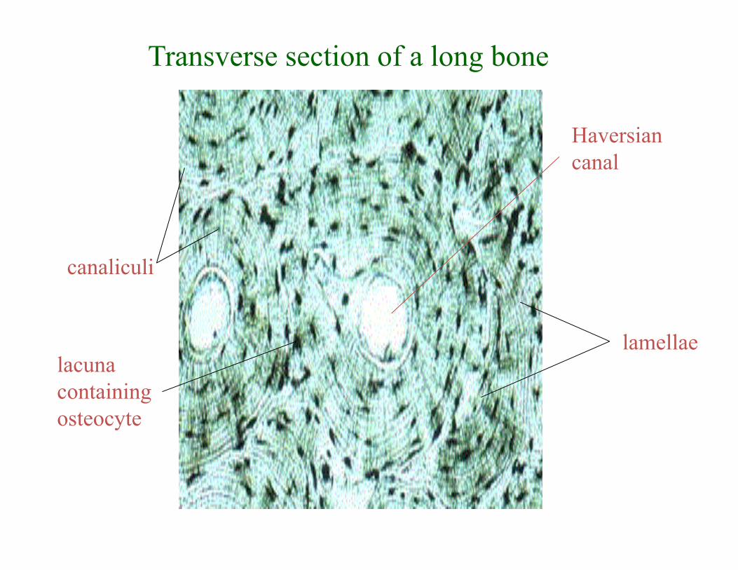
Haversian canal
lamellae
canaliculi
lacuna containing osteocyte
Transverse section of a long bone

Cross section of compact bone
Muscle TissueTissue with cells having fibers specialized for contraction.
(Striated, voluntary) Skeletal MuscleParallel elongated cells (fibers) are subdivided into sarcomeres.
multinucleated and each cell is the length of the muscle Svariable positions near the periphery of fibre.
Attached to the skeleton in the trunk , limbs and head.Pg163-164
Jenna Hellack Jan 2001

Skeletal Muscle
L.s Cs

Diagram of the arrangement of muscle fibrils

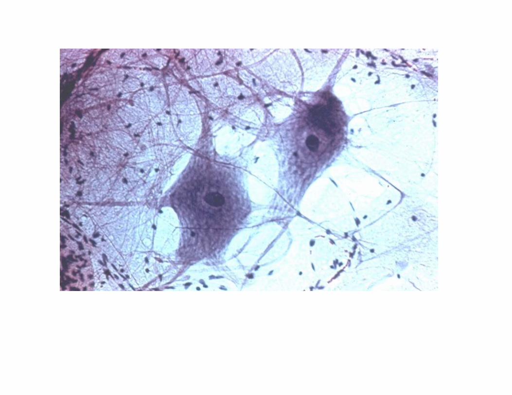
Nerve cell is called neurone.
The main function is the conduction of impulses.
Each neurone consists of a cell body with anucleus, cytoplasm. The cytoplasmic processesextending from the cell body are dendrons (carryimpulses towards the cell body) and axons (carryimpulses away from the cell body).
Pg165-166
Nervous tissue

Diagram of two neurones