histology review
DESCRIPTION
Histological imagesTRANSCRIPT

HISTOLOGY REVIEW SLIDES FROM DR YOUNGS WEBSITE THING

• What Organ Is this? • A • B • C • D Answers • Adrenal Gland • Medulla • Zona Reticulata • Zona Fasiculata • Zona Glomerulosa • Capsule

• What Organ Is this? • A • B • C • D Answers • Adrenal Gland • Endothelial Cells that
line fenestrated capillaries
• Zona Fasiculata- Steroid Producing endocrine cells
• Zona Glomerulosa • Capsule- DICT

• What Organ Is this? • A • B • C • D Answers • Adrenal Gland • Medulla- Norepi Cells • Medullar- Epi cells • Cell containing lipid
droplets for synthesizing steroid Hormones
• Endothelial cell from capillary in Zona Reticularis

• What Organ Is this? • A • B • C Answers • Adrenal Gland- Medulla • Neuronal Cell/ Ganglion Cell- Large
euchormatic nucleus and prominent Nucleolous
• Chromaffin cells- Secrete Norepi and Enkephalin
• Lumen of dialated venule

• What Organ Is this? • A • What structure do C & B make? • B • C Answers • Pancreas • Acinar Cells • Islet of Langerhan • Capillaries in Islet of Langerhan • Probably Beta Cells producing isulin- due to
the central location in I of L

• What Organ Is this? • A • B • C • D • E Answers • Thyroid Gland • Skeletal Muscle tissue- probably sterno hyoid • Connective tissue • Thyroid Gland • Dorsal Connective Tissue • Tracheal Cartilage- Hyaline Cartilage • Respiratory Epithelium- Mucos Glands seen
below

• What Organ Is this? • A • B • C • D • E Answers • Thyroid Gland • Colloid • Follicular Cell • Parafollicular Cell • Mast Cell • Fibroblast

• What Organ Is this? • A • B • C • D • E Answers • Parathyroid Gland (Top) & Thyroid Gland
(Bottom) • Parathyroid Gland • Capsule of Connective tissue for PTG • Thyroid Follicle in Thyroid Gland

• What Organ Is this? • A • B • C • D Answers • Parathyroid • Chief/ Principal Cells • Oxyphil Cells • Fibrobalsts • Adipose Cells in parathyroid gland

• What Organ Is this? • A • B • C Answers • Anterior Pituitary • Acidophil Cells
• Produce Prolactin and GH • Basophilic Cells of AP
• Produce FSH, LH, TSH, ACTH • Basal Laminae surrounding Capillaries

• What Organ Is this? • A • B • C Answers • Intermediate Pituitary/ Posterior Pituitary • Cells of Intermediate Pituitary • Pituicytes- Specialized glial cells • Herring Body- Axonal swelling containing
Homrones

• What Organ Is this? • A • B • C Answers • Intermediate/ Posterior Pituitary • Intermediate Pituitary Cells • Pituicytes • Herring Body/ Hormone accumulations
• Hormones made in Hypothalamus

• What Organ Is this? • A • B • C • D Answers • Kidney • Renal Corpuscle/ Bowmans Capsule • Portions of Proximal Convoluted Tubule
• Occluded lumen • Intralobular Artery • Medullary Rays- straight portion of Proximal
Tubule

• What Organ Is this? • A • B • C • D • E Answers • Kidney • Proximal Convoluted Tubule
• Taller cells, have uneven surfaces • Viceral Layer of Bowmans capsule
• Podocytes • Parietal Layer of Bowmans Capsule
• Simple squamous • Arteriole • Distal Convoluted Tubule
• Lower cuboidal and have smoother appearing surfaces

• What Organ Is this? • A • B • C • D • E • F • G Answers • Kidney • Proximal convoluted tubule • Distal convoluted tubule
• Shorter cells and clear lumen that lack tall microvilli
• Parietal Layer of Bowmans capsule • Afferent Arteriole • Distal Convoluted tubule
• Specifically marks MACULA DENSA cells • Juxtaglomerular Cells
• Smooth muscle cells of Afferent Arteriole
• Vascular Space within Glomerulus

• What Organ Is this? • A • B • C Answers • Kidney- Medulla • Cell of Collecting Duct • Blood Vessel of Vasa Recta
• Round cells are RBCs • Lumen of thin segment of Loop of Henle
• Maybe cant really tell

• What Organ Is this? • A • B Answers • Kidney- Medulla • Cells in Terminal Portions of Collecting Ducts
called Papillary ducts of Bellini- • Drain urine into a minor calyx
• Loose Connective Tissue

• What Organ Is this? • A • B Answers • Kidney
• Special stain prep shows proximal tubule cells maintained in culture and stained using flourecent stained antibody to epithelial intermediate filament cytokeratin.
• Nucleus • Desmosome

• What Structure Is this? • A • B • C • D • E Answers • Ureter • DICT • Smooth muscle tissue • Highly cellular LCT • Transitional Epithelium • White adipose tissue/ Adipocytes

• What Structure Is this? • A • B • C • D • E Answers • Testis
• Overall the appearance of a Seminiferous tubule
• Maturing Spermatozoa • Immature Spermatids
• Just completed second meiotic division • Leydig cells
• Between tubules • Spermatogonia • Fibroblasts

• What Structure Is this? • A • B • C • D • E Answers • Testis
• Detailed view of Seminiferous tubule • Sertoli Cells
• Maintain blood testi barrier • Pale nuclei, prominent nucleoli
• Primary spermatocytes with condensed chromosomes
• Spermatogonia • Leydig cells
• Lipid droplets • Pale, round nuclei
• Spermatids • Haploid number of chromosomes
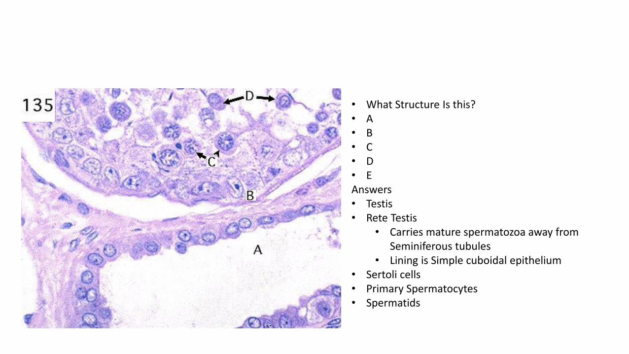
• What Structure Is this? • A • B • C • D • E Answers • Testis • Rete Testis
• Carries mature spermatozoa away from Seminiferous tubules
• Lining is Simple cuboidal epithelium • Sertoli cells • Primary Spermatocytes • Spermatids

• What Structure Is this? • A • B • C Answers • Testis • Structures that carry spermatozoa away form
seminiferous tubules • Lumen of epididymis • Lumen of Efferent Ductule
• Has cells with varying heights in lumen give it an irregular festooned outline

• What Structure Is this? • A • B • C • D • E Answers • Epididymis • Maturing spermatozoa • Stereocilia- long microvilli • Principle Cells
• Secrete proteins which facilitate sperm maturation (capacitation)
• Basal cells • Function as stem cells
• Fibroblasts

• What Structure Is this? • A • B • C Answers • Vas Deferens • Lining Epithelium of Vas Deferens • Smooth muscle tissue
• Very thick, distinguishing characteristic • DICT

• What Structure Is this? • A • B • C • D • E Answers • Vas Deferens • Spermatozoa Cells • Tall microvilli at apical surface • Pseudostratified columnar epithelium • Lamina protria layer
• LCT • Smooth muscle tissue
• Muscular nuclei are located within the center of each cell

• What Structure Is this? • A • B Answers • Seminal Vesicle • Lining epithelium
• Simple Columnar epithelium • Can vary to pseudostratified columnar • Dependent on testosterone
• Layer of DICT

• What Structure Is this? • A • B • C • D • E Answers • Prostate • Prostatic urethra • Ejaculatory Ducts • Prostati utricle
• Represents underdevelopted male homologue of Uterus
• Secretory epithelium of the prostate forms acini • Produce watery, alkaline secretion rich in
proteins, including the prostate specific antigen
• Serine protease, is overproduced by cancerous prostate glands, its detection in blood can be used as an indication of a malignant state
• Prostatic concretions • Condensed secretions

• What Structure Is this? • A • B • C • D • E • F Answers • Ovary • Epithelial Covering called Germinal
epithelium • Simple cuboidal
• Primordial Follicle • Theca Externa • Theca Interna Layer • Ganulosa Cells
• Produce fluid that accumulates to form Antrum
• Antrum

• What Structure Is this? • A Answers • Ovary
• Showing a follicle that has not been select for further development. Follicle is slowly degenerating
• Cells undergoing Apoptosis • Highly condensed nuclei containing DNA
that is being cleaved into fragments by apoptotic enzymes

• What Structure Is this? • A • B • C Answers • Ovary • Cytoplasm of a primary oocyte
• Cell plus adherent to it constitute a structure called a primordial follicle
• Ovarian stroma • LCT
• Simple squamous epithelial tissue • Diagnostic for this type of follicle

• What Structure Is this? • A • B • C • D • E • F Answers • Ovary
• Primary Follicle • Cytoplasm of a Primary Oocyte cell • Zona Pellucida
• Composed of at least 3 glycoproteins secreted by the oocyte
• Granulosa Cells • Statified columnar epithelium
• Theca interna layer • Function to synthesize steroids
• GERNIMAL EPITHELIUM • Simple cuboidal epithelial cells covering
the surface of the ovary termed GERNIMAL EPITHELIUM
• Immature primordial Follicle

• What Structure Is this? • A • B • C Answers • Ovary • Germinal epithelium • Granulosa cells • Oocyte Cell • Antrum
• Fluid filled cavity • Corpus Luteum
• Collapsing remains of follicle after ovulation and expulsion of an oocyte

• What Structure Is this? • A • B • C • D • E Answers • Ovary • Oocyte • Zona Pellucida
• Separate oocyte from surrounding cells • Antrum
• Has fluid called liquor folliculi • Granulosa cells
• Modified by liquor folliculi • Cells at D undergoing mitosis • Theca interna
• Synthesize molecules like pregnolone • Theca externa
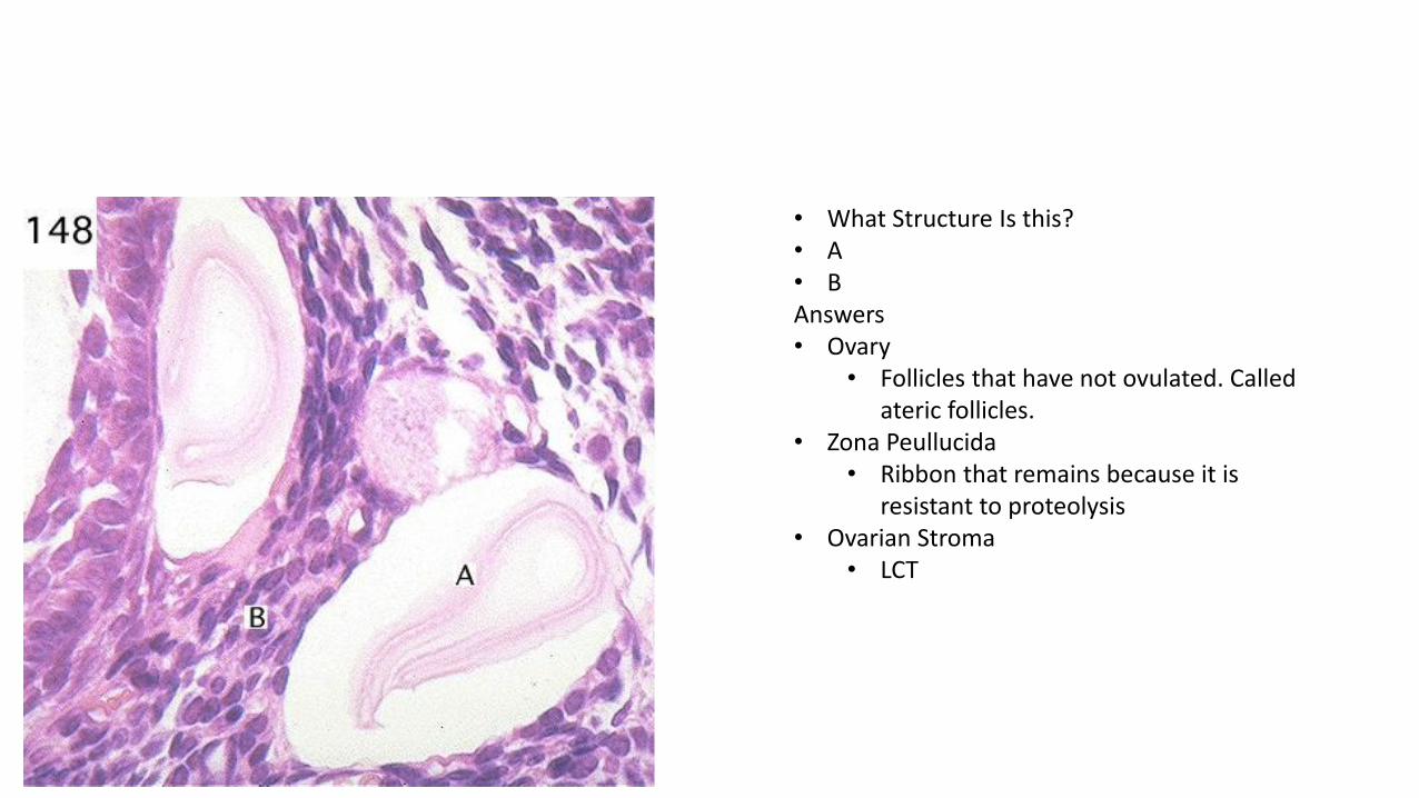
• What Structure Is this? • A • B Answers • Ovary
• Follicles that have not ovulated. Called ateric follicles.
• Zona Peullucida • Ribbon that remains because it is
resistant to proteolysis • Ovarian Stroma
• LCT

• What Structure Is this? • A • B • C Answers • Corpus Luteum • Theca Lutein • Granulosa Lutein cells
• Derived from the granulosa layer
• Connective tissue derived from Ovarian Stroma

• What Structure Is this? • A • B • C Answers • Corpus Luteum • Theca Lutein • Granulosa Lutein cells • Fribroblasts

• What Structure Is this? • A • B • C Answers • Oviduct
• Irregular lumen formed by highly folded epithelial layer
• Simple Columnar epithelium • Smooth muscle tissue • Connective tissue probably LCT

• What Structure Is this? • A • B • C Answers • Uterus
• Two main layers of the uterus • Endometrium
• Epithelium extends downward to form many glands surrounded by LCT
• Glands • Tortuous appearance of the glandular
profiles at B indicate that this tissue was obtained during the secretory phase of menstrual cycle.
• Myometrium • Smooth muscle
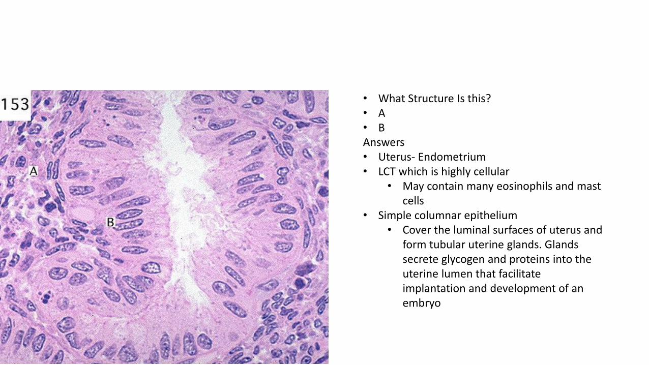
• What Structure Is this? • A • B Answers • Uterus- Endometrium • LCT which is highly cellular
• May contain many eosinophils and mast cells
• Simple columnar epithelium • Cover the luminal surfaces of uterus and
form tubular uterine glands. Glands secrete glycogen and proteins into the uterine lumen that facilitate implantation and development of an embryo

• What Structure Is this? • A • B Answers • Uterus • Blind cysts termed Nabothian Cysts
• Aggregation of epithelial cells. Found in the region of the uterine cervix
• Connective tissue

• What Structure Is this? • A • B • C • D Answers • Mammary gland • White adipose cells • DICT • LCT
• Surrounding lactiferous ducts • Stratrafied cuboidal epithelium

• What Structure Is this? • A • B • C Answers • Mammary Gland
• Non Lactating • Cells of Terminal duct- Stratified Columnar
Epithelium • Fibroblast • Plasma Cell
• Synthesize secretory IgA for delivery of this protein via transcytosis across the epithelium into milk

• What Structure Is this? • A • B • C • D • E Answers • Mammary Gland
• Lactating, shows enlargement and differentiation of mammary secretory alveoli
• Lumen of Lactiferous Duct • LCT • Secretory Alveoli

• What Structure Is this? • A • B Answers • Placenta • Chorionic Villus • Basal plate

• What Structure Is this? • A • B • C • D Answers • Placenta • Cytotrophoblast layer
• Mitotically active stem cells for the outermost layer at B
• Syncytiotrophoblast • Form placental barrier isolating the fetus
from many maternal molecules as well as transporting nutrients to the fetus and synthesizing hormones like steroids and hCG
• Labels a fibroblast in extraembryonic mesenchyme
• Embryonic blood vessels

• What Structure Is this? • A • B • C • D • E Answers • Esophagus • Lining Epithelium-Stratified Squamous
Epithelium • Lamina Propria • Muscularis Mucosa- Smooth muscle • DICT- Submucosal Layer • Muscularis Externa

• What Structure Is this? • A • B • C Answers • Esophagus • Non Keratinized Stratified Squamous
Epithelium • DICT • Small Portion of Muscularis Mucosa Layer –
smooth muscle tissue

• What Structure Is this? • A • B • C • D Answers • Stomach- Stomach lining • Mucous neck cells
• Closest to the lumen and form the openings of gastric glands
• LCT of the lamina propria layer of the stomach
• Parietal Cells • Produce acid and Intrinsic factor (for
B12) • Chief Cells
• Produce peptin and renin

• What Structure Is this? • A • B • C • D • E Answers • Stomach • Parietal cells • Enteroendocrine cell (APUD) • Smooth muscle cells of the Muscularis
Mocosae • Chief Cells • Arteriole in submucosa layer

• What Structure Is this? • A • B • C • D • E • F Answers • Small Intestine • Outermost muscularis external • Submucosa DICT • Muscularis Mucosae • Lymphatic Capillary (central Lacteal) • LCT of lamina propria • Intestinal Villus

• What Structure Is this? • A • B • C • D • E • F Answers • Small Intestine • Muscularis Externa Layer • Nerve Cells- Submucosal Plexus/ Meissners
Plexus • Muscularis Mucosae Layer • Lamina Propria • Epithelial cells of intestinal crypts • Lympatic capillary

• What Structure Is this? • A • B • C • D • E Answers • Small intestine • Simple columnar epithelium • Goblet cells • Smoth muscle cells • Plasma cells
• Abundant cytoplasm and pale staining golgi regions
• M cell • Transport antigen molecules form
intestinal lumen to present them to lymphocytes
• Lymphocytes

• What Structure Is this? • A • B • C • D Answers • Small Intestine • Lymphocytes migrating through epithelium • Simple Columnar epithelium • Goblet Cell • Microvilli- increase surface area and promote
absorption

• What Structure Is this? • A • B • C • D • E Answers • Small intestine • Smooth muscle cells of Muscularis mucosae • Enteroendocrine cells (APUD)
• Secretes hormones • Cell division of epithelial cells
• Dark staining nuclei • Submucosae
• Contains blood vessels and fibroblasts • Plasma Cells

• What Structure Is this? • A • B • C • D Answers • Small intestine • Paneth Cells
• Secrete lysozyme and defensing protect the intestine agains pathogenic bacteria
• Goblet cell in intestinal epithelium • Mast Cell • Smooth muscle in muscularis mucosae

• What Structure Is this? • A • B Answers • Small intestine • Mitotic figures of intestinal epithelium
• Chromosomes arranged in anaphase portion of mitosis
• LCT of lamina propria layer

• What Structure Is this? • A • B • C • D • E Answers • Small intestine • Brunner's glands
• Secrete alkaline mucus that protects the lining of the intestine from the stomach acid carried along with a meal
• Distinctive feature of duodenum • Muscularis mucosal layer • Epithelium

• What Structure Is this? • A • B • C Answers • Small intestine • Smooth muscle- circular
• Nuclei is in the center • Nerve cells- myenteric plexus • Smooth muscle cells of the muscularis
externa are cut in the longitudinal section

• What Structure Is this? • A • B • C • D • E Answers • Large Intestine • Muscularis externa layer • Submucosae • Muscularis Mucosae • Lamina Propria • Simple columnar epithelium
• Lack villi

• What Structure Is this? • A • B • C • D • E Answers • Gall Bladder • Simple Columnar epithelium • LCT
• Abundant fibroblasts and blood vessels
• Smooth muscle fibers • Provide for contraction of the gall
bladder when stimulated by the hormone CCK

• What Structure Is this? • A • B Answers • Gall Bladder
• Simple Columnar epithelium • Fibroblast cells in LCT

• What Structure Is this? • A • B • C • D • E Answers • Pancreas • Enteroacinar cells • Basal portion of the cells very basophilic due
to the presence of RER • Golgi apparatus- lighter staining • Beginning of intercalated duct
• Lined with simple cuboidal epithelium • Surrounding ligh areas around the acini
contain vessels

• What Structure Is this? • A • B • C • D • E Answers • Liver • Hepatocyte (parenchymal Cell) • Kuppfer cells • Central Vein
• Collects blood from the sinusoids that radiate out from it

• What Structure Is this? • A • B • C Answers • Liver • Central Vein • Hepatocyte • Kuppfer cell

• What Structure Is this? • A • B • C • D Answers • Liver • Portal vein • Hepatic artery • Bile duct
• Lining epithelium is simple columnar • Hepatocyte

• What Structure Is this? • A • B • C • D • E Answers • Liver • Hepatic Artery • Bile Duct • Hepatocyte • Endothelial cells of the sinusoids • Small bile duct
• Lining is simple cuboidal epithelium

• What Structure Is this? • A • B • C • D • E Answers • Liver • Hepatocyte • Light channel
• Probably bile canaliculus • Kuppfer cell • Ito cell
• Stores vitamin A can see the clear lipid droplets
• Endothelial cell

• What Structure Is this? • A • B Answers • Liver- PAS Stain • Central Vein • Ito Cell
• Doesn’t stain because it doesn’t have glycogen

What is the Organ? Terminal bronchiole A? Non-ciliated Clara cell B? Type II Pneumocyte C? Smooth Muscle cell (cut in cross section)
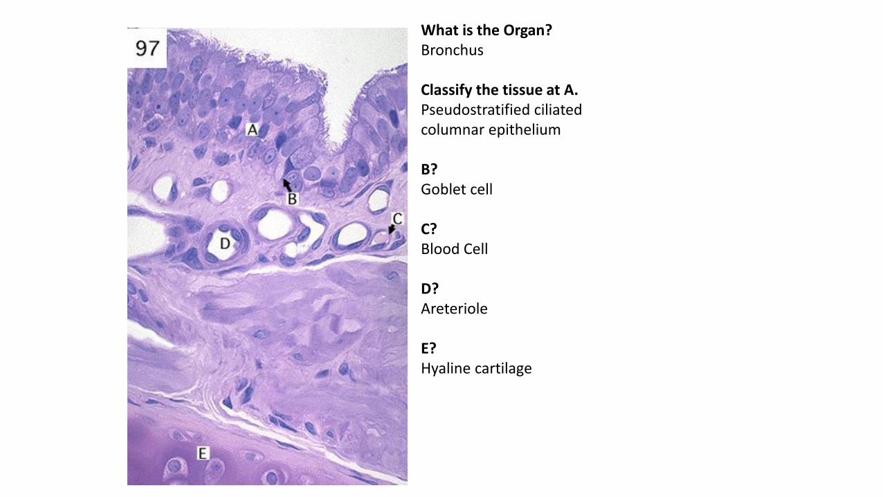
What is the Organ? Bronchus Classify the tissue at A. Pseudostratified ciliated columnar epithelium B? Goblet cell C? Blood Cell D? Areteriole E? Hyaline cartilage

What is the Organ? Lung A? Type II pneumocyte Function of cell at B? Phagocytosis (it’s an alveolar macrophage/dust cell)
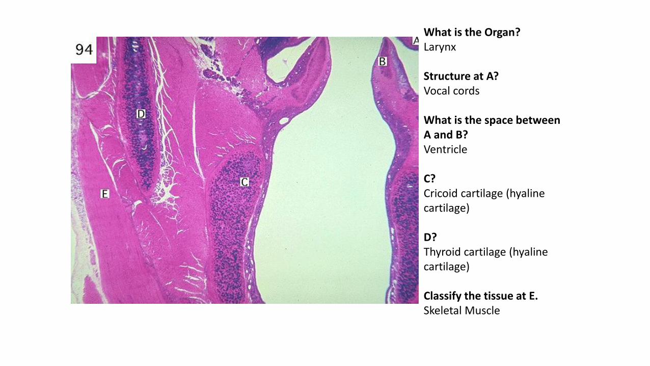
What is the Organ? Larynx Structure at A? Vocal cords What is the space between A and B? Ventricle C? Cricoid cartilage (hyaline cartilage) D? Thyroid cartilage (hyaline cartilage) Classify the tissue at E. Skeletal Muscle

What is the Organ? Trachea Classify the tissue at A. Nerve Classify the tissue at B. Adipose tissue Classify the tissue at C. Hyaline cartilage D? Vein

What is the Organ? Aveoli A? Type II pneumocyte B? Capillary Classify the epithelium at C. Simple cuboidal Classify the tissue at D Smooth muscle E is the ____ of a Type I pneumocyte through which gasses diffuse for respiration. Attenuated cytoplasm

What is the Organ? Lung (with surrounding bronchus tissue) A? Muscular artery B? bronchus C? Terminal bronchiole D? Respiratory bronchiole E? Alveoli

What is the Organ? Tongue Classify the tissue at A. Stratified Squamous Keratinized Epithelium B? Taste bud C? Areteriole Classify the tissue at D. Skeletal muscle Classify the tissue at E. Loose Connective Tissue (of Lamina Propria)

What is the Organ? Tongue Classify the tissue at A. Keratinized stratified squamous epithelium. What is the name of the structure at A? Filiform papilla Classify the tissue at B? DICT

What is the Organ/Gland? Minor salivary gland A? Serous demilume The cells at B have ___-containing cytoplasmic granules that do not stain intesely after exposure to routine stains. Mucin

What is the Organ/Gland? Parotid gland A? Serous acinus B? Striated duct The structure at B is lined by which kind of epithelium? Simple cuboidal The parotid gland is unique among salivary glands in that it contains no _____. Mucous acini

What is the Organ/Gland? Sublingual Gland A? Striated duct B? Mucous acinus Classify the epithelium at C. Interlobular duct

What is the Organ/Gland? Sublingual Gland A? Serous acinus B? Mucous acinus C? Striated duct D? White fat cell E? Interlobular duct Classify the tissue at E and the tissue surrounding it. Stratified columnar, surrounded by DICT.

What is the Organ/Gland? Taste bud What kind of papillae are visualized here? Circumvalate papillae

What is the Organ/Gland? Von Ebner’s Gland A? Taste buds Classify the tissue at B. LCT Classify the tissue at C. DICT Classify the tissue at D Skeletal muscle Classify the tissue at E Stratified columnar epithelium

What is the Organ? Kidney (medulla) Structure at A? Collecting duct B? Blood vessel of a vasa recta C is possibly the lumen of a ____. Loop of Henle (thin segment) OR vasa recta

What is the Organ? Kidney (cortex) Structure at A? Renal corpuscle Structure at B? Portions of proximal convoluted tubule (note occluded lumen) C? Intralobular artery D is part of the _____. Medullary rays. The most common tubular component shown is the _____. Straight portion of proximal tubule.

What is the Organ? Kidney Structure at A? Terminal portions of collecting ducts/papillary ducts Where do the papillary ducts drain? Minor calyx Classify the tissue at B. LCT

What is the Organ? Kidney Structure at A? Proximal convoluted tubule B and C point to the _____ and _____ layers of the Bowman’s capsule. B: visceral, C:parietal Classify the tissue at C. Simple squamous Podocytes are located at __. B

What is the Organ? Kidney Tall cells of the proximal convoluted tubule are identified at __. A C labels the _____ layer of Bowman’s capsule. parietal B and E label _____. Distal convoluted tubules. Note clear lumen and lack of border microvili. What kind of cell does the arrow at E point to? Macula densa cell What do the cells at F secrete? Renin. They’re JG cells.

What is the Organ? Ureter Classify the tissue at A. DICT Classify the tissue at B. Smooth muscle Classify the tissue at C. LCT Classify the tissue lining the lumen of this structure. Transitional epithelium Classify the tissue at E. Adipose

What is the Organ? Urinary bladder Classify the tissue at A Transitional epithelium Classify the tissue at B LCT