histology - ksu facultyfac.ksu.edu.sa/sites/default/files/1_histology.pdf · eder, et al.:...
TRANSCRIPT

Eder, et al.: Laboratory Atlas of Anatomy and Physiology, Third Edition
1. Histology Text © The McGraw−Hill Companies, 2001
1
1Histology
Simple Cuboidal Epithelium
C H A P T E R

Eder, et al.: Laboratory Atlas of Anatomy and Physiology, Third Edition
1. Histology Text © The McGraw−Hill Companies, 2001
2 C H A P T E R 1
Chromosomes
Spindle fibers
Aster
Figure 1-1Interphase Nuclear membrane intact withchromatin visible. (�250)
Figure 1-2Prophase Duplicated chromosomescondensed into visible strands; nuclearmembrane absent. (�250)
Figure 1-3Metaphase Darkly stained chromosomespositioned by microtubular framework toalign at cell equator. Spindle fibers and aster visible. (�250)
Chromatin
Nuclear membrane
Chromosome

Eder, et al.: Laboratory Atlas of Anatomy and Physiology, Third Edition
1. Histology Text © The McGraw−Hill Companies, 2001
H i s t o l o g y 3
Figure 1-4Anaphase Darkly stained chromosomesmove to opposite poles under microtubularinfluence. Spindle fibers and aster visible.(�250)
Cleavage furrow at equator
Figure 1-5Telophase Separated chromosomes lose microtubular attachments. Belt ofactinomyocin forms at equator, assists information of new cell membranes andcytokinesis. Cleavage furrow forms twodaughter cells. (�250)
Spindle fibers
Asters
Chromosomes

Eder, et al.: Laboratory Atlas of Anatomy and Physiology, Third Edition
1. Histology Text © The McGraw−Hill Companies, 2001
4 C H A P T E R 1
Figure 1-6Simple Squamous Epithelium Single layerof flat cells covering a surface. From humanomentum. (�250)
Figure 1-7Simple Squamous Epithelium Surface viewof flattened cells. Human mesothelium.(�250)
Figure 1-8Simple Cuboidal Epithelium Althoughnot strictly cube shaped, cuboidal cells areroughly equidimensional in length, width,and depth. Single layer of cells liningsurface of kidney tubules. Cross section.(�250)
Nucleus
Simple cuboidal epithelium
Lumen of tubule
Basement membrane (circled)
Fixed macrophage
Simple squamous epithelium
Nucleus
Basement membrane

Eder, et al.: Laboratory Atlas of Anatomy and Physiology, Third Edition
1. Histology Text © The McGraw−Hill Companies, 2001
H i s t o l o g y 5
Figure 1-9Simple Cuboidal Epithelium Longitudinalsection of kidney tubule. (�250)
Figure 1-10Simple Columnar Epithelium Cellularheight is much greater than width or length.Nuclei generally appear in a row. Frompancreatic duct. (�250)
Figure 1-11Pseudostratified Ciliated ColumnarEpithelium Nuclei appear to lie in tworows, but in fact all cells in single layer are in contact with basement membrane.Section shows well-defined cilia, threegoblet cells, basement membrane,underlying connective tissue. From monkey trachea. (�100)
Basement membrane
Simple cuboidal epithelium
Lumen of tubule
Nucleus
Simple columnar epithelium
Basement membrane
Nucleus
Cilia
Goblet cell
Basement membrane

Eder, et al.: Laboratory Atlas of Anatomy and Physiology, Third Edition
1. Histology Text © The McGraw−Hill Companies, 2001
Figure 1-12Pseudostratified Ciliated ColumnarEpithelium Section shows cilia, multiplelayers of nuclei, basement membrane,underlying connective tissue. From humantrachea. (�250)
Figure 1-13Stratified Squamous Epithelium Flattenedcells at surface change to less flattenedmorphology in deeper layers. Oral cavity of rabbit. (�100)
Cilia
Nucleus
Basement membrane
6 C H A P T E R 1

Eder, et al.: Laboratory Atlas of Anatomy and Physiology, Third Edition
1. Histology Text © The McGraw−Hill Companies, 2001
H i s t o l o g y 7
Figure 1-14Stratified Squamous Epithelium Flattened,keratinized cells at surface show variationsin form in deeper layers. From human skin.(�100)
Figure 1-15Transitional Epithelium from Urinary Bladder Umbrella cells stretch and flatten as bladder fills. Basement membraneseparates epithelium from underlyingconnective tissue containing blood vessels.(�250)
Umbrella cell
Basement membrane
Blood vessel lumen
Keratinized cells
Papilla

Eder, et al.: Laboratory Atlas of Anatomy and Physiology, Third Edition
1. Histology Text © The McGraw−Hill Companies, 2001
8 C H A P T E R 1
Figure 1-16Wall of Elastic Artery Extracellularelastic fibers running parallel in a plane.Structure permits tissue elasticity and recoil.From aorta. (�100)
Figure 1-17Reticular Connective Tissue Mesh ofreticular fibers appears as dark lines;provides scaffold for cellular organization of this lymph node. (�250)
Figure 1-18Loose (Areolar) Connective Tissue Pinkbands of collagen fibers run in all directionsthrough intercellular spaces of subcutaneoustissue, permit flexible resistance tomechanical stress. (�100)
Elastic fiber
Reticular fiber
Fibroblast
Elastic fiber
Collagen fibers

Eder, et al.: Laboratory Atlas of Anatomy and Physiology, Third Edition
1. Histology Text © The McGraw−Hill Companies, 2001
H i s t o l o g y 9
Figure 1-19Dense Regular Connective Tissue Bands of collagen fibers running in regular, parallelrows resist mechanical stress mainly alongcourse of fibers. Monkey tendon. (�250)
Figure 1-20Dense Irregular Connective Tissue Bands of collagen running in irregular rows givemultidirectional tensile strength. Collagen-secreting fibroblasts appear throughout.(�100)
Figure 1-21Adipose Tissue Large, empty, polyhedralvacuoles dominate small, eccentricallylocated cell nuclei of adipocytes. Finecapillaries run through tissue. (�100)
Collagen fibers
Nucleus of fibroblast
Nuclei of fibroblasts
Collagen fibers
Vacuole
Nucleus
Capillary

Eder, et al.: Laboratory Atlas of Anatomy and Physiology, Third Edition
1. Histology Text © The McGraw−Hill Companies, 2001
10 C H A P T E R 1
Figure 1-22Fibrocartilage Cell nests of chondrocytesin territorial matrix surrounded by coarseextracellular fibers. (�250)
Figure 1-23Hyaline Cartilage During interstitialgrowth, cartilage cells often form smallclusters and move apart as they secreteextracellular matrix. (�100)
Lacunae
Collagen fibers
Chondrocyte
Chondrocytes
Matrix
Lacunae

Eder, et al.: Laboratory Atlas of Anatomy and Physiology, Third Edition
1. Histology Text © The McGraw−Hill Companies, 2001
H i s t o l o g y 11
Figure 1-24Hyaline Cartilage Artifactual vacuolationforms characteristic lacunae aroundchondrocyte cell bodies. From trachea.(�250)
Figure 1-25Elastic Cartilage Extracellular matrixcontains elastic fibers that confer elasticrecoil to this tissue. (�250)
Lacuna
Chondrocyte
Chondrocyte
Elastic fiber
Lacunae

Eder, et al.: Laboratory Atlas of Anatomy and Physiology, Third Edition
1. Histology Text © The McGraw−Hill Companies, 2001
12 C H A P T E R 1
Figure 1-26Skin Thick, keratinized, multilayeredstratum corneum rests atop grainy stratumgranulosum (stratum lucidum not clearlyevident). Stratum spinosum, composed ofirregularly shaped cells with indistinctnuclei, lies atop single, clearly nucleatedstratum basale. Human palm. (�100)
Figure 1-27Skin Squamous epidermis with cornifiedlayers overlying darkly stained stratumbasale and connective tissue of underlyingdermis. Single papilla visible. Human scalp. (�100)
Figure 1-28Meissner’s Corpuscle in Dermis Elongatedoval body located in dermis just belowstratum basale is thought to be responsiblefor part of fine touch reception. (�100)
Stratum corneum
Stratum granulosum
Stratum spinosum
Stratum basale
Papilla
Epidermis
Dermis
Stratum basale (epidermis)
Meissner’s corpuscle
Dermis
Cornified layer
Eder, et al.: Laboratory Atlas of Anatomy and Physiology, Third Edition
1. Histology Text © The McGraw−Hill Companies, 2001
H i s t o l o g y 13
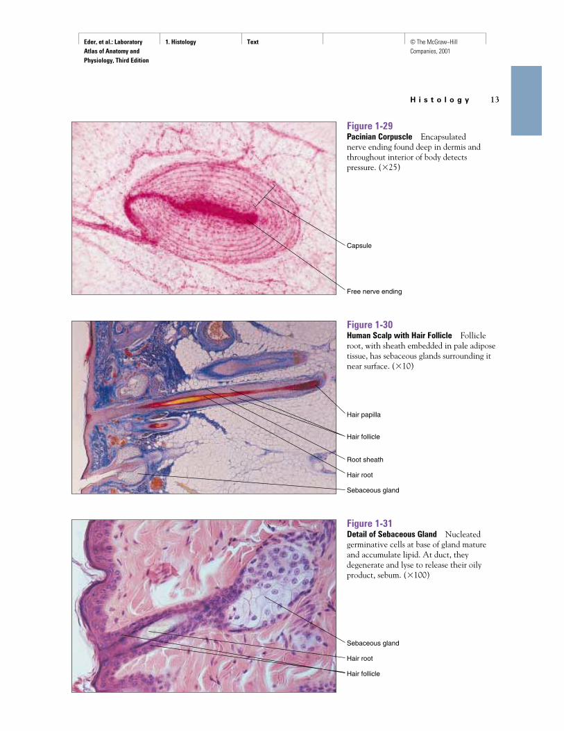
Figure 1-29Pacinian Corpuscle Encapsulated nerve ending found deep in dermis andthroughout interior of body detects pressure. (�25)
Figure 1-30Human Scalp with Hair Follicle Follicleroot, with sheath embedded in pale adiposetissue, has sebaceous glands surrounding itnear surface. (�10)
Figure 1-31Detail of Sebaceous Gland Nucleatedgerminative cells at base of gland matureand accumulate lipid. At duct, theydegenerate and lyse to release their oilyproduct, sebum. (�100)
Capsule
Free nerve ending
Hair papilla
Hair follicle
Root sheath
Hair root
Sebaceous gland
Sebaceous gland
Hair root
Hair follicle

Eder, et al.: Laboratory Atlas of Anatomy and Physiology, Third Edition
1. Histology Text © The McGraw−Hill Companies, 2001
14 C H A P T E R 1
Figure 1-32Compact Bone Center of “tree ring”structure, Haversian canal contains bloodvessel. Osteocytes imprisoned in small, darklacunae surrounding central Haversiancanal receive nutrition and communicatevia canaliculi, or little canals. Human.(�50)
Figure 1-33Detail of Compact Bone Haversian systemevident.
Figure 1-34Cancellous (Spongy) Bone Osteoblasts onspongy bone are engaged in secretion of newbony matrix. (�100)
Haversian canal
Lacunae
Haversian system (osteon)
Canaliculus
Haversian canal
Osteocyte in lacuna
Lamella
Haversian system (osteon)
Osteocyte
Resting osteoblast
Spongy bone
Osteoblast

Eder, et al.: Laboratory Atlas of Anatomy and Physiology, Third Edition
1. Histology Text © The McGraw−Hill Companies, 2001
H i s t o l o g y 15
Figure 1-35Red Bone Marrow Medullary cavity in thehead of long bones of the adult containsstem cells, precursors to red blood cells, andwhite blood cells and platelets. Human.(�250)
Figure 1-36Developing Bone at Epiphyseal PlateMiddle belt of cartilage undergoing primarycalcification is replaced by new bone.
Figure 1-37Detail of Epiphyseal Plate Epiphyseal plate cartilage at right transforms into zonesof proliferating chondrocytes with primaryossification occurring on their calcifiedremnants. Newly formed bone appears at left. (�50)
Cartilage of epiphyseal plate
Eosinophilic myelocyte
Myeloblast
Basophilic myelocyte
Neutrophilic stab cell
Neutrophil
White blood cell precursors
Erythroblasts Proerythroblast ErythroblastsRed blood cell precursors

Eder, et al.: Laboratory Atlas of Anatomy and Physiology, Third Edition
1. Histology Text © The McGraw−Hill Companies, 2001
16 C H A P T E R 1
Figure 1-38Striated (Skeletal) Muscle (Cross Section)Eccentrically located multiple nucleiaccompany individual cells (fibers), each of which contains many myofibrils. Human tongue. (�250)
Figure 1-39Striated (Skeletal) Muscle Fiber(Longitudinal Section) Banded appearancearises from regular arrangement ofoverlapping bundles of thick and thinfilaments (myosin and actin, respectively).Eccentrically located nuclei are thin and elongated. (�250)
Figure 1-40Striated (Skeletal) Muscle Fibers(Longitudinal Section) Each light (I) bandhas a dark (Z) line through it. Each dark(A) band has a light (H) zone through it.(�250)
Muscle fibers
Nucleus
Striations
Nucleus
H zone
Z line
A band
Nucleus
I band

Eder, et al.: Laboratory Atlas of Anatomy and Physiology, Third Edition
1. Histology Text © The McGraw−Hill Companies, 2001
H i s t o l o g y 17
Figure 1-41Cardiac Muscle (Longitudinal Section)Multinucleated, striated muscle fibersbranch and anastomose at junctions markedby dark intercalated disks. (�250)
Figure 1-42Smooth Muscle (Longitudinal Section)Canoe- or spindle-shaped muscle cells lackstriations, and each has a single, elongatednucleus. (�250).
Figure 1-43Innervation of Skeletal Muscle: MotorEndplate Branching nerve bundleterminates in small, specialized dents, themyoneural junctions. Nerve terminalsrelease small quantities of chemicalneurotransmitter to stimulate musclecontraction.
Nucleus
Intercalated disk
Nucleus
Terminal branches of motor neuron
Skeletal muscle fibers
Myoneural junction

Eder, et al.: Laboratory Atlas of Anatomy and Physiology, Third Edition
1. Histology Text © The McGraw−Hill Companies, 2001
18 C H A P T E R 1
Figure 1-44Astrocytes (Neuroglia) Star-shapedsupporting cells of central nervous systemmodulate ionic environment. Cytoplasmicextensions make contact with blood vessel.Cat. (Silver stain; �280)
Figure 1-45Purkinje Cells (Neurons) Numerousbranched processes (dendrites) receiveinformation for processing. Single process(axon) sends information to other neurons.Human cerebellum. (�100)
Figure 1-46Pyramidal Cells Neurons from humancerebral cortex directly receive informationfrom hundreds of other cells; sendinformation on to hundreds of others. (Fox-Golgi stain; �100)
Astrocyte
Blood vessel
Dendrites
Nucleus
Cell body
Axon
Dendrites
Axon
Eder, et al.: Laboratory Atlas of Anatomy and Physiology, Third Edition
1. Histology Text © The McGraw−Hill Companies, 2001
H i s t o l o g y 19
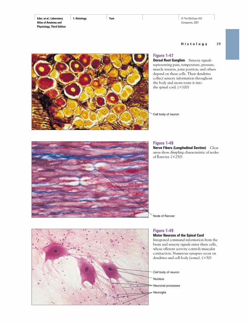
Figure 1-47Dorsal Root Ganglion Sensory signalsrepresenting pain, temperature, pressure,muscle tension, joint position, and othersdepend on these cells. Their dendrites collect sensory information throughoutthe body and axons route it into the spinal cord. (�100)
Figure 1-48Nerve Fibers (Longitudinal Section) Clearareas show dimpling characteristic of nodesof Ranvier. (�250)
Figure 1-49Motor Neurons of the Spinal CordIntegrated command information from thebrain and sensory signals enter these cells,whose efferent activity controls muscularcontraction. Numerous synapses occur ondendrites and cell body (soma). (�50)
Node of Ranvier
Cell body of neuron
Cell body of neuron
Nucleus
Neuronal processes
Neuroglia

Eder, et al.: Laboratory Atlas of Anatomy and Physiology, Third Edition
1. Histology Text © The McGraw−Hill Companies, 2001
20 C H A P T E R 1
Figure 1-50Myelinated Nerve Fibers (Cross Section)Central core stains dark; insulating myelinappears white. (�250)
Figure 1-51Spinal Cord, Lumbar Region (Cross Section)Top is dorsal, bottom is ventral. Lightcentral dot is central canal. Darkly stainingH-shaped region is grey matter of cell bodies;surrounding lighter material is composed ofmyelinated axons. Ventral horns of graymatter contain motor neurons; dorsal hornscontain cell bodies of sensory pathways.(�4)
Figure 1-52Retina Layered structure evident. Darkline of cells near top is pigment epithelium.Broad striped region representsphotoreceptors (rods and cones), whosenuclei stain heavily immediately beneath.Below receptor nuclei lie synaptic regionand a layer of nuclei belonging to bipolarcells. Bipolar cell output synapses ontoganglion cells, only a few of which appearnear bottom. Axons of ganglion cells formoptic nerve. (�100)
White matter
Dorsal horn
Central canal
Ventral horn
Pigmented epithelium
Rods and cones
Receptor nuclei
Bipolar cell nuclei
Ganglion cells
Core of nerve
Axon
Myelin sheath
Neurilemma
Capillary

Eder, et al.: Laboratory Atlas of Anatomy and Physiology, Third Edition
1. Histology Text © The McGraw−Hill Companies, 2001
H i s t o l o g y 21
Figure 1-53aOrgan of Corti Thick finger of tectorialmembrane extends from right to stimulatecomplex of four hair cells (three on left, oneon right) of central structure that rests onimportant basilar membrane. Nerve fibersfrom hair cells exit right to spiral ganglionfor processing and transmission of messagesto brain. (�100)
Figure 1-54Taste Bud Dissolved chemicals enterfungiform papilla through small pore todirectly stimulate sensory cells and initiatetaste perception. (�100)
Tectorial membrane
Nerve fibers
Hair cells
Basilar membrane
Tectorial membrane
Hair cells of Organ of Corti
Basilar membrane
Taste bud
Taste pore
Figure 1-53bThe Organ of Corti High magnification.(�500)

Eder, et al.: Laboratory Atlas of Anatomy and Physiology, Third Edition
1. Histology Text © The McGraw−Hill Companies, 2001
22 C H A P T E R 1
Figure 1-55Thyroid Gland Follicles Cuboidalepithelium surrounds endocrine follicles ofthe thyroid gland, the only gland that storessubstantial amounts of its own hormone.(�100)
Figure 1-56Parathyroid Gland LM of section throughthe parathyroid gland. (�40)
Thyroid follicle
Cuboidal cells
Colloid
Capsule
Trabecular blood vessels

Eder, et al.: Laboratory Atlas of Anatomy and Physiology, Third Edition
1. Histology Text © The McGraw−Hill Companies, 2001
H i s t o l o g y 23
Figure 1-57aPituitary Gland The pituitary glandconsists of two components: the posteriorcomponent, or neurohypophysis (lightstain), consists of mainly nervous tissue,whereas the anterior component, oradenohypophysis (dark stain) consists of aglandular epithelium. (�10)
Islet of Langerhans
Exocrine cells of pancreas
Figure 1-57bPituitary Gland The cleft between theneurohypophysis and adenohypophysis isvisible in this view of the pituitary gland.(�100)
Figure 1-58Pancreas The pancreatic islet ofLangerhans cells form the endocrine portionof the pancreas. Alpha cells secreteglucagon, beta cells secrete insulin, anddelta cells secrete somatostatin. Theexocrine portion of the pancreas secretesdigestive enzymes through a series of ducts.
Cleft
Neurohypophysis
Adenohypophysis
Pituitary gland
Cleft
Adenohypophysis
Neurohypophysis

Eder, et al.: Laboratory Atlas of Anatomy and Physiology, Third Edition
1. Histology Text © The McGraw−Hill Companies, 2001
24 C H A P T E R 1
Figure 1-59Adrenal Cortex Outer zone of roundedgroups of cells (zona glomerulosa) secretesmineralcorticosteroids (aldosterone).Middle zone of cells appearing in rows (zonafasciculata) secretes glucocorticosteroids.Innermost zone of cells arranged in ameshwork (zona reticularis) secretes mainlyandrogens. (�50)
Zona glomerulosa
Zona fasciculata
Zona reticularis
Figure 1-60Neutrophil Most numerous (65%) of the leukocytes, it is characterized by amultilobed nucleus and granular cytoplasm.Engages in phagocytosis. (Neutral dyes stain; �640)
Barr body
Nucleus

Eder, et al.: Laboratory Atlas of Anatomy and Physiology, Third Edition
1. Histology Text © The McGraw−Hill Companies, 2001
H i s t o l o g y 25
Figure 1-61Basophil Normally the rarest (1%) of theleukocytes, its kidney-shaped nucleus maybe almost obscured by cytoplasmic granules.These cells contain numerous chemicalsinvolved in inflammation. (Basic dyesstain; �640)
Figure 1-62Eosinophil Relatively rare (6%) leukocyte.Usually identifiable because of red-to-orange-staining cytoplasmic granules.Function not definitely known but elevatedespecially in allergies. (Selective eosin stain; �640)
Nucleus (two lobes)
Granules
Eder, et al.: Laboratory Atlas of Anatomy and Physiology, Third Edition
1. Histology Text © The McGraw−Hill Companies, 2001
26 C H A P T E R 1
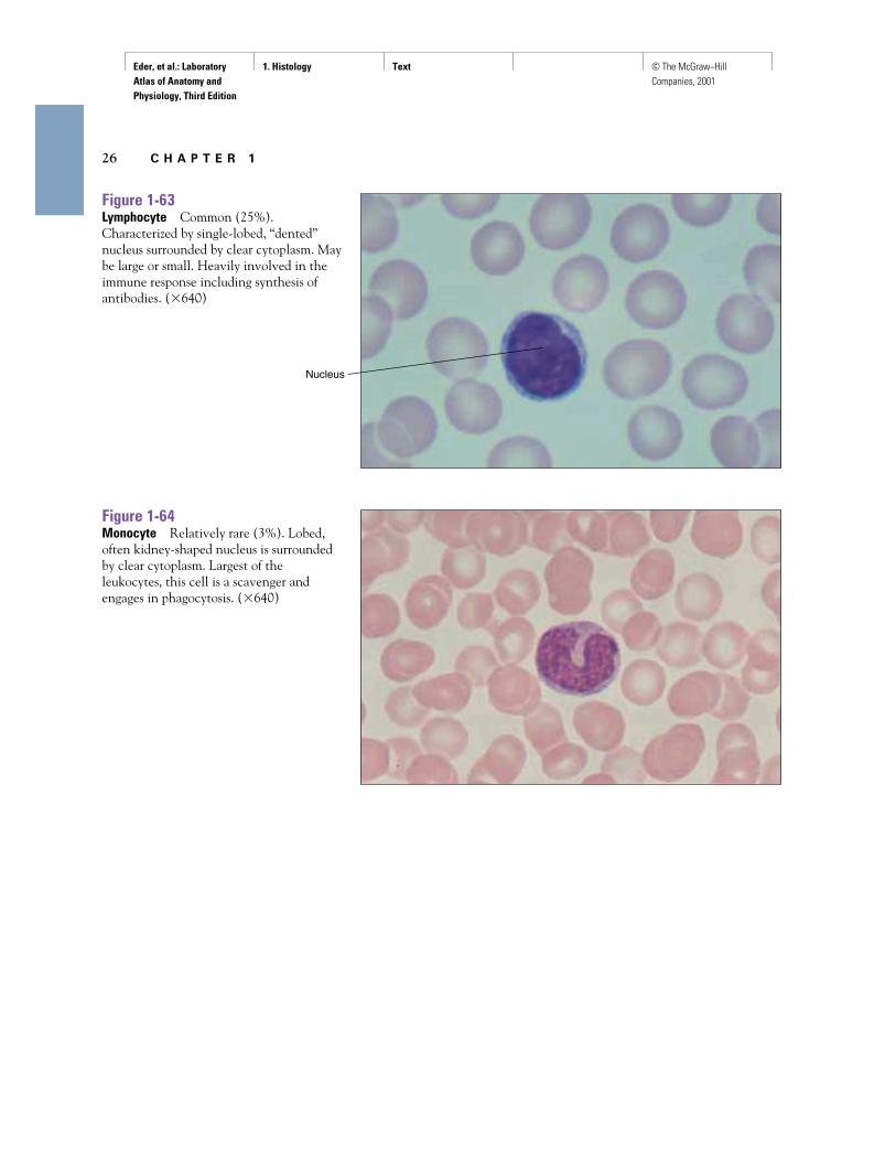
Figure 1-63Lymphocyte Common (25%).Characterized by single-lobed, “dented”nucleus surrounded by clear cytoplasm. Maybe large or small. Heavily involved in theimmune response including synthesis ofantibodies. (�640)
Figure 1-64Monocyte Relatively rare (3%). Lobed,often kidney-shaped nucleus is surroundedby clear cytoplasm. Largest of theleukocytes, this cell is a scavenger andengages in phagocytosis. (�640)
Nucleus

Eder, et al.: Laboratory Atlas of Anatomy and Physiology, Third Edition
1. Histology Text © The McGraw−Hill Companies, 2001
H i s t o l o g y 27
Figure 1-67Artery (A) and Vein (V) Blood vesselspossess a tunica intima that lines the lumen,outside of which is a muscular tunica media,and a connective tissue covering, the tunicaadventitia. The tunica media of arteries istypically much thicker than that of veins.(�100)
A
V
Tunica adventitia
Tunica media
Tunica intima
Figure 1-65Erythrocytes (Red Blood Cells) and PlateletsCirculating erythrocytes are far more commonthan any of the leukocytes. Normally theyhave no nucleus but contain the red pigmenthemoglobin, which permits them to transportoxygen and carbon dioxide throughout thebody. Typically they assume the shape of abiconcave disk. Their diameter of about7 microns is useful for comparing sizes ofother histological structures. Platelets arecellular remnants of a much larger precursor.These remnants contain numerous chemicals,including those important for clotting andinflammation. Platelets initiate blood clottingby forming a plug at wound sites. (�500)
Figure 1-66Sickle Cell Anemia Genetic alteration ofhemoglobin results in altered membranestructure and abnormal wavy or elongated,curved shape that often resembles a sickle(upper left). Oxygen-carrying capacity ismuch reduced. (�500)
Platelets
Erythrocytes

Eder, et al.: Laboratory Atlas of Anatomy and Physiology, Third Edition
1. Histology Text © The McGraw−Hill Companies, 2001
28 C H A P T E R 1
Figure 1-68aArterial Cross Section Single layer ofdarkly stained cells, the tunica intima lines the lumen. Thick tunica media iscomposed of canoe-shaped smooth musclecells. Outer adventitial layer of connectivetissue provides elastic support and strength.(�50)
Tunica media
Tunica adventitia
Lumen
Tunica intima
Lumen
Artery wall
Fatty deposit
Artery wall
Lumen filled with blood
Figure 1-68bAtherosclerosis Cross section of a healthyartery.
Figure 1-68cAtherosclerosis Cross section of an arterywith advanced atherosclerosis.

Eder, et al.: Laboratory Atlas of Anatomy and Physiology, Third Edition
1. Histology Text © The McGraw−Hill Companies, 2001
H i s t o l o g y 29
Figure 1-71Lymph Node Outer cortex containingseveral follicles surrounds medulla, with itsnarrow, dark medullary cords. Notch ishilum, through which blood and lymphaticvessels pass. (�5)
Follicle (germinal center)
Hilum
Medulla
Cortex
Figure 1-70Capillary with Red Blood Cells in Single FileCapillary wall is made of flattenedendothelial cells without complex tunics, a simple structure that facilitates theexchange of gases, nutrients, wastes, and hormones. (�400)
Endothelium
Red blood cell
Figure 1-69Detail of Arterial Wall Inner endothelialcells of tunica intima (left) lie on a basementmembrane. A thin layer of smooth musclecells and elastic tissue (lamina propria)throws this tunic into folds. The tunicamedia contains multiple layers of smoothmuscle cells regularly arranged. A wavyexternal elastic membrane separates thetunica media from the adventitia.Adventitia
Tunica media
Lamina propria
Tunica intima
External elastic membrane

Eder, et al.: Laboratory Atlas of Anatomy and Physiology, Third Edition
1. Histology Text © The McGraw−Hill Companies, 2001
30 C H A P T E R 1
Figure 1-72Valve of Lymphatic Vessel One-way flowof lymph, from left to right in this figure, isensured by valve action in lymph vessel.Vessels themselves are thin walled and lackmusculature; pumping action occursthrough compression by neighboringmuscles. (�25)
Figure 1-73aThymus Various lobules contain thick,darkly staining cortex surrounding a smaller,lighter-staining medulla. Small, roundcellular patches in medulla are Hassall’scorpuscles. In adults, much of thymusdegenerates and is replaced by adiposetissue. (�10)
Valve
Cortex
Medulla
Hassall’s corpuscle
Hassall’s (thymic) corpuscles
Figure 1-73bThymus Under higher magnification, the appearance of Hassall’s corpusclesdistinguish the thymus from other organs.Surrounding the corpuscles are reticulateepithelial cells. (�400)
Eder, et al.: Laboratory Atlas of Anatomy and Physiology, Third Edition
1. Histology Text © The McGraw−Hill Companies, 2001
H i s t o l o g y 31
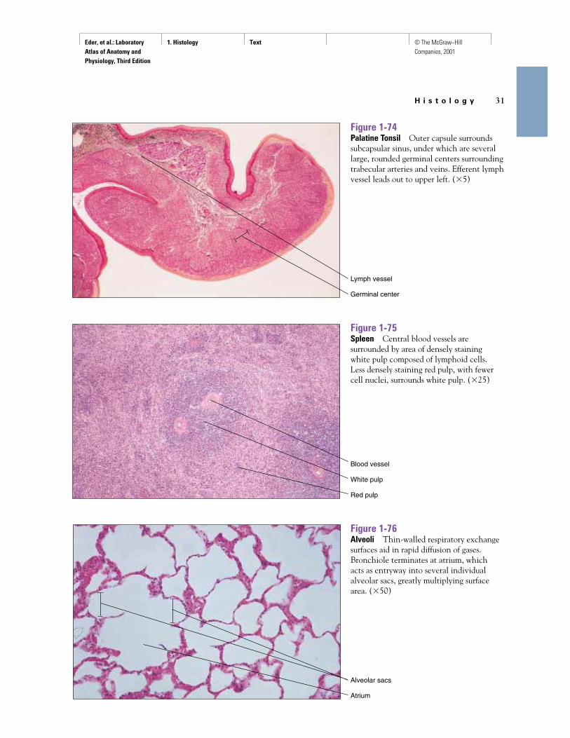
Figure 1-76Alveoli Thin-walled respiratory exchangesurfaces aid in rapid diffusion of gases.Bronchiole terminates at atrium, which acts as entryway into several individualalveolar sacs, greatly multiplying surfacearea. (�50)
Alveolar sacs
Atrium
Figure 1-75Spleen Central blood vessels aresurrounded by area of densely staining white pulp composed of lymphoid cells. Less densely staining red pulp, with fewercell nuclei, surrounds white pulp. (�25)
Figure 1-74Palatine Tonsil Outer capsule surroundssubcapsular sinus, under which are severallarge, rounded germinal centers surroundingtrabecular arteries and veins. Efferent lymphvessel leads out to upper left. (�5)
Lymph vessel
Germinal center
Blood vessel
White pulp
Red pulp

Eder, et al.: Laboratory Atlas of Anatomy and Physiology, Third Edition
1. Histology Text © The McGraw−Hill Companies, 2001
32 C H A P T E R 1
Figure 1-77Details of Alveolus Squamous cellscompose alveolar sac, which is penetratedby thin-walled blood vessels (upper left)containing erythrocytes. (�100)
Blood vessels
Free alveolar macrophage
Erythrocyte
Simple squamous epithelium
Figure 1-78Bronchiole Epithelial layer that lines thelumen is surrounded by layer of smoothmuscle, which regulates bronchiolardiameter. Round structures outside ofsmooth muscle layer are blood vessels.(�100)
Smooth muscle
Blood vessel
Lumen
Epithelium
Figure 1-79Esophagus Surrounding the lumen,esophageal structure contains, in order, thefour basic layers of the alimentary canal:mucosa (composed of epithelium, the thicklamina propria, and dark muscularis),submucosa (light with spaces, blood vessels,and lymph channels), two thick layers ofthe muscularis (circular and longitudinal),and the thin, connective adventitia on thesurface. Cross section, human. (�3)
Mucosa
Submucosa
Adventitia
Muscularis
Eder, et al.: Laboratory Atlas of Anatomy and Physiology, Third Edition
1. Histology Text © The McGraw−Hill Companies, 2001
H i s t o l o g y 33
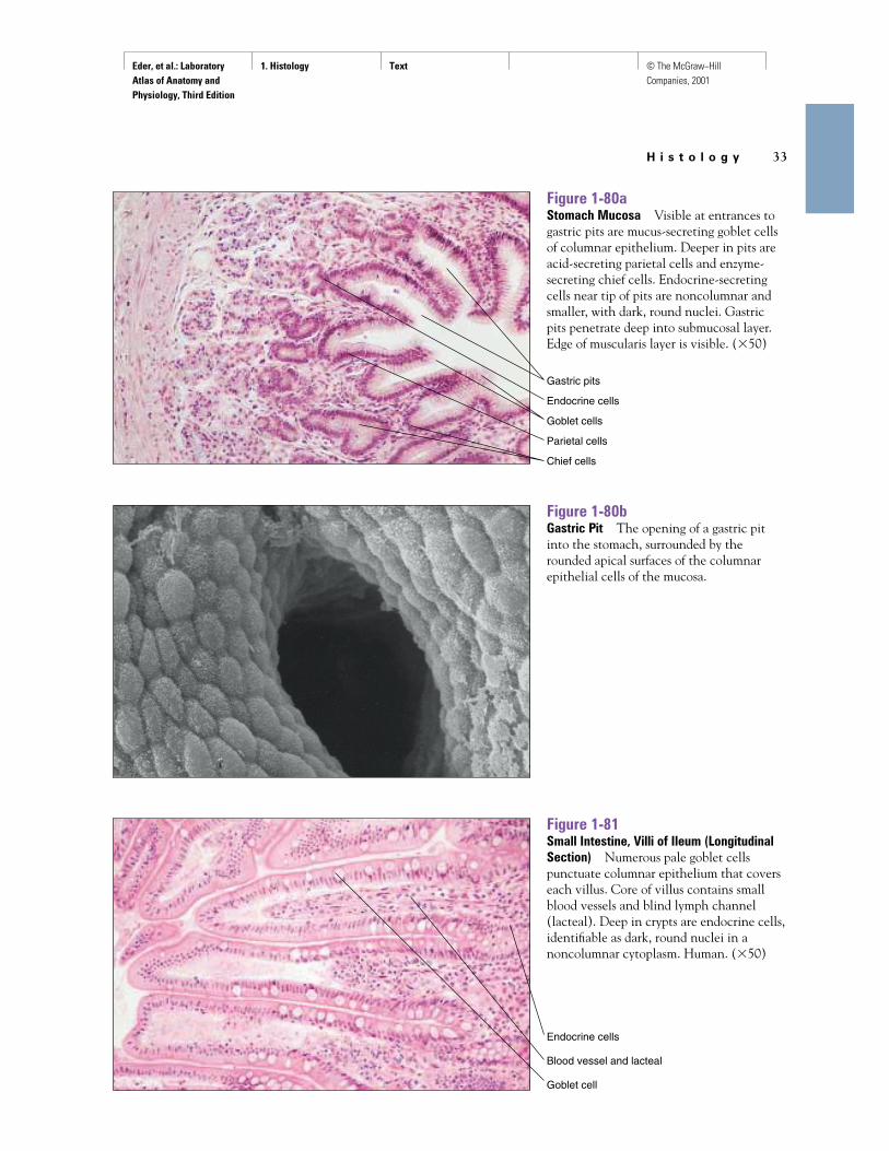
Figure 1-80aStomach Mucosa Visible at entrances togastric pits are mucus-secreting goblet cellsof columnar epithelium. Deeper in pits areacid-secreting parietal cells and enzyme-secreting chief cells. Endocrine-secretingcells near tip of pits are noncolumnar andsmaller, with dark, round nuclei. Gastric pits penetrate deep into submucosal layer.Edge of muscularis layer is visible. (�50)
Gastric pits
Endocrine cells
Goblet cells
Parietal cells
Chief cells
Figure 1-80bGastric Pit The opening of a gastric pitinto the stomach, surrounded by therounded apical surfaces of the columnarepithelial cells of the mucosa.
Figure 1-81Small Intestine, Villi of Ileum (LongitudinalSection) Numerous pale goblet cellspunctuate columnar epithelium that coverseach villus. Core of villus contains smallblood vessels and blind lymph channel(lacteal). Deep in crypts are endocrine cells,identifiable as dark, round nuclei in anoncolumnar cytoplasm. Human. (�50)
Endocrine cells
Blood vessel and lacteal
Goblet cell

Eder, et al.: Laboratory Atlas of Anatomy and Physiology, Third Edition
1. Histology Text © The McGraw−Hill Companies, 2001
34 C H A P T E R 1
Figure 1-83Large Intestine (Colon) (Cross Section)Surface is thrown into folds but devoid ofvilli. Thick submucosa contains bloodvessels and lymph channels. (�10)
Submucosa
Blood vessel
Lymph channel
Figure 1-82Small Intestine, Villi of Ileum (Cross Section)Goblet cells emptying contents throughbrush border surface are evident. Core ofvillus contains blood vessels, lymphchannels, and lymphocytes. Human.(�100)
Core of villus
Brush border
Goblet cell
Lymphocyte
Mucosa
Muscularis
Eder, et al.: Laboratory Atlas of Anatomy and Physiology, Third Edition
1. Histology Text © The McGraw−Hill Companies, 2001
H i s t o l o g y 35
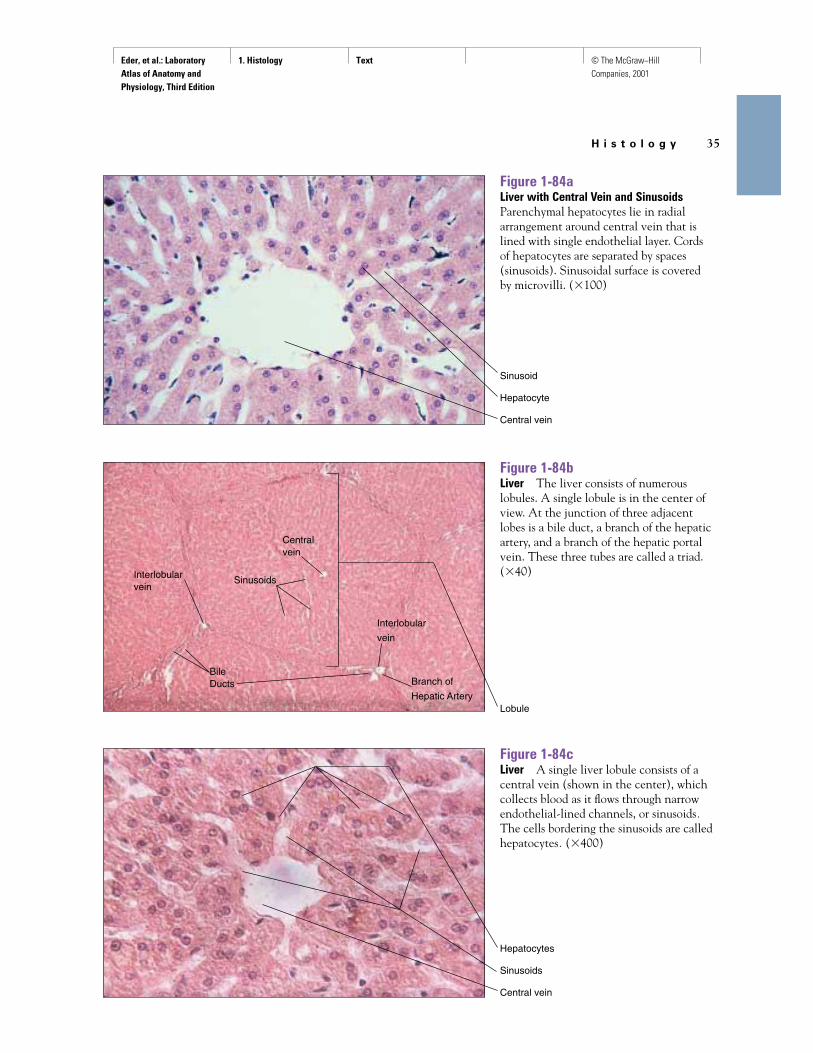
Figure 1-84aLiver with Central Vein and SinusoidsParenchymal hepatocytes lie in radialarrangement around central vein that islined with single endothelial layer. Cords of hepatocytes are separated by spaces(sinusoids). Sinusoidal surface is covered by microvilli. (�100)
Figure 1-84bLiver The liver consists of numerouslobules. A single lobule is in the center ofview. At the junction of three adjacentlobes is a bile duct, a branch of the hepaticartery, and a branch of the hepatic portalvein. These three tubes are called a triad.(�40)
Sinusoid
Hepatocyte
Central vein
Hepatocytes
Sinusoids
Central vein
Lobule
Figure 1-84cLiver A single liver lobule consists of acentral vein (shown in the center), whichcollects blood as it flows through narrowendothelial-lined channels, or sinusoids.The cells bordering the sinusoids are calledhepatocytes. (�400)
Interlobularvein
BileDucts
Interlobular
vein
Sinusoids
Centralvein
Branch of
Hepatic Artery

Eder, et al.: Laboratory Atlas of Anatomy and Physiology, Third Edition
1. Histology Text © The McGraw−Hill Companies, 2001
36 C H A P T E R 1
Figure 1-85Gallbladder Mucosal folds are covered byepithelium with well-developed microvilli.Lamina propria contains blood vessels.(�25)
Muscularis
Lamina propria
Blood vessel
Figure 1-86Vermiform Appendix (Cross Section)Overall structure resembles that of colon.Large, darkly staining structures arelymphoid follicles, the size and number ofwhich decrease with age. Human. (�3)
Lymphoid follicle(germinal center)
Figure 1-87Sublingual Salivary Gland Large, pale,mucus-secreting cells, some with caps ofserous demilunes, secrete their contents into ducts that may be lined with striatedepithelial cells indicative of ion exchangeactivity. (�100)
Epithelial cells
Salivary duct
Serous demilunes
Eder, et al.: Laboratory Atlas of Anatomy and Physiology, Third Edition
1. Histology Text © The McGraw−Hill Companies, 2001
H i s t o l o g y 37
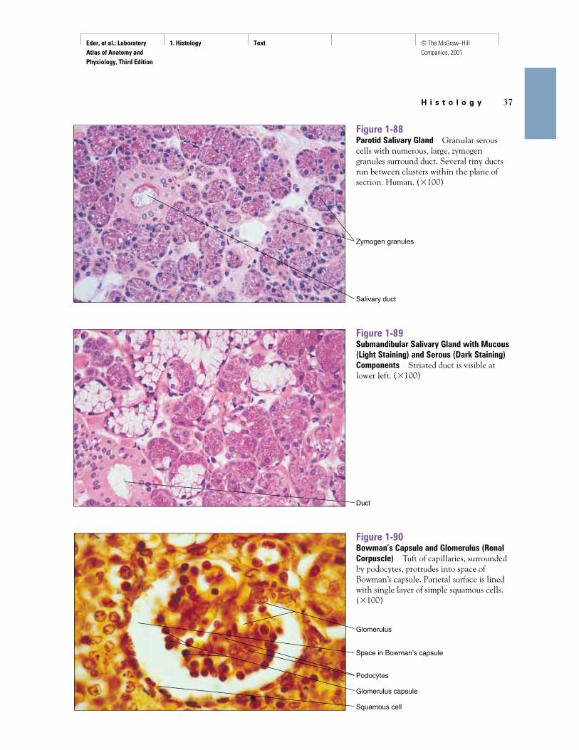
Figure 1-90Bowman’s Capsule and Glomerulus (RenalCorpuscle) Tuft of capillaries, surroundedby podocytes, protrudes into space ofBowman’s capsule. Parietal surface is linedwith single layer of simple squamous cells.(�100)
Glomerulus
Space in Bowman’s capsule
Podocytes
Glomerulus capsule
Squamous cell
Figure 1-88Parotid Salivary Gland Granular serouscells with numerous, large, zymogengranules surround duct. Several tiny ductsrun between clusters within the plane ofsection. Human. (�100)
Figure 1-89Submandibular Salivary Gland with Mucous(Light Staining) and Serous (Dark Staining)Components Striated duct is visible atlower left. (�100)
Zymogen granules
Salivary duct
Duct

Eder, et al.: Laboratory Atlas of Anatomy and Physiology, Third Edition
1. Histology Text © The McGraw−Hill Companies, 2001
38 C H A P T E R 1
Figure 1-91Two Glomeruli and Bowman’s Capsules“Lacy” edges of glomerulus on left showscharacteristics of pregnancy-inducedhypertension (PIH), here inducedexperimentally in a pregnant rat. (�50)
Figure 1-92Distal Convoluted Tubules Lined withCuboidal Epithelium Cross section of rat.(�400)
Cuboidal cell
Figure 1-93Ureter Star-shaped lumen is lined withtransitional epithelium that varies inthickness to change shape as lumenstretches. Delicate lamina propria separates epithelium from alternating layers of circular and longitudinal smoothmuscle. (�25)
Transitional epithelium
Smooth muscle and adventitial connective tissue

Eder, et al.: Laboratory Atlas of Anatomy and Physiology, Third Edition
1. Histology Text © The McGraw−Hill Companies, 2001
H i s t o l o g y 39
Figure 1-96Seminiferous Tubules of Testis Lined withSertoli Cells and Germinativum in VariousStages of Development Tunica propriasurrounds each tubule. Interstitial spacescontain blood vessels and clumps ofinterstitial (Leydig) cells that secretetestosterone. (�50)
Spermatozoa
Tunica propria
Basement membrane
Interstitial cells
Spermatocytes
Sertoli cells
Figure 1-94Urinary Bladder Umbrella cells oftransitional epithelium stretch and flatten as bladder fills. Basement membraneseparates epithelium from underlyingconnective tissue containing blood vessels.Monkey. (�100)
Figure 1-95Urethra (within Penis) Lumen is lined with transitional epithelium and isembedded in corpus spongiosum of thepenis. Paraurethral glands located above the lumen in the figure secrete mucus into the urethra. A smooth muscle layer(tunica muscularis) surrounds the urethral structures. (�10)
Umbrella cells
Lumen of bladder
Basement membrane
Paraurethral glands
Corpus spongiosum
Tunica muscularis
Lumen

Eder, et al.: Laboratory Atlas of Anatomy and Physiology, Third Edition
1. Histology Text © The McGraw−Hill Companies, 2001
40 C H A P T E R 1
Figure 1-97Spermatozoa Head contains numerousenzymes and nucleus with DNA. Thickmidpiece just behind head is packed withmitochondria. (�250)
Figure 1-98Epididymis Tall, pseudostratified columnarepithelium with microvilli surrounds alumen packed with clumps of spermatozoa.Narrow band of smooth muscle cellsencircles each tubule.
Pseudostratified columnar epithelium
Smooth muscle
Figure 1-99Ductus Deferens Ciliated columnarepithelial cells line a spermatozoa-filledlumen. Three layers of smooth muscle cellssurround mucosa, a circular layer betweentwo longitudinal ones. (�50)
Columnar epithelium of mucosa
Smooth muscle
Head of sperm
Midpiece
Tail
Eder, et al.: Laboratory Atlas of Anatomy and Physiology, Third Edition
1. Histology Text © The McGraw−Hill Companies, 2001
H i s t o l o g y 41
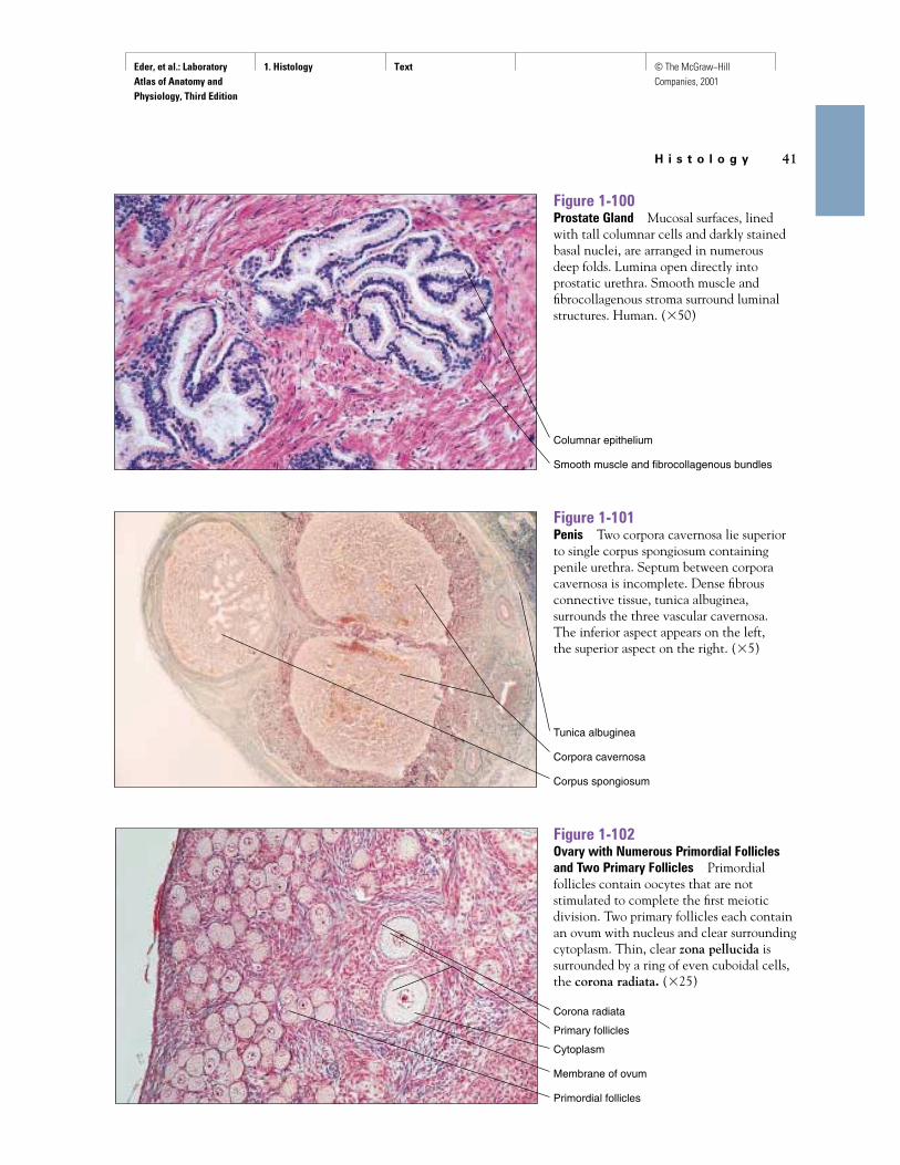
Figure 1-102Ovary with Numerous Primordial Folliclesand Two Primary Follicles Primordialfollicles contain oocytes that are notstimulated to complete the first meioticdivision. Two primary follicles each containan ovum with nucleus and clear surroundingcytoplasm. Thin, clear zona pellucida issurrounded by a ring of even cuboidal cells,the corona radiata. (�25)
Corona radiata
Primary follicles
Cytoplasm
Membrane of ovum
Primordial follicles
Figure 1-100Prostate Gland Mucosal surfaces, linedwith tall columnar cells and darkly stainedbasal nuclei, are arranged in numerous deep folds. Lumina open directly intoprostatic urethra. Smooth muscle andfibrocollagenous stroma surround luminalstructures. Human. (�50)
Columnar epithelium
Smooth muscle and fibrocollagenous bundles
Figure 1-101Penis Two corpora cavernosa lie superiorto single corpus spongiosum containingpenile urethra. Septum between corporacavernosa is incomplete. Dense fibrousconnective tissue, tunica albuginea,surrounds the three vascular cavernosa. The inferior aspect appears on the left, the superior aspect on the right. (�5)
Tunica albuginea
Corpora cavernosa
Corpus spongiosum

Eder, et al.: Laboratory Atlas of Anatomy and Physiology, Third Edition
1. Histology Text © The McGraw−Hill Companies, 2001
42 C H A P T E R 1
Figure 1-103Detail of Oocyte in Primordial FollicleClear nucleus contains well-definednucleolus. Neither zona pellucida norcorona radiata is evident. (�250)
Figure 1-104Secondary Ovarian Follicle with OvumBright zona pellucida surrounds outermembrane of ovum and in turn issurrounded by dark, cellular corona radiata.A large antrum has formed where the egg is not anchored to the follicular wall ofgranulosa cells. (�100)
Nucleolus
Nucleus
Stratified cuboidal epithelium
Nucleus
Membrane of ovum
Zona pellucida
Corona radiata
Antrum

Eder, et al.: Laboratory Atlas of Anatomy and Physiology, Third Edition
1. Histology Text © The McGraw−Hill Companies, 2001
H i s t o l o g y 43
Figure 1-105Fallopian (Uterine) Tube Extensive foldingof mucosa, lined with ciliated columnarepithelium, is common. Epithelium rests on thin basement membrane and flatconnective tissue layer. Rhythmic beating of cilia helps transport ovum toward uterus;cell structure also suggests secretoryfunction. Human. (�100)
Columnar epithelium
Connective tissue
Figure 1-106Uterus Endometrial lining (right) duringproliferative phase of uterine cycle showsthickening of epithelial surfaces andnumerous coiled glandular ducts. (�25)
Endothelial lining
Glandular ducts