highly efficient bienzyme functionalized nanocomposite ... · pdf file9.3%ofthecarboxyl(cooh)...
TRANSCRIPT
Highly Efficient Bienzyme FunctionalizedNanocomposite-Based MicrofluidicsBiosensor Platform for BiomedicalApplicationMd. Azahar Ali1,2, Saurabh Srivastava1, Pratima R. Solanki1,5, Venu Reddy3, Ved V. Agrawal1,CheolGi Kim3, Renu John2 & Bansi D. Malhotra1,3,4
1Department of Science and Technology Centre on Biomolecular Electronics, Biomedical Instrumentation Section, CSIR-NationalPhysical Laboratory, Dr. K. S. Krishnan Marg, New Delhi-110012, India, 2Indian Institute of Technology Hyderabad, OrdnanceFactory Estate, Yeddumailaram, Hyderabad, Andhra Pradesh 502205, India, 3Center for NanoBioengineering and SpinTronics,Department of Materials Science and Engineering, Chungnam National University, Daejeon 305-764, South Korea, 4Department ofBiotechnology, Delhi Technological University, Shahbad Daulatpur, Main Bawana Road, Delhi1 110042, India, 5Special Centrefor Nanosciences, Jawaharlal Nehru University, New Delhi-110067, India.
This report describes the fabrication of a novel microfluidics nanobiochip based on a composite comprisingof nickel oxide nanoparticles (nNiO) and multiwalled carbon nanotubes (MWCNTs), as well as the chip’s usein a biomedical application. This nanocomposite was integrated with polydimethylsiloxane (PDMS)microchannels, which were constructed using the photolithographic technique. A structural andmorphological characterization of the fabricated microfluidics chip, which was functionalized with abienzyme containing cholesterol oxidase (ChOx) and cholesterol esterase (ChEt), was accomplished usingX-ray photoelectron spectroscopy (XPS) and scanning electron microscopy. The XPS studies revealed that9.3% of the carboxyl (COOH) groups present in the nNiO-MWCNT composite are used to form amide bondswith the NH2 groups of the bienzyme. The response studies on this nanobiochip reveal good reproducibilityand selectivity, and a high sensitivity of 2.2 mA/mM/cm2. This integrated microfluidics biochip provides apromising low-cost platform for the rapid detection of biomolecules using minute samples.
The ability to monitor desired biomolecules in real time via a microfluidics biochip or a micro-total analyticalsystem (m-TAS) has aroused considerable interest from the point-of-care (POC) diagnostics community1,2.The small geometry of a microfluidics biochip offers many advantages over macroelectrode-based biome-
dical devices due to its improved mass transport and diffusion, high signal-to-noise ratio, and low detectionlimit3,4. The characteristics of nanocomposites and the intrinsic benefits of microfluidics, including their laminarflow, low consumption of costly reagents and power, portability, minimal need for handling biohazardousmaterials, fast response time, multiplexing, and parallelization, are advantageous for the fabrication of a bio-chip5,6. The precise liquid control that occurs in a microfluidic platform is essential for the fabrication of micro-channels. Polydimethylsiloxane (PDMS) is an attractive polymeric material for the fabrication of microchannels,which can be temporarily sealed with a glass substrate via conformal contact mediated by Van der Waals force.Additionally, the hydrophobic properties of PDMS provide good chemical compatibility with organic solventsand cause negligible swelling5,6.
The microfluidics biochip is known to have many applications, such as enzymatic kinetics and immunoassayanalyses; DNA amplification; and cell sorting, culturing, and counting7. However, the integration of the micro-fluidics biochip with a nanostructured material continues to be a challenge. In this context, 1D structures, such asnanotubes, nanowires, and nanocomposites, composed of carbon materials can play an important role in medi-cinal chemistry and diagnostics, including the creation of biochips for in vitro and in vivo investigations3,8–13.Multiwalled carbon nanotubes (MWCNTs), allotrope of carbon play an important role towards the developmentof biochips because of their high carrier mobility and tensile strength, as well as their high aspect ratio, which leadsto quantum electron transport12. In addition, MWCNTs are non-reactive (like graphite) except at the nanotubecaps (the tips when they are not cut), where the dangling bonds and edge-plane-like sites located at both ends are
OPEN
SUBJECT AREAS:ELECTROCATALYSIS
BIOSENSORS
NANOSENSORS
MICROFLUIDICS
Received22 May 2013
Accepted27 August 2013
Published27 September 2013
Correspondence andrequests for materials
should be addressed toV.V.A. (ved.varun@
gmail.com;agrawalvv@nplindia.
org); C.K. ([email protected]); R.J. (renujohn@
iith.ac.in) or B.D.M.(bansi.malhotra@
gmail.com)
SCIENTIFIC REPORTS | 3 : 2661 | DOI: 10.1038/srep02661 1
open to reactions14. The MWCNTs are known to produce changes inenergy bands close to the Fermi level15,16. The exciting electronicproperties and high electrochemical reactivity of MWCNTs suggestthat fast electron transfer reaction occurs when they are used as theelectrode in an electrochemical biochip15,16. Lin et al. have developeda microfluidics electrochemical sensor for on-site, non-invasivemonitoring of lead and chlorophenols17. Wisitsoraat et al. havedeveloped an electrochemical biochip for cholesterol detection thathas a sensitivity of 0.0512 nA/mg/dl, which is attributed to the directgrowth of CNT on glass18. However, MWCNTs are known toagglomerate via Van der Waals interactions, resulting in poor film-forming ability. To overcome this problem, nanostructured metaloxides (NMOx) may be used to control the agglomeration ofMWCNTs19. The covalent binding (or sidewall functionalization)of biomolecules (e.g., proteins, enzymes, and nucleic acids) to carb-oxyl-functionalized MWCNTs via diimide-activated amidation mayprovide improved stability and reproducibility20–24. In such a case, the
large surface area of the MWCNTs and the presence of abundantfunctional groups may offer a suitable platform for biofunctionaliza-tion20–25. Additionally, MWCNTs may facilitate continuous conduct-ing pathways to transport the charge carriers, allowing for a highersensitivity25. Shim et al. have used functionalized CNT for biomole-cular recognition in a streptavidin/biotin approach to investigate theadsorption of proteins on the sidewalls of carbon nanotubes20.
A biosensor based on nanostructured nickel oxide (nNiO) hasrecently been explored to detect biomolecules such as DNA, anti-body-antigen interactions, glucose, and cholesterol26,27. However,nNiO-based biosensors have limited applications due to the inher-ently poor electrical conductivity of nNiO26. The non-covalentimmobilization of enzymes onto nNiO-based biochip has recentlybeen found to result in poor stability of the desired biomolecules26–28.To improve the characteristics of a biosensing device, nNiO can beintegrated with MWCNTs29,30. Zhang et al. have used CNT-NMOx todevelop solar cells and gas sensors21. The sp2 hybridization and
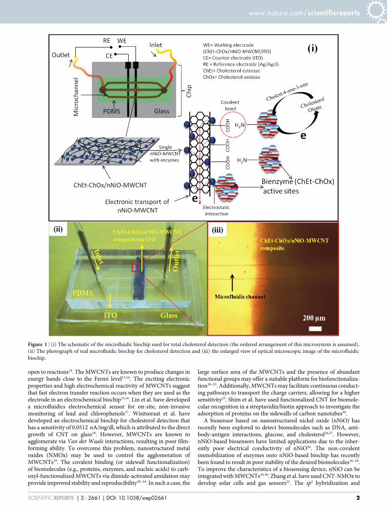
Figure 1 | (i) The schematic of the microfluidic biochip used for total cholesterol detection (the ordered arrangement of this microsystem is assumed).
(ii) The photograph of real microfluidic biochip for cholesterol detection and (iii) the enlarged view of optical microscopic image of the microfluidic
biochip.
www.nature.com/scientificreports
SCIENTIFIC REPORTS | 3 : 2661 | DOI: 10.1038/srep02661 2
electronic properties of nNiO coupled with the specific recognitionproperties of immobilized MWCNTs could lead to a miniaturizedbiochip29,30. Chen et al. have reported the creation of a non-enzymaticelectrochemical glucose sensor based on a MnO2/MWCNT matrix29.Zhu et al. have utilized MWCNTs with ZnO nanoparticles for ultra-fast, nonlinear optical switching19.
In this study, we discuss the fabrication of a novel nNiO-MWCNTnanocomposite-based biochip for total cholesterol estimation. Thebiochip is immobilized with a bienzyme, composed of cholesterolesterase (ChEt) and cholesterol oxidase (ChOx), and integrated withPDMS microchannels. Unlike conventional methods, our procedurecreates bienzyme functionalization through the carboxylation andamidation of the nNiO-MWCNT nanocomposite that serves as thesensing interface.
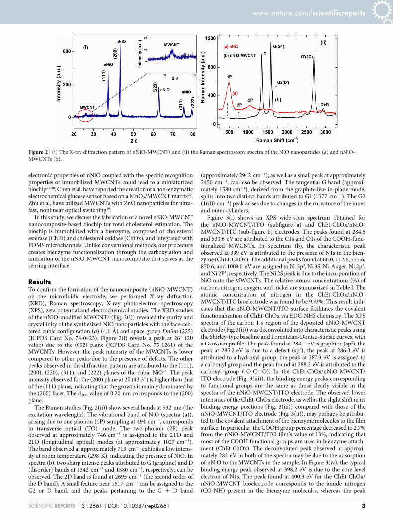
ResultsTo confirm the formation of the nanocomposite (nNiO-MWCNT)on the microfluidic electrode, we performed X-ray diffraction(XRD), Raman spectroscopy, X-ray photoelectron spectroscopy(XPS), zeta potential and electrochemical studies. The XRD studiesof the nNiO-modified MWCNTs (Fig. 2(i)) revealed the purity andcrystallinity of the synthesized NiO nanoparticles with the face-cen-tered cubic configuration (a) (4.1 A) and space group Fm3m (225)(JCPDS Card No. 78-0423). Figure 2(i) reveals a peak at 26u (2hvalue) due to the (002) plane (JCPDS Card No. 75-1261) of theMWCNTs. However, the peak intensity of the MWCNTs is lowercompared to other peaks due to the presence of defects. The otherpeaks observed in the diffraction pattern are attributed to the (111),(200), (220), (311), and (222) planes of the cubic NiO26. The peakintensity observed for the (200) plane at 2h (43.3u) is higher than thatof the (111) plane, indicating that the growth is mainly dominated bythe (200) facet. The d200 value of 0.20 nm corresponds to the (200)plane.
The Raman studies (Fig. 2(ii)) show several bands at 532 nm (theexcitation wavelength). The vibrational band of NiO (spectra (a)),arising due to one phonon (1P) sampling at 494 cm21, correspondsto transverse optical (TO) mode. The two-phonon (2P) peakobserved at approximately 746 cm21 is assigned to the 2TO and2LO (longitudinal optical) modes (at approximately 1027 cm21).The band observed at approximately 713 cm21 exhibits a low intens-ity at room temperature (298 K), indicating the presence of NiO. Inspectra (b), two sharp intense peaks attributed to G (graphite) and D(disorder) bands at 1342 cm21 and 1580 cm21, respectively, can beobserved. The 2D band is found at 2695 cm21 (the second order ofthe D band). A small feature near 1617 cm21 can be assigned to theG2 or D band, and the peaks pertaining to the G 1 D band
(approximately 2942 cm21), as well as a small peak at approximately2450 cm21, can also be observed. The tangential G band (approxi-mately 1580 cm21), derived from the graphite-like in-plane mode,splits into two distinct bands attributed to G1 (1577 cm21). The G2(1610 cm21) peak arises due to changes in the curvature of the innerand outer cylinders.
Figure 3(i) shows an XPS wide-scan spectrum obtained forthe nNiO-MWCNT/ITO (subfigure a) and ChEt-ChOx/nNiO-MWCNT/ITO (sub-figure b) electrodes. The peaks found at 284.6and 530.6 eV are attributed to the C1s and O1s of the COOH-func-tionalized MWCNTs. In spectrum (b), the characteristic peakobserved at 399 eV is attributed to the presence of N1s in the bien-zyme (ChEt-ChOx). The additional peaks found at 66.0, 112.6, 777.6,870.6, and 1009.0 eV are assigned to Ni 3p3, Ni 3S, Ni-Auger, Ni 2p3,and Ni 2P1, respectively. The Ni 2S peak is due to the incorporation ofNiO onto the MWCNTs. The relative atomic concentrations (%) ofcarbon, nitrogen, oxygen, and nickel are summarized in Table I. Theatomic concentration of nitrogen in the ChEt-ChOx/nNiO-MWCNT/ITO bioelectrode was found to be 9.93%. This result indi-cates that the nNiO-MWCNT/ITO surface facilitates the covalentfunctionalization of ChEt-ChOx via EDC-NHS chemistry. The XPSspectra of the carbon 1 s region of the deposited nNiO-MWCNTelectrode (Fig. 3(ii)) was deconvoluted into characteristic peaks usingthe Shirley-type baseline and Lorentzian-Doniac-Sunsic curves, witha Gaussian profile. The peak found at 284.1 eV is graphitic (sp2), thepeak at 285.2 eV is due to a defect (sp3), the peak at 286.3 eV isattributed to a hydroxyl group, the peak at 287.3 eV is assigned toa carbonyl group and the peak found at 288.2 eV is attributed to thecarboxyl group (-O-C5O). In the ChEt-ChOx/nNiO-MWCNT/ITO electrode (Fig. 3(iii)), the binding energy peaks correspondingto functional groups are the same as those clearly visible in thespectra of the nNiO-MWCNT/ITO electrode. The observed lowerintensities of the ChEt-ChOx electrode, as well as the slight shift in itsbinding energy positions (Fig. 3(iii)) compared with those of thenNiO-MWCNT/ITO electrode (Fig. 3(ii)), may perhaps be attribu-ted to the covalent attachment of the bienzyme molecules to the filmsurface. In particular, the COOH group percentage decreased to 2.7%from the nNiO-MWCNT/ITO film’s value of 13%, indicating thatmost of the COOH functional groups are used in bienzyme attach-ment (ChEt-ChOx). The deconvoluted peak observed at approxi-mately 282 eV in both of the spectra may be due to the adsorptionof nNiO to the MWCNTs in the sample. In Figure 3(iv), the typicalbinding energy peak observed at 398.2 eV is due to the core-levelelectron of N1s. The peak found at 400.3 eV for the ChEt-ChOx/nNiO-MWCNT bioelectrode corresponds to the amide nitrogen(CO-NH) present in the bienzyme molecules, whereas the peak
Figure 2 | (i) The X-ray diffraction pattern of nNiO-MWCNTs and (ii) the Raman spectroscopy spectra of the NiO nanoparticles (a) and nNiO-
MWCNTs (b).
www.nature.com/scientificreports
SCIENTIFIC REPORTS | 3 : 2661 | DOI: 10.1038/srep02661 3

observed at 401.7 eV, attributed to the ;N species, confirms cova-lent functionalization. The value of the average surface potential ofcolloidal NiO nanoparticles was estimated to be 122.7 mV usingzeta potential measurements. This value reveals that the NiO nano-particles are positively charged (Supporting Information, Fig. S1(a)).The average surface potential of the MWCNTs was measured to be26.7 mV, indicating the presence of negatively charged groups onthe sidewalls of the carbon nanotubes (Supporting Information, Fig.S1(b)). The positively charged nNiO provides strong electrostaticinteractions that link it to the negatively charged MWCNTs.
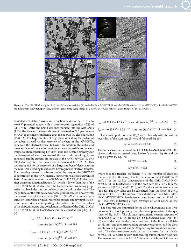
The scanning electron micrograph (SEM) studies (Fig. S2) werecarried out as a morphological observation study at an acceleratingvoltage of 20 kV. The SEM image (Supporting Information, Fig.S2(i)) shows COOH-functionalized MWCNTs that are likely to berandomly oriented on the ITO substrate. The in situ incorporatedMWCNTs, with deposited nNiO, are shown in Figure S2(ii). TheNiO nanoparticles appear to be attached to the MWCNTs, which arenot agglomerated, indicating that the presence of NiO nanoparticlesprevented their agglomeration. The transmission electron micro-scopy (TEM) image shows spherical NiO nanoparticles with an aver-age size of ,70 nm (Fig. 4(i)). The high-resolution image of theindividual MWCNTs shows outer and inner diameters of 34 and
12 nm, respectively. The selected area electron diffraction (SAED)pattern shows that graphitic (002) and (004) reflections are present inthe MWCNTs (the inset in Figure 4(ii)). Figure 4(iii) shows that thewell-dispersed COOH-functionalized tubular-shaped MWCNTs areentangled with NiO nanoparticles and randomly oriented. It appearsthat some of the NiO nanoparticles are aggregated on the MWCNTsurfaces. A few NiO nanoparticles can be observed inside the carbonnanotubes (Fig. 4(iii), marked with dotted lines). The length range ofthe MWCNTs is several tens of micrometers, and they have anexternal diameter of approximately 20–80 nm. From the atomic-scale image of the nNiO-MWCNTs (Fig. 4(iv)), it can be concludedthat this nanocomposite is crystalline in nature, with an interlayerspacing of 3.41 A (d002 for MWCNT, shown in the subfigure inset).The lattice fringe spacing of the NiO nanocrystal is estimated to be2.0 A, which agrees with the results of the XRD studies. These resultsclearly reveal a successful formation of the nNiO-MWCNT compos-ite, which can be used to fabricate the desired biochip.
The electrochemical behavior of the biochip, both before and afterundergoing surface modification by the bienzyme (ChEt-ChOx), wasinvestigated using cyclic voltammetry (CV) in phosphate buffer sal-ine (PBS, pH 7.0) containing ferro/ferri cyanide (as a mediator) at ascan rate of 30 mV/s (Fig. 5(A)). The CV of the nNiO/ITO electrode
Figure 3 | (i) The wide-scan X-ray photoelectron spectra (XPS) of various films. (ii) The XPS spectra of the C1s region of the nNiO-MWCNT film after
deconvolution; (iii) the C1s region of the ChEt-ChOx/nNiO-MWCNT/ITO film; and (iv) the N1s core-level spectra of the ChEt-ChOx/nNiO-MWCNT/
ITO film.
Table I | The atomic concentration (%) of the elements C, Ni, O, and N present in the nNiO-MWCNT/ITO and ChEt-ChOx/nNiO-MWCNT/ITO films obtained from XPS analysis
Elements/atomic concentration (%) C Ni O N
nNiO-MWCNT/ITO 12.52 33.72 53.76ChEt-ChOx/nNiO-MWCNT/ITO 59.11 3.88 27.08 9.93
www.nature.com/scientificreports
SCIENTIFIC REPORTS | 3 : 2661 | DOI: 10.1038/srep02661 4
exhibited well-defined oxidation/reduction peaks in the 20.8 V to10.9 V potential range, with a peak-to-peak separation (DE) of0.115 V (a). After the nNiO was incorporated into the MWCNTs(1.0%) (b), the electrochemical current increased to 28.0 mA becauseMWCNTs are more conductive than the nNiO/ITO electrode alone(23.0 mA). The large number of edge plane sites along the surface ofthe tubes, as well as the presence of defects in the MWCNTs,enhanced the electrochemical behavior. In addition, the outer andinner surfaces of the carbon nanotubes were accessible to the elec-trolyte solution containing Fe31/Fe21 ions and became pathways forthe transport of electrons toward the electrode, resulting in anenhanced faradic current. In the case of the nNiO-MWCNT(2.0%)/ITO electrode (c), the peak current increased to 31.2 mA. Thisincrease is due to the presence of a large number of defect sites inthe MWCNTs, leading to enhanced heterogeneous electron transfer.The resulting current can be controlled by varying the MWCNTconcentration in the nNiO matrix. Furthermore, a redox current of14.0 mA was obtained for the nNiO-MWCNT(2.0%)/ITO electrodeafter bienzyme functionalization (d), which is lower than that of thenNiO-MWCNT/ITO electrode; the bienzyme has insulating prop-erties that block the transport of electrons toward the electrode. Themagnitudes of the cathodic and anodic peaks increased linearly withthe square root of the scan rate (30 to 100 mV s21), indicating adiffusion-controlled or quasi-reversible process and favourable elec-tron transfer kinetics (Supporting Information, Fig. S3). The valuesof the slope, intercept, and correlation coefficient of the ChEt-ChOx/nNiO-MWCNT/ITO bioelectrode can be estimated using Eq. (1)–(4).
Ipa~6:71 mAz3:55 mAmV{1s� �1=2
scan rate mV=sð Þ½ �1=2; R2~0:999ð1Þ
Ipc~{0:137 mA{2:56 mAmV{1s� �1=2
scan rate mV=sð Þ½ �1=2; R2~0:997ð2Þ
Epa~0:368 Vz1:95 s1=2 scan rate mV=sð Þ½ �1=2; R2~0:998 ð3Þ
Epc~{0:235 V{1:54 s1=2 scan rate mV=sð Þ½ �1=2; R2~0:993 ð4Þ
The anodic peak potential (Epa) varied linearly with the naturallogarithm of the scan rate (ln n) and followed Eq. (5).
Epa~0:232 ln nz1:509 ð5Þ
The surface concentration of the ChEt-ChOx/nNiO-MWCNT/ITObioelectrode was estimated using Laviron’s theory (Eq. 6), and theslope is given by Eq. (7):
RT=anF~0:232 ð6Þ
Ip~n2F2I��
4RT ð7Þ
where a is the transfer coefficient, n is the number of electronstransferred (1 in this case), F is the Faraday constant (96485.34 C/mol), I* is the surface concentration of the ChEt-ChOx/nNiO-MWCNT/ITO bioelectrode, n is the scan rate (mV s21), R is thegas constant (8.314 J mol21 K21), and T is the absolute temperature(298 K). The ip/n value can be calculated from the slope of the ipversus n plot. The total surface concentration of the ChEt-ChOx/nNiO-MWCNT/ITO bioelectrode was found to be 11.07 3
1026 mol/cm2, indicating a high coverage of ChEt-ChOx on thenNiO-MWCNT/ITO surface.
The flow rate was optimized for the ChEt-ChOx/nNiO-MWCNTmicrofluidics biochip using the chronoamperometric technique(inset of Fig. 5(A)). The chronoamperometric current response ofthe nNiO-MWCNT/ITO (i) and ChEt-ChOx/nNiO-MWCNT/ITO(ii) electrodes was obtained as a function of the flow rate (0.01–10 mL/min) and the corresponding current versus time, and the plotsare shown in Figures S4 and S5 (Supporting Information), respect-ively. The chronoamperometric current increases for the nNiO-MWCNT/ITO electrode with an increasing buffer solution flow rate.The maximum current is 0.1 mL/min, after which point it reaches
Figure 4 | The HR-TEM analysis of (i) the NiO nanoparticles, (ii) an individual MWCNT (inset: the SAED pattern of the MWCNT), (iii) the MWCNTs
modified with NiO nanoparticles, and (iv) an atomic-scale image of a nNiO-MWCNT (inset: lattice fringes of the MWCNT).
www.nature.com/scientificreports
SCIENTIFIC REPORTS | 3 : 2661 | DOI: 10.1038/srep02661 5
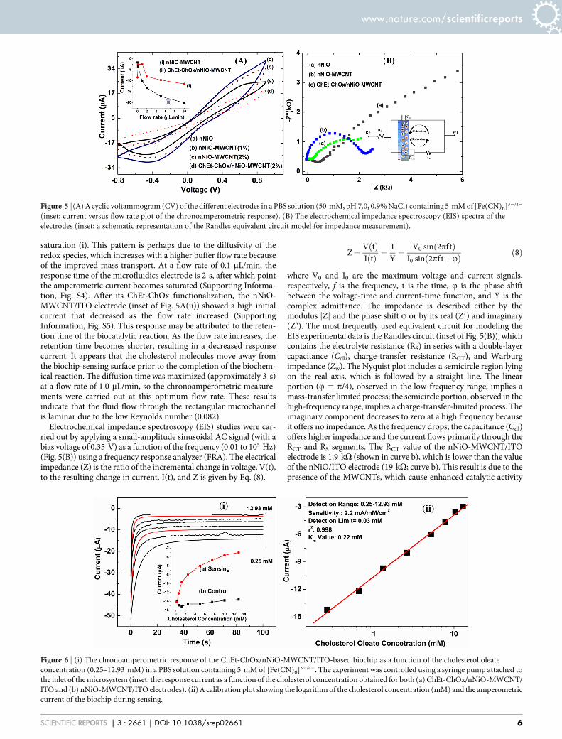
saturation (i). This pattern is perhaps due to the diffusivity of theredox species, which increases with a higher buffer flow rate becauseof the improved mass transport. At a flow rate of 0.1 mL/min, theresponse time of the microfluidics electrode is 2 s, after which pointthe amperometric current becomes saturated (Supporting Informa-tion, Fig. S4). After its ChEt-ChOx functionalization, the nNiO-MWCNT/ITO electrode (inset of Fig. 5A(ii)) showed a high initialcurrent that decreased as the flow rate increased (SupportingInformation, Fig. S5). This response may be attributed to the reten-tion time of the biocatalytic reaction. As the flow rate increases, theretention time becomes shorter, resulting in a decreased responsecurrent. It appears that the cholesterol molecules move away fromthe biochip-sensing surface prior to the completion of the biochem-ical reaction. The diffusion time was maximized (approximately 3 s)at a flow rate of 1.0 mL/min, so the chronoamperometric measure-ments were carried out at this optimum flow rate. These resultsindicate that the fluid flow through the rectangular microchannelis laminar due to the low Reynolds number (0.082).
Electrochemical impedance spectroscopy (EIS) studies were car-ried out by applying a small-amplitude sinusoidal AC signal (with abias voltage of 0.35 V) as a function of the frequency (0.01 to 105 Hz)(Fig. 5(B)) using a frequency response analyzer (FRA). The electricalimpedance (Z) is the ratio of the incremental change in voltage, V(t),to the resulting change in current, I(t), and Z is given by Eq. (8).
Z~V tð ÞI tð Þ ~
1Y
~V0 sin 2pf tð Þ
I0 sin 2pf tzQð Þ ð8Þ
where V0 and I0 are the maximum voltage and current signals,respectively, f is the frequency, t is the time, Q is the phase shiftbetween the voltage-time and current-time function, and Y is thecomplex admittance. The impedance is described either by themodulus Zj j and the phase shift Q or by its real (Z9) and imaginary(Z0). The most frequently used equivalent circuit for modeling theEIS experimental data is the Randles circuit (inset of Fig. 5(B)), whichcontains the electrolyte resistance (RS) in series with a double-layercapacitance (Cdl), charge-transfer resistance (RCT), and Warburgimpedance (Zw). The Nyquist plot includes a semicircle region lyingon the real axis, which is followed by a straight line. The linearportion (Q 5 p/4), observed in the low-frequency range, implies amass-transfer limited process; the semicircle portion, observed in thehigh-frequency range, implies a charge-transfer-limited process. Theimaginary component decreases to zero at a high frequency becauseit offers no impedance. As the frequency drops, the capacitance (Cdl)offers higher impedance and the current flows primarily through theRCT and RS segments. The RCT value of the nNiO-MWCNT/ITOelectrode is 1.9 kV (shown in curve b), which is lower than the valueof the nNiO/ITO electrode (19 kV; curve b). This result is due to thepresence of the MWCNTs, which cause enhanced catalytic activity
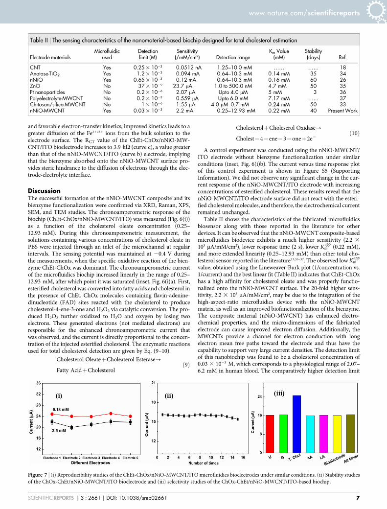
Figure 6 | (i) The chronoamperometric response of the ChEt-ChOx/nNiO-MWCNT/ITO-based biochip as a function of the cholesterol oleate
concentration (0.25–12.93 mM) in a PBS solution containing 5 mM of [Fe(CN)6]32/42. The experiment was controlled using a syringe pump attached to
the inlet of the microsystem (inset: the response current as a function of the cholesterol concentration obtained for both (a) ChEt-ChOx/nNiO-MWCNT/
ITO and (b) nNiO-MWCNT/ITO electrodes). (ii) A calibration plot showing the logarithm of the cholesterol concentration (mM) and the amperometric
current of the biochip during sensing.
Figure 5 | (A) A cyclic voltammogram (CV) of the different electrodes in a PBS solution (50 mM, pH 7.0, 0.9% NaCl) containing 5 mM of [Fe(CN)6]32/42
(inset: current versus flow rate plot of the chronoamperometric response). (B) The electrochemical impedance spectroscopy (EIS) spectra of the
electrodes (inset: a schematic representation of the Randles equivalent circuit model for impedance measurement).
www.nature.com/scientificreports
SCIENTIFIC REPORTS | 3 : 2661 | DOI: 10.1038/srep02661 6
and favorable electron-transfer kinetics; improved kinetics leads to agreater diffusion of the Fe21/31 ions from the bulk solution to theelectrode surface. The RCT value of the ChEt-ChOx/nNiO-MW-CNT/ITO bioelectrode increases to 3.9 kV (curve c), a value greaterthan that of the nNiO-MWCNT/ITO (curve b) electrode, implyingthat the bienzyme absorbed onto the nNiO-MWCNT surface pro-vides steric hindrance to the diffusion of electrons through the elec-trode-electrolyte interface.
DiscussionThe successful formation of the nNiO-MWCNT composite and itsbienzyme functionalization were confirmed via XRD, Raman, XPS,SEM, and TEM studies. The chronoamperometric response of thebiochip (ChEt-ChOx/nNiO-MWCNT/ITO) was measured (Fig. 6(i))as a function of the cholesterol oleate concentration (0.25–12.93 mM). During this chronoamperometric measurement, thesolutions containing various concentrations of cholesterol oleate inPBS were injected through an inlet of the microchannel at regularintervals. The sensing potential was maintained at 20.4 V duringthe measurements, when the specific oxidative reaction of the bien-zyme ChEt-ChOx was dominant. The chronoamperometric currentof the microfluidics biochip increased linearly in the range of 0.25–12.93 mM, after which point it was saturated (inset, Fig. 6(i)a). First,esterified cholesterol was converted into fatty acids and cholesterol inthe presence of ChEt. ChOx molecules containing flavin-adenine-dinucleotide (FAD) sites reacted with the cholesterol to producecholesterol-4-ene-3-one and H2O2 via catalytic conversion. The pro-duced H2O2 further oxidized to H2O and oxygen by losing twoelectrons. These generated electrons (not mediated electrons) areresponsible for the enhanced chronoamperometric current thatwas observed, and the current is directly proportional to the concen-tration of the injected esterified cholesterol. The enzymatic reactionsused for total cholesterol detection are given by Eq. (9–10).
Cholesterol OleatezCholeaterol Esterase?
Fatty AcidzCholesterolð9Þ
CholesterolzCholeserol Oxidase?
Cholest{4{ene{3{onez2e{ð10Þ
A control experiment was conducted using the nNiO-MWCNT/ITO electrode without bienzyme functionalization under similarconditions (inset, Fig. 6(i)b). The current versus time response plotof this control experiment is shown in Figure S5 (SupportingInformation). We did not observe any significant change in the cur-rent response of the nNiO-MWCNT/ITO electrode with increasingconcentrations of esterified cholesterol. These results reveal that thenNiO-MWCNT/ITO electrode surface did not react with the esteri-fied cholesterol molecules, and therefore, the electrochemical currentremained unchanged.
Table II shows the characteristics of the fabricated microfluidicsbiosensor along with those reported in the literature for otherdevices. It can be observed that the nNiO-MWCNT composite-basedmicrofluidics biodevice exhibits a much higher sensitivity (2.2 3
103 mA/mM/cm2), lower response time (2 s), lower Kappm (0.22 mM),
and more extended linearity (0.25–12.93 mM) than other total cho-lesterol sensor reported in the literature33,35–37. The observed low Kapp
m
value, obtained using the Lineweaver-Burk plot (1/concentration vs.1/current) and the best linear fit (Table II) indicates that ChEt-ChOxhas a high affinity for cholesterol oleate and was properly functio-nalized onto the nNiO-MWCNT surface. The 20-fold higher sens-itivity, 2.2 3 103 mA/mM/cm2, may be due to the integration of thehigh-aspect-ratio microfluidics device with the nNiO-MWCNTmatrix, as well as an improved biofunctionalization of the bienzyme.The composite material (nNiO-MWCNT) has enhanced electro-chemical properties, and the micro-dimensions of the fabricatedelectrode can cause improved electron diffusion. Additionally, theMWCNTs provide a channel for electron conduction with longelectron mean free paths toward the electrode and thus have thecapability to support very large current densities. The detection limitof this nanobiochip was found to be a cholesterol concentration of0.03 3 1023 M, which corresponds to a physiological range of 2.07–6.2 mM in human blood. The comparatively higher detection limit
Table II | The sensing characteristics of the nanomaterial-based biochip designed for total cholesterol estimation
Electrode materialsMicrofluidic
usedDetectionlimit (M)
Sensitivity(/mM/cm2) Detection range
Km Value(mM)
Stability(days) Ref.
CNT Yes 0.25 3 1023 0.0512 nA 1.25–10.0 mM …… …… 18Anatase-TiO2 Yes 1.2 3 1023 0.094 mA 0.64–10.3 mM 0.14 mM 35 34nNiO Yes 0.65 3 1023 0.12 mA 0.64–10.3 mM 0.16 mM 60 26ZnO No 37 3 1029 23.7 mA 1.0 to 500.0 nM 4.7 mM 50 35Pt nanoparticles No 0.2 3 1026 2.07 mA Upto 4.0 mM 5 mM 3 36Polyelectrolyte-MWCNT No 0.2 3 1023 0.559 mA Upto 6.0 mM 7.17 mM …… 37Chitosan/silica-MWCNT No 1 3 1026 1.55 mA 4.0 mM–0.7 mM 0.24 mM 50 33nNiO-MWCNT Yes 0.03 3 1023 2.2 mA 0.25–12.93 mM 0.22 mM 40 Present Work
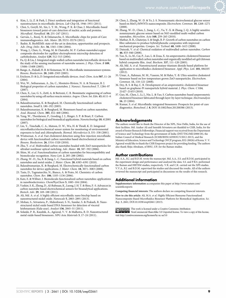
Figure 7 | (i) Reproducibility studies of the ChEt-ChOx/nNiO-MWCNT/ITO microfluidics bioelectrodes under similar conditions. (ii) Stability studies
of the ChOx-ChEt/nNiO-MWCNT/ITO bioelectrode and (iii) selectivity studies of the ChOx-ChEt/nNiO-MWCNT/ITO-based biochip.
www.nature.com/scientificreports
SCIENTIFIC REPORTS | 3 : 2661 | DOI: 10.1038/srep02661 7

of the ChEt-ChOx/nNiO-MWCNT/ITO microfluidics total choles-terol sensor may be caused by either insufficient electrical conduc-tivity of the nNiO or the morphological changes arising as a result ofinteractions between the bienzyme (ChEt-ChOx) and the nNiO-MWCNT nanocomposite surface18,26,34,37.
Our biochip shows good reproducibility for cholesterol concen-trations of 2.59 and 5.18 mM (Fig. 7(i)) as is evidenced by the lowstandard deviations (RSD) found (4.19% and 2.13%, n 5 5), indi-cating good precision. The ChEt-ChOx/nNiO-MWCNT/ITO bioe-lectrode also shows good repeatability, as evidenced by its low RSD of1.12% (n 5 15) for a cholesterol concentration of 2.50 mM. Nosignificant decrease in the current was observed after 12 uses; infurther procedures, the bioelectrode showed slight decreases in itscurrent response due to the denaturation of the biomolecules (Fig. 7(ii)).
The selectivity of the ChEt-ChOx/nNiO-MWCNT/ITO bioelec-trode was demonstrated using lactic acid (LA), glucose (G), ascorbicacid (AA), uric acid (UA), and other interferents in a buffer solution.We found that the present bioelectrode is highly specific to totalcholesterol (5.18 mM) and shows a negligible change in its responsecurrent in the presence of other analytes (interferents) (Fig. 7(iii)).The shelf life of this biochip was determined by measuring the changein the current response at regular 5-day intervals for approximately 3months; the chip exhibited a 93.7% response after approximately 40days (data not shown). The ChEt-ChOx/nNiO-MWCNT/ITO bioe-lectrode was stored at 4uC when not in use. This integrated micro-fluidics nanobiochip reached 92% of its steady-state current in lessthan 2 s, indicating that a fast electron exchange was occurringbetween the active sites of the bienzyme and the nNiO-MWCNT/ITO electrode.
In summary, our miniaturized microfluidics-integrated biochipbased on a nNiO-MWCNT composite allows for the rapid detectionof biomolecules at a low cost. The nNiO serves as a support for thedispersion of MWCNTs and helps exfoliate smaller bundles or indi-vidual nanotubes. The covalent functionalization through reactionsof desired chemical groups onto the p-conjugated skeleton of thisnNiO-MWCNT composite matrix offers considerable advantagesfor the development of a nanobiochip to detect molecules such ascholesterol. In particular, the COOH groups on the MWCNTs pro-vide simple biofunctionalization and a higher loading capacity ofbiomolecules on the microfluidic transducer surface. The 9.3% ofthe carboxyl groups that were present on the nNiO-MWCNT surfacewere used to covalently bind with the bienzyme. The synergisticelectrocatalytic activity of the MWCNTs provides straight conduct-ing pathways for carriers such as electrons and ions, resulting in ahigher sensitivity than nNiO-based biochips. This bienzyme functio-nalized and integrated microfluidics biochip was highly sensitive tocholesterol oxidation and was both highly reproducible and stable,allowing it to be utilized for total cholesterol monitoring. Our plat-form exhibits improved detection limits and a faster response time.This highly efficient composite microfluidics biochip could be usedto create an array-based biochip that simultaneously monitors multi-analytes, including low-density lipoproteins and triglycerides, mak-ing it a promising platform for biomedical applications.
MethodsNickel nitrate [Ni(NO3)2]?H2O and potassium hydroxide (KOH) were procuredfrom Sigma-Aldrich, USA. N-hydroxisuccimide (NHS), N-ethyl-N9-(3-dymethyla-minopropyl carbodiimide) (EDC), cholesterol oxidase (ChOx), cholesterol esterase(ChEt), and cholesterol oleate were purchased from Sigma-Aldrich, USA. Differentconcentrations of cholesterol oleate, from 0.25 to 12.93 mM, were prepared in 0.9%NaCl solution. SU8-100 negative photoresist and SU-8 developer were purchasedfrom Microchem (Newton, MA, USA).
Instrumentation. The nanostructured nickel oxide (nNiO) and the nNiO-MWCNTmatrices were characterized using X-ray diffraction (XRD, Cu Ka radiation, Rigaku),Raman spectroscopy, and transmission electron microscopy (TEM; JEOL JEM-2000EX). The XPS measurements were performed using an X-ray photoelectronspectrometer (XPS, Multilab 2000, Thermo Scientific) equipped with an alpha 110
hemispherical electron energy analyzer and an X-ray source. The XPS measurementswere made in the binding energy range of 0–1100 eV to confirm the ChEt-ChOxfunctionalization of the nNiO-MWCNT matrix. The electrochemical studies wereperformed using an electrochemical analyzer (AUT-84275) in PBS at pH 7.0containing 5 mM of [Fe(CN)6]32/42 as a redox species.
PDMS microchannel fabrication. The three rectangular PDMS microchannels werefabricated using the soft lithography technique26. The channel height and width wereeach 200 mm, and the length was 2.0 cm. The microchannels had reservoirs at theend, which were fabricated by punching holes in the PDMS slab at the desiredposition. The three fabricated channels were connected to reservoirs via a single inletand outlet (Fig. 1(i)). The principal reason to use three channels is to increase thesensor surface area of the biochip.
Microelectrode fabrication. The indium tin oxide (ITO)-coated glass substrate (witha thickness of approximately 150–300 A) had a sheet resistance of 70–100 V and wasused to fabricate the microelectrode (0.6 3 0.26 cm2) via wet chemical etching. Themasked ITO was dipped in ITO etchant solution (HNO3:HCl:H2O in a 1510510ratio) for 15 min, and ITO was selectively etched from the glass substrate. The ITOelectrode was cleaned with acetone and then sonicated with acetone (10 min) andwater (2 min) several times. The glass substrate containing the electrodes washydrolyzed using a mixture of H2O5H2O25NH3 (55151) and washed with deionizedwater. It was then dried in an oven at 100uC for approximately 4 h.
Synthesis of the nNiO-MWCNT composite. The MWCNTs (90%) were synthesizedby catalytic chemical vapor deposition using a mixture of ferrocene as the catalyst andtoluene as the hydrocarbon source31. These MWCNTs were purified andfunctionalized by refluxing in a concentrated solution of nitric acid and sulfuric acid,which generated a large number of COOH groups on the nanotube surfaces32. Thedrop-wise addition of 0.2 M KOH solution and COOH-terminated MWCNTs to asolution of 0.5 M Ni(NO3)2?6H2O (nickel nitrate) in de-ionized water with constantstirring results in a blackish-green precipitate of nickel hydroxide, Ni(OH)2, at a pH ofapproximately 11.8. Excess solvents were evaporated to obtain the precipitate, whichwas dried at 80uC for 24 h to create a transparent viscous solution of nickelhydroxide-modified MWCNTs. This precipitate was maintained at a pH of 10.0. Thethick gel-like Ni(OH)2-modified MWCNT solution was deposited onto patternedITO electrodes on the glass substrate via dip coating. The electrodes were initiallydried at approximately 110uC for 1 h, and they were then annealed at 400uC underambient conditions for approximately 2 h to remove any solvent, causing the nNiO-MWCNTs (weight 1.0%) and nNiO-MWCNTs (weight 2.0%) to adhere to the ITOsurface.
Functionalization of the bienzyme (ChEt-ChOx). A mixture of ChEt (1 mg/ml) andChOx (1 mg/ml) in a 151 ratio was spread onto the nNiO-MWCNT/ITOmicroelectrode through physical absorption (Fig. 1(i)); the electrode was then kept ina humid chamber for 4 h at room temperature (298 K). The carboxyl-terminatedMWCNTs and the NiO nanoparticles are negatively and positively charged,respectively, resulting in increased electrostatic interactions. However, the carboxylicgroups can undergo an amidation reaction with the amino acids of proteins. Oneapproach to covalently bind proteins or enzymes utilizes the diimide-activatedamidation of carboxylic acid functionalized carbon nanotubes. The COOH terminalgroup was activated using EDC (0.4 M) as the coupling agent and NHS (0.1 M) as theactivator. The formation of strong covalent (C-N) amide bonds between the COOHgroups of the MWCNTs and the NH2 groups of the bienzyme (Fig. 1(i)) wasconfirmed via XPS studies. It is possible that the nNiO molecules with a highisoelectric point (an IEP of 10.0) interact with the bienzyme directly throughelectrostatic interactions due to their low IEP (5.5).
Microfluidics biochip integration. A lithographically fabricated rectangular PDMSmicrochannel slab was sealed temporarily (irreversible) with the ITO glass substrateusing a conformal contact38 that contained the modified nNiO-MWCNT and ChEt-ChOx electrodes (Fig. 1(i). The photograph of real microfluidic nanobiochip forcholesterol detection is shown in Fig. 1(ii). Fig. 1(iii) shows the enlarged view ofoptical microscopic image of this microfluidic nanobiochip. The PDMS surfaceprovides an adhesive layer that plays an important role, making the bonding with theglass substrate irreversible through weak Van der Waals forces38. Again, the inletreservoir of the PDMS microchannel was used to introduce the syringe pump, whichwas based on a stepped motor, to control the fluid flow in the microfluidics biochip.The outlet reservoir was used to accommodate an Ag/AgCl wire that served as thereference electrode (RE). The bare ITO acted as the counter electrode (CE), and theChEt-ChOx/nNiO-MWCNT/ITO bioelectrode was used as a sensor for totalcholesterol detection. All of these three electrodes contained a glass substrate andPDMS microchannels and were embedded on the same biochip.
1. Chovan, T. & Guttman, A. Microfabricated devices in biotechnology andbiochemical processing. Trends Biotechnol. 20, 116–122 (2002).
2. Hansen, C. & Quake, S. R. Microfluidics in structural biology: smaller, faster.better. Curr. Opin. Struct. Biol. 13, 538–544 (2003).
3. Crevillen, A. G., Avilla, M., Pumera, M., Gonzalez, M. C. & Escarpa, A. Foodanalysis on microfluidic devices using ultrasensitive carbon nanotubes detectors.Anal. Chem. 79, 7408–7415 (2007).
www.nature.com/scientificreports
SCIENTIFIC REPORTS | 3 : 2661 | DOI: 10.1038/srep02661 8
4. Kim, J., Li, Z. & Park, I. Direct synthesis and integration of functionalnanostructures in microfluidic devices. Lab Chip 11, 1946–1951 (2011).
5. Choi, S., Goryll, M., Sin, L. Y. M., Wong, P. K. & Chae, J. Microfluidic-basedbiosensors toward point-of-care detection of nucleic acids and proteins.Microfluid. Nanofluid. 10, 231–247 (2011).
6. Gervais, L., Rooij, N. & Delamarche, E. Microfluidic chips for point-of-Careimmunodiagnostics. Adv. Mater. 23, H151–H176 (2011).
7. Bashir, R. BioMEMS: state-of-the-art in detection, opportunities and prospects.Adv. Drug. Deliv. Rev. 56, 1565–1586 (2004).
8. Wang, J., Chen, G., Wang, M. & Chatrathi, M. P. Carbon-nanotube/coppercomposite electrodes for capillary electrophoresis microchip detection ofcarbohydrates. Analyst 129, 512–515 (2004).
9. Fu, Q. & Liu, J. Integrated single-walled carbon nanotube/microfluidic devices forthe study of the sensing mechanism of nanotube sensors. J. Phys. Chem. B 109,13406–13408 (2005).
10. Bange, A., Halsall, H. B. & Heineman, W. R. Microfluidic immunosensor systems.Biosens. Bioelectron. 20, 2488–2503 (2005).
11. Erickson, D. & Li, D. Integrated microfluidic devices. Anal. Chim. Acta 507, 11–26(2004).
12. Wei, W., Sethuraman, A., Jin, C., Monteiro-Riviere, N. A. & Narayan, R. J.Biological properties of carbon nanotubes. J. Nanosci. Nanotechnol. 7, 1284–97(2007).
13. Chen, X., Lee, G. S., Zettl, A. & Bertozzi, C. R. Biomimetic engineering of carbonnanotubes by using cell surface mucin mimics. Angew. Chem. Int. Edn. 43, 6111–6(2004).
14. Balasubramanian, K. & Burghard, M. Chemically functionalized carbonnanotubes. Small 1, 180–192 (2005).
15. Balasubramanian, K. & Burghard, M. Biosensors based on carbon nanotubes.Anal. Bioanal. Chem. 385, 452–468 (2006).
16. Yang, W., Thordarson, P., Gooding, J. J., Ringer, S. P. & Braet, F. Carbonnanotubes for biological and biomedical applications. Nanotechnology 18, 412001(2007).
17. Lin, Y., Timchalk, C. A., Matson, D. W., Wu, H. & Thrall, K. D. Integratedmicrofluidics/electrochemical sensor system for monitoring of environmentalexposures to lead and chlorophenols. Biomed. Microdevices 3, 331–338 (2001).
18. Wisitsoraat, A. et al. Fast cholesterol detection using flow injection microfluidicdevice with functionalized carbon nanotubes based electrochemical sensor.Biosens. Bioelectron. 26, 1514–1520 (2010).
19. Zhu, Y. et al. Multiwalled carbon nanotubes beaded with ZnO nanoparticles forultrafast nonlinear optical switching. Adv. Mater. 18, 587–592 (2006).
20. Shim, M. et al. Functionalization of carbon nanotubes for biocompatibility andbiomolecular recognition. Nano Lett. 2, 285–288 (2002).
21. Zhang, W.-D., Xu, B. & Jiang, L.-C. Functional hybrid materials based on carbonnanotubes and metal oxides. J. Mater. Chem. 20, 6383–6391 (2010).
22. Balasubramanian, K. & Burghard, M. Electrochemically functionalized carbonnanotubes for device applications. J. Mater. Chem. 18, 3071–3083 (2008).
23. Tasis, D., Tagmatarchis, N., Bianco, A. & Prato, M. Chemistry of carbonnanotubes. Chem. Rev. 106, 1105–1136 (2006).
24. Katz, E. & Willner, I. Biomolecule-functionalized carbon nanotubes: applicationsin nanobioelectronics. ChemPhysChem 5, 1085–104 (2004).
25. Vashist, S. K., Zheng, D., Al-Rubeaan, K., Luong, J. H. T. & Sheu, F.-S. Advances incarbon nanotube based electrochemical sensors for bioanalytical applications.Biotech. Adv. 29, 169–188 (2011).
26. Ali, Md. A. et al. A highly efficient microfluidic nano biochip based onnanostructured nickel oxide. Nanoscale 5, 2883–2891 (2013).
27. Mohan, S., Srivastava, P., Maheshwari, S. N., Sundar, S. & Prakash, R. Nano-structured nickel oxide based DNA biosensor for detection of visceralleishmaniasis (Kala-azar). Analyst 136, 2845–51 (2011).
28. Solanki, P. R., Kaushik, A., Agrawal, V. V. & Malhotra, B. D. Nanostructuredmetal oxide-based biosensors. NPG Asia Materials 3, 17–24 (2011).
29. Chen, J., Zhang, W.-D. & Ye, J.-S. Nonenzymatic electrochemical glucose sensorbased on MnO2/MWNTs nanocomposite. Electrochem. Commun. 10, 1268–1271(2008).
30. Zhang, W.-D., Chen, J., Jiang, L.-C., Yu, Y.-X. & Zhang, J.-Q. A highly sensitivenonenzymatic glucose sensor based on NiO-modified multi-walled carbonnanotubes. Microchim. Acta 168, 259–265 (2010).
31. Mathur, R. B., Chaterjee, S. & Singh, B. P. Growth of carbon nanotubes on carbonfibre substrates to produce hybrid/phenolic composites with improvedmechanical properties. Compos. Sci. Technol. 68, 1608–1615 (2008).
32. Datsyuk, V. et al. Chemical oxidation of multiwalled carbon nanotubes. Carbon46, 833–840 (2008).
33. Tan, X., Li, M., Cai, P., Luo, L. & Zoua, X. An amperometric cholesterol biosensorbased on multiwalled carbon nanotubes and organically modified sol-gel/chitosanhybrid composite film. Anal. Biochem. 337, 111–120 (2005).
34. Ali, Md. A. et al. Nanostructured anatase-titanium dioxide based platform forapplication to microfluidics cholesterol biosensor. Appl. Phys. Lett. 101, 084105(2012).
35. Umar, A., Rahman, M. M., Vaseem, M. & Hahn, Y.-B. Ultra-sensitive cholesterolbiosensor based on low-temperature grown ZnO nanoparticles. Electrochem.Commun. 11, 118–121 (2008).
36. Dey, R. S. & Raj, C. R. Development of an amperometric cholesterol biosensorbased on graphene-Pt nanoparticle hybrid material. J. Phys. Chem. C 114,21427–21433 (2010).
37. Guo, M., Chen, J., Li, J., Nie, L. & Yao, S. Carbon nanotubes-based amperometriccholesterol biosensor fabricated through layer-by-layer technique. Electroanalysis16, 23 (2004).
38. Kumar, S. et al. Microfluidic-integrated biosensors: Prospects for point-of-carediagnostics. Biotechnol. J. 8, DOI 10.1002/biot.201200386 (2013).
AcknowledgementsThe authors would like to thank the Director of the NPL, New Delhi, India, for the use oftheir facilities. Md. Azahar Ali and Saurabh Srivastava are thankful to CSIR, India, for theaward of Senior Research Fellowships. Financial support was received from the Departmentof Science and Technology from the government of India (DST/TSG/ME/2008/18), theIndian Council of Medical Research (ICMR/RHN/ADHOC/5/2012-2013), and theMinistry of Education, Science and Technology (WCP program, R32-20026) of Korea. V. V.Agrawal would like to thank the CSIR Empower project for providing funding. The authorsalso thank Shiju Abraham, of BHU, UP, for the Raman studies.
Author contributionsMd. A.A., R.J. and B.D.M. wrote the manuscript. Md. A.A., S.S. and B.D.M. participated inthe experiment design and performance and analyzed the data. S.S. and P.R.S. performedthe Raman and HRTEM studies, respectively. V.R. and C.K. carried out the XPS studies.V.V.A., R.J. and B.D.M. supervised the studies and discussed the results. All of the authorsreviewed the manuscript and participated in discussions on the results of this research.
Additional informationSupplementary information accompanies this paper at http://www.nature.com/scientificreports
Competing financial interests: The authors declare no competing financial interests.
How to cite this article: Ali, M.A. et al. Highly Efficient Bienzyme FunctionalizedNanocomposite-Based Microfluidics Biosensor Platform for Biomedical Application. Sci.Rep. 3, 2661; DOI:10.1038/srep02661 (2013).
This work is licensed under a Creative Commons Attribution-NonCommercial-ShareAlike 3.0 Unported license. To view a copy of this license,
visit http://creativecommons.org/licenses/by-nc-sa/3.0
www.nature.com/scientificreports
SCIENTIFIC REPORTS | 3 : 2661 | DOI: 10.1038/srep02661 9