highlightphysics highlightphysics - lansce.lanl.gov · highlightphysics highlightphysics prad...
TRANSCRIPT
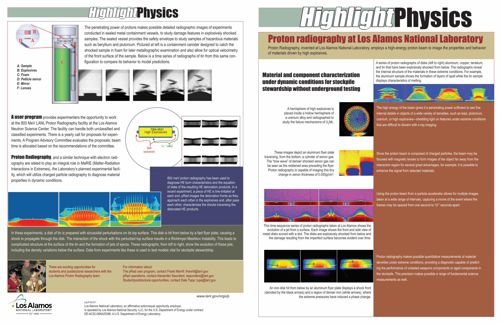
PhysicsHighlightProton radiography at Los Alamos National LaboratoryProton Radiography, invented at Los Alamos National Laboratory, employs a high-energy proton beam to image the properties and behavior of materials driven by high explosives.
A series of proton radiographs of disks (left to right) aluminum, copper, tantalum, and tin that have been explosively shocked from below. The radiographs reveal the internal structure of the materials in these extreme conditions. For example, the aluminum sample shows the formation of layers of spall while the tin sample displays characteristics of melting.
Material and component characterization under dynamic conditions for stockpile stewardship without underground testing
The high energy of the beam gives it a penetrating power sufficient to see fine internal details in objects of a wide variety of densities, such as lead, plutonium, uranium, or high explosives—shedding light on features under extreme conditions that are difficult to discern with x-ray imaging.
Since the proton beam is composed of charged particles, the beam may be focused with magnetic lenses to form images of the object far away from the interaction region for several great advantages, for example, it is possible to enhance the signal from selected materials.
Using the proton beam from a particle accelerator allows for multiple images taken at a wide range of intervals, capturing a movie of the event where the frames may be spaced from one second to 10-7 seconds apart.
Proton radiography makes possible quantitative measurements of material densities under extreme conditions, providing a diagnostic capable of predict-ing the performance of untested weapons components or aged components in the stockpile. This precision makes possible a range of fundamental science measurements as well.
A hemisphere of high explosives is placed inside a hollow hemisphere of a uranium alloy and radiographed to
study the failure mechanisms of U6Nb.
These images depict an aluminum flyer plate traversing, from the bottom, a cylinder of xenon gas.
The “bow wave” of denser shocked xenon gas can be seen as the reddened area preceding the flyer. Proton radiography is capable of imaging this tiny
change in xenon thickness of 0.055g/cm2.
This time-sequence series of proton radiographs taken at Los Alamos shows the evolution of a jet from a surface. Each image shows the front and side view of
metal disks scored with a slot. The disks are explosively shocked from below and the damage resulting from the imperfect surface becomes evident over time.
An iron disk hit from below by an aluminum flyer plate displays a shock front (denoted by the black arrows) and a region of denser iron (white arrows), where
the extreme pressures have induced a phase change.LALP-09-071Los Alamos National Laboratory, an affirmative action/equal opportunity employer, is operated by Los Alamos National Security, LLC, for the U.S. Department of Energy under contract DE-AC52-06NA25396. A U.S. Department of Energy Laboratory.
www.lanl.gov/orgs/p
PhysicsHighlightThe penetrating power of protons makes possible detailed radiographic images of experiments conducted in sealed metal containment vessels, to study damage features in explosively shocked samples. The sealed vessel provides the safety envelope to study samples of hazardous materials such as beryllium and plutonium. Pictured at left is a containment canister designed to catch the shocked sample in foam for later metallographic examination and also allow for optical velocimetry of the front surface of the sample. Below is a time series of radiographs of tin from this same con-figuration to compare its behavior to model predictions.
800 meV proton radiography has been used to diagnose HE burn characteristics and the equation of state of the resulting HE detonation products. In a recent experiment, a piece of HE is line-initiated at each end. pRad images the detonation fronts as they approach each other in the explosives and, after pass each other, characterizes the shocks traversing the detonated HE products.
A: SampleB: ExplosivesC: FoamD: Pellicle mirrorE: MirrorF: Lenses
A user program provides experimenters the opportunity to work at the 800 MeV LANL Proton Radiography facility at the Los Alamos Neutron Science Center. The facility can handle both unclassified and classified experiments. There is a yearly call for proposals for experi-ments. A Program Advisory Committee evaluates the proposals; beam time is allocated based on the recommendations of the committee.
Proton Radiography, and a similar technique with electron radi-ography are slated to play an integral role in MaRIE (Matter-Radiation Interactions in Extremes), the Laboratory’s planned experimental facil-ity, which will utilize charged particle radiography to diagnose material properties in dynamic conditions.
In these experiments, a disk of tin is prepared with sinusoidal perturbations on its top surface. This disk is hit from below by a fast flyer plate, causing a shock to propagate through the disk. The interaction of the shock with the perturbed top surface results in a Richtmyer-Meshkov instability. This leads to complicated structure at the surface of the tin and the formation of jets of ejecta. These radiographs, from left to right, show the evolution of these jets, including the density variations below the surface. Data from experiments like these is used to test models vital for stockpile stewardship.
There are exciting opportunities for students and postdoctoral researchers with the Los Alamos Proton Radiography team.
For information about: The pRad user program, contact Frank Merrill: [email protected] operations, contact Alexander Saunders: [email protected]/postdoctoral opportunities, contact Dale Tupa: [email protected]
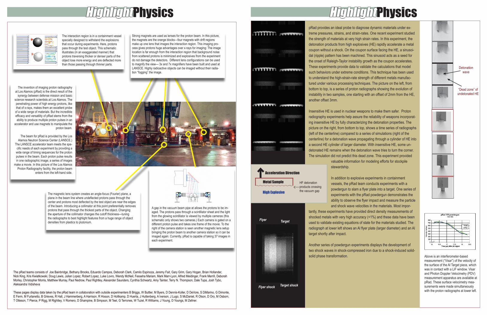
PhysicsHighlight PhysicsHighlightpRad provides an ideal probe to diagnose dynamic materials under ex-treme pressures, strains, and strain-rates. One recent experiment studied the strength of materials at very high strain rates. In this experiment, the detonation products from high explosives (HE) rapidly accelerate a metal coupon without a shock. On the coupon surface facing the HE, a sinusoi-dal (ripple) pattern has been machined. This sinusoid acts as a seed for the onset of Raleigh-Taylor instability growth as the coupon accelerates. These experiments provide data to validate the calculations that model such behaviors under extreme conditions. This technique has been used to understand the high-strain-rate strength of different metals manufac-tured under various processing techniques. The picture on the left, from bottom to top, is a series of proton radiographs showing the evolution of instability in two samples, one starting with an offset of 2mm from the HE, another offset 3mm.
Insensitive HE is used in nuclear weapons to make them safer. Proton radiography experiments help assure the reliability of weapons incorporat-ing insensitive HE by fully characterizing the detonation properties. The picture on the right, from bottom to top, shows a time series of radiographs (left of the centerline) compared to a series of simulations (right of the centerline) for a detonation wave propagating through a cylinder of HE into a second HE cylinder of larger diameter. With insensitive HE, some un-detonated HE remains when the detonation wave tries to turn the corner. The simulation did not predict this dead zone. This experiment provided
valuable information for modeling efforts for stockpile stewardship.
In addition to explosive experiments in containment vessels, the pRad team conducts experiments with a powdergun to slam a flyer plate into a target. One series of experiments with the pRad powdergun demonstrates the ability to observe the flyer impact and measure the particle and shock wave velocities in the materials. Most impor-
tantly, these experiments have provided direct density measurements of shocked metals with very high accuracy (<1%) and these data have been used to validate existing equations of state for the materials studied. The radiograph at lower left shows an Al flyer plate (larger diameter) and an Al target shortly after impact.
Another series of powdergun experiments displays the development of two shock waves in shock-compressed iron due to a shock-induced solid-solid phase transformation. Above is an interferometer-based
measurement (“Visar”) of the velocity of the surface of the Al Target piece, which was in contact with a LiF window. Visar and Photon Doppler Velocimetry (PDV) measurement apparatus are available at pRad. These surface velocimetry mea-surements were made simultaneously with the proton radiographs at lower left.
“Dead zone” of undetonated HE
Detonation wave
Flyer Target
Target shockFlyer shock
The pRad teams consists of Joe Bainbridge, Bethany Brooks, Eduardo Campos, Deborah Clark, Camilo Espinoza, Jeremy Fait, Gary Grim, Gary Hogan, Brian Hollander, Nick King, Kris Kwiatkowski, Doug Lewis, Julian Lopez, Robert Lopez, Luke Lovro, Wendy McNeil, Fesseha Mariam, Mark Marr-Lyon, Alfred Meidinger, Frank Merrill, Deborah Morley, Christopher Morris, Matthew Murray, Paul Nedrow, Paul Rightley, Alexander Saunders, Cynthia Schwartz, Amy Tainter, Terry N. Thompson, Dale Tupa, Josh Tybo, Aleksandra Vidisheva
These pages display data taken by the pRad team in collaboration with outside experimenters B Briggs, W Buttler, M Byers, D Dennis-Koller, D DeVore, S DiMarino, G Dimonte, E Ferm, M Furlanetto, B Grieves, R Hall, J Hammerberg, A Harrison, R Hixson, D Holtkamp, D Huerta, J Huttenberg, A Iverson, J Lugo, S McDaniel, R Olson, D Oro, M Osborn, T Otteson, T Pierce, P Rigg, M Rightley, V Romero, D Shampine, B Simpson, M Teel, G Terrones, W Tuzel, R Williams, J Young, D Youngs, M Zellner.
The invention of imaging proton radiography at Los Alamos (pRad) is the direct result of the
synergy between defense mission and basic science research scientists at Los Alamos. The penetrating power of high energy protons, like that of x-rays, makes them an excellent probe
of a wide range of materials. But the incredible efficacy and versatility of pRad stems from the ability to produce multiple proton pulses in an
accelerator and use magnets to manipulate the proton beam.
The beam for pRad is provided by the Los Alamos Neutron Science Center (LANSCE.)
The LANSCE accelerator team meets the spe-cific needs of each experiment by providing a wide range of timing sequences for the proton pulses in the beam. Each proton pulse results in one radiographic image; a series of images
make a movie. In this picture of the Los Alamos Proton Radiography facility, the proton beam
enters from the left-hand side.
Strong magnets are used as lenses for the proton beam. In this picture, the magnets are the orange blocks—four magnets with drift regions make up one lens that images the interaction region. This imaging pro-cess gives protons huge advantages over x-rays for imaging: The image location is far enough from the interaction region that background noise from scattered protons is minimized and explosives from the experiment do not damage the detectors. Different lens configurations can be used to magnify the view— 3x and 7x magnifiers have been built and used at LANSCE. Highly radioactive objects can be imaged without their radia-tion “fogging” the image.
The interaction region is in a containment vessel specially designed to withstand the explosions that occur during experiments. Here, protons pass through the test object. This schematic illustrates (in an exaggerated manner) that protons traversing thicker or denser parts of the object lose more energy and are deflected more than those passing through thinner parts.
The magnetic lens system creates an angle-focus (Fourier) plane, a plane in the beam line where undeflected protons pass through the center and protons most deflected by the test object are near the edges of the beam. Introducing a collimator at this point preferentially removes protons that pass through the thickest parts of the object. Changing the aperture of the collimator changes the cutoff thickness—tuning the radiographs to best highlight features from a huge range of object densities from plastics to plutonium.
A gap in the vacuum beam pipe at allows the protons to be im-aged. The protons pass through a scintillator sheet and the light from the glowing scintillator is viewed by multiple cameras (this schematic only shows two cameras.) Each camera is gated on a different proton pulse and takes one frame of the movie. To the right of the camera station is seen another magnetic lens setup bringing the proton beam to another camera station so it can be imaged again. Currently, pRad is capable of taking 37 images in each experiment.
Acceleration Direction
High Explosive
HF detonation products crossingthe vacuum gap
Metal Sample